title: identification of key differentially expressed ... · title: identification of key...
TRANSCRIPT
1
Title: Identification of Key Differentially Expressed MicroRNAs in
Cancer Patients Through Pan-cancer Analysis
Yu Hu1, Hayley Dingerdissen1, Samir Gupta2, Robel Kahsay1, Vijay Shanker2, Quan Wan1, Cheng Yan1,
Raja Mazumder1,3,*
1The Department of Biochemistry & Molecular Medicine, The George Washington University Medical
Center, Washington, DC 20037, United States of America 2Department of Computer and Information Science, University of Delaware, Newark, Delaware 19716,
United States of America
3The McCormick Genomic and Proteomic Center, The George Washington University, Washington, DC
20037, United States of America
*Corresponding author: address: Ross Hall, 2300 Eye Street N.W., Washington, DC 20037
E-mail: [email protected] (Hu Y), [email protected] (Dingerdissen H),
[email protected] (Gupta S), [email protected] (Kahsay R), [email protected] (Shanker V),
[email protected] (Wan Q), [email protected] (Yan C), [email protected]
(Mazumder R)
Running title: Pan-cancer Analysis of MiRNAs and Their Targets
.CC-BY-NC-ND 4.0 International licensecertified by peer review) is the author/funder. It is made available under aThe copyright holder for this preprint (which was notthis version posted August 9, 2018. . https://doi.org/10.1101/388892doi: bioRxiv preprint
2
Abstract
A number of microRNAs (miRNAs) functioning in gene silencing have been associated with
cancer progression. However, common expression patterns of abnormally expressed miRNAs
and their potential roles in multiple cancer types have not yet been evaluated. To minimize the
difference of patients, we collected miRNA sequencing data of 575 patients with tumor and
adjacent non-tumorous tissues from 14 cancer types from The Cancer Genome Atlas (TCGA),
and performed differential expression analysis using DESeq2 and edgeR. The results showed
that cancer types can be grouped based on the distribution of miRNAs with different expression
patterns. We found 81 significantly differentially expressed miRNAs (SDEmiRNAs) unique to
one of the 14 cancers may affect patient survival rate, and 21 key SDEmiRNAs (nine over-
expressed and 12 under-expressed) associated with at least eight cancers and enriched in more
than 60% of patients per cancer, including four newly identified SDEmiRNAs (hsa-mir-4746,
hsa-mir-3648, hsa-mir-3687, and hsa-mir-1269a). The downstream effect of these 21
SDEmiRNAs on cellular functions was evaluated through enrichment and pathway analysis of
7,186 protein-coding gene targets from literature mining with known differential expression
profiles in cancers. It enables identification of their functional similarity in cell proliferation control
across a wide range of cancers and to build common regulatory networks over cancer-related
pathways. This is validated by construction of a regulatory network in PI3K pathway. This study
provides evidence of the value of further analysis on SDEmiRNAs as potential biomarkers and
therapeutic targets for cancer diagnosis and treatment.
KEYWORDS: miRNA; differential expression analysis; pan-cancer; miRNA target; functional
analysis
.CC-BY-NC-ND 4.0 International licensecertified by peer review) is the author/funder. It is made available under aThe copyright holder for this preprint (which was notthis version posted August 9, 2018. . https://doi.org/10.1101/388892doi: bioRxiv preprint
3
Introduction
MicroRNAs (miRNAs) are small non-coding RNAs with two forms, the premature miRNA (length
50-125 bp) and the processed, mature form (length 18-24 bp)[1]. miRNAs function in gene
silencing by binding to mRNAs, causing either mRNA destabilization or inhibition of
translation[2]. From the beginning of 21st century, pioneer researchers have increasingly
catalogued the close relationship between miRNAs and cancer development. For example,
Calin et al.[3] discovered the deletion or down-regulation of miR15/miR16 in chronic lymphocytic
leukemia, Takamizawa et al.[4] showed under-expressed hsa-let-7 is related to lower survival
rate in lung cancer, Chan et al.[5] knocked down hsa-mir-21 in glioblastoma cells which
activated apoptosis, and Li et al.[6] found hsa-mir-10b enabled cell metastasis in breast cancer.
These studies not only laid the foundation for a number of studies on the roles of these miRNAs
in different cancer types, but also paved the way to the increasing identification of cancer-
associated miRNAs[7].
Furthermore, it is believed that there are significantly fewer known human miRNAs (about
1,881 pre-mature and 2,588 mature in miRBase as of 2016[1]) than total mRNAs because each
miRNA has multiple target mRNAs. Due to this one-to-many relationship, studying miRNA
expression profiles could contribute more efficiently to construction of the molecular regulatory
network as a whole, especially in characterizing cancer-associated regulation of molecular
pathways or biological processes. miRNAs are classified as tumor suppressors or oncogenes
(oncomirs) according to their expression profiles in cancer[8]. In breast cancer, at least 20
oncogenic miRNAs or cluster families have been identified to promote cell proliferation,
invasion, migration, or angiogenesis[9]. Cluster families are groups of several miRNAs
transcribed together and co-expressed. Similarly, more than 30 tumor suppressive miRNAs or
families have been observed to play a role in cell apoptosis and negative control of cell
proliferation or migration[9]. In cancer metastasis, these miRNAs can change the expression of
genes essential for cellular homeostasis and for robustness of cell fate decisions, often through
various signaling pathways[10]. Therefore, to gain a comprehensive view of the cancer
landscape, it is imperative to characterize miRNAs on a large scale and integrate evidence that
has been observed across multiple cancer types to build more inclusive regulatory networks.
.CC-BY-NC-ND 4.0 International licensecertified by peer review) is the author/funder. It is made available under aThe copyright holder for this preprint (which was notthis version posted August 9, 2018. . https://doi.org/10.1101/388892doi: bioRxiv preprint
4
In large-scale studies, roles of miRNAs have been found to be important in cancer
diagnosis, prognosis, and therapy for multiple cancer types[10-12]. Each cancer type has a
unique miRNA expression signature that can be classified into different prognostic groups[13].
With an increasing number of studies about cancer incidence, there has been a corresponding
increase in observations of tumors with different molecular mechanisms even within a single
cancer type[14, 15]. Cancer subtypes can also be identified by miRNA expression profiling, as in
the case of non-small cell lung cancer where miRNAs have classified three subtypes by different
driving factors (ALK, EGFR, and KRAS)[16]. The diagnostic and prognostic roles of miRNAs
further indicate their potential as therapeutic targets and as candidates for clinical trials[17].
Recently, miRNAs have been gradually introduced into personalized medicine approaches for
cancer or other disease therapies[18, 19]. Based on the apparent importance of miRNAs in
cancer, a number of studies have been conducted on miRNA expression profiles in which
statistical analysis was used to determine changes between experimental and control samples
to characterize cancer-specific miRNAs in one or two cancers. However, pan-cancer analysis of
miRNA expression has not been explored to identify similarity and differences across multiple
cancers.
With the introduction of newer, more sensitive procedures in library sample preparation,
accompanied by decreasing costs of next-generation sequencing (NGS) methods[20],
researchers can now acquire a comprehensive miRNA abundance profiling through deep
sequencing[21], despite known complications like low abundant miRNAs and miRNA processing
complexity. Several normalization methods for miRNA-seq have been widely used in previous
studies[22, 23], including edgeR (Trimmed Mean of M values, TMM)[24] and DESeq[25]. In this
study, we performed differential expression analysis by both DESeq2[26] and edgeR[24] for
paired miRNA-seq data (tumor and surrounding non-tumorous tissues) from The Cancer
Genome Atlas (TCGA: http://cancergenome.nih.gov; https://portal.gdc.cancer.gov/), following
the methods described by Yang S et al[27] and used in the study of Metpally RP et al[20].
Although previous studies have demonstrated critical roles of miRNAs in the prognosis of
cancer types and subtypes, our research singled out key miRNAs observed to have significantly
differential expression (SDEmiRNAs) for 14 TCGA cancer types independently. The distribution
of these SDEmiRNAs with different expression trends enables us to group the 14 cancers and
.CC-BY-NC-ND 4.0 International licensecertified by peer review) is the author/funder. It is made available under aThe copyright holder for this preprint (which was notthis version posted August 9, 2018. . https://doi.org/10.1101/388892doi: bioRxiv preprint
5
based on different potential developmental mechanisms of the 14 cancers. Individually,
comparing the expression profiles of these SDEmiRNAs, we were able to screen out a list of
miRNAs which are significantly differential expressed in only one of the 14 cancers. These
unique SDEmiRNAs specific to one cancer could be considered as biomarkers of each cancer,
and may have influence in patient survival rate, regardless of their driven factors. We also
identified a subset of key SDEmiRNAs that are significantly over- or under-expressed with high
patient frequencies in at least eight cancer types (over-expression implies functional up-
regulation). With the functional and enrichment analyses of their cancer-related target genes
and pathways, these SDEmiRNAs might be commonly functional and executive factors
promoting multiple cancer development via similar molecular mechanisms. As an example, we
constructed common regulatory networks of several SDEmiRNAs in signaling pathways across
different cancer types, despite the great difference among their target genes in multiple tissues.
Notably, some of the identified SDEmiRNAs are barely studied in cancer development due to
their relatively low expression levels but have been found to be significantly differentially
expressed in a wide range of cancer types, showing their potential vital roles in cancer
development. Thus, this study evaluates the expression of all human miRNAs in 14 cancer
types, widens the roles of individual SDEmiRNAs to cancer types where they have not been
studied, classifies the functions of SDEmiRNAs in different tissues to small number of molecular
mechanisms, and therefore, provides a means for improved discovery of mechanisms of cancer
development that could improve downstream clinical studies and trials by prioritizing miRNA and
targets for translational study. The results are integrated in BioXpress v3.0
(https://hive.biochemistry.gwu.edu/bioxpress) and open-source to support further analysis.
Results and Discussion
Data collection and overall evaluation before differential expression analysis
The TCGA project provides a comprehensive database with a unified data analysis pipeline for
collecting and analyzing samples. We were able to collect tumor-only expression data for 1,904
miRNAs in 32 TCGA/ICGC cancer types generated by the HiSeq platform (7,994 TCGA patients
.CC-BY-NC-ND 4.0 International licensecertified by peer review) is the author/funder. It is made available under aThe copyright holder for this preprint (which was notthis version posted August 9, 2018. . https://doi.org/10.1101/388892doi: bioRxiv preprint
6
and 168 ICGC patients) and 1,668 miRNAs in 12 TCGA cancer types generated by Genome
Analyzer (1,439 TCGA patients), from a total of 9,601 patients. Within these data, 22 TCGA
cancer types containing paired data (tumor data with matched adjacent non-tumorous data)
from the HiSeq platform, we set a threshold of patient counts (greater than 10, covering more
than 95% of total patients with paired data), resulting in 14 cancer types to be used in further
analysis (BLCA, BRCA, PRAD, STAD, KIRP, KIRC, KICH, LUAD, LUSC, LIHC, THCA, ESCA,
UCEC, and HNSC – please see Table 1 for full cancer names). These cancer types involved a
total of 575 TCGA-only patients (1,152 paired samples) with an average of 1,393 expressed
miRNAs for each cancer.
In order to overview the expression profiles of miRNAs in the paired samples from the 14
cancer types, RPM values of each miRNA (pre-calculated by TCGA) in each TCGA patient were
first used to calculate regular fold change (FC) values between cancer and corresponding non-
tumorous samples. After comparing these FC values, we found that the total number of over-
expressed miRNAs is two-fold higher than that of under-expressed miRNAs. However, when
considering only those miRNAs with FC > 2 (FC < 0.5 for under-expressed miRNAs), and with
relatively high frequencies in patients in at least eight cancers, the trends of the numbers of
miRNAs with over- and under-expression are similar to each other across more than 35% of
patients in each cancer (Figure 1). This suggests that many over-expressed miRNAs may be
effectors driven by other factors and vary from patient to patient.
This expression distribution also points out 27 miRNAs (11 over-expressed miRNAs and 16
under-expressed miRNAs) in more than 60% of patients (Figure 1) in eight or more cancers.
Most of these miRNAs have been previously reported in at least one publication associated with
cancer, some of which are well-studied tumor suppressors, including has-mir-133b, and hsa-
mir-139, or contribute to cell invasion, such as hsa-mir-301b[28, 29] hsa-mir-1247 was newly
found to be a prognostic marker in pancreatic cancer as a suppressor[28], and hsa-mir-1258
targets heparanase (HPSE) to suppress breast and non-small cell lung cancer[30, 31]. hsa-mir-
1269a, hsa-mir-3687, and hsa-mir-3648 were also newly identified to be involved in cancer
development[32-34], but their molecular mechanisms in cancers need to be researched further.
Besides them, two miRNAs reported in 2011[35], hsa-mir-4746 and hsa-mir-4664, which are
over-expressed in 11 and eight cancers with average of patient frequencies, 74.42% (average
.CC-BY-NC-ND 4.0 International licensecertified by peer review) is the author/funder. It is made available under aThe copyright holder for this preprint (which was notthis version posted August 9, 2018. . https://doi.org/10.1101/388892doi: bioRxiv preprint
7
FC = 3.95) and 75.49% (average FC = 8.07), respectively, although their average expression
levels are lower than those well-studied miRNAs, which may lead to difficulties in quantification
and qualification.
These results validate the reliability and confidence of our data, and suggest the potential
similar roles of the subset of 27 miRNAs in multiple cancer development, based on their similar
expression profiles. It is also important to note that after statistical analysis in the following
sections, 21 out of these 27 miRNAs were found to be significantly differentially expressed
(SDEmiRNAs) in the same cancer types.
miRNA differential expression analysis
SDEmiRNAs could be used in distinguishing cancer types
DESeq2[26] and edgeR[24] were used to perform differential expression analysis, applying
statistical tests to minimize the potential false positive errors from the differences among
individuals and samples. Data from all patients for a single cancer type were analyzed together
resulting in reporting average differential expression levels by cancer type. Both DESeq2 and
edgeR provide p values and adjusted p values (or FDR in edgeR). The results of the two tools
showed a similar ranking of significantly differentially expressed miRNAs (SDEmiRNAs) when
ranked by adjusted p values. We found 10 to 15% of miRNAs with significant differential
expression in each cancer type reported by both DESeq2 and edgeR (Table 1). As shown in
Figure 2A, of all 1,870 miRNAs in our integrated database, the specific distributions of the
quantities and types of 656 SDEmiRNAs vary between cancer types, suggesting the existence
of unique SDEmiRNA patterns for each of these cancer types. These unique patterns of
SDEmiRNAs potentially reduce the amount of miRNAs used for prognosis of cancer types,
based on the findings of Lu et al[13]. The result of differential expression analysis of each
miRNA is available in BioXpress, shown by both figures and table (Figure S1).
Moreover, similar SDEmiRNA expression patterns are shown in LUAD and LUSC from lung
tissue, as well as in KICH, KIRP, and KIRC from kidney tissue, as expected. However, among
387 SDEmiRNAs in either LUAD or LUSC (Figure S2A), 190 have log2FC values greater than 1
.CC-BY-NC-ND 4.0 International licensecertified by peer review) is the author/funder. It is made available under aThe copyright holder for this preprint (which was notthis version posted August 9, 2018. . https://doi.org/10.1101/388892doi: bioRxiv preprint
8
(∆log2FC > 1), 140 (36.2% of 387) of which have the opposite expression trend for both
cancers, suggesting they could be key factors underlying the differences between LUAD and
LUSC. We also performed a pairwise comparison among KICH, KIRP, and KIRC using the set
of 330 SDEmiRNAs with ∆log2FC > 1 in at least one of the three cancer types. It shows that
KIRP and KIRC have more similar SDEmiRNA expression patterns than KIRP and KICH, and
KICH and KIRC (Figure S2B-D). This suggests that the development of KICH may be
mechanistically more distant than the development of KIRC and KIRP.
Expression pattern of SDEmiRNAs groups cancer type based on different patterns of
mechanisms in different cancer development
We counted the number of SDEmiRNAs that have the same expression trend in at least 80% of
patients (by log2FC values) for each cancer type (Table 1, and the corresponding patient
frequencies in Figure S3). These results showed different cancer types have different
proportions of SDEmiRNAs: some cancers have more over-expressed SDEmiRNAs than under-
expressed; some have a balance between over- and under-expressed SDEmiRNAs; and other
cancer types have more under-expressed SDEmiRNAs (Figure S3). Similar results are shown
when the log2FC values of all SDEmiRNAs were used to display their distributions in all cancer
types (Figure 2C).
In four cancer types (KICH, KIRP, LIHC, and THCA), the numbers of under-expressed
SDEmiRNAs are much higher than those of over-expressed SDEmiRNAs, while for most of the
cancer types (BLCA, ESCA, KIRC, LUAD, LUSC, PRAD, STAD, and UCEC), the median
expression values of their SDEmiRNAs are higher than zero. Because the main function of
miRNA is to suppress target genes, the significant number of under-expressed miRNAs
corresponds to the up-regulation of oncogenes, which may play a more important role in the
occurrence and progression of the four cancers characterized predominantly by under-
expression of miRNAs. Conversely, the down-regulation of tumor suppression genes may be
more important in cancer occurrence and progression for those cancers characterized
predominantly by over-expression of miRNAs. Cancers like BRCA, HNSC, and KIRC have an
almost symmetric distribution of SDEmiRNAs about log2FC = 0 (Figure 2C), suggesting
.CC-BY-NC-ND 4.0 International licensecertified by peer review) is the author/funder. It is made available under aThe copyright holder for this preprint (which was notthis version posted August 9, 2018. . https://doi.org/10.1101/388892doi: bioRxiv preprint
9
regulation of both oncogenes and tumor suppression genes could be equally important for
disease progression. However, it could also be possible that the similar distribution between
over- and under-expressed miRNAs indicates that these miRNAs are equally unimportant to
disease and may suggest other mechanisms are primarily at work for these cancer types.
The distribution of SDEmiRNAs is able to group cancer types to three categories based on
the potential different mechanisms during cancer initiation and development. It allows high
efficiency of further research on exploring mechanisms of cancers.
SDEmiRNAs that are unique to one of the 14 cancer types might be driver miRNAs
Although we are mostly interested in SDEmiRNAs implicated in multiple cancers (as discussed
in depth below), SDEmiRNAs that only exist in a single cancer type may be considered potential
driver miRNAs or biomarkers specific for that cancer type. Therefore, we set up screening
conditions to identify those SDEmiRNAs affecting a single cancer type: 1) SDEmiRNA should be
reported to be significant by both DESeq2 and edgeR; 2) this SDEmiRNA should not exist in the
remaining 13 cancers; 3) the patient frequency of this SDEmiRNA should be more than 50% in
this cancer; 4) the log2FC value of this SDEmiRNA should be greater than 1, or smaller than -1.
As shown in Table S1, we identified a total of 81 SDEmiRNAs with significant expression
trends in a single cancer type, 57 of which have patient frequencies higher than 60% (Table 2).
Most of these SDEmiRNAs have much lower expression in tissues compared with other well-
studied miRNAs, and have not been functionally studied in cancer development, especially in
their corresponding cancer types. However, this does not negate their potential cancer-related.
In order to validate the results, as an example, we performed survival analysis on four
SDEmiRNAs unique to BRCA (absolute log2FC values > 1) by using miRpower[36]. Except hsa-
mir-329-2 which is not in miRpower, the rest three SDEmiRNAs suggest significantly survival
difference (log-rank p value < 0.01) between groups with under-/over-expression of each
SDEmiRNA, after optimizing the cutoff of the patients (Figure S4): hsa-mir-4784 (Hazard Ratio =
1.82), hsa-mir-1262 (HR = 0.58), and hsa-mir-320c-1 (HR = 0.68). The difference of survival
rates between patients within under- and over-expression were consistent with our results. Even
though the input datasets were not exactly matched with this study, it still shows the potential
.CC-BY-NC-ND 4.0 International licensecertified by peer review) is the author/funder. It is made available under aThe copyright holder for this preprint (which was notthis version posted August 9, 2018. . https://doi.org/10.1101/388892doi: bioRxiv preprint
10
vital roles of these unique SDEmiRNAs in the development of BRCA. Similarly, other
SDEmiRNAs may also have the potential as signatures of different cancer types, and are of
great importance and prospect to have additional analysis.
Pan-cancer analysis of key SDEmiRNAs
As shown in Figure 2B, after unsupervised clustering of all SDEmiRNAs, several SDEmiRNAs
follow the same trend of either over-expression or under-expression across most of the cancer
types, suggesting that these SDEmiRNAs may function similarly across different cancer types.
To further study the key SDEmiRNAs affecting multiple cancers, we identified a subset of 90
miRNAs that have significant differential expression in at least eight cancer types (58% of total
cancer types).
Selected SDEmiRNAs were validated by experimentally validated databases and text mining
tool
Before proceeding with the analysis, we first confirmed these 90 key SDEmiRNAs generated
from DESeq2 and edgeR using four experimentally validated databases. Among 2,280
significantly differentially expressed miRNA-cancer correlations, 208 (9.12%) correlations were
recorded in the union of the four databases (HMDD, Mir2disease, miRCancer, and miRiaD) to
be exclusively over- or under-expressed in certain types of cancer. Furthermore, we randomly
picked 24 of 90 SDEmiRNAs (a total of 226 miRNA-cancer correlations) as the input of our text
mining tool (DEXTER). After mapping and manual curation, we extracted 157 (69.47%)
interactions (Table S2). None of the 90 SDEmiRNAs were found by the databases of DEXTER
with opposite expression trends compared to the trends we identified in our study. Based on
these observations, we consider our differential expression analysis results to be reliable.
Key SDEmiRNAs with at least 60% patient frequencies were considered reliable and may have
similar functions across multiple cancers
.CC-BY-NC-ND 4.0 International licensecertified by peer review) is the author/funder. It is made available under aThe copyright holder for this preprint (which was notthis version posted August 9, 2018. . https://doi.org/10.1101/388892doi: bioRxiv preprint
11
From the 90 selected SDEmiRNAs, we defined a subset of them based on their expression
trend: 47 SDEmiRNAs are all over-expressed and 18 are all under-expressed in eight or more
cancer types (a total of 65 SDEmiRNAs with the same significantly differential expression trend
in eight or more cancers) (Table S3). Beside 11 SDEmiRNAs, the rest 54 SDEmiRNAs belong
to 40 miRNA families, according to MirGeneDB v2.0[37]. Among them, all members of 22
families are included in the list, although most of the families have only one or two members.
However, the similar expression trends of these SDEmiRNAs suggest their high consistence of
activities and potential functions across 58% of investigated cancers, and therefore, these small
families can be classified to larger ones (Table S3). Individually, six newly reported miRNAs
were included in the 65 SDEmiRNAs: hsa-mir-3170, hsa-mir-3677, hsa-mir-4326, hsa-mir-4652,
hsa-mir-7706, and hsa-mir-105-2 (Table S3). Among them, hsa-mir-4652 is the top one
significantly over-expressed miRNAs with largest log2FC value (average log2FC = 5.34, patient
frequency larger than 60% in seven cancer types). We found no cancer-related studies that
have explored the mechanisms of these six miRNAs in cancers, suggesting their high potential
for involvement in cancer development, especially hsa-mir-4652.
Comparing the 65 SDEmiRNAs identified by differential expression analysis to the 27
miRNAs with regular FC > 2 or FC < 0.5 across 60% of patients (in ‘Data collection and
evaluation’ section, Figure 1) in at least eight cancer types, we found an overlap of nine over-
expressed and 12 under-expressed SDEmiRNAs between the two datasets (a total of 21
miRNAs) (Table 3). This subset not only includes well-studied miRNAs such as hsa-mir-145 and
hsa-mir-210, but also newly identified miRNAs such as hsa-mir-4746 (in ‘Data collection and
evaluation’ section), hsa-mir-3648, hsa-mir-3687, and hsa-mir-1269a. Two of the over-
expressed and five of the under-expressed key miRNAs are in the top five significantly over-
and under-expressed miRNAs of the 65 ranked by average log2FC values (Table 3).
Molecular mechanisms for the newly identified hsa-mir-1269a have rarely been published for
the cancer types we report here, but Min et al. have recently found a single nucleotide variation
in hsa-mir-1269a that could contribute to the occurrence of hepatocellular carcinoma[38]. In our
results, hsa-mir-1269a has log2FC value consistently larger than 2.20 in each of its eight
associated cancer types (average log2FC = 5.18), with the average frequency of affected
patients, greater than 64.58% for a strict fold change requirement (FC > 5 for each patient).
.CC-BY-NC-ND 4.0 International licensecertified by peer review) is the author/funder. It is made available under aThe copyright holder for this preprint (which was notthis version posted August 9, 2018. . https://doi.org/10.1101/388892doi: bioRxiv preprint
12
Other members of the 21 key SDEmiRNAs (Table 3) include hsa-mir-183 and hsa-mir-96, which
belong to the mir-183-96-182 cluster, which functions to induce cell proliferation and cancer
development[39]. Notably, unlike the other two miRNAs from the same cluster, hsa-mir-182 only
shows significantly differential expression but not high patient frequencies. Considering our
relatively stringent criteria on FC values (FC > 2 or FC < 0.5), the changes of hsa-mir-182 in
most patients are not as strong as that of hsa-mir-183 and hsa-mir-96.
Overall, our study groups 14 cancer types with potential different molecular mechanisms of
development based on the distribution of SDEmiRNAs, as well as proposes some SDEmiRNAs
previously indicated to play a role in distinct cancer types may function similarly in the
development of additional cancer types, and the classification of small miRNA families into
larger ones. Moreover, the results correspond well with those of previously published studies,
and include evaluation of the 21 key SDEmiRNAs with high patient frequencies in a
comprehensive pan-cancer analysis. Newly identified miRNAs hsa-mir-4746, hsa-mir-3648, hsa-
mir-3687, and hsa-mir-1269a could also be important factors in multiple cancer types, although
their relations to cancer were rarely reported by previous studies due to their low expression
levels compared to others.
SDEmiRNA targets enrichment and functional analysis
Each of the 65 SDEmiRNAs discussed above (with the same significantly differential expression
trend in eight or more cancer types) participates in some form of molecular regulation, either
inducing or inhibiting cell proliferation and cancer metastasis in different cancer types. In order
to better understand the general pathways or biological processes by which SDEmiRNAs
contribute to the occurrence and development of multiple cancer types, we extracted
experimentally validated non-tissue-specific targets for those SDEmiRNAs without tissue or
cancer information, as well as tissue-specific targets.
Distribution and functional analysis of non-tissue-specific targets of 65 SDEmiRNAs
.CC-BY-NC-ND 4.0 International licensecertified by peer review) is the author/funder. It is made available under aThe copyright holder for this preprint (which was notthis version posted August 9, 2018. . https://doi.org/10.1101/388892doi: bioRxiv preprint
13
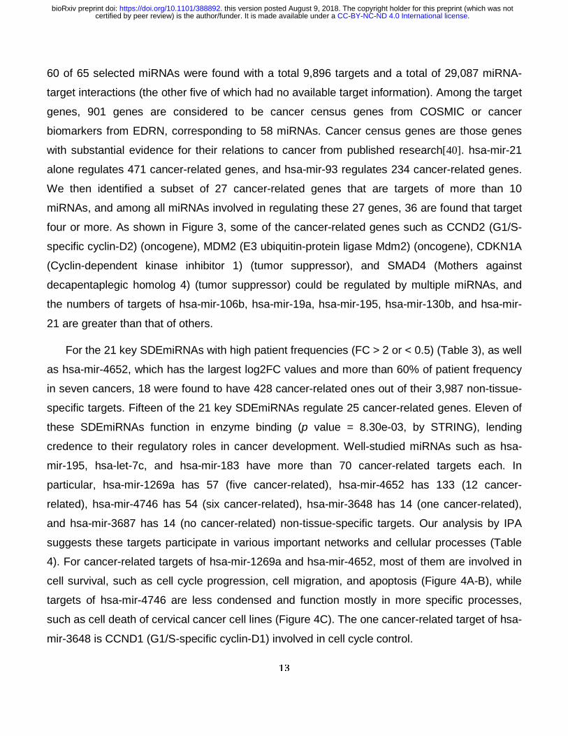
60 of 65 selected miRNAs were found with a total 9,896 targets and a total of 29,087 miRNA-
target interactions (the other five of which had no available target information). Among the target
genes, 901 genes are considered to be cancer census genes from COSMIC or cancer
biomarkers from EDRN, corresponding to 58 miRNAs. Cancer census genes are those genes
with substantial evidence for their relations to cancer from published research[40]. hsa-mir-21
alone regulates 471 cancer-related genes, and hsa-mir-93 regulates 234 cancer-related genes.
We then identified a subset of 27 cancer-related genes that are targets of more than 10
miRNAs, and among all miRNAs involved in regulating these 27 genes, 36 are found that target
four or more. As shown in Figure 3, some of the cancer-related genes such as CCND2 (G1/S-
specific cyclin-D2) (oncogene), MDM2 (E3 ubiquitin-protein ligase Mdm2) (oncogene), CDKN1A
(Cyclin-dependent kinase inhibitor 1) (tumor suppressor), and SMAD4 (Mothers against
decapentaplegic homolog 4) (tumor suppressor) could be regulated by multiple miRNAs, and
the numbers of targets of hsa-mir-106b, hsa-mir-19a, hsa-mir-195, hsa-mir-130b, and hsa-mir-
21 are greater than that of others.
For the 21 key SDEmiRNAs with high patient frequencies (FC > 2 or < 0.5) (Table 3), as well
as hsa-mir-4652, which has the largest log2FC values and more than 60% of patient frequency
in seven cancers, 18 were found to have 428 cancer-related ones out of their 3,987 non-tissue-
specific targets. Fifteen of the 21 key SDEmiRNAs regulate 25 cancer-related genes. Eleven of
these SDEmiRNAs function in enzyme binding (p value = 8.30e-03, by STRING), lending
credence to their regulatory roles in cancer development. Well-studied miRNAs such as hsa-
mir-195, hsa-let-7c, and hsa-mir-183 have more than 70 cancer-related targets each. In
particular, hsa-mir-1269a has 57 (five cancer-related), hsa-mir-4652 has 133 (12 cancer-
related), hsa-mir-4746 has 54 (six cancer-related), hsa-mir-3648 has 14 (one cancer-related),
and hsa-mir-3687 has 14 (no cancer-related) non-tissue-specific targets. Our analysis by IPA
suggests these targets participate in various important networks and cellular processes (Table
4). For cancer-related targets of hsa-mir-1269a and hsa-mir-4652, most of them are involved in
cell survival, such as cell cycle progression, cell migration, and apoptosis (Figure 4A-B), while
targets of hsa-mir-4746 are less condensed and function mostly in more specific processes,
such as cell death of cervical cancer cell lines (Figure 4C). The one cancer-related target of hsa-
mir-3648 is CCND1 (G1/S-specific cyclin-D1) involved in cell cycle control.
.CC-BY-NC-ND 4.0 International licensecertified by peer review) is the author/funder. It is made available under aThe copyright holder for this preprint (which was notthis version posted August 9, 2018. . https://doi.org/10.1101/388892doi: bioRxiv preprint
14
By targeting a wide range of cancer-related genes, those 65 SDEmiRNAs, especially 21 key
SDEmiRNAs, participate in multiple processes, contributing to cancer development. Moreover,
those newly identified SDEmiRNAs could be equally important with others, despite their
relatively lower expression levels. They may be able to achieve their contributions to cancer
through regulating targets within small regions or participating in short-term stress response,
which requires less dosage.
Distribution of tissue-specific targets of 65 SDEmiRNAs and functional analysis of them
We also collected tissue-specific targets for the 65 SDEmiRNAs (significantly expressed in at
least eight cancer types) (Table S3) and explored the potential molecular mechanisms of those
tissue-specific targets in light of their observed expression trends and biological processes in
which they participate. All selected miRNAs and their targets were mapped to 11 cancer types
by DOIDs. A total of 9,572 target genes were found, of which 7,325 were mapped to reviewed
UniProtKB accession numbers (Table S4). 7,186 of the implicated significant, reviewed target
genes are included in BioXpress v3.0, which reports genes with significant expression changes
derived from paired tumor and non-tumorous data from TCGA: currently, there are 5,971 genes
showing significant differential expression in multiple cancer types in BioXpress (Table S5).
miRNA target genes for lung cancer and kidney cancer are a combination of those identified for
TCGA cancer types LUAD and LUSC, KICH and KIRP and KIRC, respectively, and have the
same expression trend across these TCGA cancer types.
Mapping to Gene Ontology (GO) terms and PANTHER pathway IDs resulted in retrieval of
12,467 GO terms and 130 PANTHER terms. According to the screening conditions described in
the methods section, 99 records were selected in Table S6 (22 from PANTHER terms and 77
from GO terms). The criteria of the screening were such that we expected to lose potentially
valuable GO or PANTHER terms, but we chose to follow a conservative method to reduce
possible false positive results. The most common molecular functions these miRNAs and their
targets participate in cancers are negative regulation of ERK1 and ERK2 cascade
(GO:0070373), canonical Wnt signaling pathway (GO:0060070), and positive regulation of
sequence-specific DNA binding transcription factor activity (GO:0051091). Each of these
.CC-BY-NC-ND 4.0 International licensecertified by peer review) is the author/funder. It is made available under aThe copyright holder for this preprint (which was notthis version posted August 9, 2018. . https://doi.org/10.1101/388892doi: bioRxiv preprint
15
functions are involved in ten types of cancer, with more than 40 SDEmiRNAs, and more than 30
targets, respectively. This result not only is consistent with Gosline et al.[41], which suggested
the strong regulation of miRNAs on transcription factors in mice, but also suggests most of
these SDEmiRNAs could promote the activity of transcription factors.
These terms suggest the participation in regulation of cell proliferation and apoptosis
through different mechanisms, and frequently implicated cancer pathways were found in our list,
including the p53 pathway, Wnt signaling pathway, TGF-β/SMAD signaling pathway, and others.
We also found that the occurrence and development of these cancer types may share similar
molecular mechanisms with other diseases or processes, including Alzheimer’s disease (AD)
(with involved amyloid secretase and presenilin pathways), for which the product the key gene
implicated in AD, the amyloid precursor protein, is over-expressed in multiple cancer types[42]
and other neurodegenerative diseases.
For the 21 key SDEmiRNAs (with both significant differential expression and at least 60%
patient frequencies in at least eight cancer types) (Table 3), as well as hsa-mir-4652, 19 were
found to have tissue-specific targets. We identified the top one or two genes that are targets of
the most SDEmiRNAs in each cancer type. In breast cancer, CCND1 and IGF1R (Insulin-like
growth factor 1 receptor) are found to be the targets of nine and seven key SDEmiRNAs. IGF1R
is also the most popular target of eight SDEmiRNAs in lung cancer, and of seven SDEmiRNAs
in head and neck cancer. CCND1 promotes G1/S transition during mitotic cell cycle by
regulating CDK4 (cyclin-dependent kinase 4), and is up-regulated in various cancer types
including breast cancer[43, 44]. IGF1R is also a well-studied oncogene that allows cell
proliferation and growth, and inhibits apoptosis[45]. Our RNA-seq analysis in BioXpress v3.0
shows that CCND1 is significantly over-expressed in BRCA, while IGF1R has significant over-
expression in BRCA, LUSC, and HNSC. The roles of CCND1 and IGF1R could be vital and
essential, compared to other cancer-related ones, for the development of these cancer types,
considering such huge compensation mechanisms over their regulation.
In addition, in breast cancer hsa-mir-1269a has one target (EN2, Homeobox protein
engrailed-2) which is a sequence-specific DNA binding protein positively regulating transcription
from RNA polymerase II promoter. hsa-mir-4652 has two targets (GALNT3, EIF4EBP1) in lung
cancer. GALNT3 (Polypeptide N-acetylgalactosaminyltransferase 3) functions as an iron binding
.CC-BY-NC-ND 4.0 International licensecertified by peer review) is the author/funder. It is made available under aThe copyright holder for this preprint (which was notthis version posted August 9, 2018. . https://doi.org/10.1101/388892doi: bioRxiv preprint
16
enzyme in glycosylation, while EIF4EBP1 (Eukaryptic translation initiation factor 4E-binding
protein 1) binds to IRES-dependent translational initiation to repress protein translation and
participates in insulin receptor signaling pathway and G1/S transition of mitotic cell cycle (Table
4). hsa-mir-4652 has another target (H2AFX, Histone H2AX) involved in DNA and histone
binding in head and neck cancer. CancerMiner predicted 12 targets of hsa-mir-3648 in head and
neck cancer (Table S4). Their functions include ATP binding, cell cycle control, lipid metabolism,
protein folding, and nucleobase biosynthesis. hsa-mir-4746 has a common target, PPM1D
(Protein phosphatase 1D), in both breast and lung cancer. Because of its function in protein
dephosphorylation, it is involved in G2/M transition of mitotic cell cycle and suppression of cell
proliferation in cancer[46]. The importance of the functions of their target genes in gene/cellular
regulation suggests abnormally expressed hsa-mir-1269a, hsa-mir-4652, and hsa-mir-4746 may
be able to contribute to the development of corresponding cancer types.
Construction of regulatory networks of SDEmiRNAs and their targets
Suppression of a negative regulator will result in up-regulation of the regulator’s target molecule.
Similarly, because miRNAs function to suppress gene expression, miRNA inhibition of a
negative regulator will also result in up-regulation of the target molecule. There are some cases
in which a miRNA can have two targets such that one target regulates the other. In these cases,
up-regulated targets of over-expressed miRNAs or down-regulated targets of under-expressed
miRNAs are considered to be indirect targets: miRNAs either inhibit the negative regulators or
activate the positive regulators of the differentially expressed target genes, respectively.
Across eight cancer types, there are 34 SDEmiRNAs that regulate 14 potential target genes
in the angiogenic patterning of blood vessels (GO:0001569), which is essential for establishing
abnormal tumor blood vessels[47]. Our results here show hsa-mir106b, hsa-mir-93, and hsa-
mir-21, which are over-expressed in six cancers and target eight genes (out of the 14 possible
targets), play important roles in tumor blood vessel generation. All three SDEmiRNAs directly
target one key tumor suppressor gene, TGFBR2 (TGF-beta receptor II)[48], leading to its under-
expression. Unlike over-expressed miRNAs with a relatively large number of targets, most
under-expressed miRNAs only target central genes of angiogenesis, including TGFBR2
.CC-BY-NC-ND 4.0 International licensecertified by peer review) is the author/funder. It is made available under aThe copyright holder for this preprint (which was notthis version posted August 9, 2018. . https://doi.org/10.1101/388892doi: bioRxiv preprint
17
(indirect), VEGFA (vascular endothelial growth factor A) (direct), and CTNNB1 (beta-catenin)
(direct). These trends could suggest that during patterning of blood vessels in these eight
cancer types, most tumor suppressive miRNAs regulating angiogenic central genes are
repressed, while the three oncogenic miRNAs above activate a variety of relevant genes,
efficiently promoting the tumorigenic process.
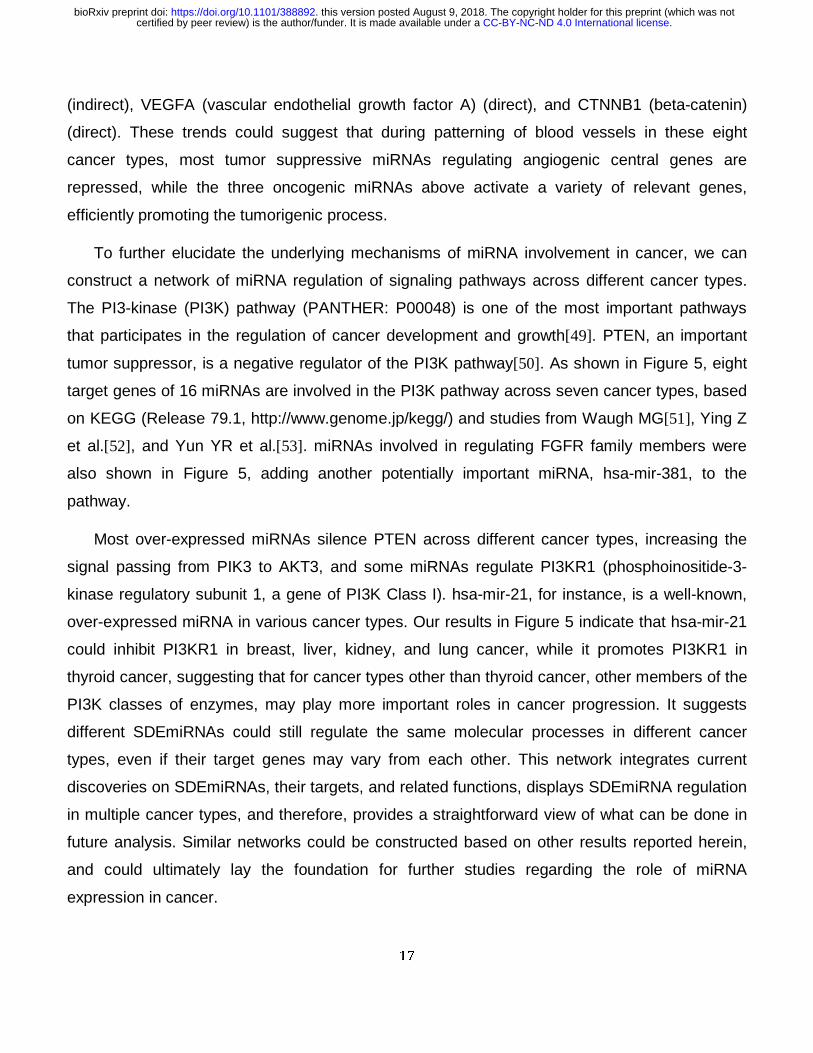
To further elucidate the underlying mechanisms of miRNA involvement in cancer, we can
construct a network of miRNA regulation of signaling pathways across different cancer types.
The PI3-kinase (PI3K) pathway (PANTHER: P00048) is one of the most important pathways
that participates in the regulation of cancer development and growth[49]. PTEN, an important
tumor suppressor, is a negative regulator of the PI3K pathway[50]. As shown in Figure 5, eight
target genes of 16 miRNAs are involved in the PI3K pathway across seven cancer types, based
on KEGG (Release 79.1, http://www.genome.jp/kegg/) and studies from Waugh MG[51], Ying Z
et al.[52], and Yun YR et al.[53]. miRNAs involved in regulating FGFR family members were
also shown in Figure 5, adding another potentially important miRNA, hsa-mir-381, to the
pathway.
Most over-expressed miRNAs silence PTEN across different cancer types, increasing the
signal passing from PIK3 to AKT3, and some miRNAs regulate PI3KR1 (phosphoinositide-3-
kinase regulatory subunit 1, a gene of PI3K Class I). hsa-mir-21, for instance, is a well-known,
over-expressed miRNA in various cancer types. Our results in Figure 5 indicate that hsa-mir-21
could inhibit PI3KR1 in breast, liver, kidney, and lung cancer, while it promotes PI3KR1 in
thyroid cancer, suggesting that for cancer types other than thyroid cancer, other members of the
PI3K classes of enzymes, may play more important roles in cancer progression. It suggests
different SDEmiRNAs could still regulate the same molecular processes in different cancer
types, even if their target genes may vary from each other. This network integrates current
discoveries on SDEmiRNAs, their targets, and related functions, displays SDEmiRNA regulation
in multiple cancer types, and therefore, provides a straightforward view of what can be done in
future analysis. Similar networks could be constructed based on other results reported herein,
and could ultimately lay the foundation for further studies regarding the role of miRNA
expression in cancer.
.CC-BY-NC-ND 4.0 International licensecertified by peer review) is the author/funder. It is made available under aThe copyright holder for this preprint (which was notthis version posted August 9, 2018. . https://doi.org/10.1101/388892doi: bioRxiv preprint
18
Materials and Methods
Data
miRNA sequencing data
This study started with miRNA sequencing data integration and filtering, and performed
expression analysis and downstream functional analysis (Figure 6). All miRNAseq data (both
counts of raw reads and Reads Per Million miRNA mapped (RPM) values, annotated to miRNA
names level) were downloaded from TCGA (Release-2016-03) (http://cancergenome.nih.gov)
by using TCGA-Assembler[54] with default parameters (Figure 6). Data was then divided into
two groups: paired data—data with both tumor and adjacent non-tumorous samples from the
same patient, and tumor-only expression data—data coming exclusively from all tumor samples
in all TCGA cancer types with miRNAseq data. Paired data were limited to those generated on
the HiSeq platform which generated enough matched samples than other platforms, whereas
tumor-only expression data were focused on data generated either on HiSeq or Genome
Analyzer (GA) of Illumina[55] systems, analyzed in separate groups for data from each platform.
Two additional projects (MALY, Malignant Lymphoma done by Germany, and OV, Ovarian
Cancer done by Australia, Release 20), available only through the International Cancer Genome
Consortium (ICGC, https://icgc.org/) and generated by Illumina-HiSeq system, were imported
into the tumor-only expression dataset. Fold change value (FC) was calculated by (RPM)tumor /
(RPM)non-tumor for one miRNA in one cancer type. Each miRNA was mapped to its corresponding
HGNC ID (HUGO Gene Nomenclature Committee, Issued in Autumn/Winter 2015:
http://www.genenames.org), miRBase ID (Release 21)[1], and Ensembl ID (Release 82)[56]
using customized python (v2.7) and R (3.3) scripts.
Databases for Validation of miRNA
To validate the selected differentially expressed miRNAs, we used four experimentally validated
databases generated from literature mining: HMDD (v2.0)[57], Mir2disease (2008)[58],
miRCancer (March2016)[59], and miRiaD (2016)[60]. We also applied our literature mining tool,
DEXTER, to extract miRNA-cancer correlations from publications[61]. The basic search term is
.CC-BY-NC-ND 4.0 International licensecertified by peer review) is the author/funder. It is made available under aThe copyright holder for this preprint (which was notthis version posted August 9, 2018. . https://doi.org/10.1101/388892doi: bioRxiv preprint
19
“[miRNA name] is [over-expressed / under-expressed / significantly higher / lower] in [cancer
tissue/sample] compared to [normal / non-tumorous sample]”. This tool is currently being
benchmarked, and the results were manually curated before reported as validate records. The
cancer terms for each reported interaction between miRNA and specific cancer types were then
mapped to Disease Ontology[62] cancer slim in order to unify the integrated dataset[63]. We
also incorporated information regarding miRNA expression changes in cancer types (up/down-
regulation) from these databases, when available.
miRNA Targets
Five experimentally validated miRNA target databases generated from literature mining were
used in our study: miRTarbase (Release 6.0)[64], miRecords (Apr 2013)[65], miRWalk 2.0
(miRNA-target interaction databases)[66], miRTex (May 2015)[67], and Tarbase-DIANA lab
v6.0[68]. These databases were generated by different algorithms but with a common motive,
which is to extract miRNA-target-cancer/tissue interactions from published papers. Most of
these published papers were validating the interactions or stating that the interactions were
commonly accepted by previous studies. CancerMiner (1.0), a database for tissue-specific
miRNA target predictions based on TCGA data[69], was also included in the integrated miRNA
target database for this study. This database was built upon multivariate linear model for
expression association of miRNA and mRNA from TCGA, and rank-based association
recurrence score that showed the possible association of miRNA-target interactions and cancer
types[69].
GO (Gene Ontology) IDs[70] and PANTHER pathway IDs and terms were downloaded from
UniProt (http://www.uniprot.org/, Release 2016_08) and PANTHER ((Protein ANalysis THrough
Evolutionary Relationships, Release 11.0: http://www.pantherdb.org/)[71] for each target in the
resulting database.
Data Normalization and Differential Expression Analysis
.CC-BY-NC-ND 4.0 International licensecertified by peer review) is the author/funder. It is made available under aThe copyright holder for this preprint (which was notthis version posted August 9, 2018. . https://doi.org/10.1101/388892doi: bioRxiv preprint
20
We used DESeq2 version 1.8.1[26] and edgeR version 3.10.2[24] (R packages) to normalize
paired miRNA sequencing data and to analyze the differential expression of miRNAs in each
cancer type. These tools are both based on a negative binomial distribution assumption[27, 72],
and both tools have been proven to generate normalized data of high quality and to perform
similarly in differential expression analyses, as well as in fold change estimates[73,
74]_ENREF_19_ENREF_23. DESeq2 fits a generalized linear model for each miRNA with the fold
change estimate shrunken by empirical Bayes[26]. A Wald test was used in DESeq2 for
statistical analysis and calculating p values for the significance of differentially expressed
miRNAs in each cancer type. edgeR, however, uses the TMM method for normalization of
sequencing data. To estimate parameters for the negative binomial model, we applied common
dispersion, trended dispersion, and tagwise dispersion for our multi-factor cases. These
dispersions were shrunken by an empirical Bayes procedure towards a consensus value[24],
and edgeR applied Fisher's Exact Test to determine differential expression[24]. False discovery
rate (FDR) was reported by edgeR for the significance of multiple comparisons conducted. The
input data for the two tools were raw read counts (without normalization), excluding any miRNAs
with no expression among all patients. For both tools, we designed the model matrix with two
categories, one with levels “cancer” and “non-tumor”, the other with TCGA patient IDs. The
cutoff for the significant adjusted p-value of each gene is 0.05/n (using Bonferroni’s Approach,
where n is the total number of expressed miRNAs in each cancer type), considering the
difference of cancers. miRNAs with significant adjusted p-values generated by both DESeq2
and edgeR were considered to be differentially expressed in a given cancer type. Results from
differential expression analysis for paired and tumor-only expression data were integrated into
BioXpress version 3.0[75]. The Heatmap with clustering for significantly differentially expressed
miRNAs across different cancer types was generated by R package gplots 3.0.1.
Survival Analysis
miRpower[36] was used to suggest the influence of miRNAs on patient survival rate in breast
cancer. This platform is continuously updated with TCGA datasets, and has been cited by
dozens of studies. By providing “auto select best cutoff”, miRpower is able to optimize the cutoff
.CC-BY-NC-ND 4.0 International licensecertified by peer review) is the author/funder. It is made available under aThe copyright holder for this preprint (which was notthis version posted August 9, 2018. . https://doi.org/10.1101/388892doi: bioRxiv preprint
21
of the patient groups for different investigated miRNAs. In our study here, TCGA was selected
as the only dataset to be considered, and therefore, miRNAs from breast cancer (BRCA) were
under survival analysis (1,061 TCGA patients in total).
miRNA Target Data Integration and Enrichment
After extracting all targets for selected miRNAs (those significantly over- or under-expressed in
at least eight cancer types), we separated these targets into two categories: tissue-specific
targets (records clearly including the miRNA-target connection in a specific tissue or cancer
type), and non-tissue-specific targets (records containing only a miRNA-target connection).
Tarbase provided the majority of records for the tissue-specific target group. We excluded
associations derived from microarray studies and integrated those records with cancer
metastasis annotation. Reported regulatory changes (up/down) of miRNAs were also retrieved.
Selective records from miRWalk 2.0 were included in the tissue-specific target group if the
records were involved in miRNAs-organ interactions or miRNA-gene-OMIM interactions
generated by miRWalk text-mining search on PubMed database. To include more tissue-
specific targets, we also integrated data from a predicted database, CancerMiner, which applied
mRNA and miRNA expression, DNA copy number, and promoter methylation data from TCGA
to predict miRNA-mRNA-cancer type associations[69]. CancerMiner data were included only for
records not found in Tarbase or miRWalk. Expression changes of all tissue-specific target genes
in corresponding cancer types were extracted from BioXpress v3.0[75]. For the non-tissue-
specific target group, we first excluded those targets that were also in the tissue-specific target
section. Some records from Tarbase that do not contain tissue or cancer information were
included. Gene names of the records from miRTex were first manually curated and mapped to
synonyms available in UniProtKB, and records with miRNA-gene associations were selected
while miRNA-miRNA interactions were excluded. miRNA-gene targets from miRWalk 2.0 and
records from miRTarbase and miRecords were all classified into the non-tissue-specific target
group. All cancer types from those databases were mapped to DOIDs.
Tissue-specific target genes were mapped to UniProtKB accession numbers[76],
maintaining only those genes that have been manually annotated and reviewed in
.CC-BY-NC-ND 4.0 International licensecertified by peer review) is the author/funder. It is made available under aThe copyright holder for this preprint (which was notthis version posted August 9, 2018. . https://doi.org/10.1101/388892doi: bioRxiv preprint
22
UniProtKB/Swiss-Prot. For each GO or PANTHER term, we merged all the target genes
involved and their regulating miRNAs. In order to reduce false positive errors and to exclude
highly generic terms of GO, GO terms of genes associated more than eight cancer types and
fewer than 50 target genes were selected. We also collected non-tissue-specific targets that are
considered to be cancer census genes from COSMIC (version 78)[77] and biomarkers from
EDRN (Downloaded in Feb, 2016, https://edrn.nci.nih.gov/). Enrichment analysis of these non-
tissue-specific targets was done using STRING (version 10.0)[78] and Ingenuity Pathway
Analysis (IPA) (March 2017).
Conclusion
Different cancer types have unique expression patterns of significantly differentially expressed
miRNAs (SDEmiRNAs). Our study here sought to identify prevalent expression-based or
mechanistic features of SDEmiRNAs associated with cancer development, even though the
analysis here did not separate cancer samples based on clinical information, such as tumor
stage or patient metadata. Applying our miRNA analysis findings, we propose to narrow down
the number of miRNAs needed to classify the 14 cancer types, as well as cell type cancers from
the same location. Moreover, based on the inhibition role of miRNA, development of different
cancer types may rely on either more tumor suppressors or more oncogenes, resulting in three
categories of cancers. We also identified 81 unique SDEmiRNAs with significantly differential
expression in only one out of the 14 cancer types: these SDEmiRNAs might have unique
functions in each cancer, even including effect on patient survival rates, although most of them
have not been studied in their corresponding cancer type and their expression levels are lower
than well-studied ones.
We then identified a set of 21 key SDEmiRNAs (including four relatively newly identified
ones, hsa-mir-4746, hsa-mir-3648, hsa-mir-3687, and hsa-mir-1269a) that have high enrichment
in patients of at least eight cancer types, as well as hsa-mir-4652 with high enrichment in seven
cancer types. These key SDEmiRNAs may have much wider and more similar regulatory
mechanisms across multiple cancer types than what researchers have found in one or two
.CC-BY-NC-ND 4.0 International licensecertified by peer review) is the author/funder. It is made available under aThe copyright holder for this preprint (which was notthis version posted August 9, 2018. . https://doi.org/10.1101/388892doi: bioRxiv preprint
23
cancers so far. Our study also shows the necessary to study further on those newly identified
ones, which might be equally important with others. To explore the possible molecular
mechanisms of these key SDEmiRNAs, enrichment analysis of the SDEmiRNAs and their
targets in different cancer types was applied, which suggested that different miRNAs participate
in a limited amount of cancer-associated molecular processes/pathways through regulating
different targets. This means they may have similar mechanisms in development of these
cancer types.
The construction of regulatory network of SDEmiRNAs with their experimentally validated
targets combines results of current studies and shows the potential research directions for
further analysis. Our study here provides a rationale for the continued exploration and validation
of the functional roles of key SDEmiRNAs in cancers, and therefore, pushes all the results on
BioXpress to allow further analysis.
.CC-BY-NC-ND 4.0 International licensecertified by peer review) is the author/funder. It is made available under aThe copyright holder for this preprint (which was notthis version posted August 9, 2018. . https://doi.org/10.1101/388892doi: bioRxiv preprint
24
Authors’ Contributions
R.M. supervised the study. Y.H. and R.M. contributed to conception and design. Y.H., Q.W. and
C.Y. were involved in methodology and acquisition of data. Y.H. analyzed and interpreted data.
Y.H., R.K., S.G. and V.S. participated in constructing database or data support. Y.H., H.D., S.G.,
R.K., V.S., Q.W., C.Y. and R.M. wrote and reviewed the manuscript.
Competing Interest
None.
Acknowledgments
The authors thank Yu Fan and Ting-Chia Chang for helpful discussion. Yu Hu earned an MS in
the Bioinformatics Track of the Bioinformatics and Molecular Biochemistry Program at the
George Washington University. This work is from a dissertation to be presented to the above
program in partial fulfillment of the requirements for the Master of Science degree.
Funding
This project was partially funded by NCI/EDRN Associate Member contract #156620 and The
McCormick Genomic and Proteomic Center (MGPC), the George Washington University.
Additional Information
Results of miRNA differential expression analysis are available in:
https://hive.biochemistry.gwu.edu/bioxpress
.CC-BY-NC-ND 4.0 International licensecertified by peer review) is the author/funder. It is made available under aThe copyright holder for this preprint (which was notthis version posted August 9, 2018. . https://doi.org/10.1101/388892doi: bioRxiv preprint
25
References
[1] Kozomara A, Griffiths-Jones S. miRBase: annotating high confidence microRNAs using deep sequencing data. Nucleic Acids Res 2014;42:D68-73. [2] Zempleni J, Baier SR, Howard KM, Cui J. Gene regulation by dietary microRNAs. Can J Physiol Pharmacol 2015;93:1097-102. [3] Calin GA, Dumitru CD, Shimizu M, Bichi R, Zupo S, Noch E, et al. Frequent deletions and down-regulation of micro- RNA genes miR15 and miR16 at 13q14 in chronic lymphocytic leukemia. Proc Natl Acad Sci U S A 2002;99:15524-9. [4] Takamizawa J, Konishi H, Yanagisawa K, Tomida S, Osada H, Endoh H, et al. Reduced expression of the let-7 microRNAs in human lung cancers in association with shortened postoperative survival. Cancer Res 2004;64:3753-6. [5] Chan JA, Krichevsky AM, Kosik KS. MicroRNA-21 is an antiapoptotic factor in human glioblastoma cells. Cancer Res 2005;65:6029-33. [6] Ma L, Teruya-Feldstein J, Weinberg RA. Tumour invasion and metastasis initiated by microRNA-10b in breast cancer. Nature 2007;449:682-8. [7] Gonzalez-Quintana V, Palma-Berre L, Campos-Parra AD, Lopez-Urrutia E, Peralta-Zaragoza O, Vazquez-Romo R, et al. MicroRNAs are involved in cervical cancer development, progression, clinical outcome and improvement treatment response (Review). Oncol Rep 2016;35:3-12. [8] Tutar L, Tutar E, Ozgur A, Tutar Y. Therapeutic Targeting of microRNAs in Cancer: Future Perspectives. Drug Dev Res 2015;76:382-8. [9] Wang W, Luo YP. MicroRNAs in breast cancer: oncogene and tumor suppressors with clinical potential. J Zhejiang Univ Sci B 2015;16:18-31. [10] Hayes J, Peruzzi PP, Lawler S. MicroRNAs in cancer: biomarkers, functions and therapy. Trends Mol Med 2014;20:460-9. [11] Thomas J, Ohtsuka M, Pichler M, Ling H. MicroRNAs: Clinical Relevance in Colorectal Cancer. Int J Mol Sci 2015;16:28063-76. [12] Khan MA, Zubair H, Srivastava SK, Singh S, Singh AP. Insights into the Role of microRNAs in Pancreatic Cancer Pathogenesis: Potential for Diagnosis, Prognosis, and Therapy. Adv Exp Med Biol 2015;889:71-87. [13] Lu J, Getz G, Miska EA, Alvarez-Saavedra E, Lamb J, Peck D, et al. MicroRNA expression profiles classify human cancers. Nature 2005;435:834-8. [14] Ren Z, Wang W, Li J. Identifying molecular subtypes in human colon cancer using gene expression and DNA methylation microarray data. Int J Oncol 2016;48:690-702. [15] Sisti JS, Collins LC, Beck AH, Tamimi RM, Rosner BA, Eliassen AH. Reproductive risk factors in relation to molecular subtypes of breast cancer: Results from the nurses' health studies. Int J Cancer 2016;138:2346-56. [16] Gasparini P, Cascione L, Landi L, Carasi S, Lovat F, Tibaldi C, et al. microRNA classifiers are powerful diagnostic/prognostic tools in ALK-, EGFR-, and KRAS-driven lung cancers. Proc Natl Acad Sci U S A 2015;112:14924-9. [17] Monroig-Bosque Pdel C, Rivera CA, Calin GA. MicroRNAs in cancer therapeutics: "from the bench to the bedside". Expert Opin Biol Ther 2015;15:1381-5. [18] Sethi S, Ali S, Sarkar FH. MicroRNAs in personalized cancer therapy. Clin Genet 2014;86:68-73. [19] Peng F, Zhang Y, Wang R, Zhou W, Zhao Z, Liang H, et al. Identification of differentially expressed miRNAs in individual breast cancer patient and application in personalized medicine. Oncogenesis 2016;5:e194.
.CC-BY-NC-ND 4.0 International licensecertified by peer review) is the author/funder. It is made available under aThe copyright holder for this preprint (which was notthis version posted August 9, 2018. . https://doi.org/10.1101/388892doi: bioRxiv preprint
26
[20] Metpally RP, Nasser S, Malenica I, Courtright A, Carlson E, Ghaffari L, et al. Comparison of Analysis Tools for miRNA High Throughput Sequencing Using Nerve Crush as a Model. Front Genet 2013;4:20. [21] Humphreys DT, Suter CM. miRspring: a compact standalone research tool for analyzing miRNA-seq data. Nucleic Acids Res 2013;41:e147. [22] Lopes-Ramos CM, Habr-Gama A, Quevedo Bde S, Felicio NM, Bettoni F, Koyama FC, et al. Overexpression of miR-21-5p as a predictive marker for complete tumor regression to neoadjuvant chemoradiotherapy in rectal cancer patients. BMC Med Genomics 2014;7:68. [23] Cobos Jimenez V, Willemsen AM, Bradley EJ, Baas F, van Kampen AH, Kootstra NA. Next-generation sequencing of microRNAs in primary human polarized macrophages. Genom Data 2014;2:181-3. [24] Robinson MD, McCarthy DJ, Smyth GK. edgeR: a Bioconductor package for differential expression analysis of digital gene expression data. Bioinformatics 2010;26:139-40. [25] Anders S, Huber W. Differential expression analysis for sequence count data. Genome Biol 2010;11:R106. [26] Love MI, Huber W, Anders S. Moderated estimation of fold change and dispersion for RNA-seq data with DESeq2. Genome Biol 2014;15:550. [27] Yang S, Guo L, Shao F, Zhao Y, Chen F. A Systematic Evaluation of Feature Selection and Classification Algorithms Using Simulated and Real miRNA Sequencing Data. Comput Math Methods Med 2015;2015:178572. [28] Funamizu N, Lacy CR, Parpart ST, Takai A, Hiyoshi Y, Yanaga K. MicroRNA-301b promotes cell invasiveness through targeting TP63 in pancreatic carcinoma cells. Int J Oncol 2014;44:725-34. [29] Chang YY, Kuo WH, Hung JH, Lee CY, Lee YH, Chang YC, et al. Deregulated microRNAs in triple-negative breast cancer revealed by deep sequencing. Mol Cancer 2015;14:36. [30] Tang D, Zhang Q, Zhao S, Wang J, Lu K, Song Y, et al. The expression and clinical significance of microRNA-1258 and heparanase in human breast cancer. Clin Biochem 2013;46:926-32. [31] Liu H, Chen X, Gao W, Jiang G. The expression of heparanase and microRNA-1258 in human non-small cell lung cancer. Tumour Biol 2012;33:1327-34. [32] Bu P, Wang L, Chen KY, Rakhilin N, Sun J, Closa A, et al. miR-1269 promotes metastasis and forms a positive feedback loop with TGF-beta. Nat Commun 2015;6:6879. [33] Gonul O, Aydin HH, Kalmis E, Kayalar H, Ozkaya AB, Atay S, et al. Effects of Ganoderma lucidum (Higher Basidiomycetes) Extracts on the miRNA Profile and Telomerase Activity of the MCF-7 Breast Cancer Cell Line. Int J Med Mushrooms 2015;17:231-9. [34] Emmadi R, Canestrari E, Arbieva ZH, Mu W, Dai Y, Frasor J, et al. Correlative Analysis of miRNA Expression and Oncotype Dx Recurrence Score in Estrogen Receptor Positive Breast Carcinomas. PLoS One 2015;10:e0145346. [35] Persson H, Kvist A, Rego N, Staaf J, Vallon-Christersson J, Luts L, et al. Identification of new microRNAs in paired normal and tumor breast tissue suggests a dual role for the ERBB2/Her2 gene. Cancer Res 2011;71:78-86. [36] Lanczky A, Nagy A, Bottai G, Munkacsy G, Szabo A, Santarpia L, et al. miRpower: a web-tool to validate survival-associated miRNAs utilizing expression data from 2178 breast cancer patients. Breast Cancer Res Treat 2016;160:439-46. [37] Fromm B, Billipp T, Peck LE, Johansen M, Tarver JE, King BL, et al. A Uniform System for the Annotation of Vertebrate microRNA Genes and the Evolution of the Human microRNAome. Annu Rev Genet 2015;49:213-42.
.CC-BY-NC-ND 4.0 International licensecertified by peer review) is the author/funder. It is made available under aThe copyright holder for this preprint (which was notthis version posted August 9, 2018. . https://doi.org/10.1101/388892doi: bioRxiv preprint
27
[38] Min P, Li W, Zeng D, Ma Y, Xu D, Zheng W, et al. A single nucleotide variant in microRNA-1269a promotes the occurrence and process of hepatocellular carcinoma by targeting to oncogenes SPATS2L and LRP6. Bull Cancer 2017;104:311-20. [39] Ma Y, Liang AJ, Fan YP, Huang YR, Zhao XM, Sun Y, et al. Dysregulation and functional roles of miR-183-96-182 cluster in cancer cell proliferation, invasion and metastasis. Oncotarget 2016. [40] Futreal PA, Coin L, Marshall M, Down T, Hubbard T, Wooster R, et al. A census of human cancer genes. Nat Rev Cancer 2004;4:177-83. [41] Gosline SJ, Gurtan AM, JnBaptiste CK, Bosson A, Milani P, Dalin S, et al. Elucidating MicroRNA Regulatory Networks Using Transcriptional, Post-transcriptional, and Histone Modification Measurements. Cell Rep 2016;14:310-9. [42] Woods NK, Padmanabhan J. Inhibition of amyloid precursor protein processing enhances gemcitabine-mediated cytotoxicity in pancreatic cancer cells. J Biol Chem 2013;288:30114-24. [43] Long J, Ou C, Xia H, Zhu Y, Liu D. MiR-503 inhibited cell proliferation of human breast cancer cells by suppressing CCND1 expression. Tumour Biol 2015;36:8697-702. [44] Reis-Filho JS, Savage K, Lambros MB, James M, Steele D, Jones RL, et al. Cyclin D1 protein overexpression and CCND1 amplification in breast carcinomas: an immunohistochemical and chromogenic in situ hybridisation analysis. Mod Pathol 2006;19:999-1009. [45] Werner H, Sarfstein R. Transcriptional and epigenetic control of IGF1R gene expression: implications in metabolism and cancer. Growth Horm IGF Res 2014;24:112-8. [46] Ogasawara S, Kiyota Y, Chuman Y, Kowata A, Yoshimura F, Tanino K, et al. Novel inhibitors targeting PPM1D phosphatase potently suppress cancer cell proliferation. Bioorg Med Chem 2015;23:6246-9. [47] Nagy JA, Chang SH, Dvorak AM, Dvorak HF. Why are tumour blood vessels abnormal and why is it important to know? Br J Cancer 2009;100:865-9. [48] Xu JB, Bao Y, Liu X, Liu Y, Huang S, Wang JC. Defective expression of transforming growth factor beta type II receptor (TGFBR2) in the large cell variant of non-small cell lung carcinoma. Lung Cancer 2007;58:36-43. [49] Baselga J. Targeting the phosphoinositide-3 (PI3) kinase pathway in breast cancer. Oncologist 2011;16 Suppl 1:12-9. [50] Chalhoub N, Baker SJ. PTEN and the PI3-kinase pathway in cancer. Annu Rev Pathol 2009;4:127-50. [51] Waugh MG. Chromosomal Instability and Phosphoinositide Pathway Gene Signatures in Glioblastoma Multiforme. Mol Neurobiol 2016;53:621-30. [52] Ying Z, Xie X, Chen M, Yi K, Rajagopalan S. Alpha-lipoic acid activates eNOS through activation of PI3-kinase/Akt signaling pathway. Vascul Pharmacol 2015;64:28-35. [53] Yun YR, Won JE, Jeon E, Lee S, Kang W, Jo H, et al. Fibroblast growth factors: biology, function, and application for tissue regeneration. J Tissue Eng 2010;2010:218142. [54] Zhu Y, Qiu P, Ji Y. TCGA-assembler: open-source software for retrieving and processing TCGA data. Nat Methods 2014;11:599-600. [55] Minoche AE, Dohm JC, Himmelbauer H. Evaluation of genomic high-throughput sequencing data generated on Illumina HiSeq and genome analyzer systems. Genome Biol 2011;12:R112. [56] Yates A, Akanni W, Amode MR, Barrell D, Billis K, Carvalho-Silva D, et al. Ensembl 2016. Nucleic Acids Res 2016;44:D710-6. [57] Li Y, Qiu C, Tu J, Geng B, Yang J, Jiang T, et al. HMDD v2.0: a database for experimentally supported human microRNA and disease associations. Nucleic Acids Res 2014;42:D1070-4.
.CC-BY-NC-ND 4.0 International licensecertified by peer review) is the author/funder. It is made available under aThe copyright holder for this preprint (which was notthis version posted August 9, 2018. . https://doi.org/10.1101/388892doi: bioRxiv preprint
28
[58] Jiang Q, Wang Y, Hao Y, Juan L, Teng M, Zhang X, et al. miR2Disease: a manually curated database for microRNA deregulation in human disease. Nucleic Acids Res 2009;37:D98-104. [59] Xie B, Ding Q, Han H, Wu D. miRCancer: a microRNA-cancer association database constructed by text mining on literature. Bioinformatics 2013;29:638-44. [60] Hinske LC, Franca GS, Torres HA, Ohara DT, Lopes-Ramos CM, Heyn J, et al. miRIAD-integrating microRNA inter- and intragenic data. Database (Oxford) 2014;2014. [61] Gupta S, Dingerdissen H, Ross KE, Hu Y, Wu CH, Mazumder R, et al. DEXTER: Disease-Expression Relation Extraction from Text. Database (Oxford) 2018;2018. [62] Kibbe WA, Arze C, Felix V, Mitraka E, Bolton E, Fu G, et al. Disease Ontology 2015 update: an expanded and updated database of human diseases for linking biomedical knowledge through disease data. Nucleic Acids Res 2015;43:D1071-8. [63] Wu TJ, Schriml LM, Chen QR, Colbert M, Crichton DJ, Finney R, et al. Generating a focused view of disease ontology cancer terms for pan-cancer data integration and analysis. Database (Oxford) 2015;2015:bav032. [64] Chou CH, Chang NW, Shrestha S, Hsu SD, Lin YL, Lee WH, et al. miRTarBase 2016: updates to the experimentally validated miRNA-target interactions database. Nucleic Acids Res 2016;44:D239-47. [65] Xiao F, Zuo Z, Cai G, Kang S, Gao X, Li T. miRecords: an integrated resource for microRNA-target interactions. Nucleic Acids Res 2009;37:D105-10. [66] Dweep H, Gretz N. miRWalk2.0: a comprehensive atlas of microRNA-target interactions. Nat Methods 2015;12:697. [67] Li G, Ross KE, Arighi CN, Peng Y, Wu CH, Vijay-Shanker K. miRTex: A Text Mining System for miRNA-Gene Relation Extraction. PLoS Comput Biol 2015;11:e1004391. [68] Vergoulis T, Vlachos IS, Alexiou P, Georgakilas G, Maragkakis M, Reczko M, et al. TarBase 6.0: capturing the exponential growth of miRNA targets with experimental support. Nucleic Acids Res 2012;40:D222-9. [69] Jacobsen A, Silber J, Harinath G, Huse JT, Schultz N, Sander C. Analysis of microRNA-target interactions across diverse cancer types. Nat Struct Mol Biol 2013;20:1325-32. [70] Consortium GO. Gene Ontology Consortium: going forward. Nucleic Acids Res 2015;43:D1049-56. [71] Mi H, Poudel S, Muruganujan A, Casagrande JT, Thomas PD. PANTHER version 10: expanded protein families and functions, and analysis tools. Nucleic Acids Res 2016;44:D336-42. [72] Varet H, Brillet-Gueguen L, Coppee JY, Dillies MA. SARTools: A DESeq2- and EdgeR-Based R Pipeline for Comprehensive Differential Analysis of RNA-Seq Data. PLoS One 2016;11:e0157022. [73] Dillies MA, Rau A, Aubert J, Hennequet-Antier C, Jeanmougin M, Servant N, et al. A comprehensive evaluation of normalization methods for Illumina high-throughput RNA sequencing data analysis. Brief Bioinform 2013;14:671-83. [74] Tam S, Tsao MS, McPherson JD. Optimization of miRNA-seq data preprocessing. Brief Bioinform 2015;16:950-63. [75] Dingerdissen HM, Torcivia-Rodriguez J, Hu Y, Chang TC, Mazumder R, Kahsay R. BioMuta and BioXpress: mutation and expression knowledgebases for cancer biomarker discovery. Nucleic Acids Res 2018;46:D1128-D36. [76] Consortium U. UniProt: a hub for protein information. Nucleic Acids Res 2015;43:D204-12. [77] Forbes SA, Bindal N, Bamford S, Cole C, Kok CY, Beare D, et al. COSMIC: mining complete cancer genomes in the Catalogue of Somatic Mutations in Cancer. Nucleic Acids Res 2011;39:D945-50. [78] Szklarczyk D, Franceschini A, Wyder S, Forslund K, Heller D, Huerta-Cepas J, et al. STRING v10: protein-protein interaction networks, integrated over the tree of life. Nucleic Acids Res 2015;43:D447-52.
.CC-BY-NC-ND 4.0 International licensecertified by peer review) is the author/funder. It is made available under aThe copyright holder for this preprint (which was notthis version posted August 9, 2018. . https://doi.org/10.1101/388892doi: bioRxiv preprint
29
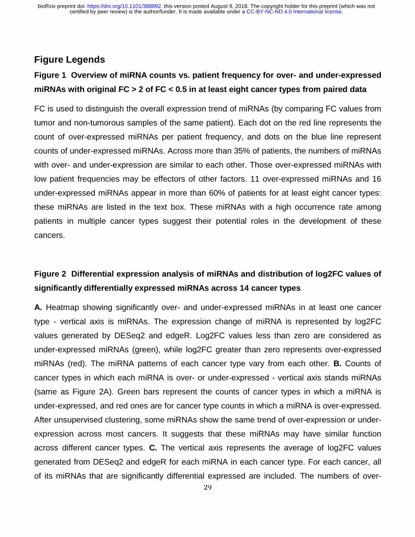
Figure Legends
Figure 1 Overview of miRNA counts vs. patient frequency for over- and under-expressed
miRNAs with original FC > 2 of FC < 0.5 in at least eight cancer types from paired data
FC is used to distinguish the overall expression trend of miRNAs (by comparing FC values from
tumor and non-tumorous samples of the same patient). Each dot on the red line represents the
count of over-expressed miRNAs per patient frequency, and dots on the blue line represent
counts of under-expressed miRNAs. Across more than 35% of patients, the numbers of miRNAs
with over- and under-expression are similar to each other. Those over-expressed miRNAs with
low patient frequencies may be effectors of other factors. 11 over-expressed miRNAs and 16
under-expressed miRNAs appear in more than 60% of patients for at least eight cancer types:
these miRNAs are listed in the text box. These miRNAs with a high occurrence rate among
patients in multiple cancer types suggest their potential roles in the development of these
cancers.
Figure 2 Differential expression analysis of miRNAs and distribution of log2FC values of
significantly differentially expressed miRNAs across 14 cancer types
A. Heatmap showing significantly over- and under-expressed miRNAs in at least one cancer
type - vertical axis is miRNAs. The expression change of miRNA is represented by log2FC
values generated by DESeq2 and edgeR. Log2FC values less than zero are considered as
under-expressed miRNAs (green), while log2FC greater than zero represents over-expressed
miRNAs (red). The miRNA patterns of each cancer type vary from each other. B. Counts of
cancer types in which each miRNA is over- or under-expressed - vertical axis stands miRNAs
(same as Figure 2A). Green bars represent the counts of cancer types in which a miRNA is
under-expressed, and red ones are for cancer type counts in which a miRNA is over-expressed.
After unsupervised clustering, some miRNAs show the same trend of over-expression or under-
expression across most cancers. It suggests that these miRNAs may have similar function
across different cancer types. C. The vertical axis represents the average of log2FC values
generated from DESeq2 and edgeR for each miRNA in each cancer type. For each cancer, all
of its miRNAs that are significantly differential expressed are included. The numbers of over-
.CC-BY-NC-ND 4.0 International licensecertified by peer review) is the author/funder. It is made available under aThe copyright holder for this preprint (which was notthis version posted August 9, 2018. . https://doi.org/10.1101/388892doi: bioRxiv preprint
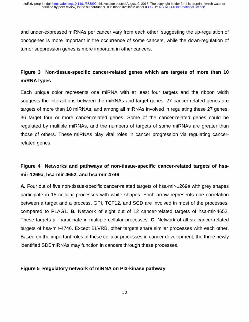
30
and under-expressed miRNAs per cancer vary from each other, suggesting the up-regulation of
oncogenes is more important in the occurrence of some cancers, while the down-regulation of
tumor suppression genes is more important in other cancers.
Figure 3 Non-tissue-specific cancer-related genes which are targets of more than 10
miRNA types
Each unique color represents one miRNA with at least four targets and the ribbon width
suggests the interactions between the miRNAs and target genes. 27 cancer-related genes are
targets of more than 10 miRNAs, and among all miRNAs involved in regulating these 27 genes,
36 target four or more cancer-related genes. Some of the cancer-related genes could be
regulated by multiple miRNAs, and the numbers of targets of some miRNAs are greater than
those of others. These miRNAs play vital roles in cancer progression via regulating cancer-
related genes.
Figure 4 Networks and pathways of non-tissue-specific cancer-related targets of hsa-
mir-1269a, hsa-mir-4652, and hsa-mir-4746
A. Four out of five non-tissue-specific cancer-related targets of hsa-mir-1269a with grey shapes
participate in 15 cellular processes with white shapes. Each arrow represents one correlation
between a target and a process. GPI, TCF12, and SCD are involved in most of the processes,
compared to PLAG1. B. Network of eight out of 12 cancer-related targets of hsa-mir-4652.
These targets all participate in multiple cellular processes. C. Network of all six cancer-related
targets of hsa-mir-4746. Except BLVRB, other targets share similar processes with each other.
Based on the important roles of these cellular processes in cancer development, the three newly
identified SDEmiRNAs may function in cancers through these processes.
Figure 5 Regulatory network of miRNA on PI3-kinase pathway
.CC-BY-NC-ND 4.0 International licensecertified by peer review) is the author/funder. It is made available under aThe copyright holder for this preprint (which was notthis version posted August 9, 2018. . https://doi.org/10.1101/388892doi: bioRxiv preprint
31
Blue arrows indicate the regulation among genes in the PI3-kinase pathway (arrows with vertical
short line segments represent inhibition role of up-stream genes). Red arrows indicate
regulation interaction between miRNAs and PI3-kinase pathway. Minus sign suggests inhibition,
and plus sign promotion. Eight target genes of 16 miRNAs are involved in the PI3K pathway and
regulation of PTEN gene across seven cancer types. Different miRNAs in different cancers
could regulate the same molecular processes through targeting their components. These
miRNAs still could function similarly across at least eight cancer types.
Figure 6 Cancer-related significantly differential expression of miRNA workflow
Paired miRNAs (tumorous and adjacent non-tumorous samples) were retrieved from TCGA
database and analyzed by DESeq2 and edgeR for differential expression. We then screened
out those significantly differentially expressed miRNAs (SDEmiRNAs). After validation by
literature mining databases and our text-mining tool, DEXTER, we proposed the possibility to
use these SDEmiRNAs in classification of cancer types or cancer subtypes, and screened out
unique SDEmiRNAs for one single cancer. We also kept those key SDEmiRNAs significantly
expressed in at least eight cancers with high patient frequencies. Moreover, we collected both
tissue-specific targets and non-tissue-specific targets from experimentally validated databases,
and did functional/enrichment analysis with those key SDEmiRNAs.
.CC-BY-NC-ND 4.0 International licensecertified by peer review) is the author/funder. It is made available under aThe copyright holder for this preprint (which was notthis version posted August 9, 2018. . https://doi.org/10.1101/388892doi: bioRxiv preprint
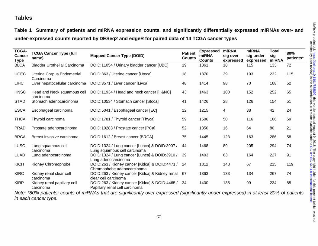
32
Tables
Table 1 Summary of patients and miRNA expression counts, and significantly differentially expressed miRNAs over- and
under-expressed counts reported by DESeq2 and edgeR for paired data of 14 TCGA cancer types
Note: *80% patients: counts of miRNAs that are significantly over-expressed (significantly under-expressed) in at least 80% of patients in each cancer type.
TCGA-Cancer Type
TCGA Cancer Type (full name) Mapped Cancer Type (DOID) Patient
Counts
Expressed miRNA Counts
miRNA sig over-expressed
miRNA sig under-expressed
Total sig miRNA
80% patients*
BLCA Bladder Urothelial Carcinoma DOID:11054 / Urinary bladder cancer [UBC] 19 1361 18 115 133 72
UCEC Uterine Corpus Endometrial Carcinoma
DOID:363 / Uterine cancer [Uteca] 18 1370 39 193 232 115
LIHC Liver hepatocellular carcinoma DOID:3571 / Liver cancer [Livca] 48 1414 98 70 168 52
HNSC Head and Neck squamous cell carcinoma
DOID:11934 / Head and neck cancer [H&NC] 43 1463 100 152 252 65
STAD Stomach adenocarcinoma DOID:10534 / Stomach cancer [Stoca] 41 1426 28 126 154 51
ESCA Esophageal carcinoma DOID:5041 / Esophageal cancer [EC] 12 1215 4 38 42 24
THCA Thyroid carcinoma DOID:1781 / Thyroid cancer [Thyca] 59 1506 50 116 166 59
PRAD Prostate adenocarcinoma DOID:10283 / Prostate cancer [PCa] 52 1350 16 64 80 21
BRCA Breast invasive carcinoma DOID:1612 / Breast cancer [BRCA] 75 1445 123 163 286 58
LUSC Lung squamous cell carcinoma
DOID:1324 / Lung cancer [Lunca] & DOID:3907 / Lung squamous cell carcinoma
44 1468 89 205 294 74
LUAD Lung adenocarcinoma DOID:1324 / Lung cancer [Lunca] & DOID:3910 / Lung adenocarcinoma
39 1403 63 164 227 91
KICH Kidney Chromophobe DOID:263 / Kidney cancer [Kidca] & DOID:4471 / Chromophobe adenocarcinoma
24 1312 148 67 215 119
KIRC Kidney renal clear cell carcinoma
DOID:263 / Kidney cancer [Kidca] & Kidney renal clear cell carcinoma
67 1363 133 134 267 74
KIRP Kidney renal papillary cell carcinoma
DOID:263 / Kidney cancer [Kidca] & DOID:4465 / Papillary renal cell carcinoma
34 1400 135 99 234 85
.C
C-B
Y-N
C-N
D 4.0 International license
certified by peer review) is the author/funder. It is m
ade available under aT
he copyright holder for this preprint (which w
as notthis version posted A
ugust 9, 2018. .
https://doi.org/10.1101/388892doi:
bioRxiv preprint
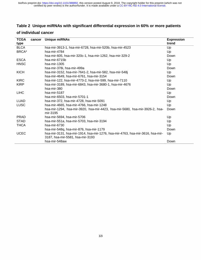
33
Table 2 Unique miRNAs with significant differential expression in 60% or more patients
of individual cancer
TCGA cancer type
Unique miRNAs Expression trend
BLCA hsa-mir-3913-1, hsa-mir-6728, hsa-mir-520b, hsa-mir-4523 Up BRCA* hsa-mir-4784 Up hsa-mir-605, hsa-mir-320c-1, hsa-mir-1262, hsa-mir-329-2 Down ESCA hsa-mir-6715b Up HNSC hsa-mir-1305 Up hsa-mir-378i, hsa-mir-499a Down KICH hsa-mir-3152, hsa-mir-7641-2, hsa-mir-582, hsa-mir-548j Up hsa-mir-4649, hsa-mir-6761, hsa-mir-3154 Down KIRC hsa-mir-122, hsa-mir-4773-2, hsa-mir-599, hsa-mir-7110 Up KIRP hsa-mir-3189, hsa-mir-6843, hsa-mir-3680-1, hsa-mir-4676 Up hsa-mir-380 Down LIHC hsa-mir-5187 Up hsa-mir-6503, hsa-mir-5701-1 Down LUAD hsa-mir-372, hsa-mir-4728, hsa-mir-5091 Up LUSC hsa-mir-4665, hsa-mir-4766, hsa-mir-1248 Up hsa-mir-1294, hsa-mir-3920, hsa-mir-4423, hsa-mir-5680, hsa-mir-3926-2, hsa-
mir-3195 Down
PRAD hsa-mir-5694, hsa-mir-5706 Up STAD hsa-mir-551a, hsa-mir-5703, hsa-mir-3194 Up THCA hsa-mir-6730 Up hsa-mir-548q, hsa-mir-876, hsa-mir-1179 Down UCEC hsa-mir-3131, hsa-mir-1914, hsa-mir-1276, hsa-mir-4763, hsa-mir-3616, hsa-mir-
3187, hsa-mir-5581, hsa-mir-3193 Up
hsa-mir-548aw Down
.CC-BY-NC-ND 4.0 International licensecertified by peer review) is the author/funder. It is made available under aThe copyright holder for this preprint (which was notthis version posted August 9, 2018. . https://doi.org/10.1101/388892doi: bioRxiv preprint
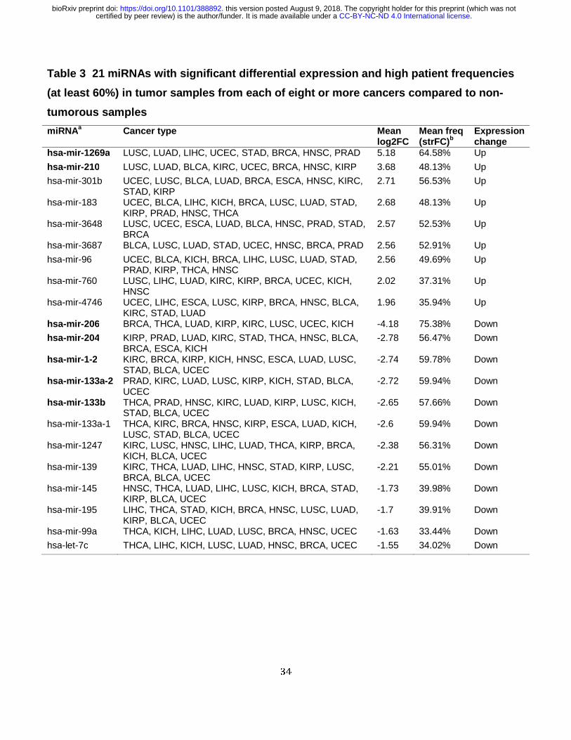
34
Table 3 21 miRNAs with significant differential expression and high patient frequencies
(at least 60%) in tumor samples from each of eight or more cancers compared to non-
tumorous samples
miRNAa Cancer type Mean log2FC
Mean freq (strFC)b
Expression change
hsa-mir-1269a LUSC, LUAD, LIHC, UCEC, STAD, BRCA, HNSC, PRAD 5.18 64.58% Up
hsa-mir-210 LUSC, LUAD, BLCA, KIRC, UCEC, BRCA, HNSC, KIRP 3.68 48.13% Up hsa-mir-301b UCEC, LUSC, BLCA, LUAD, BRCA, ESCA, HNSC, KIRC,
STAD, KIRP 2.71 56.53% Up
hsa-mir-183 UCEC, BLCA, LIHC, KICH, BRCA, LUSC, LUAD, STAD, KIRP, PRAD, HNSC, THCA
2.68 48.13% Up
hsa-mir-3648 LUSC, UCEC, ESCA, LUAD, BLCA, HNSC, PRAD, STAD, BRCA
2.57 52.53% Up
hsa-mir-3687 BLCA, LUSC, LUAD, STAD, UCEC, HNSC, BRCA, PRAD 2.56 52.91% Up
hsa-mir-96 UCEC, BLCA, KICH, BRCA, LIHC, LUSC, LUAD, STAD, PRAD, KIRP, THCA, HNSC
2.56 49.69% Up
hsa-mir-760 LUSC, LIHC, LUAD, KIRC, KIRP, BRCA, UCEC, KICH, HNSC
2.02 37.31% Up
hsa-mir-4746 UCEC, LIHC, ESCA, LUSC, KIRP, BRCA, HNSC, BLCA, KIRC, STAD, LUAD
1.96 35.94% Up
hsa-mir-206 BRCA, THCA, LUAD, KIRP, KIRC, LUSC, UCEC, KICH -4.18 75.38% Down
hsa-mir-204 KIRP, PRAD, LUAD, KIRC, STAD, THCA, HNSC, BLCA, BRCA, ESCA, KICH
-2.78 56.47% Down
hsa-mir-1-2 KIRC, BRCA, KIRP, KICH, HNSC, ESCA, LUAD, LUSC, STAD, BLCA, UCEC
-2.74 59.78% Down
hsa-mir-133a-2 PRAD, KIRC, LUAD, LUSC, KIRP, KICH, STAD, BLCA, UCEC
-2.72 59.94% Down
hsa-mir-133b THCA, PRAD, HNSC, KIRC, LUAD, KIRP, LUSC, KICH, STAD, BLCA, UCEC
-2.65 57.66% Down
hsa-mir-133a-1 THCA, KIRC, BRCA, HNSC, KIRP, ESCA, LUAD, KICH, LUSC, STAD, BLCA, UCEC
-2.6 59.94% Down
hsa-mir-1247 KIRC, LUSC, HNSC, LIHC, LUAD, THCA, KIRP, BRCA, KICH, BLCA, UCEC
-2.38 56.31% Down
hsa-mir-139 KIRC, THCA, LUAD, LIHC, HNSC, STAD, KIRP, LUSC, BRCA, BLCA, UCEC
-2.21 55.01% Down
hsa-mir-145 HNSC, THCA, LUAD, LIHC, LUSC, KICH, BRCA, STAD, KIRP, BLCA, UCEC
-1.73 39.98% Down
hsa-mir-195 LIHC, THCA, STAD, KICH, BRCA, HNSC, LUSC, LUAD, KIRP, BLCA, UCEC
-1.7 39.91% Down
hsa-mir-99a THCA, KICH, LIHC, LUAD, LUSC, BRCA, HNSC, UCEC -1.63 33.44% Down
hsa-let-7c THCA, LIHC, KICH, LUSC, LUAD, HNSC, BRCA, UCEC -1.55 34.02% Down
.CC-BY-NC-ND 4.0 International licensecertified by peer review) is the author/funder. It is made available under aThe copyright holder for this preprint (which was notthis version posted August 9, 2018. . https://doi.org/10.1101/388892doi: bioRxiv preprint
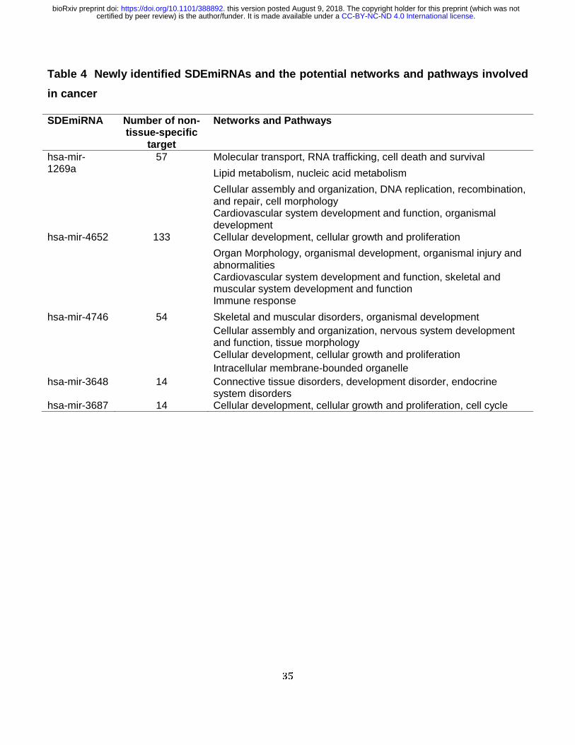
35
Table 4 Newly identified SDEmiRNAs and the potential networks and pathways involved
in cancer
SDEmiRNA Number of non-tissue-specific
target
Networks and Pathways
hsa-mir-1269a
57 Molecular transport, RNA trafficking, cell death and survival
Lipid metabolism, nucleic acid metabolism
Cellular assembly and organization, DNA replication, recombination, and repair, cell morphology Cardiovascular system development and function, organismal development
hsa-mir-4652 133 Cellular development, cellular growth and proliferation
Organ Morphology, organismal development, organismal injury and abnormalities Cardiovascular system development and function, skeletal and muscular system development and function Immune response
hsa-mir-4746 54 Skeletal and muscular disorders, organismal development Cellular assembly and organization, nervous system development and function, tissue morphology Cellular development, cellular growth and proliferation Intracellular membrane-bounded organelle
hsa-mir-3648 14 Connective tissue disorders, development disorder, endocrine system disorders
hsa-mir-3687 14 Cellular development, cellular growth and proliferation, cell cycle
.CC-BY-NC-ND 4.0 International licensecertified by peer review) is the author/funder. It is made available under aThe copyright holder for this preprint (which was notthis version posted August 9, 2018. . https://doi.org/10.1101/388892doi: bioRxiv preprint
36
Supplementary Materials
Figure S1 Webpage of each miRNA in BioXpress v3.0
Results of differential expression analysis of human whole miRNAs are shown by both table and
figures in BioXpress v3.0. Each miRNA is also mapped to corresponding Ensembl, miRbase,
RefSeq, and HGNC identifiers to allow users to search. Cancer types are also mapped to
Disease Oncology IDs.
Figure S2 Identification of different cell types within one organ cancer by miRNA
A. Lung cancer - there are 190 significantly differentially expressed miRNAs with ∆log2FC > 1
between LUAD (Lung adenocarcinoma) and LUSC (Lung squamous cell carcinoma), including
50 having the same expression change in these two cell types. The remaining 140 miRNAs with
opposite expression patterns are considered to be key factors underlying the differences
between LUAD and LUSC. B-D. Kidney cancer - Pairwise comparisons were made between
KIRP (Kidney renal papillary cell carcinoma) and KIRC (Kidney renal clear cell carcinoma) (B),
KICH (Kidney Chromophobe) and KIRP (C), and KIRC and KICH (D). There are 330
significantly differentially expressed miRNAs with ∆log2FC > 1 between any two of the three
subtypes. miRNA types and order are preserved across the three comparisons of kidney cancer.
From the comparison, the development of KIRC and KIRP may be mechanistically closer with
each other, compared to the development of KICH.
Figure S3 Significantly differentially expressed miRNAs that are over-expressed in at
least 80 percent of patient or under-expressed per cancer
Log2FC values were used to distinguish the over- or under-expression of miRNAs per patient.
Green bars with values larger than 80 percent indicate miRNAs are over-expressed in at least
80% of patient, while red bars with values larger than 80 present miRNAs (green bars lower
than 20) that are under-expressed in at least 80% of patient per cancer. In the figure, some
cancers have more over-expressed miRNAs than under-expressed ones, while others have the
opposite trend. This suggests different cancer types can have different enrichment patterns of
.CC-BY-NC-ND 4.0 International licensecertified by peer review) is the author/funder. It is made available under aThe copyright holder for this preprint (which was notthis version posted August 9, 2018. . https://doi.org/10.1101/388892doi: bioRxiv preprint
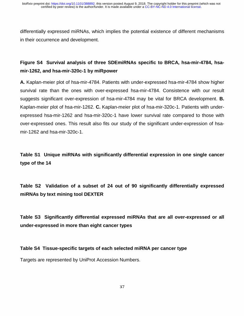
37
differentially expressed miRNAs, which implies the potential existence of different mechanisms
in their occurrence and development.
Figure S4 Survival analysis of three SDEmiRNAs specific to BRCA, hsa-mir-4784, hsa-
mir-1262, and hsa-mir-320c-1 by miRpower
A. Kaplan-meier plot of hsa-mir-4784. Patients with under-expressed hsa-mir-4784 show higher
survival rate than the ones with over-expressed hsa-mir-4784. Consistence with our result
suggests significant over-expression of hsa-mir-4784 may be vital for BRCA development. B.
Kaplan-meier plot of hsa-mir-1262. C. Kaplan-meier plot of hsa-mir-320c-1. Patients with under-
expressed hsa-mir-1262 and hsa-mir-320c-1 have lower survival rate compared to those with
over-expressed ones. This result also fits our study of the significant under-expression of hsa-
mir-1262 and hsa-mir-320c-1.
Table S1 Unique miRNAs with significantly differential expression in one single cancer
type of the 14
Table S2 Validation of a subset of 24 out of 90 significantly differentially expressed
miRNAs by text mining tool DEXTER
Table S3 Significantly differential expressed miRNAs that are all over-expressed or all
under-expressed in more than eight cancer types
Table S4 Tissue-specific targets of each selected miRNA per cancer type
Targets are represented by UniProt Accession Numbers.
.CC-BY-NC-ND 4.0 International licensecertified by peer review) is the author/funder. It is made available under aThe copyright holder for this preprint (which was notthis version posted August 9, 2018. . https://doi.org/10.1101/388892doi: bioRxiv preprint

38
Table S5 Significantly differential expressed miRNA in at least eight cancer types and
their target counts for each cancer type
Table S6. Selected GO and PANTHER terms involved in miRNAs and their tissue-specific
targets. Cancer types are represented by DOIDs (Disease Ontology ID).
.CC-BY-NC-ND 4.0 International licensecertified by peer review) is the author/funder. It is made available under aThe copyright holder for this preprint (which was notthis version posted August 9, 2018. . https://doi.org/10.1101/388892doi: bioRxiv preprint
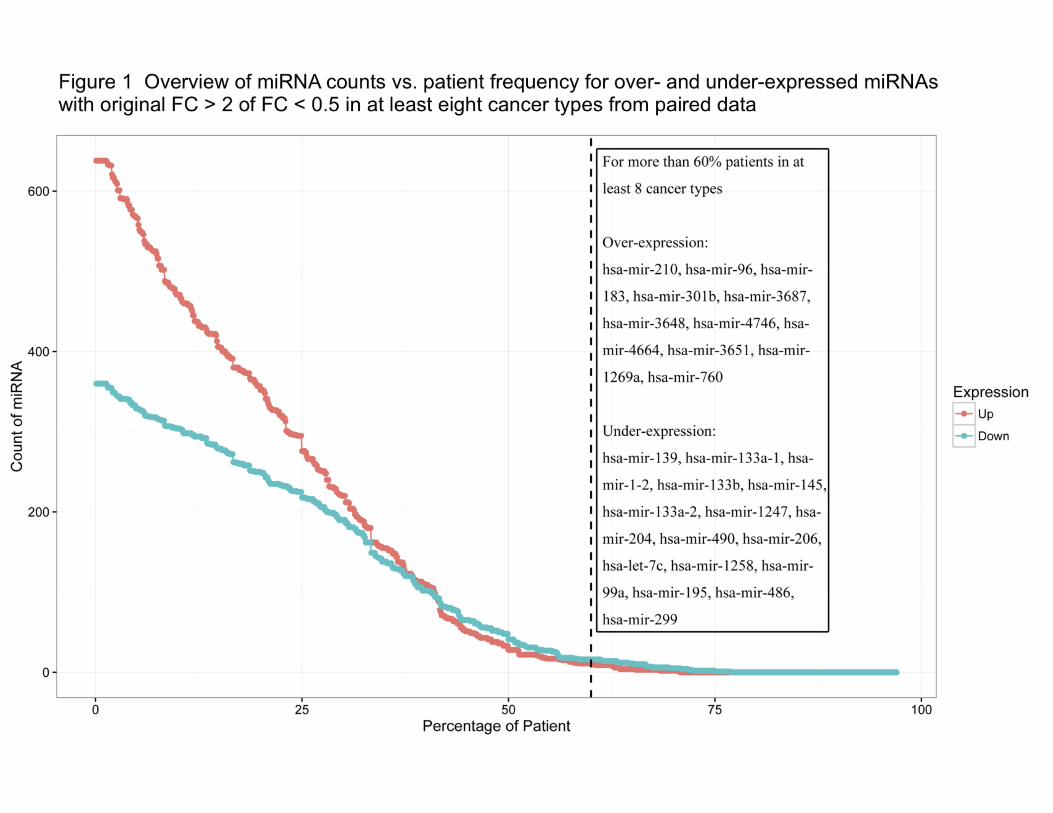
.CC-BY-NC-ND 4.0 International licensecertified by peer review) is the author/funder. It is made available under aThe copyright holder for this preprint (which was notthis version posted August 9, 2018. . https://doi.org/10.1101/388892doi: bioRxiv preprint
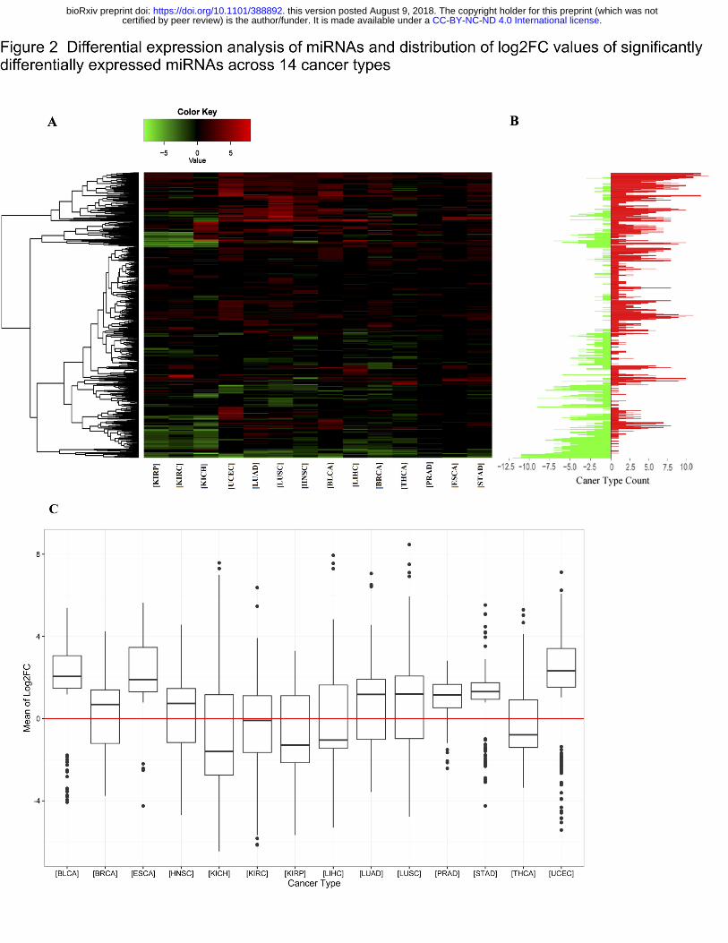
.CC-BY-NC-ND 4.0 International licensecertified by peer review) is the author/funder. It is made available under aThe copyright holder for this preprint (which was notthis version posted August 9, 2018. . https://doi.org/10.1101/388892doi: bioRxiv preprint
.CC-BY-NC-ND 4.0 International licensecertified by peer review) is the author/funder. It is made available under aThe copyright holder for this preprint (which was notthis version posted August 9, 2018. . https://doi.org/10.1101/388892doi: bioRxiv preprint
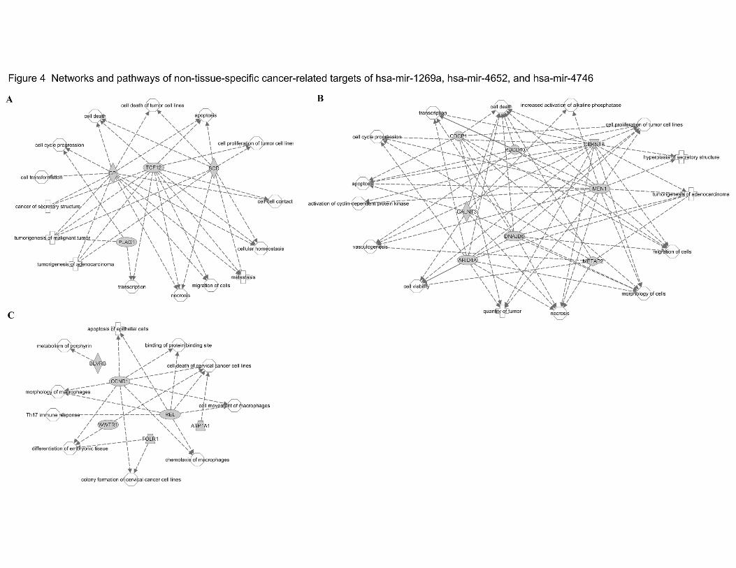
.CC-BY-NC-ND 4.0 International licensecertified by peer review) is the author/funder. It is made available under aThe copyright holder for this preprint (which was notthis version posted August 9, 2018. . https://doi.org/10.1101/388892doi: bioRxiv preprint
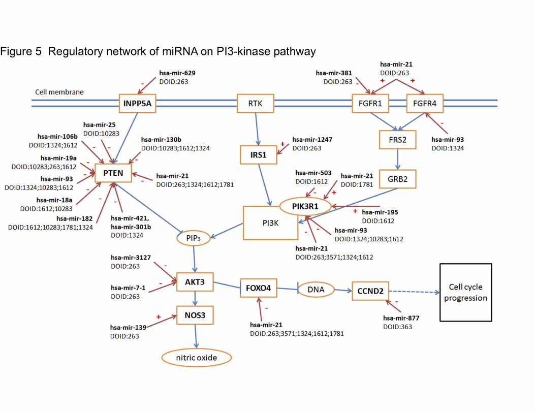
.CC-BY-NC-ND 4.0 International licensecertified by peer review) is the author/funder. It is made available under aThe copyright holder for this preprint (which was notthis version posted August 9, 2018. . https://doi.org/10.1101/388892doi: bioRxiv preprint