thyroid hormone concentrations and inhibition of thyroid stimulating hormone secretion in euthyroid...
TRANSCRIPT
European Journal of Clinical Investigation (1 977) 7,42 1-426
Thyroid hormone concentrations and inhibition of thyroid stimulating hormone secretion in euthyroid subjects with autonomous thyroid nodules A. CARPI, R. BIANCHI, G. C. ZUCCHELLI, M. G. TONI, A. COLI, C. LEVANT1 & G. MARIANI, Centre for Nuclear Medicine, Department of Metabolic Diseases, and the C.N.R. Clinical Physiology Laboratory, University of Pisa, and the Town Hospital, La Spezia, Italy
Received 7 June 1976 and in revised form 14 February 1977
Abstract. Sixty-four euthyroid patients with autonomous thyroid nodules and normal thyroxine (T4) concentra- tions and tri-iodothyronine resin uptake have been studied. The serum tri-iodothyronine (T3) concen- tration of the patients was 2.24 (k0.67) ng/ml, signifi- cantly higher than in a group of fifty-seven euthyroid control subjects (1.58 f 0.30 ng/ml). When no extra- nodular tissue was visible on the basal thyroid scan, the T3 was 2.31 (k0.63) ng/ml, significantly higher than in patients with some extranodular uptake on the basal scan (1.91 f 0.42 ng/ml). There was no significant differ- ence in the serum T4 concentrations of the patients (7.37 * 2.10 pg/IOO ml) compared to the control group (6.88 f I .89 pg/IOO ml). The T4 concentrations were not correlated with total or partial inhibition of the extranodular tissue. The thyroid hormone concentra- tions were not directly correlated to the size of the nodule assessed by scan imaging. The thyroid stimulating hormone (TSH) concentration of the patients (I .52 k 0.38 pU/ml) was significantly lower than in normals (2.49 k 0.96 pU/ml). No significant difference was found in the TSH concentrations of patients with partiaf or total inhibition of extranodular tissue irrespective of the T3 concentration. A thyrotrophin releasing hormone stimulation test in twelve patients did not increase the serum TSH, irrespectively of the T3 concentration.
These data show the high frequency of elevated serum T3 concentrations despite normal serum T4 con- centration in euthyroid patients with autonomous thyroid nodules. They confirm that inhibition of TSH secretion can occur when thyroid hormone concentra- tions are in the normal range.
Key words. T3, T4, TSH, inhibition of TSH secretion, autonomous thyroid nodule with euthyroidism.
Introduction
Euthyroid subjects with autonomous thyroid nodules have been described recently to have low serum thyroid stimulating hormone (TSH) concentrations [ I ] and
Correspondence: Dr Angelo Carpi, Centro di Medicina Nucleare, c/o Patologia Speciale Medica I , Spedali Riuniti S. Chiara, Via Roma, 56100 Pisa, Italy.
markedly reduced or absent TSH responses to thyro- trophin releasing hormone (TRH) stimulation [ I , 21. These findings suggest that TSH secretion is somehow suppressed in this condition. This conclusion is sup- ported by a normal TSH metabolic clearance rate measured in one patient with autonomous thyroid nodule [ I ] ; such limited observation, however, cannot be con- sidered conclusive evidence of the state of TSH secretion rates in all such patients.
Contradictory results are reported in respect of tri- iodothyronine (T3) concentrations in these patients in the literature so far available. While normal thyroxine (T4) levels have been observed usually, elevated T3 con- centrations are reported in some series [2, 31, and values within the normal range, in spite of very low TSH con- centrations, in other studies [ I ] . Increased T3 values could explain the suppressed TSH secretion in the first instance [2]. On the other hand, TSH suppression in patients with normal T3 concentrations could be due to increased, but still normal, concentrations of thyroid hormones, or to a TSH-inhibiting substance other than iodothyronine secreted by the thyroid nodule [ I ] . Marsden et al. [4] recently reported nine euthyroid patients with autonomous thyroid nodules who had normal T3 and T4 concentrations. They did not present TSH values in these patients.
The few observations so far reported of the fre- quency with which elevated T3 concentrations occur despite normal T4 concentrations in these conditions are insufficient [ 5 ] . The aim of this study was to clarify the relation of the thyroid hormones to TSH in euthyroid patients with autonomous thyroid nodules. To do this, we measured the T3, T4 and TSH concentrations in sixty- four patients. In twelve, a TRH stimulation test was also performed.
Materials and Methods
Patients. Sixty-four patients with one or more palpable thyroid nodules positive on the 1311 scan and partial or total absence of I3'I uptake in the extranodular paren- chyma ('hot nodules') (fifty-eight females and six males, aged 21 -72 years) but without clinical hyperthyroidism
42 1

422 A CARPI et al.
were studied. None had a clinical history and/or sero- logical findings of previous diffuse or localized thyroi- ditis. The criteria for the diagnosis of an ‘autonomous thyroid nodule’ were those of Marsden et al. [4] This diagnosis was confirmed in twelve of the patients by a test of TRH stimulation, once the synthetic hormone became available to us (Serono Pharmaceuticals, Italy). In approximately half the remaining patients the auto- nomy of the nodule was proved by a suppression test. The main clinical and laboratory data of these patients are shown in Table 1.
T3, T4, TSH concentrations, and T3 resin uptake were measured on the same blood sample taken between 08.00
and 09.00 hours. The T3 resin uptake was always normal. The TRH stimulation test was performed in twelve patients by measuring the plasma TSH concentration basally, and at 5, 15, 30, 45 and 60 min after the i.v. injection of 250 pg synthetic TRH.
Thyroid scan with 1311 was performed in all the patients under the same geometrical conditions to obtain comparable images both before and after TSH stimula- tion (5 I.U. of bovine TSH i.m. for 4 days before the study).
A thyroid suppression test was performed in twenty- four patients by T3 administration before the control scan (100 c(g daily, for 8 days).
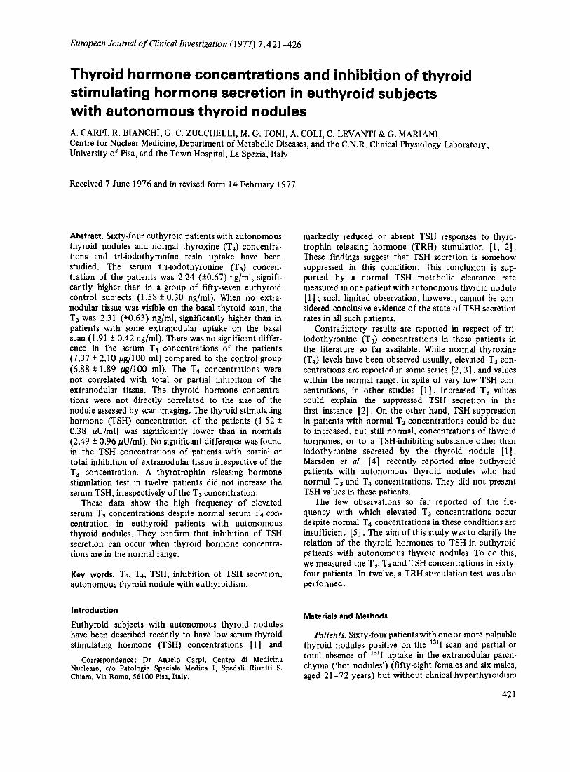
Table 1 . Clinical and laboratory data of buthyroid patients with autonomous thyroid nodule Solitary autonomous nodules
Patient Sex Age Nodular diameter Inhibition of T3 T4 TSH (years) on scan image (em) extranodular tissue (ndml) bd100 ml) O1U/ml)
F.I. F <4 P 1.92 V.E. B.A. P.M. G.R. S.P. V.L. M.M. P.F. K.A. O.M. B.M. T.G. T.G. B.M. B.C. P.A. N.M. V.B. G.A. G.I. F.O. S.D. M.M. B.A. D.L. P.A. C.G. L.F. T.S. S.C. M.M. D.A. A.S. C.R. C.D. S.W. M.L. P.D. P.C. C.P. B.B. C.M. L.G. C.M. M.M. C.P.
F F F F F F M F F F F M F F M F F F F F M F F F F F F F F F F F F F F F M F F F M F F
F F
F‘
54 38 4 1 49 53 47 44 24 30 34 23 5 1 66 64 57 47 44 3 1 53 36 65 46 46 35 63 4 0 50 34 27 38 32 30 36 45 47 6 2 34 37 34 70 21 45 4 0 49 37 37 50
<4 <4 <4 >4 <4 >4 >4 >4 <4 >4 <4 <4 <4 <4 >4 >4 >4 <4 <4 <4 >4 <4 <4 <4 <4 >4 >4 <4 <4 <4 >4 <4 >4 >4 <4 >4 <4 <4 <4 >4 >4 <4 >4 <4 <4 >4
T P T P T P T T P T T T T P T T P T P T P P P T P P P T T T P T T P T P T T T T T T P T T T
1.50 1.55 2.00 2.57 3.42 2.00 2.65 2.52 1.95 2.17 2.45 1.85 2.35 2.15 2.30 3.15 1.43 2.67 1.95 3.45 1.61 2.38 1.49 3.8 1.82 2.27 1.75 2.65 1.95 1.2 1.95 2.95 2.25 1.20 2.50 1.55 1.82 1.97 1.75 1.9 1.97 1.75 2.88 1.37 1.9 2.7
5.5 6.2 8.82 7.6 5.7
10.4 5.2 7.2 8 6.4 4.9 9.6 4.1 7.4 7.3 9.7 5.5 3.2 6.99 9.93
3.5 7.28 7.13 9.04 6.4 6.62 3.9 8.2 5.2 7.2 8.4 9.8 5 8.1 9.9 5 8.6 8 9.6 8.5 5.8 8.6
12.2 5.9
10.7 4.3
10
1.9 1.72 1.6
1.45 1.42
1.05 1.8 1.9 1.2 0.48 0.74
-
-
- -
- -
1.75 1.65 1.7 1.37 1.85 1.9 1.7 2.1 1.6 1.75 1.97 1.47 1.65 1.4 0.3 0.82 1.4 1.4 0.97 1.5 0.3 1.8 1.45 1.35 1.42 1.4 1.65 1.8 1.45 1.55
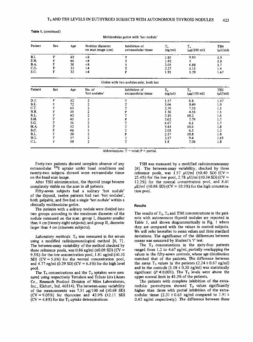
T3 AND TSH LEVELS IN EUTHYROID SUBJECTS WITH AUTONOMOUS THYROID NODULES 423
Table 1. (continued)
Multinodular goitre with ‘hot nodule’
Patient Sex Age Nodular diameter Inhibition of T3 T4 TSH on scan image (cm) extranodular tissue (ng/ml) (Ccg/lOO ml) OcU/ml)
B.I. F 49 <4 T 1.85 9.93 1.9 S.M. F 46 <4 T 1.92 5 1.8 B.A. F 30 <4 T 2.05 6.88 1.7 C.G. F 32 >4 T 2.27 5.15 1.4 S.G. F 32 <4 P 1.95 5.29 1.67
Goitre with two nodules only, both hot
Patient Sex Age No. of Inhibition of T3 T4 TSH ‘hot nodules’ extranodular tissue (ng/mU ( d l 0 0 ml) bU/ml)
D.T. F 52 2 T 1.57 6.8 1.57 B.E. F 12 2 T 3.04 9.49 1.9 C.T. F 63 2 T 2.70 7.35 1.5 N.R. F 35 2 P 1.36 4.56 1.5 R.L. F 42 2 T 3.10 10.2 1.6 S.M. F 45 2 P 3.02 7.79 1.7 S.G. F 31 2 T 4.67 6.1 1.7 M.A. F 52 2 T 3.45 10.6 1.8 B.C. F 48 2 T 2.05 6.5 1.2 B.L. F 38 2 P 2.57 10.8 1.8 M.L. F 51 2 T 2.57 9.4 1.65 C.L. F 59 2 T 1.8 7.06 1.8
Abbreviations: T = total; P = partial.
Forty-two patients showed complete absence of any extranodular 1311 uptake under basal conditions and twenty-two subjects showed some extranodular tissue on the basal scan image.
After TSH administration, the thyroid image became completely visible on the scan in all patients.
Fifty-seven subjects had a solitary ‘hot nodule’ of the thyroid, twelve patients had two ‘hot nodules’, both palpable, and five had a single ‘hot nodule’ within a clinically multinodular goitre,
The patients with a solitary nodule were divided into two groups according to the maximum diameter of the nodule measured on the scan: group I, diameter smaller than 4 cm (twentyeight subjects); and group 11, diameter larger than 4 cm (ninetekn subjects).
Laboratory methods. T3 was measured in the serum using a modified radioimmunological method [6, 71. The between-assay variability of the method checked by three reference pools, was 0.86 ng/ml (k0.08 SD) (CV = 9.5%) for the low ooncentration pool, 1.81 ng/ml (kO.10 SD) (CV = 5.6%) for the normal concentration pool, and 4.77 ng/ml (0.29 SD) (CV = 6.1%) for the high level
The T4 concentrations and the T3 uptakes were mea- sured using respectively Tetralute and Trilute kits (Ames Co., Research Product Division of Miles Laboratories, Inc., Elkhart, Ind. 46514). The between-assay variability of the measurements was 7.51 pg/IOO ml (k0.68 SD) (CV=9.05%) for thyroxine and 43.9% (k2.11 SD) (CV = 4.8%) for the T3 uptake determinations.
pool.
TSH was measured by a modified radioimmunoassay [8] . The between-assay variability, checked by three reference pools, was 1.57 pU/ml (k0.40 SD) (CV= 25.4%) for the low pool, 2.78 pU/ml (k0.34 SD) (CV = 12.2%) for the normal concentration pool, and 8.41 pU/ml (k0.88 SD) (CV = 10.5%) for the high concentra- tion pool.
Results
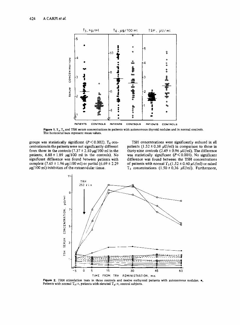
The results of T3, T4 and TSH concentrations in the pati- ents with autonomous thyroid nodules are reported in Table 1, and shown diagrammatically in Fig. 1 where they are compared with the values in control subjects. We will refer hereafter to mean values and their standard deviations. The significance of the differences between means was measured by Student’s ‘t’ test.
The T3 concentrations in the sixty-four patients ranged from 1.2 to 4.67 ng/ml, partially overlapping the values in the fifty-seven controls, whose age distribution matched that of the patients. The difference between the mean T3 values in the patients (2.24 ?r 0.67 nglml) and in the controls (1.58 f 0.30 ng/ml) was statistically significant (P Q 0.001). The T3 levels were above the upper normal limit in 45.3% of the patients.
The patients with complete inhibition of the extra- nodular parenchyma showed T3 values significantly higher than those with partial inhibition of the extra- nodular tissue (2.31 + 0.63 ng/ml compared to 1.91 + 0.42 ng/ml respectively). The difference between these
424 A CARPI et al.
2 0 k 4 a 2
u 2 0 0
k
W
5 w U)
3
4
Y
3 c
0
i 0
0
8, 4 : 0 0
a 0 0
5 0
4 0
0 0
0 0
I 3
PATIENTS CONTROLS PATIENTS CONTROLS PATIENTS C O N T R O L S
Figure l .T3, T, and TSH serum concentrations in patients with autonomous thyroid nodules and in normal controls. The horizontal bars represent mean values.
groups was statistically significant (P < 0.002). T4 con- centrationsin the patients were not significantly different from those in the controls (7.37 f 2.10 pg/lOO ml in the patients; 6.88 * 1.89 pg/IOO ml in the controls). No significant difference was found between patients with complete (7.65 f 1.96 pg/IOO ml) or partial (6.69 f 2.29 pg/lOO ml) inhibition of the extranodular tissue.
TSH concentrations were significantly reduced in all patients (1.52 * 0.38 pU/ml) in comparison to those in thirty-nine controls (2.49 ? 0.96 pU/ml). The difference was statistically significant (P< 0.001). No significant difference was found between the TSH concentrations of patients with normal T3 (1.52 f 0.40 pU/ml) or raised T3 concentrations ( I S O * 0.36 pU/ml). Furthermore,
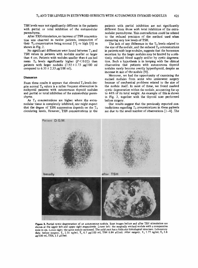
"1 TRH
..
- 5 0 5 15 30 45 60 TIME FROM T R H ADMINISTRATION, min
Figure 2. TRH stimulation tests in three controls and twelve euthyroid patients with autonomous Patients with normal T,; 0, patients with elevated T,; 0, control subjects.
nodules. e,
T3 AND TSH LEVELS IN EUTHYROID SUBJECTS WITH AUTONOMOUS THYROID NODULES 425
TSH levels were not significantly different in the patients with partial or total inhibition of the extranodular parenchyma.
After TRH stimulation, no increase of TSH concentra- tion was observed in twelve patients, irrespective of their T3 concentration being normal [7] or high [5] as shown in Fig. 2 .
No significant differences were found between T3 and TSH values in patients with nodules smaller or larger than 4 cm. Patients with nodules smaller than 4 cm had mean T4 levels significantly higher (P<O.O2) than patients with larger nodules (7.92 f 1.73 pg/lOO ml compared to 6.35 f 2.33 pg/lOO ml).
Discussion
From these results it appears that elevated T3 levels des- pite normal T4 values is a rather frequent observation in euthyroid patients with autonomous thyroid nodules and partial or total inhibition of the extranodular paren- chyma.
As T3 concentrations are higher where the extra- nodular tissue is completely inhibited, one might expect that the degree of TSH suppression depends on the T3 circulating levels. However, TSH concentrations in the
patients with partial inhibition are not significantly different from those with total inhibition of the extra- nodular parenchyma. This contradiction could be related to the reduced precision of the method used when measuring very low levels of TSH.
The lack of any difference in the T3 levels related to the size of the nodule, and the reduced T4 concentrations in patients with large nodules, suggests that the hormones secretion by the larger nodules may be limited by a rela- tively reduced blood supply and/or by cystic degenera- tion. Such a hypothesis is in keeping with the clinical observation that patients with autonomous thyroid nodules rarely become overtly hyperthyroid, despite an increase in size of the nodule [9] .
Moreover, we had the opportunity of examining the excised nodules from some who underwent surgery because of mechanical problems related to the size of the nodule itself. In most of these, we found marked cystic degeneration within the nodule, accounting for up to 44% of its total weight. An example of this is shown in Fig. 3 , together with the thyroid scan performed before surgery.
Our results suggest that the previously reported con- tradictions regarding T3 concentrations in these patients are due to the small number of observations [I -41. The
Figure 3. Partial cystic degeneration of an autonomous nodule. Scan images before and after TSH stimulation are shown at the upper left and upper right respectively. Lower left: the surgically excised nodule with a comparative scale in cm. Lower right: the same nodule sectioned. The solid part has a follicular histological structure. Laboratory data: before surgery, T, 2.35 ng/ml, T, 6.7 pg/100 ml, TSH 0.84 /.tU/ml. After surgery, T, 1.77 ng/ml, T, 5.8 pg/100 ml, TSH, 1.5 /.tU/ml.
426 A CARPI et al,
present series, larger than any previous one of which we are aware, shows that T3 concentrations can be either normal or increased. Moreover, evidence is given that suppression of TSH secretion is not, or not always, due to elevated T3 concentrations, as low TSH values can be associated with normal concentrations of the thyroid hormones. Experiments in which T3 and T4 have been administered to normal or hypothyroid subjects have shown that small increments of thyroid hormones, even within the normal range, can inhibit TSH secretion [ 101 . On the other hand, the hypothesis that substances other than iodothyronine, secreted by the nodular tissue, are able to inhibit TSH secretion cannot be excluded, though this does not have a good experimental basis
It is currently believed that the selective increase of T3 concentration in patients with autonomous nodules is due to preferential production of T3 by the nodular tissue [ l l , 121.
In conclusion, further studies, including measurement of the nodule hormonal content and identification of substances other than iodothyronine able to inhibit TSH secretion, are necessary to clarify better the pathophysio- logy of this disease.
References
[ I 1 *
1 Ridgway E.C., Weintraub B.D., Cevallos J.L., Rack M.C. & Moloof F. (1973) Suppression of pituitary TSH secretion in the patient with a hyperfunctioning thyroid nodule. J Clin Invest 5 2 , 2783-2792.
2 Evered D.C., Clark F. & Petersen V.B. (1974) Thyroid func- tion in euthyroid subjects with autonomous thyroid nodules. Clin Endocrinol ( O x f ) 3, 149-154.
3 Nouel J.P., Brunelle P. & Segond G. (1973) Le dosage de la triiodothyronine sgrique dans les nodules autonomes hyper- fixants thyroidiens. Nouv Presse Med 2, 1912.
4 Marsden P., Facer P., Acosta M. & McKennon C.G. (1975) Serum triiodothyronine in solitary autonomous nodules of the thyroid. Clin Endocrinol (Oxf ) 4,327 -330.
5 Miller J.M. (1975) Plummer’s disease. Symposium on current concepts of thyroid disease. Med Clin North A m 59, 1203- 1216.
6 Galbiati A., Malvano R. & Zucchelli G.C. (1974) Assessment of some methodological aspects of radioimmunoassay of tri- iodothyronine. JNucl BioZMed 18,160-169.
7 Malvano R., Galbiati A. & Zucchelli G.C. (1975) Effects of plasma proteins on the radioimmunoassay of triiodothyronine. JNucI Biol Med 19,94-104.
8 Franchimont P. & Hendrick J.C. (1475) Radioimmunoassay of glycoprotein hormones. Radioimmunoassay and Related Procedures in Medicine, Vol. 1, p. 195. International Atomic Energy Agency, Publishing Section, Vienna.
9 Hamburger J.I. (1975) Solitary autonomously functioning thyroid lesions; diagnosis, clinical features and pathogenetic considerations. Am JMed 58,740-748.
10 Snyder P.J. & Utiger R.D. (1972) Inhibition of thyrotropin response to thyrotropin-releasing hormone by small quanti- ties of thyroid hormones. J CIin Invest 5 1, 2077-2084.
11 Shimaoka K. (1963) Toxic adenoma of the thyroid with tri- iodothyronine as the principal circulating thyroid hormone. Acta Endocrinol43,285-293.
12 Hollander C.S., Mitsuma T., Nihei N., Shenkman L., Burday S.Z. & Blum M. (1972) Clinical and laboratory observations in cases of tri-iodothyronine toxicosis confirmed by radio- immunoassay. Lancet i, 609-611.