thymidine kinase 2 enzyme kinetics elucidate the mechanism of thymidine-induced mitochondrial dna...
TRANSCRIPT
Thymidine Kinase 2 Enzyme Kinetics Elucidate the Mechanism ofThymidine-Induced Mitochondrial DNA DepletionRen Sun and Liya Wang*
Department of Anatomy, Physiology and Biochemistry, Swedish University of Agricultural Sciences, Box 7011, SE-750 07 Uppsala,Sweden
ABSTRACT: Mitochondrial thymidine kinase 2 (TK2) is a nuclear gene-encoded protein, synthesized in the cytosol and subsequently translocated intothe mitochondrial matrix, where it catalyzes the phosphorylation of thymidine(dT) and deoxycytidine (dC). The kinetics of dT phosphorylation exhibitsnegative cooperativity, but dC phosphorylation follows hyperbolic Michaelis−Menten kinetics. The two substrates compete with each other in that dT is acompetitive inhibitor of dC phosphorylation, while dC acts as a noncompetitiveinhibitor of dT phosphorylation. In addition, TK2 is feedback inhibited by dTTPand dCTP. TK2 also phosphorylates a number of pyrimidine nucleosideanalogues used in antiviral and anticancer therapy and thus plays an importantrole in mitochondrial toxicities caused by nucleoside analogues. Deficiency inTK2 activity due to genetic alterations causes devastating mitochondrial diseases,which are characterized by mitochondrial DNA (mtDNA) depletion or multiple deletions in the affected tissues. Severe TK2deficiency is associated with early-onset fatal mitochondrial DNA depletion syndrome, while less severe deficiencies result in late-onset phenotypes. In this review, studies of the enzyme kinetic behavior of TK2 enzyme variants are used to explain themechanism of mtDNA depletion caused by TK2 mutations, thymidine overload due to thymidine phosphorylase deficiency, andmitochondrial toxicity caused by antiviral thymidine analogues.
Mitochondria are dynamic organelles that undergoconstant changes, e.g., fission and fusion, during the
lifetime of the cell. Mitochondria contain multiple copies ofcircular mitochondrial DNA (mtDNA) molecules, whichencode 13 essential subunits of the mitochondrial respiratorychain complexes. To maintain a sustainable mtDNA copynumber, a constant supply of precursors, i.e., dNTPs, isessential because mtDNA replicates in a manner that isindependent of the cell cycle. A limitation of one or moredNTPs will stall mtDNA synthesis and result in mtDNAdepletion, which leads to mitochondrial diseases.1 In dividingcells and/or tissues, mitochondrial dNTPs are synthesized viathe cytosolic de novo pathway and the salvage pathway;however, in nondividing cells, the dNTPs required for mtDNAreplication are mainly generated in organello via thymidinekinase 2 (TK2) and deoxyguanosine kinase (dGK). TK2 anddGK catalyze the phosphorylation of all four naturaldeoxynucleosides to their respective monophosphates usingATP or other nucleoside triphosphates as phosphate donors.Both enzymes are essential for mtDNA precursor synthesis andmitochondrial function in many cell and tissue types.2
Thymidine kinase 2 (TK2, EC 27.1.21) catalyzes the transferof the γ-phosphate group from ATP to the 5′-hydroxyl group ofthymidine (dT), deoxycytidine (dC), or deoxyuridine (dU) toform their corresponding monophosphates. TK2 also phos-phorylates a number of pyrimidine nucleoside analogues, suchas zidovudine (AZT) used in anti-HIV therapy, and thus mayplay an important role in the mitochondrial toxicity observed inantiviral and anticancer therapies using nucleoside ana-
logues.3−5 TK2 is present in all cells that contain mitochondria,and the levels of TK2 correlated to the mitochondrial contentof the cells or tissues.2 A recent study showed that TK2 isupregulated during the stationary phase growth of culturedcells.6
Deficiency in TK2 activity due to genetic mutations causesdevastating mitochondrial diseases, which are characterized astissue-specific mtDNA depletion and/or deletion. Mitochon-drial DNA depletion syndrome (MDS) is characterized by asevere and tissue-specific reduction in the mtDNA copynumber in the absence of qualitative defects in mtDNA.7
MDS caused by TK2 mutations affected mainly liver andskeletal muscle, but in some cases, multiple tissues wereinvolved.8−10 Mutations in the TK2 gene are also the geneticcauses of late-onset autosomal recessive progressive externalophthalmoplegia (arPEO).11 The subject of this review is thesubstrate specificity, enzyme kinetics, and regulation of the TK2enzyme, as well as the mechanism of thymidine overload-induced mtDNA depletion.
■ TK2 ENZYME KINETICS
The first extensive kinetic analysis was performed using thepurified enzyme obtained from human leukemic spleen.12 Itwas shown that with dC and dU as substrates, the reactions
Received: June 4, 2014Revised: September 11, 2014Published: September 12, 2014
Current Topic
pubs.acs.org/biochemistry
© 2014 American Chemical Society 6142 dx.doi.org/10.1021/bi5006877 | Biochemistry 2014, 53, 6142−6150

followed hyperbolic Michaelis−Menten kinetics, with Km(micromolar) and Vmax/Km (units per milligram per micro-molar) values of 36 and 25 for dC and 6 and 115 for dU,respectively. However, the phosphorylation of dT exhibitednegative cooperativity with Hill coefficients in the range of 0.3−0.5, indicating that the affinity of dT for TK2 decreases with anincrease in concentration as shown by the biphasic Hofsteeplot. The Km value was 0.3 μM when the dT concentration was<8 μM, while at higher concentrations, the Km value for dT was16 μM, which gives a kcat/Km value of ∼2.6 × 10⁴ s−1 M−1 athigh dT concentrations and a kcat/Km value of 2.7 × 10⁵ s−1
M−1 at low dT concentrations. Thus, the efficiency of dTphosphorylation is lower at high dT concentrations (>8 μM)than at low dT concentrations (Figure 1). The specific activity
with dT phosphorylation is significantly higher than that withdC phosphorylation at physiologically relevant substrateconcentrations (<0.5 μM) because of the negative cooperativitywith dT (Figure 1B). AZT phosphorylation also showednegative cooperativity.12 Similar results were obtained withrecombinant human TK2.13
In organello studies with mitochondria isolated from rat liver,heart, and brain showed that the phosphorylation of dT alsoexhibited negative cooperativity, and the catalytic efficienciesdiffered in mitochondria isolated from different tissues.Negative cooperative kinetic behavior was also observed withAZT phosphorylation in heart and liver mitochondria, but notin brain mitochondria. Instead, in brain mitochondria, AZTphosphorylation followed hyperbolic Michaelis−Menten ki-netics, but negative cooperativity was observed with dCydphosphorylation.14−18 Thus, TK2 enzyme kinetics are complexand tissue-specific.
TK2 belongs to the enzyme family that includes mitochon-drial deoxyguanosine kinase (dGK), cytosolic deoxycytidinekinase (dCK), and Drosophila melanogaster deoxynucleosidekinase (Dm-dNK). The three-dimensional (3D) structures ofdGK, dCK, and Dm-dNK have been determined but not theTK2 structure. Substrate and inhibitor binding sites have beenstudied in detail with dGK, dCK, and Dm-dNK.19−22 However,there is a fundamental lack of structural information aboutTK2; hence, the number of substrate binding sites per activeenzyme is not known, which is a real barrier to achieving a clearunderstanding of its biological function.
■ REGULATION OF TK2 ACTIVITY BY SUBSTRATES,SUBSTRATE ANALOGUES, AND FEEDBACKINHIBITORS
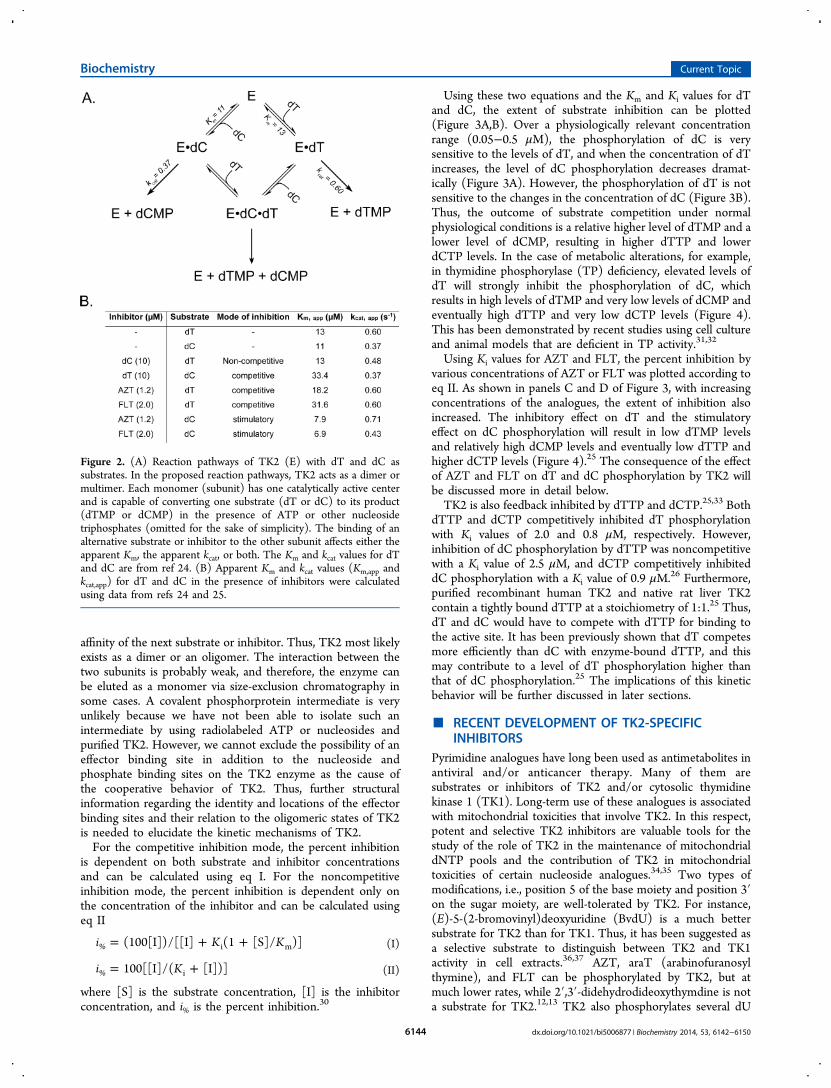
Substrate dT and dC phosphorylations are regulated by adifferent kinetic mechanisms, and thus, the enzyme hasdifferent catalytic efficiencies for dT and dC, as describedabove. The negative cooperative kinetics with dT suggest thatunder physiological conditions, e.g., low dT concentrations(<0.05 μM), the TK2 activity will be several hundred-foldhigher than with a noncooperative enzyme with the same Kmvalue (Figure 1B).23 In cells, both substrates coexist and acompetition between dT and dC for the same enzyme isunavoidable. In the presence of both substrates, dC acted as anoncompetitive inhibitor of dT phosphorylation with a Ki valueof 40 μM and dT behaved as a competitive inhibitor toward dCphosphorylation with a Ki value of 4.9 μM,24 resulting inchanges in apparent Km or kcat values for dT and dC in thepresence of alternative substrates (Figure 2).Thymidine analogues such as AZT and FLT strongly
inhibited dT phosphorylation with Ki values of 3 and 1.4 μM,respectively. AZT and FLT bind to the enzyme in the same wayas dT and thus acted as competitive inhibitors of dTphosphorylation. In the absence of these analogues, recombi-nant human TK2 phosphorylates dC with a Km value of 11 ± 1μM and a kcat value of 0.37 ± 0.01 s−1.24 However, addition ofAZT or FLT resulted in an increased level of dCphosphorylation; for instance, in the presence of 1.2 μMAZT, the Km and kcat values for dC were changed to 7.9 ± 0.7μM and 0.71 ± 0.03 s−1, respectively, and in the presence of 2.0μM FLT, the Km and kcat values for dC were changed to 6.9 ±0.5 μM and 0.43 ± 0.02 s−1, respectively (Figure 2). Thisstimulatory effect was shown to be due to the presence of afeedback inhibitor, e.g., dTTP in the TK2 enzyme.25
TK2 exists in monomer, dimer, or tetramer forms.12,24,26−28
Each monomer is likely to have one nucleoside binding site andone nucleotide (phosphate donor) binding site, as predicted bythe structural model of TK2, which was built on the basis ofamino acid sequence homology to and the 3D structure of Dm-dNK.4 Dm-dNK strictly follows Michaelis−Menten kineticswith all of its substrates29 but not TK2. Therefore, the TK2kinetic behavior may involve a third molecule (inhibitor orsubstrate) that acts through one of the following mechanisms:(1) a protein−protein interaction in a multimeric state, (2) acovalent phosphoprotein intermediate that transfers thephosphate group to the nucleoside, or (3) an uncharacterizedeffector binding site on each monomer.Protein−protein or subunits interactions are most likely
because the kinetic studies with natural substrates andinhibitors described above suggest that the binding of thefirst substrate/inhibitor to one subunit induced conformationalchanges in the other subunit, leading to changes in the binding
Figure 1. (A) TK2 substrate saturation curves of V (velocity) vs S(substrate concentration). The curves were drawn by using Km, Vmax,S0.5, and n values from ref 12. (B) Substrate saturation curves at lowconcentrations.
Biochemistry Current Topic
dx.doi.org/10.1021/bi5006877 | Biochemistry 2014, 53, 6142−61506143
affinity of the next substrate or inhibitor. Thus, TK2 most likelyexists as a dimer or an oligomer. The interaction between thetwo subunits is probably weak, and therefore, the enzyme canbe eluted as a monomer via size-exclusion chromatography insome cases. A covalent phosphorprotein intermediate is veryunlikely because we have not been able to isolate such anintermediate by using radiolabeled ATP or nucleosides andpurified TK2. However, we cannot exclude the possibility of aneffector binding site in addition to the nucleoside andphosphate binding sites on the TK2 enzyme as the cause ofthe cooperative behavior of TK2. Thus, further structuralinformation regarding the identity and locations of the effectorbinding sites and their relation to the oligomeric states of TK2is needed to elucidate the kinetic mechanisms of TK2.For the competitive inhibition mode, the percent inhibition
is dependent on both substrate and inhibitor concentrationsand can be calculated using eq I. For the noncompetitiveinhibition mode, the percent inhibition is dependent only onthe concentration of the inhibitor and can be calculated usingeq II
= + +i K K(100[I])/[[I] (1 [S]/ )]% i m (I)
= +i K100[[I]/( [I])]% i (II)
where [S] is the substrate concentration, [I] is the inhibitorconcentration, and i% is the percent inhibition.30
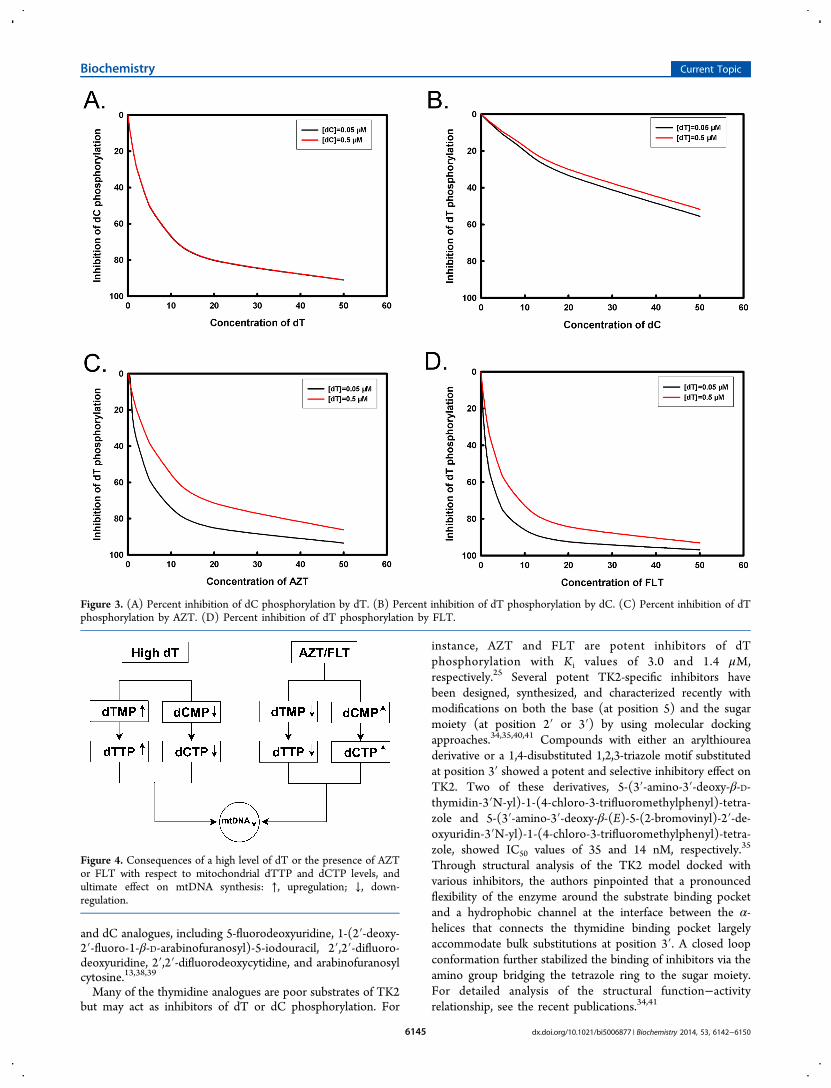
Using these two equations and the Km and Ki values for dTand dC, the extent of substrate inhibition can be plotted(Figure 3A,B). Over a physiologically relevant concentrationrange (0.05−0.5 μM), the phosphorylation of dC is verysensitive to the levels of dT, and when the concentration of dTincreases, the level of dC phosphorylation decreases dramat-ically (Figure 3A). However, the phosphorylation of dT is notsensitive to the changes in the concentration of dC (Figure 3B).Thus, the outcome of substrate competition under normalphysiological conditions is a relative higher level of dTMP and alower level of dCMP, resulting in higher dTTP and lowerdCTP levels. In the case of metabolic alterations, for example,in thymidine phosphorylase (TP) deficiency, elevated levels ofdT will strongly inhibit the phosphorylation of dC, whichresults in high levels of dTMP and very low levels of dCMP andeventually high dTTP and very low dCTP levels (Figure 4).This has been demonstrated by recent studies using cell cultureand animal models that are deficient in TP activity.31,32
Using Ki values for AZT and FLT, the percent inhibition byvarious concentrations of AZT or FLT was plotted according toeq II. As shown in panels C and D of Figure 3, with increasingconcentrations of the analogues, the extent of inhibition alsoincreased. The inhibitory effect on dT and the stimulatoryeffect on dC phosphorylation will result in low dTMP levelsand relatively high dCMP levels and eventually low dTTP andhigher dCTP levels (Figure 4).25 The consequence of the effectof AZT and FLT on dT and dC phosphorylation by TK2 willbe discussed more in detail below.TK2 is also feedback inhibited by dTTP and dCTP.25,33 Both
dTTP and dCTP competitively inhibited dT phosphorylationwith Ki values of 2.0 and 0.8 μM, respectively. However,inhibition of dC phosphorylation by dTTP was noncompetitivewith a Ki value of 2.5 μM, and dCTP competitively inhibiteddC phosphorylation with a Ki value of 0.9 μM.26 Furthermore,purified recombinant human TK2 and native rat liver TK2contain a tightly bound dTTP at a stoichiometry of 1:1.25 Thus,dT and dC would have to compete with dTTP for binding tothe active site. It has been previously shown that dT competesmore efficiently than dC with enzyme-bound dTTP, and thismay contribute to a level of dT phosphorylation higher thanthat of dC phosphorylation.25 The implications of this kineticbehavior will be further discussed in later sections.
■ RECENT DEVELOPMENT OF TK2-SPECIFICINHIBITORS
Pyrimidine analogues have long been used as antimetabolites inantiviral and/or anticancer therapy. Many of them aresubstrates or inhibitors of TK2 and/or cytosolic thymidinekinase 1 (TK1). Long-term use of these analogues is associatedwith mitochondrial toxicities that involve TK2. In this respect,potent and selective TK2 inhibitors are valuable tools for thestudy of the role of TK2 in the maintenance of mitochondrialdNTP pools and the contribution of TK2 in mitochondrialtoxicities of certain nucleoside analogues.34,35 Two types ofmodifications, i.e., position 5 of the base moiety and position 3′on the sugar moiety, are well-tolerated by TK2. For instance,(E)-5-(2-bromovinyl)deoxyuridine (BvdU) is a much bettersubstrate for TK2 than for TK1. Thus, it has been suggested asa selective substrate to distinguish between TK2 and TK1activity in cell extracts.36,37 AZT, araT (arabinofuranosylthymine), and FLT can be phosphorylated by TK2, but atmuch lower rates, while 2′,3′-didehydrodideoxythymdine is nota substrate for TK2.12,13 TK2 also phosphorylates several dU
Figure 2. (A) Reaction pathways of TK2 (E) with dT and dC assubstrates. In the proposed reaction pathways, TK2 acts as a dimer ormultimer. Each monomer (subunit) has one catalytically active centerand is capable of converting one substrate (dT or dC) to its product(dTMP or dCMP) in the presence of ATP or other nucleosidetriphosphates (omitted for the sake of simplicity). The binding of analternative substrate or inhibitor to the other subunit affects either theapparent Km, the apparent kcat, or both. The Km and kcat values for dTand dC are from ref 24. (B) Apparent Km and kcat values (Km,app andkcat,app) for dT and dC in the presence of inhibitors were calculatedusing data from refs 24 and 25.
Biochemistry Current Topic
dx.doi.org/10.1021/bi5006877 | Biochemistry 2014, 53, 6142−61506144
and dC analogues, including 5-fluorodeoxyuridine, 1-(2′-deoxy-2′-fluoro-1-β-D-arabinofuranosyl)-5-iodouracil, 2′,2′-difluoro-deoxyuridine, 2′,2′-difluorodeoxycytidine, and arabinofuranosylcytosine.13,38,39
Many of the thymidine analogues are poor substrates of TK2but may act as inhibitors of dT or dC phosphorylation. For
instance, AZT and FLT are potent inhibitors of dTphosphorylation with Ki values of 3.0 and 1.4 μM,respectively.25 Several potent TK2-specific inhibitors havebeen designed, synthesized, and characterized recently withmodifications on both the base (at position 5) and the sugarmoiety (at position 2′ or 3′) by using molecular dockingapproaches.34,35,40,41 Compounds with either an arylthioureaderivative or a 1,4-disubstituted 1,2,3-triazole motif substitutedat position 3′ showed a potent and selective inhibitory effect onTK2. Two of these derivatives, 5-(3′-amino-3′-deoxy-β-D-thymidin-3′N-yl)-1-(4-chloro-3-trifluoromethylphenyl)-tetra-zole and 5-(3′-amino-3′-deoxy-β-(E)-5-(2-bromovinyl)-2′-de-oxyuridin-3′N-yl)-1-(4-chloro-3-trifluoromethylphenyl)-tetra-zole, showed IC50 values of 35 and 14 nM, respectively.35
Through structural analysis of the TK2 model docked withvarious inhibitors, the authors pinpointed that a pronouncedflexibility of the enzyme around the substrate binding pocketand a hydrophobic channel at the interface between the α-helices that connects the thymidine binding pocket largelyaccommodate bulk substitutions at position 3′. A closed loopconformation further stabilized the binding of inhibitors via theamino group bridging the tetrazole ring to the sugar moiety.For detailed analysis of the structural function−activityrelationship, see the recent publications.34,41
Figure 3. (A) Percent inhibition of dC phosphorylation by dT. (B) Percent inhibition of dT phosphorylation by dC. (C) Percent inhibition of dTphosphorylation by AZT. (D) Percent inhibition of dT phosphorylation by FLT.
Figure 4. Consequences of a high level of dT or the presence of AZTor FLT with respect to mitochondrial dTTP and dCTP levels, andultimate effect on mtDNA synthesis: ↑, upregulation; ↓, down-regulation.
Biochemistry Current Topic
dx.doi.org/10.1021/bi5006877 | Biochemistry 2014, 53, 6142−61506145
■ ROLE OF TK2 IN MITOCHONDRIAL DTTP ANDDCTP POOL MAINTENANCE AND MTDNADEPLETION AND DELETION
In mitochondria, the levels of DNA precursors, e.g., dNTPs,play a crucial role in mtDNA replication. A limitation of one ormore of the four dNTPs will stall mtDNA replication and resultin mtDNA depletion or deletion1 and lead to mitochondrialdiseases. Currently, 12 of >200 nuclear genes encoding proteinsinvolved in mtDNA replication and mtDNA precursormetabolism are associated with mitochondrial diseases,including TK2, DGUOK (deoxyguanosine kinase), and TYMP(thymidine phosphorylase).10,42
Progressive external ophthalmoplegia (PEO) manifests asmultiple mtDNA deletions, which accumulate with age andcause late-onset symptoms in postmitotic tissues, such as thenervous system and skeletal muscle. These symptoms usuallyare weakness of the external eye muscles, bilateral ptosis,exercise intolerance, proximal muscle weakness, wasting,
hearing loss, hypogonadism, optic atrophy, and Parkinsonism.Mutations in DGUOK and TK2 are linked to autosomalrecessive PEO (arPEO).11,43,44
TK2 Kinetics Elucidate the Mechanism of MDS Causedby TK2 Mutations. MDS was initially described by Moraes etal. in 1991, and 10 years later, the first report assigning TK2mutations to MDS was published.7,8 The two missensemutations in the TK2 gene, His90Asn and Ile181Asn, werefound in four individuals with devastating isolated skeletalmyopathy, mtDNA depletion, and death at an early age.8 Sincethen, more than 50 individuals suffering from MDS with both amolecularly confirmed diagnosis and histological or biochem-ical analysis have been linked to a variety of genetic alternationsin the TK2 gene.45 In addition to myopathy, neurologicalphenotypes and multitissue pathology have been discovered inpatients carrying TK2 mutations46 (Table 1) (see recentarticles for more details5,10,45,47).The first kinetic characterization of TK2 mutations identified
in MDS patients showed that the His90Asn mutant exhibited
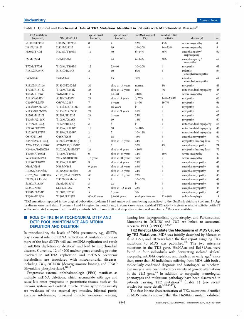
Table 1. Clinical and Biochemical Data of TK2 Mutations Identified in Patients with Mitochondrial Diseasesa
TK2 mutation(reported) NM_004614.4
age at onset(months)
age at death(months)
mtDNA content(%)
residual TK2activity disease(s) ref
<H90N/H90N H121N/H121N 0 4 22% 45% severe myopathy 8I181N/I181N I212N/I212N 8 19 16−20% 14−23% severe myopathy 8H90N/T77M H121N/T108M 12 40 6−14% 36% encephalopathy/
nephropathy62
I22M/I22M I53M/I53M 1 24 6−14% 28% encephalopathy/myopathy
62
T77M/T77M T108M/T108M 12 23−40 10−20% b myopathy 63R183G/R254X R183G/R254X 2 10 60% b infantile
encephalomyopathy64
E48fsX149 E48fsX149 5 15 37% b infantileencephalomyopathy
64
R152G/K171del R183G/K202del 36 alive at 14 years normal 1% myopathy 49T77M/R161 K T108M/R192K 28 alive at 12 years 8% 7% mitochondrial myopathy 48T64M/R183W T64M/R183W 11 24−28 <10% b severe myopathy 65A181V/A181V A139V/A139V 24 alive at 5 years 5, 70% 14.8−25.9% myopathy 66C108W/L257P C66W/L215P 7 6 years 8−9% 19.7% myopathy 66V113fs20X/I212N V113fs20X/I212N 24 16 years b b myopathy 67V113fs20X/N93S V113fs20X/N93S 24 alive at 9 years 21% b myopathy 67R120R/H121N R120R/H121N 24 6 years 25% b myopathy 67T108M/Q125X T108M/Q125X 7 19 18% b myopathy 67Y154N/R172Q Y112N/R130Q b b b b mitochondrial myopathy 68R225W/R225W R183W/R183W 18 38 5−10% b mitochondrial myopathy 46R172W/R172W R130W/R130W 2 7 10−12% b mitochondrial myopathy 46Q87X/N100S Q45X/N58S 3 18 <5% b encephalomyopathy 69K85NfsX9/R172Q K43NfsX9/R130Q 12 alive at 13 years 24% b myopathy, hearing loss 70A73fs.X230/R130W A73fsX230/R130W 1 3 20% 4% encephalomyopathy 71K244del/S93IfsX99 K202del/S51IfsX57 24 alive at 6 years <10% b myopathy, hearing loss 72T108M/T108M T108M/T108M 0 alive at 44 years 34% 40% severe myopathy 47W4V.fsX40/R90C W4V.fsX40/R90C 13 years alive at 31 years 39% b severe myopathy 47R183W/R183W R183W/R183W 9 alive at 4 years 25% b encephalomyopathy 45N58S/N58S N58S/N58S 9 years alive at 28 years 86% b encephalomyopathy 45R130Q/K44Nfsx9 R130Q/K44Nfsx9 24 alive at 15 years 24% b encephalomyopathy 45c.157_2A> G/R196S c.157_2A>G/R196S 48 alive at 10 years 73% b encephalomyopathy 45I212N/5.8 kb del I212N/5.8 kb del 3 8 10−20% b encephalomyopathy 45S135L/R183W S135L/R183W 5 22 b b encephalomyopathy 45S135L/N58S S135L/N58S 9 alive at 1.2 years 22% b encephalomyopathy 45T108M/L233P T108M/L233P 5 3 years 5% b encephalomyopathy 45T230A/R225W T230A/R225W 40−50 years 55−60 years multiple deletion 22−40% ophthalmoplegia 11
aTK2 mutations reported in the original publication (column 1) and amino acid numbering normalized to the GenBank database (column 2). Agefor disease onset and death (columns 3 and 4) is given in months and, in some cases, years. Residual TK2 activity is given as relative activity (with dTas the substrate) compared with healthy controls. fsXx, frame shift and stop after amino acid number x. bData not available.
Biochemistry Current Topic
dx.doi.org/10.1021/bi5006877 | Biochemistry 2014, 53, 6142−61506146
normal activity with dT but strongly reduced activity with dC(65% reduction) under conditions of excess substrates. TheHis90Asn mutant also lost negative cooperativity with dT,which indicated that at physiological dT concentrations (<0.05μM), the His90Asn mutant would only have <7% of activitywith dT compared to that with the wild-type enzyme. Thisdifference would cause a severe reduction in the size of thedTTP and/or dCTP pool and thus a reduced level of mtDNAsynthesis. The Ile181Asn mutant enzyme had only 0.6% activitywith dT and 0.01% activity with dC compared with that of thewild-type enzyme, which would ultimately result in a depletionof dTTP and dCTP pools and depletion of mtDNA.24 Thecharacterization of two other mutations in TK2 found inpatients, e.g., T77M, showed not only drastically reducedoverall efficiency with dT and dC but also altered cooperativityfor dT and dC; i.e., the presence of dT enhanced dCphosphorylation, while dC functions as a strong inhibitor of dTphosphorylation. Such an alteration exaggerated the effects ofreduced TK2 activity and thus, severe mtDNA depletion.48 Thetwo TK2 mutations, e.g., T230A and R225W, causing late-onsetarPEO, showed significantly reduced activity with both dT anddC and an altered ratio of dC/dT phosphorylation.11 Thesestudies suggested that a severe reduction in TK2 activity or analteration of substrate specificity or kinetic behavior resulted ina disruption and limitation of mitochondrial dTTP and dCTPproduction, which caused mtDNA depletion and/or deletion.Many of the TK2 mutations identified in MDS patients have
not been characterized; however, from the data available todate, we concluded that the level of residual TK2 activity in theaffected tissues is vital for survival (Table 1). However, theresidual TK2 levels measured using a single substrate cannotprovide adequate information regarding mtDNA depletioncaused by the TK2 mutations, as was exemplified with theHis90Asn mutant. Thus, kinetic studies of mutant TK2 areimportant for understanding the mechanism underlyingmtDNA depletion. In most cases, the severity of TK2deficiency is correlated with the patient phenotype, namely,severe impairment of TK2 resulting in encephalomyopathywith multiple-organ involvement, whereas partial reduction ofTK2 was associated with myopathy.5 Thus, relatively high levelsof residual TK2 activity are associated with late-onset disease.However, in one case, the patient survived longer with severeTK2 deficiency (1% of controls),49 and it was suggested that analternative compensatory mechanism could be involved.TK2 Kinetics Underlie Thymidine Overload-Induced
mtDNA Depletion. High plasma dT levels were found inpatients with defective thymidine phosphorylase (TP), whichpresented itself as mitochondrial neurogastrointestinal ence-phalomyopathy (MNGIE) with multiple mtDNA deletions anddepletions.50,51 TP is an enzyme that catalyzes the reversiblephosphorolysis of dT to thymine and deoxyribose 1-phosphateand plays an important role in maintaining a low plasma level ofdT and dU.52 Deficiency in TP activity resulted in anaccumulation of dT and dU in patients, which has beensuggested to be the cause of the mtDNA deletion and depletionobserved in MNGIE patients.24,26 In the plasma of healthyindividuals, dT is undetectable; however, in TP patients, theplasma level of dT and dU ranged from 3.9 to 17.7 μM,51,53
which indicates several hundred-fold increases in dT and dUconcentrations. According to eq I, dT alone will inhibit dCphosphorylation of up to 80% (Figure 3A). In mitochondria,elevated dT levels will eventually result in an increased level ofdTTP and a diminished dCTP pool, as illustrated in Figure 4,
and ultimately to mtDNA deletion or depletion. It wasdemonstrated experimentally that high levels of dT and dU,because of TP deficiency, resulted in higher levels of dTTP butlower levels of dCTP in TP-knockout mouse brain mitochon-dria, which resulted in mtDNA instability, e.g., mtDNA deletionand depletion.54 In vitro studies also showed that limited dCTPlevels due to dT overload could cause mtDNA depletion.31 Onthe basis of the TK2 kinetics described above, mtDNAdepletion was prevented by deoxynucleoside (dC) supplemen-tation or inhibition of dC catabolism in a cell culture model anda mouse model with defective TP. Taken together, thesefindings point to a potential treatment for mtDNA depletioncaused by defects in dNTP metabolism.32
TK2 Kinetics Shed Light on Thymidine Analogue-Induced mtDNA Depletion. In early chemotherapy againstHIV, AZT was administered alone at a relatively large dose andoften resulted in severe mitochondrial dysfunction. In addition,mtDNA depletion was observed in muscles, peripheral blood,and other tissues of treated patients.55 Although the doses ofAZT are much smaller in a modern HAART regime, long-termexposure to this analogue still causes adverse effects, includinganemia, lactic acidosis, neutropenia, skeletal muscle myopathy,and cardiomyopathy with mtDNA depletion.56 These sideeffects were attributed to AZT-mediated mitochondrial toxicity,which was thought to be a class-wide major adverse effectassociated with nucleoside analogues. Previously, the mecha-nism of AZT toxicity had been identified as the inhibition ofmtDNA polymerase-γ, i.e., the DNA pol-γ hypothesis.57
However, to reach 50% inhibition of pol-γ, at least 100 μMAZT-TP is required, which is far greater than the concentrationthat can be clinically achieved using the plasma concentrationof AZT.18
Thus, an alternative mechanism underlying AZT-associatedmitochondrial toxicity has been suggested; AZT is a potentinhibitor of dT phosphorylation catalyzed by TK2, which mayresult in a depleted mitochondrial dTTP pool and limitmtDNA replication, thereby ultimately resulting in mtDNAdepletion.15,17 Compared with proliferating tissues, mostpostmitotic tissues, such as skeletal muscle, mainly rely onthe TK2-catalyzed dT/dC phosphorylation for the main-tenance of dTTP and dCTP pools and, thus, exhibit greatersensitivity to AZT and other pyrimidine analogues. As shown inpanels C and D of Figure 3, dT phosphorylation by TK2 isstrongly inhibited by AZT or FLT. Assuming that themitochondrial AZT concentration is within the same range ofplasma concentration, i.e., ∼3 μM,18 AZT would inhibit dTphosphorylation by >50% (Figure 3C), and FLT at aconcentration of 3 μM will inhibit dT phosphorylation by70% (Figure 3D). On the other hand, dC phosphorylation willbe stimulated. Such opposite effects will alter the ratio of dTand dC phosphorylation and result in a significantly decreasedlevel of dTMP and an increased level of dCMP and eventuallysmall dTTP and relatively large dCTP pools25 (Figure 4). A>50% reduction of the dTTP pool has been reported inperfused rat hearts with short-term AZT exposure.58 Recentstudies using peripheral blood mononuclear cells fromindividuals who were infected with HIV and have been treatedwith antiviral therapy showed significant depletions of bothribonucleotide and deoxynucleotide pools.59 Thus, thesefindings strongly suggest that perturbation of intramitochon-drial dT and dC phosphorylation by TK2 contributes to themitochondrial toxicity of AZT and other related pyrimidine
Biochemistry Current Topic
dx.doi.org/10.1021/bi5006877 | Biochemistry 2014, 53, 6142−61506147
nucleoside analogue drugs in a manner independent of mtDNApolymerase-γ inhibition.
■ CONCLUSIONMitochondrial diseases are multifactorial, and interactions ofdefective gene(s) or proteins with other factors are involved indisease development and contribute to the tissue specificity ofthe disease. However, the kinetics and tissue-specific distribu-tion of TK2 have helped to explain the mechanism of tissue-specific mtDNA deletion in many patients with TK2 mutations.TK2 kinetic studies have also been used to design therapy forthe treatment of TP deficiency and may be used to weaken theside effects of nucleoside analogues and to develop novelnucleoside analogue drugs. In some patients, mtDNA depletionhas become less apparent as the disease progresses, despiteclinical and biochemical deterioration, and in a few cases, anincrease or lack of change in mtDNA copy number has beenreported.49,60,61 Thus, further biochemical characterization ofmutant TK2 enzymes and their interactions with other factorsand/or proteins are required to fully understand themechanism of mtDNA copy number alteration and to developtherapeutic strategies for the treatment of mitochondrialdiseases caused by defects in dNTP metabolism.
■ AUTHOR INFORMATIONCorresponding Author*Department of Anatomy, Physiology and Biochemistry,Swedish University of Agricultural Sciences, Box 7011, SE-750 07 Uppsala, Sweden. E-mail: [email protected]. Telephone:+46 18 672820.FundingThis work was supported by a grant from the Swedish ResearchCouncil.NotesThe authors declare no competing financial interest.
■ ACKNOWLEDGMENTSWe thank Prof. Staffan Eriksson for valuable discussions.
■ ABBREVIATIONSTK2, thymidine kinase 2; mtDNA, mitochondrial DNA; MDS,mtDNA depletion syndrome; dT, 2′-deoxythymidine; dC, 2′-deoxycytidine; dU, 2′-deoxyuridine; AZT, 3′-azido-2′,3′-dideoxythymidine; FLT, 3′-fluoro-2′,3′-dideoxythymidine;dNTP, deoxynucleoside triphosphate; TK1, thymidine kinase1; TP, thymidine phosphorylase.
■ REFERENCES(1) Taanman, J.-W., Muddle, J. R., and Muntau, A. C. (2003)Mitochondrial DNA depletion can be prevented by dGMP and dAMPsupplementation in a resting culture of deoxyguanosine kinase-deficient fibroblasts. Hum. Mol. Genet. 12, 1839−1845.(2) Arner, E. S. J., and Eriksson, S. (1995) Mammaliandeoxyribonucleoside kinases. Pharmacol. Ther. 67, 155−186.(3) Eriksson, S., and Wang, L. (2002) The role of the cellulardeoxynucleoside kinases in activation of nucleoside analogs used inchemotherapy. Recent Advances in Nucleosides: Chemistry and Chemo-therapy (Chu, C. K., Ed.) pp 455−475, Elsevier, Amsterdam.(4) Eriksson, S., Munch-Petersen, B., Johansson, K., and Eklund, H.(2002) Structure and function of cellular deoxynucleoside kinases.Cell. Mol. Life Sci. 59, 1327−1346.(5) Wang, L., and Eriksson, S. (2010) Tissue specific distribution ofpyrimidine deoxynucleoside salvage enzymes shed light on the
mechanism of mitochondrial DNA depletion. Nucleosides, NucleotidesNucleic Acids 29, 400−403.(6) Sun, R., Eriksson, S., and Wang, L. (2014) Mitochondrialthymidine kinase 2 but not deoxyguanosine kinase is up-regulatedduring stationary growth phase of the cultured cells. Nucleosides,Nucleotides Nucleic Acids 33, 282−286.(7) Moraes, C., Shanske, S., Tritschler, H., Aprille, J., Andreetta, F.,Bonilla, E., Schon, E., and DiMauro, S. (1991) mtDNA depletion withvariable tissue expression: A novel genetic abnormality in mitochon-drial diseases. Am. J. Hum. Genet. 48, 492−501.(8) Saada, A., Shaag, A., Mandel, H., Nevo, Y., Eriksson, S., andElpeleg, O. (2001) Mutant mitochondrial thymidine kinase inmitochondrial DNA depletion myopathy. Nat. Genet. 29, 342−344.(9) Eriksson, S., and Wang, L. (2008) Molecular mechanisms ofmitochondrial DNA depletion diseases caused by deficiencies inenzymes in purine and pyrimidine metabolism. Nucleosides, NucleotidesNucleic Acids 27, 800−808.(10) Wang, L. (2010) Deoxynucleoside salvage enzymes and tissuespecific mitochondrial DNA depletion. Nucleosides, Nucleotides NucleicAcids. 29, 370−381.(11) Tyynismaa, H., Sun, R., Ahola-Erkkila, S., Almusa, H.,Poyhonen, R., Korpela, M., Honkaniemi, J., Isohanni, P., Paetau, A.,Wang, L., and Suomalainen, A. (2012) Thymidine kinase 2 mutationsin autosomal recessive progressive external ophthalmoplegia withmultiple mitochondrial DNA deletions. Hum. Mol. Genet. 21, 66−75.(12) Munch-Petersen, B., Cloos, L., Tyrsted, G., and Eriksson, S.(1991) Diverging substrate specificity of pure human thymidinekinases 1 and 2 against antiviral dideoxynucleosides. J. Biol. Chem. 266,9032−9038.(13) Wang, L., Munch-Petersen, B., Herrstrom Sjoberg, A., Hellman,U., Bergman, T., Jornvall, H., and Eriksson, S. (1999) Humanthymidine kinase 2: Molecular cloning and characterisation of theenzyme activity with antiviral and cytostatic nucleoside substrates.FEBS Lett. 443, 170−174.(14) McKee, E. E., Bentley, A. T., Hatch, M., Gingerich, J., andSusan-Resiga, D. (2004) Phosphorylation of thymidine and AZT inheart mitochondria: Elucidation of a novel mechanism of AZTcardiotoxicity. Cardiovasc. Toxicol. 4, 155−167.(15) Lynx, M. D., Bentley, A. T., and McKee, E. E. (2006) 3′-Azido-3′-deoxythymidine (AZT) inhibits thymidine phosphorylation inisolated rat liver mitochondria: A possible mechanism of AZThepatotoxicity. Biochem. Pharmacol. 71, 1342−1348.(16) Lynx, M. D., and McKee, E. E. (2006) 3′-Azido-3′-deoxythymidine (AZT) is a competitive inhibitor of thymidinephosphorylation in isolated rat heart and liver mitochondria. Biochem.Pharmacol. 72, 239−243.(17) Susan-Resiga, D., Bentley, A. T., Lynx, M. D., Laclair, D. D., andMcKee, E. E. (2007) Zidovudine inhibits thymidine phosphorylationin the isolated perfused rat heart. Antimicrob. Agents Chemother. 51,1142−1149.(18) McCann, K., Williams, D., and McKee, E. E. (2012) Metabolismof deoxypyrimidines and deoxypyrimidine antiviral analogs in isolatedbrain mitochondria. J. Neurochem. 122, 126−137.(19) Johansson, K., Ramaswamy, S., Ljungcrantz, C., Knecht, W.,Piskur, J., Munch-Petersen, B., Eriksson, S., and Eklund, H. (2001)Structural basis for substrate specificities of cellular deoxyribonucleo-side kinases. Nat. Struct. Biol. 8, 616−620.(20) Sabini, E., Ort, S., Monnerjahn, C., Konrad, M., and Lavie, A.(2003) Structure of human dCK suggests strategies to improveanticancer and antiviral therapy. Nat. Struct. Biol. 10, 513−519.(21) Mikkelsen, N. E., Johansson, K., Karlsson, A., Knecht, W.,Andersen, G., Piskur, J., Munch-Petersen, B., and Eklund, H. (2003)Structural basis for feedback inhibition of the deoxyribonucleosidesalvage pathway: Studies of the Drosophila deoxyribonucleoside kinase.Biochemistry 42, 5706−5712.(22) Sabini, E., Hazra, S., Ort, S., Konrad, M., and Lavie, A. (2008)Structural basis for substrate promiscuity of dCK. J. Mol. Biol. 378,607−621.
Biochemistry Current Topic
dx.doi.org/10.1021/bi5006877 | Biochemistry 2014, 53, 6142−61506148
(23) Munch-Petersen, B. (2010) Enzymatic regulation of cytosolicthymidine kinase 1 and mitochondrial thymidine kinase 2: A minireview. Nucleosides, Nucleotides Nucleic Acids 29, 363−369.(24) Wang, L., Saada, A., and Eriksson, S. (2003) Kinetic propertiesof mutant human thymidine kinase 2 suggest a mechanism formitochondrial DNA depletion myopathy. J. Biol. Chem. 278, 6963−6968.(25) Wang, L., Sun, R., and Eriksson, S. (2011) The kinetic effects onthymidine kinase 2 by enzyme bound dTTP may explain themitochondrial side effects of antiviral nucleoside analogs. Antimicrob.Agents Chemother. 55, 2552−2558.(26) Wang, L., and Eriksson, S. (2000) Cloning and characterizationof full length mouse thymidine kinase 2: The N-terminal sequencedirects import of the precursor protein into mitochondria. Biochem. J.351, 469−476.(27) Barroso, J. F., Carvalho, R. N., and Flatmark, T. (2005) Kineticanalysis and ligand-induced conformational changes in dimeric andtetrameric forms of human thymidine kinase 2. Biochemistry 44, 4886−4896.(28) Radivoyevitch, T., Munch-Petersen, B., Wang, L., and Eriksson,S. (2011) A mathematical model of human thymdine kinase 2.Nucleosides, Nucleotides Nucleic Acids 30, 203−209.(29) Munch-Petersen, B., Knecht, W., Lenz, C., Sondergaard, L., andPiskur, J. (2000) Functional expression of a multisubstratedeoxyribonucleoside kinase from Drosophila melanogaster and its C-terminal deletion mutants. J. Biol. Chem. 275, 6673−6679.(30) Segel, I. (1993) Enzyme Kinetics, Wiley Classics Library Edition,pp 100−132, John Wiley & Sons, Inc., New York.(31) Gonzalez-Vioque, E., Torres-Torronteras, J., Andreu, A., andMarti, R. (2011) Limited dCTP availability accounts for mitochondrialDNA depletion in mitochondrial neurogastrointestinal encephalomy-opathy (MNGIE). PLoS Genet. 7, e1002035.(32) Camara, Y., Gonzalez-Vioque, E., Scarpelli, M., Torres-Torronteras, J., Caballero, A., Hirano, M., and Ramon, M. (2013)Administration of deoxyribonucleosides or inhibition of theircatabolism as a pharmacological approach for mitochondrial DNAdepletion syndrome. Hum. Mol. Genet. 23, 2459−2467.(33) Barroso, J. F., Elholm, M., and Flatmark, T. (2003) Tightbinding of deoxyribonucleotide triphosphates to human thymidinekinase 2 expressed in Escherichia coli. Purification and partialcharacterization of its dimeric and tetrameric forms. Biochemistry 42,15158−15169.(34) Perez-Perez, M., Priego, E., Hernandez, A., Familiar, O.,Camarsa, M., Negri, A., Gago, F., and Balzarini, J. (2008) Structure,physiological role, and specific inhibitors of human thymidine kinase 2(TK2): Present and future. Med. Res. Rev. 28, 797−820.(35) Van Poecke, S., Negri, A., Janssen, J., Solaroli, N., Karlsson, A.,Gago, F., Balzarini, J., and Van Calenbergh, S. (2011) Synthesis,modeling and evaluation of 3′-(1-aryl-1H-tetrazol-5-ylamino)-substi-tuted 3′-deoxythymidine derivaties as potent and selective humanmitochondrial thymidine kinase inhibitors. Org. Biomol. Chem. 9, 892−901.(36) Franzolin, E., Rampazzo, C., -Perez, M. J., Hernandez, A. I.,Balzarini, J., and Bianchi, V. (2006) Bromovinyl-deoxyuridine: Aselective substrate for mitochondrial thymidine kinase in cell extracts.Biochem. Biophys. Res. Commun. 344, 30−36.(37) Wang, L., and Eriksson, S. (2008) 5-Bromovinyl 2′-deoxyuridinephosphorylation by mitochondrial and cytosolic thymidine kinase(TK2 and TK1) and its use in selective measurement of TK2 activityin crude extracts. Nucleosides, Nucleotides Nucleic Acids 27, 858−862.(38) Wang, J., and Eriksson, S. (1996) Phosphorylation of the anti-hepatitis B nucleoside analog 1-(2′-deoxy-2′-fluoro-1-β-D-arabinofur-anosyl)-5-iodouracil (FIAU) by human cytosolic and mitochondrialthymidine kinase and implication for cytotoxicity. Antimicrob. AgentsChemother. 40, 1555−1557.(39) Eriksson, S., and Wang, L. (1997) Substrate specificities,expression, and primary sequences of deoxynucleoside kinases;implications for chemotherapy. Nucleosides Nucleotides 16, 653−659.
(40) Ciliberti, N., Manfredini, S., Angusti, A., Durini, E., Solaroli, N.,Vertuani, S., Buzzoni, L., Bonache, M., Ben-Shalom, E., Karlsson, A.,Saada, A., and Balzarini, J. (2007) Novel selective humanmitochondrial kinase inhibitors: Design, synthesis and enzymaticactivity. Bioorg. Med. Chem. 15, 3065−3081.(41) Priego, E., Karlsson, A., Gago, F., Camarasa, M., Balzarini, J., andPerez-Perez, M. (2012) Recent advances in thymidine kinase 2 (TK2)inhibitors and new perspectives for potential applications. Curr. Pharm.Des. 18, 2981−2994.(42) Suomalainen, A., and Isohanni, P. (2010) Mitochondrial DNAdepletion syndromes: Many genes, common mechanisms. Neuro-muscular Disorders 20, 429−437.(43) Copeland, W. C. (2012) Defects in mitochondrial DNAreplication and human disease. Crit. Rev. Biochem. Mol. Biol. 47, 64−74.(44) Ronchi, D., Garone, C., Bordoni, A., Rios, P., Calvo, S.,Ripolone, M., Ranieri, M., Rizzuti, M., Villa, L., Magri, F., Corti, S.,Bresolin, N., Mootha, V., Moggio, M., DiMauro, S., Comi, G., andSciacco, M. (2012) Next-generation sequencing reveals DGUOKmutations in adult patients with mitochondrial DNA multipledeletions. Brain 135, 3404−3415.(45) Chanprasert, S., Wang, J., Weng, S., Enns, G., Boue, D., Wong,B., Mendell, J., Perry, D., Sahenk, Z., Craigen, W., Alcala, F., Pascual, J.,Melancon, S., Zhang, V., Scaglia, F., and Wong, L. (2013) Molecularand clinical characterization of the myopathic form of mitochondrialDNA depletion syndrome caused by mutations in the thymidinekinase (TK2) gene. Mol. Genet. Metab. 110, 153−161.(46) Gotz, A., Isohanni, P., Pihko, H., Paetau, A., Herva, R.,Saarenpaa-Heikkila, O., Valanne, L., Marjavaara, S., and Suomalainen,A. (2008) Thymidine kinase 2 defects can cause multi-tissue mtDNAdepletion syndrome. Brain 131, 2841−2850.(47) Behin, A., Jardel, C., Claeys, K., Faggart, J., Louha, M., Romero,N., Laforet, P., Eymard, B., and Lombes, A. (2012) Adult cases ofmitochondrial DNA depletion due to TK2 defect: An expandingspectrum. Neurology 78, 644−648.(48) Wang, L., Limongelli, A., Vila, M., Carrara, F., Zeviani, M., andEriksson, S. (2005) Molecular insight into mitochondrial DNAdepletion syndrome in two patients with novel mutations in thedeoxyguanosine kinase and thymidine kinase 2 genes. Mol. Genet.Metab. 84, 75−82.(49) Vila, M., Segovia-Silvestre, T., Gamez, J., Marina, A., Naini, A.,Meseguer, A., Lombes, A., Bonilla, E., DiMauro, S., Hirano, M., andAndreu, A. (2003) Reversion of mtDNA depletion in a patient withTK2 deficiency. Neurology 60, 1203−1205.(50) Nishino, I., Spinazzola, A., and Hirano, M. (1999) Thymidinephosphorylase gene mutations in MNGIE, a human mitochondrialdisorder. Science 283, 689−692.(51) Valentino, M., Marti, R., Tadesse, S., Lopez, L., Manes, J., Lyzak,J., Hahn, A., Carelli, V., and Hirano, M. (2007) Thymidine anddeoxyuridine accumulation in tissues of patients with mitochondrialneurogastrointestinal encephalomyopathy (MNGIE). FEBS Lett. 581,3410−3414.(52) Spinazzola, A., Marti, R., Nishino, I., Andreu, A., Naini, A.,Tadesse, S., Pela, I., Zammarchi, E., Donati, M., Oliver, J., and Hirano,M. (2002) Altered thymidine metabolism due to defects of thymidinephosphorylase. J. Biol. Chem. 277, 4128−4133.(53) Marti, R., Nishigaki, Y., and Hirano, M. (2003) Elevated plasmadeoxyurindine in patients with thymidine phosphorylase deficiency.Biochem. Biophys. Res. Commun. 303, 14−18.(54) Lopez, L., Akman, H., Garcia-Cazola, A., Dorado, B., Marti, R.,Nishino, I., Tadesse, S., Pizzorno, G., Shungu, D., Bonilla, E., Tanji, K.,and Hirano, M. (2009) Unbalanced deoxynucleotide pools causemitochondrial DNA instability in thymidine phosphorylase-deficientmice. Hum. Mol. Genet. 18, 714−722.(55) Dalakas, M., Illa, I., Pezeshkpour, G., Laukaitis, J., Cohen, B., andGriffin, J. (1990) Mitochondrial myopathy caused by long-termzidovudine therapy. N. Engl. J. Med. 322, 1098−1105.(56) Scruggs, E., and Dirks Naylor, A. (2008) Mechanisms ofzidovudine-induced mitochondrial toxicity and myopathy. Pharmacol-ogy 82, 83−88.
Biochemistry Current Topic
dx.doi.org/10.1021/bi5006877 | Biochemistry 2014, 53, 6142−61506149
(57) Lewis, W., and Dalakas, M. C. (1995) Mitochondrial toxicity ofantiviral drugs. Nat. Med. 1, 417−422.(58) Morris, G. W., Iams, T. A., Slepchenko, K. G., and McKee, E. E.(2009) Origin of pyrimidine deoxynucleotide pools in perfused ratheart implications for 3′-azido-3′-deoxythymidine-dependent cardio-toxicity. Biochem. J. 422, 513−520.(59) Selvaraj, S., Ghebremichael, M., Li, M., Foli, Y., Langs-Barlow,A., Ogbuagu, A., Barakat, L., Tubridy, E., Edifor, R., Lam, W., Cheng,Y.-C., and Paintsil, E. (2014) Antiretroviral therapy-inducedmitochondrial toxicity: Potential mechanisms beyond polymerase-γinhibition. Clin. Pharmacol. Ther. 96, 110−120.(60) Leshinsky-Silver, E., Michelson, M., Cohen, S., Ginsberg, M.,Sadeh, M., Barash, V., Lerman-Sagie, T., and Lev, D. (2008) A defectin the thymidine kinase 2 gene causing isolated mitochondrialmyopathy without mtDNA depletion. European Journal of PaediatricNeurology 12, 309−313.(61) Vila, M., Villarroya, J., Garcia-Arumi, E., Castellote, A.,Meseguer, A., Hirano, M., and Roig, M. (2008) Selective musclefiber loss and molecular compensation in mitochondrial myopathy dueto TK2 deficiency. J. Neurol. Sci. 267, 137−141.(62) Mancuso, M., Salviati, L., Sacconi, S., Otaegui, D., Camano, P.,Marina, A., Bacman, S., Moraes, C., Carlo, J., Garcia, M., Garcia-Alvarez, M., Monzon, L., Naini, A., Hirano, M., Bonilla, E., Taratuto,A., DiMauro, S., and Vu, T. (2002) Mitochondrial DNA depletion:Mutations in thymidine kinase gene with myopathy and SMA.Neurology 59, 1197−1202.(63) Mancuso, M., Filosto, M., Bonilla, E., Hirano, M., Shanske, S.,Vu, T., and DiMauro, S. (2003) Mitochondrial myopathy of childhoodassociated with mitochondrial DNA depletion and a homozygousmutation (T77M) in the TK2 gene. Arch. Neurol. 60, 1007−1009.(64) Carrozzo, R., Bornstein, B., Lucioli, S., Campos, Y., de la Pena,P., Petit, N., Dionisi-Vici, C., Vilarinho, L., Rizza, T., Bertini, E.,Garesse, R., Santorelli, F., and Arenas, J. (2003) Mutation analysis in16 patients with mtDNA depletion. Hum. Mutat. 21, 453−454.(65) Tulinius, M., Moslemi, A., Darin, N., Holme, E., and Oldfors, A.(2005) Novel mutations in the thymidine kinase 2 gene (TK2)associated with fatal mitochondrial myopathy and mitochondrial DNAdepletion. Neuromuscular Disorders 15, 412−415.(66) Galbiati, S., Bordoni, A., Papadimitriou, D., Toscano, A.,Rodolico, C., Katsarrou, E., Sciacco, M., Garufi, A., Prelle, A.,Aguennouz, M., Bonsignore, M., Crimi, M., Martinuzzi, A., Bresolin,N., Papadimitriou, A., and Comi, G. (2006) New mutations in TK2gene associated with mitochondrial DNA depletion. PaediatricNeurology 34, 177−185.(67) Oskoui, M., Davidzon, G., Pascual, J., Erazo, R., Gurgel-Giannetti, J., Krishna, S., Bonilla, E., De Vivo, D., Shanske, S., andDiMauro, S. (2006) Clinical spectrum of mitochondrial DNAdepletion due to mutations in the thymidine kinase 2 gene. Arch.Neurol. 63, 1122−1126.(68) Alberio, S., Mineri, R., Tiranti, V., and Zeviani, M. (2007)Depeltion of mtDNA: Syndromes and genes. Mitochondrion 7, 6−12.(69) Blakely, E., He, L., Gardner, J., Hudson, G., Walter, J., Hughes,I., Turnbull, D., and Taylor, R. (2008) Novel mutations in the TK2gene associated with fatal mitochondrial DNA depletion myopathy.Neuromuscular Disorders 18, 557−560.(70) Collins, J., Bove, K., Dimmock, D., Morehart, P., Wong, L., andWong, B. (2009) Progressive myofiber loss with extensive fibro-fattyreplacement in a child with mitochondrial DNA depeltion syndromend novel thymidine kinase 2 gene mutations. Neuromuscular Disorders19, 784−787.(71) Lesko, N., Naess, K., Wibom, R., Solaroli, N., Nennesmo, I., vonDobeln, U., Karlsson, A., and Larsson, N. G. (2010) Two novelmutations in thymidine kinase 2 cause early onset fatal encephalomy-opathy and severe mtDNA depletion. Neuromuscular Disorders 20,198−203.(72) Marti, R., Nascimento, A., Colomer, J., Lara, M., Lopez-Gallardo, E., Ruiz-Pesini, E., Montoya, J., Andreu, A., Briones, P., andPineda, M. (2010) Hearing loss in a patient with the myopathic form
of mitochondrial DNA depletion syndrome and a novel mutation inthe TK2 gene. Pediatr. Res. 68, 151−154.
Biochemistry Current Topic
dx.doi.org/10.1021/bi5006877 | Biochemistry 2014, 53, 6142−61506150