thrombocytosis:diagnosticevaluation,thromboticrisk...
TRANSCRIPT

Hindawi Publishing CorporationThrombosisVolume 2011, Article ID 536062, 16 pagesdoi:10.1155/2011/536062
Review Article
Thrombocytosis: Diagnostic Evaluation, Thrombotic RiskStratification, and Risk-Based Management Strategies
Jonathan S. Bleeker and William J. Hogan
Division of Hematology, Department of Medicine, Mayo Clinic, 200 First Street SW, Rochester, MN 55905, USA
Correspondence should be addressed to William J. Hogan, [email protected]
Received 12 January 2011; Accepted 17 March 2011
Academic Editor: G. Pineo
Copyright © 2011 J. S. Bleeker and W. J. Hogan. This is an open access article distributed under the Creative CommonsAttribution License, which permits unrestricted use, distribution, and reproduction in any medium, provided the original work isproperly cited.
Thrombocytosis is a commonly encountered clinical scenario, with a large proportion of cases discovered incidentally. Thedifferential diagnosis for thrombocytosis is broad and the diagnostic process can be challenging. Thrombocytosis can bespurious, attributed to a reactive process or due to clonal disorder. This distinction is important as it carries implications forevaluation, prognosis, and treatment. Clonal thrombocytosis associated with the myeloproliferative neoplasms, especially essentialthrombocythemia and polycythemia vera, carries a unique prognostic profile, with a markedly increased risk of thrombosis.This risk is the driving factor behind treatment strategies in these disorders. Clinical trials utilizing targeted therapies inthrombocytosis are ongoing with new therapeutic targets waiting to be explored. This paper will outline the mechanismsunderlying thrombocytosis, the diagnostic evaluation of thrombocytosis, complications of thrombocytosis with a special focus onthrombotic risk as well as treatment options for clonal processes leading to thrombocytosis, including essential thrombocythemiaand polycythemia vera.
1. Diagnostic Evaluation of Thrombocytosis
The threshold for clinically significant thrombocytosis isvariable from patient to patient, and the exact definitionof thrombocytosis also varies in the literature, although aplatelet count of ≥450 × 109/L is a generally accepted value[1]. A cohort study evaluating 10,000 Italian patients founda platelet count greater than 409 × 109/L for women and381 × 109/L for men represented the 99th percentile in thispopulation [2]. In this cohort, 99 patients (0.99%) had aplatelet count greater than 400× 109/L on first measurement,with only 8 of these 99 exhibiting persistent thrombocytosison re-evaluation 8 months later, reinforcing the importanceof re-evaluation for persistence of thrombocytosis. Throm-bocytosis has a multitude of potential etiologies and thusevaluation of a patient with thrombocytosis requires carefulconsideration of patient history, comorbid conditions, otherhematologic parameters, and past platelet counts. In general,causes of thrombocytosis can be described as spurious,reactive, or clonal in nature (Table 1) [3].
A number of population studies have examined thedegree of thrombocytosis as well as the frequency of variousetiologies of thrombocytosis when it occurs. Reactive causesare by far the most common etiology of thrombocytosis inthese population studies, comprising 88–97% of cases inadults in two large case series [4, 5] and 100% of pediatriccases in a single case series [6]. Extreme thrombocytosis,defined as a platelet count >1,000 × 109/L is quite rare,as only 2–5.8% of patients demonstrate this degree ofthrombocytosis upon presentation [4–6]. Although oftenthought to be more common in clonal processes, extremethrombocytosis can also be due to reactive causes, with82% of cases of extreme thrombocytosis in one series beingreactive in nature [7].
1.1. Spurious Thrombocytosis. Spurious thrombocytosis is anextremely rare cause of apparent thrombocytosis, althoughit is likely underrecognized and characterized as reactivethrombocytosis in many cases as it often occurs along

2 Thrombosis
Table 1: Causes of thrombocytosis (adapted from Harrison et al.).
Clonal Reactive Spurious
Essential thrombocythemia Infection Microspherocytes
Polycythemia vera Inflammation Cryoglobulinemia
Primary myelofibrosis Tissue damage Neoplastic cell fragments
Myelodysplasia with del (5q) Hyposplenism Schistocytes
Refractory anemia with ringed Post-operative Bacteria
sideroblasts associated with
marked thrombocytosis (RARS-T)
Chronic myeloid leukemia Iron deficiency
Chronic myelomonocytic leukemia Malignancy
Atypical chronic myeloid leukemia Hemolysis
MDS/MPN-U Drug effect
POEMS syndrome “Rebound” following myelosuppression
Familial thrombocytosis
with processes associated with reactive thrombocytosis.Spurious thrombocytosis is characterized by the presenceof nonplatelet structures in the peripheral blood which arecounted as platelets by the automated counters used inmodern complete blood counts. A variety of such structures,including needle-like cryoglobulin crystals [8], cytoplasmicfragments of circulating leukemic cells [9], bacteria [10], andred blood cell microvesicles following massive burn injury[11] are examples of the wide variety of structures that canmimic platelets when analyzed by automated cell counters.Peripheral blood smear evaluation is a simple methodto confirm the veracity of a diagnosis of thrombocytosisand should be a part of every evaluation for a cause ofthrombocytosis.
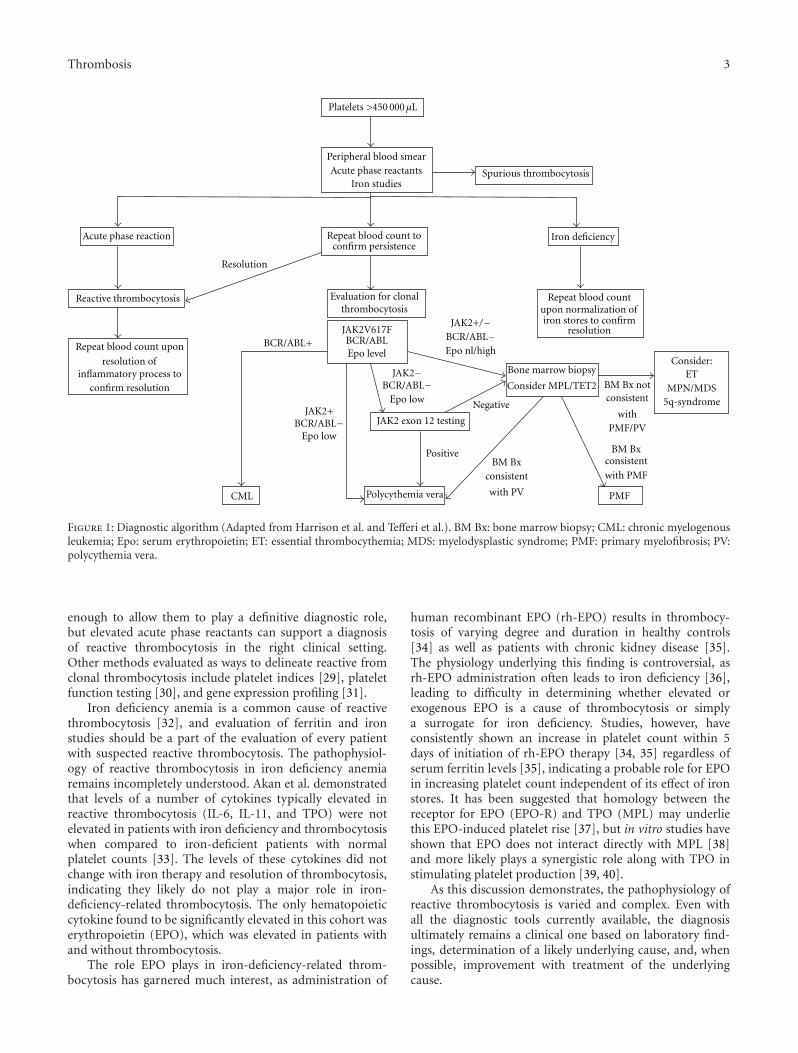
1.2. Reactive Thrombocytosis. Once the diagnosis of throm-bocytosis is confirmed by peripheral blood smear review,the diagnostic evaluation turns to determining whetherthe process is reactive or clonal in nature (Figure 1). Animportant initial step in this determination is familiarity withthe underlying causes of reactive thrombocytosis (Table 1).In adults, infection (typically acute), tissue damage, chronicinflammatory disorders, and malignancy are the most com-mon causes of reactive thrombocytosis, with one or moreof these processes present in >75% of cases of reactivethrombocytosis [4, 5]. In children, the most common causesof reactive thrombocytosis are similar, although hemolyticanemias, especially thalassemia, were a relatively commonetiology in at least one Middle Eastern study [6]. A thoroughhistory and physical examination should allow for theexclusion of multiple of the most common causes of reactivethrombocytosis.
The presence of a potential cause of reactive throm-bocytosis does not rule out a concomitant clonal process,especially in persistent thrombocytosis. Much work has beendone in an effort to come up with affordable, reliable, andrapid laboratory evaluation which can distinguish clonalprocesses from reactive thrombocytosis. The basis of many
of these proposed evaluations is based on the mechanisms ofreactive thrombocytosis.
Thrombocytopoiesis occurs in the setting of a complexcytokine milieu. Thrombopoietin (TPO) is the primaryregulator of platelet production [12, 13], but many othercytokines such as IL-1 [14–16], IL-4 [17], IL-6 [14–22], IL-11 [23], and TNF [15] play important roles inthrombocytopoiesis. Many of these same cytokines alsoplay a critical role in the body’s response to inflammatoryconditions [24, 25]. Evaluation of patients with reactiveand clonal thrombocytosis has consistently demonstratedthat circulating levels of multiple cytokines, most notablyIL-6 [14, 17, 19, 20, 22, 25, 26] are elevated in patientswith reactive thrombocytosis but not in those with clonalthrombocytosis or normal controls. Evaluation of circulatingthrombopoietin levels as a discriminant between reactive andclonal processes has proved less informative, as results havenot been consistent [18, 21, 22, 25–28]. One difficulty inutilizing circulating cytokine levels as a diagnostic tool liesin the finding that the rise in cytokine levels seems to precedethe clinical finding of thrombocytosis, with levels returningto normal or near normal by the time thrombocytosis occurs[18, 21]. This fact, along with the difficulty in bringing suchtests to clinical use, has led to a search for surrogate markerswhich may correlate with elevated cytokine levels, especiallyIL-6. Many other markers of the acute phase reaction,including C-reactive protein (CRP) [14, 19], ferritin [14],and erythrocyte sedimentation rate (ESR) [14], are alsosignificantly elevated in patients with reactive as opposed toclonal thrombocytosis.
Tefferi et al. [19] showed a correlation between IL-6 andCRP levels in a study of 91 patients with thrombocytosis,regardless of etiology. 76% of patients in this study withreactive thrombocytosis had an elevated CRP (>1.0 mg/dL),compared to 10% of patient with clonal thrombocytosis.Thus, measurement of CRP and other acute phase reactantscan serve as easily obtained surrogates for measurement ofcytokines important in thrombocytopoiesis and should bea part of any evaluation where reactive thrombocytosis issuspected. These surrogates are neither sensitive nor specific
Thrombosis 3
Peripheral blood smearAcute phase reactants
Iron studies
Repeat blood count toconfirm persistence
Iron deficiency
Reactive thrombocytosis
Repeat blood count upon
resolution ofinflammatory process to
confirm resolution
Repeat blood countupon normalization ofiron stores to confirm
resolution
Evaluation for clonalthrombocytosis
JAK2V617FBCR/ABLEpo level
CML
JAK2+BCR/ABL−
Epo low
Polycythemia vera
JAK2−BCR/ABL−
Epo low
JAK2 exon 12 testing
Positive
JAK2+/−BCR/ABL−Epo nl/high
Bone marrow biopsy
Consider MPL/TET2
PMF
Consider:ET
MPN/MDS5q-syndrome
BM Bxconsistent
with PMF
BCR/ABL+
BM Bx notconsistent
withPMF/PV
Resolution
Negative
Spurious thrombocytosis
BM Bxconsistent
with PV
Platelets >450 000µL
Acute phase reaction
Figure 1: Diagnostic algorithm (Adapted from Harrison et al. and Tefferi et al.). BM Bx: bone marrow biopsy; CML: chronic myelogenousleukemia; Epo: serum erythropoietin; ET: essential thrombocythemia; MDS: myelodysplastic syndrome; PMF: primary myelofibrosis; PV:polycythemia vera.
enough to allow them to play a definitive diagnostic role,but elevated acute phase reactants can support a diagnosisof reactive thrombocytosis in the right clinical setting.Other methods evaluated as ways to delineate reactive fromclonal thrombocytosis include platelet indices [29], plateletfunction testing [30], and gene expression profiling [31].
Iron deficiency anemia is a common cause of reactivethrombocytosis [32], and evaluation of ferritin and ironstudies should be a part of the evaluation of every patientwith suspected reactive thrombocytosis. The pathophysiol-ogy of reactive thrombocytosis in iron deficiency anemiaremains incompletely understood. Akan et al. demonstratedthat levels of a number of cytokines typically elevated inreactive thrombocytosis (IL-6, IL-11, and TPO) were notelevated in patients with iron deficiency and thrombocytosiswhen compared to iron-deficient patients with normalplatelet counts [33]. The levels of these cytokines did notchange with iron therapy and resolution of thrombocytosis,indicating they likely do not play a major role in iron-deficiency-related thrombocytosis. The only hematopoieticcytokine found to be significantly elevated in this cohort waserythropoietin (EPO), which was elevated in patients withand without thrombocytosis.
The role EPO plays in iron-deficiency-related throm-bocytosis has garnered much interest, as administration of
human recombinant EPO (rh-EPO) results in thrombocy-tosis of varying degree and duration in healthy controls[34] as well as patients with chronic kidney disease [35].The physiology underlying this finding is controversial, asrh-EPO administration often leads to iron deficiency [36],leading to difficulty in determining whether elevated orexogenous EPO is a cause of thrombocytosis or simplya surrogate for iron deficiency. Studies, however, haveconsistently shown an increase in platelet count within 5days of initiation of rh-EPO therapy [34, 35] regardless ofserum ferritin levels [35], indicating a probable role for EPOin increasing platelet count independent of its effect of ironstores. It has been suggested that homology between thereceptor for EPO (EPO-R) and TPO (MPL) may underliethis EPO-induced platelet rise [37], but in vitro studies haveshown that EPO does not interact directly with MPL [38]and more likely plays a synergistic role along with TPO instimulating platelet production [39, 40].
As this discussion demonstrates, the pathophysiology ofreactive thrombocytosis is varied and complex. Even withall the diagnostic tools currently available, the diagnosisultimately remains a clinical one based on laboratory find-ings, determination of a likely underlying cause, and, whenpossible, improvement with treatment of the underlyingcause.
4 Thrombosis
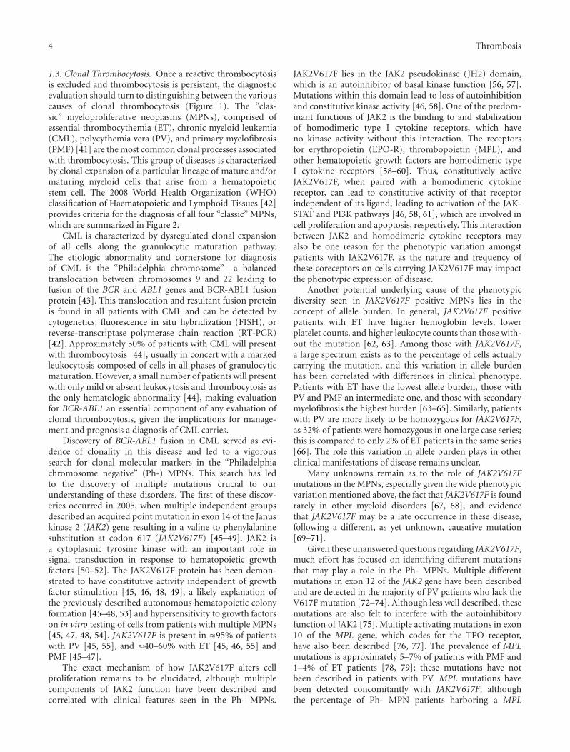
1.3. Clonal Thrombocytosis. Once a reactive thrombocytosisis excluded and thrombocytosis is persistent, the diagnosticevaluation should turn to distinguishing between the variouscauses of clonal thrombocytosis (Figure 1). The “clas-sic” myeloproliferative neoplasms (MPNs), comprised ofessential thrombocythemia (ET), chronic myeloid leukemia(CML), polycythemia vera (PV), and primary myelofibrosis(PMF) [41] are the most common clonal processes associatedwith thrombocytosis. This group of diseases is characterizedby clonal expansion of a particular lineage of mature and/ormaturing myeloid cells that arise from a hematopoieticstem cell. The 2008 World Health Organization (WHO)classification of Haematopoietic and Lymphoid Tissues [42]provides criteria for the diagnosis of all four “classic” MPNs,which are summarized in Figure 2.
CML is characterized by dysregulated clonal expansionof all cells along the granulocytic maturation pathway.The etiologic abnormality and cornerstone for diagnosisof CML is the “Philadelphia chromosome”—a balancedtranslocation between chromosomes 9 and 22 leading tofusion of the BCR and ABL1 genes and BCR-ABL1 fusionprotein [43]. This translocation and resultant fusion proteinis found in all patients with CML and can be detected bycytogenetics, fluorescence in situ hybridization (FISH), orreverse-transcriptase polymerase chain reaction (RT-PCR)[42]. Approximately 50% of patients with CML will presentwith thrombocytosis [44], usually in concert with a markedleukocytosis composed of cells in all phases of granulocyticmaturation. However, a small number of patients will presentwith only mild or absent leukocytosis and thrombocytosis asthe only hematologic abnormality [44], making evaluationfor BCR-ABL1 an essential component of any evaluation ofclonal thrombocytosis, given the implications for manage-ment and prognosis a diagnosis of CML carries.
Discovery of BCR-ABL1 fusion in CML served as evi-dence of clonality in this disease and led to a vigoroussearch for clonal molecular markers in the “Philadelphiachromosome negative” (Ph-) MPNs. This search has ledto the discovery of multiple mutations crucial to ourunderstanding of these disorders. The first of these discov-eries occurred in 2005, when multiple independent groupsdescribed an acquired point mutation in exon 14 of the Januskinase 2 (JAK2) gene resulting in a valine to phenylalaninesubstitution at codon 617 (JAK2V617F) [45–49]. JAK2 isa cytoplasmic tyrosine kinase with an important role insignal transduction in response to hematopoietic growthfactors [50–52]. The JAK2V617F protein has been demon-strated to have constitutive activity independent of growthfactor stimulation [45, 46, 48, 49], a likely explanation ofthe previously described autonomous hematopoietic colonyformation [45–48, 53] and hypersensitivity to growth factorson in vitro testing of cells from patients with multiple MPNs[45, 47, 48, 54]. JAK2V617F is present in ≈95% of patientswith PV [45, 55], and ≈40–60% with ET [45, 46, 55] andPMF [45–47].
The exact mechanism of how JAK2V617F alters cellproliferation remains to be elucidated, although multiplecomponents of JAK2 function have been described andcorrelated with clinical features seen in the Ph- MPNs.
JAK2V617F lies in the JAK2 pseudokinase (JH2) domain,which is an autoinhibitor of basal kinase function [56, 57].Mutations within this domain lead to loss of autoinhibitionand constitutive kinase activity [46, 58]. One of the predom-inant functions of JAK2 is the binding to and stabilizationof homodimeric type I cytokine receptors, which haveno kinase activity without this interaction. The receptorsfor erythropoietin (EPO-R), thrombopoietin (MPL), andother hematopoietic growth factors are homodimeric typeI cytokine receptors [58–60]. Thus, constitutively activeJAK2V617F, when paired with a homodimeric cytokinereceptor, can lead to constitutive activity of that receptorindependent of its ligand, leading to activation of the JAK-STAT and PI3K pathways [46, 58, 61], which are involved incell proliferation and apoptosis, respectively. This interactionbetween JAK2 and homodimeric cytokine receptors mayalso be one reason for the phenotypic variation amongstpatients with JAK2V617F, as the nature and frequency ofthese coreceptors on cells carrying JAK2V617F may impactthe phenotypic expression of disease.
Another potential underlying cause of the phenotypicdiversity seen in JAK2V617F positive MPNs lies in theconcept of allele burden. In general, JAK2V617F positivepatients with ET have higher hemoglobin levels, lowerplatelet counts, and higher leukocyte counts than those with-out the mutation [62, 63]. Among those with JAK2V617F,a large spectrum exists as to the percentage of cells actuallycarrying the mutation, and this variation in allele burdenhas been correlated with differences in clinical phenotype.Patients with ET have the lowest allele burden, those withPV and PMF an intermediate one, and those with secondarymyelofibrosis the highest burden [63–65]. Similarly, patientswith PV are more likely to be homozygous for JAK2V617F,as 32% of patients were homozygous in one large case series;this is compared to only 2% of ET patients in the same series[66]. The role this variation in allele burden plays in otherclinical manifestations of disease remains unclear.
Many unknowns remain as to the role of JAK2V617Fmutations in the MPNs, especially given the wide phenotypicvariation mentioned above, the fact that JAK2V617F is foundrarely in other myeloid disorders [67, 68], and evidencethat JAK2V617F may be a late occurrence in these disease,following a different, as yet unknown, causative mutation[69–71].
Given these unanswered questions regarding JAK2V617F,much effort has focused on identifying different mutationsthat may play a role in the Ph- MPNs. Multiple differentmutations in exon 12 of the JAK2 gene have been describedand are detected in the majority of PV patients who lack theV617F mutation [72–74]. Although less well described, thesemutations are also felt to interfere with the autoinhibitoryfunction of JAK2 [75]. Multiple activating mutations in exon10 of the MPL gene, which codes for the TPO receptor,have also been described [76, 77]. The prevalence of MPLmutations is approximately 5–7% of patients with PMF and1–4% of ET patients [78, 79]; these mutations have notbeen described in patients with PV. MPL mutations havebeen detected concomitantly with JAK2V617F, althoughthe percentage of Ph- MPN patients harboring a MPL

Thrombosis 5
• Bone marrow trilineage myeloproliferation• Subnormal serum erythropoietin level• Endogenous erythroid colony growth
•Platelet count ≥450× 109/L•Megakaryocyte proliferation with large and mature morphology
• Presence of JAK2 V617F or other clonal marker and no evidenceof reactive thrombocytosis
•Megakaryocyte proliferation and atypia accompanied by reticulinand/or collagen fibrosis; or in the absence of overt fibrosis, themegakaryocyte changes must be accompanied by increased
primary myelofibrosis)
other myeloid neoplasm
• Presence of JAK2 V617F or other clonal marker and no evidence
of reactive marrow fibrosis
Primary myelofibrosis minor criteria• Leukoerythroblastosis• Increased serum lactate dehydrogenase
• Anemia
• Palpable splenomegaly
Polycythemia vera major criteria•Hemoglobin >18.5 g/dL (men) >16.5 g/dL (women)‡
• Presence of JAK2 V617F or similar mutation
Polycythemia vera minor criteria
‡ Hemoglobin or hematocrit values above the 99th percentile of the
reference range for age, sex, or altitude of residence; or red cell mass >25%above mean normal predicted or hemoglobin >17 g/dL (men), or >15 g/dL(women) if associated with a sustained increase of ≥2 g/dL from baseline thatcannot be attributed to correction of iron deficiency.Diagnosis of polycythemia vera requires meeting either both major criteriaand one minor criterion or the first major criterion and two minor criteria
Essential thrombocythemia major criteria
• Not meeting WHO criteria for CML, PV, PMF,MDS or other myeloid neoplasm
Diagnosis of essential thrombocythemia requires meeting all four major criteria
Primary myelofibrosis major criteria
marrow cellularity and granulocytic proliferation (i.e., prefibrotic
• Not meeting WHO criteria for CML, PV, MDS, or
Diagnosis of primary myelofibrosis requires meeting all three major criteria and two minor criteria
Figure 2: 2008 WHO diagnostic criteria for PV, ET, and PMF. CML: chronic myelogenous leukemia; ET: essential thrombocythemia; MDS:myelodysplastic syndrome; PMF: primary myelofibrosis; PV: polycythemia vera.
mutation is higher in those without a JAK2 mutation [78].Patients with MPL mutations do have a slightly differentclinical phenotype, as they typically have lower hemoglobinlevels and higher platelet counts than those without MPLmutations [80].
Another gene of interest in the MPNs is TET2, aputative tumor suppressor gene located on chromosome4q24. Mutations of TET2 have been detected in a varietyof myeloid disorders, including Ph- MPNs. Approximately13% of Ph- MPN patients harbor a TET2 mutation [81],and it appears to be more common in patients >60 yearsold [82]. Clonal analysis also suggests that TET2 mutationscan occur prior to or following JAK2 mutation in patientswhere the two mutations exist concomitantly, indicating thatTET2 mutation is not a prerequisite for JAK2 mutation [83].Although the role of testing for novel mutations such as MPLand TET2 as part of the diagnostic evaluation for the Ph-MPNs is less clear than that of JAK2 evaluation, testing forthese mutations has been shown to increase sensitivity whenutilizing the WHO diagnostic criteria [84].
Even as the utility of molecular data in the diagnosisof Ph- MPNs increases, the diversity of disease present inthe face of these mutations has required the diagnosis ofthe Ph- MPNs to remain primarily a clinical one, based onthe 2008 WHO guidelines [42], summarized in Figure 2.Diagnosis of PV is based on the demonstration of increased
red blood cell volume, depressed serum erythropoietin levels,and the presence of a clonal marker (JAK2 mutation)[85]. Thrombocytosis is also present in ≈50% of casesof PV [86], and the presence of thrombocytosis in theabsence of fever or infection is a minor criterion in thePolycythemia Vera Study Group diagnostic criteria for PV[86]. Although thrombocytosis is not a part of the WHOdiagnostic criteria for PV, its presence, especially in thesetting of evidence of elevated red cell volume, is entirelyconsistent with the diagnosis. Thrombocytosis can also bethe only hematologic manifestation of PV, as the expectederythrocytosis can be masked by volume expansion orconcomitant iron deficiency [87–89]. Diagnosis of PMF isbased on the presence of typical findings on bone marrowbiopsy, most notably reticulin fibrosis and megakaryocyticproliferation. Thrombocytosis is present in ≈30% of PMFpatients at diagnosis, with this number decreasing as thedisease progresses and splenomegaly often leads to throm-bocytopenia [90]. Diagnosis of essential thrombocythemia isessentially one of exclusion when no other diagnosis can bemade in the setting of persistent clonal thrombocytosis.
2. Thrombotic Complications
Reactive thrombocytosis is generally felt thought to be aself-limited process which resolves with resolution of the
6 Thrombosis
underlying disorder when possible. The risk of thromboticcomplications with reactive thrombocytosis is felt to below, as 1.6% of patients with reactive thrombocytosis hadthrombotic complications in one large case series [4]. Allof these thrombotic events were venous in location andoccurred in patients with other risk factors (postoperativesetting or underlying malignancy). Even in cases of extremereactive thrombocytosis, the risk of thrombotic complicationis relatively low (4–6%) [91, 92], although reactive thrombo-cytosis has been shown to be an independent risk factor forthrombosis in patients otherwise at high risk for thrombosis,including trauma patients [93] and following coronary arterybypass grafting [94]. Patients in these settings have manyother concurrent prothrombotic risk factors which likelycontribute as much if not more to thrombotic risk than thepresence of reactive thrombocytosis.
In clonal thrombocytosis, especially in ET and PV,thrombotic complications are a major cause of morbidityand mortality and the primary factor in determining treat-ment strategy. Although thrombosis can be an issue in othercauses of clonal thrombocytosis, it is most common andmost thoroughly investigated in PV and ET, and thus thesediseases will be the focus of discussion.
2.1. Microvascular Thrombotic Disease. Many patients withclonal thrombocytosis will experience vasomotor symptomsincluding headache, syncope, chest pain, erythromelalgia,acrocyanosis, and visual changes. These symptoms are due tomicrovascular inflammation, platelet aggregation and arteri-olar microthrombi formation [95, 96] and are more commonin ET than PV, with some vasomotor symptoms presentin 29–40% of ET patients at presentation [97–99]. Thefrequency of erythromelalgia, the most common vasomotorcomplication of PV and ET, is not directly correlated withhigher platelet counts, and in fact, vasomotor symptomsare essentially never present in reactive thrombocytosis.These facts underlie the important role that qualitativeplatelet abnormalities [96] and increased thromboxane-induced platelet activation [100] play in causing vasomotorsymptoms, as erythromelalgia has been described in patientswith relatively normal platelet counts in a multitude of non-hematologic conditions [101]. The lack of specificity of thesesymptoms makes estimating prevalence difficult, but thetypical prompt response of ET-related vasomotor symptomsto aspirin can be a useful diagnostic as well as therapeuticintervention.
2.2. Macrovascular Thrombotic Disease. The rate of macro-vascular thrombotic complications at diagnosis ranges 11–25% in ET and 12–39% in PV [102–106], with arterialthrombosis comprising the majority of events. The cere-brovascular circulation, either in the form of stroke ortransient ischemic attack, is the most common site of arterialthrombotic disease, followed by the coronary arteries andperipheral vasculature [102, 105, 107, 108]. The arterial pre-dominance of thrombotic events is more marked in ET thanin PV, where up to ≈40% of thrombotic events are venousin nature [102, 105, 109]. Of special significance are venous
thromboses in unusual locations such as the splanchnic veinsand cerebral sinuses, as >50% of venous thrombotic eventsin PV and ET occur in these locations [110, 111]; this isespecially true in younger patients [112]. Given the frequencyof thrombotic events in these unusual locations, strongconsideration should be given to evaluation for occult Ph-MPN in any patient presenting with splanchnic or cerebralsinus thrombosis. Case series have reported that 23–51% ofpatients suffering from splanchnic thrombosis without anyother risk factors can be diagnosed with an underlying Ph-MPN at the time of thrombosis [113, 114], and JAK2V617Fmutations have been demonstrated in a number of suchpatients, many of which only met full criteria for a Ph- MPNlater in their course [115, 116].
The risk for thrombosis does not stop with diagnosis,and large case series have demonstrated a significant riskof thrombotic events following diagnosis even in patientstreated with cytoreductive and antiplatelet therapy. Wolan-skyj et al. compiled a historic cohort of 322 patients with ETwith 70% having >10 years of followup [106]. In this cohort,26% of patients presented with evidence of thrombosis;the cumulative probability of thrombosis at 5, 10, and 20years was 32%, 42%, and 52%, respectively. 82% of thesepatients received cytoreductive therapy and 62% receivedaspirin as part of their treatment regimen. Similarly, in alarge series of 1213 Italian patients with ET [105] 34% ofpatients either presented with thrombosis (20%) or had ahistory of thrombosis prior to presentation (14%). After amedian of 5 years of followup, an additional 19% of patientshad suffered a thrombotic event, with a cumulative incidenceof thrombosis of 53%.
2.3. Risk Factors for Thrombosis. Age is a significant riskfactor for thrombosis in the general population [117],and multiple different epidemiologic studies have shownincreasing risk for thrombosis in PV and ET with increasingage [102, 105–107]. Another consistently demonstrated riskfactor for thrombosis in patients with PV and ET is a priorthrombotic event [98, 102, 105–107]. The combination ofage >65 and a prior thrombotic event is associated witha markedly increased risk of thrombosis in PV, with athrombosis rate in the European Collaboration on Low-Dose Aspirin in Polycythemia Vera (ECLAP) trial of 10.9events/100 persons/year as compared to 2.5 events/100persons/year in those without either risk factor [102].
Another more recently described independent risk factorfor thrombosis in PV and ET is leukocytosis [106, 118–120]. The precise mechanisms behind this association are notentirely clear, although it is likely that qualitative leukocyteabnormalities play as large a role as quantitative increasesin these diseases. The relative importance of qualitativeleukocyte abnormalities is also supported by the fact thatpatients with CML, who typically have the highest leukocytecounts of all the MPNs, have a much lower rate of thrombosisthan any of the Ph- MPNs [121]. Activated granulocytesare known to play an important role in platelet activationand endothelial injury [104], and patients with ET andPV have evidence of increased granulocyte activation as
Thrombosis 7
compared to normal controls [122]; an association betweenJAK2V617F and constitutive activation of granulocytes hasalso been suggested [64]. Another feature of this increasedrisk which speaks to a qualitative component is the degreeof leukocytosis which confers an excess risk. Multiple serieshave shown that a leukocyte count of >8.7 × 10⁹/L, wellwithin the normal range, is associated with excess thromboticrisk in both PV and ET [118, 120], with ROC analysisin one study showing a leukocyte count of 9.4 × 10⁹/Lhaving the best sensitivity and specificity for demarcatinghigh-and low-risk patients [118]. The implications of thisassociation in regards to treatment of these disorders remaincontroversial, although it may provide a rationale for theefficacy of cytoreductive therapy and a leukocyte countof <10 × 109/L has been integrated into recently revisedEuropean LeukemiaNet response criteria for PV and ET[123].
In contrast to the increased risk in leukocytosis, anincreased risk of thrombotic events with increasing plateletcount has not been consistently demonstrated. Multiplestudies have failed to show a direct correlation betweenincreasing platelet count and thrombotic risk [91, 107,124], although ≈80% of thrombotic events occur in thosewith a platelet count >600 × 109/L [125]. Cytoreductivetherapy leading to decreased platelet counts has beenshown to decrease microcirculatory vasomotor symptomsand thrombosis [95, 125, 126], but it is unclear if thisis directly due to decreases in the platelet count or toother effects of cytoreductive therapy such as a decrease inleukocyte count. Likely more important than the absolutenumber of platelets are the multiple qualitative abnormalitieswhich have been noted in PV and ET, including decreasedresponse to adenosine diphosphate and epinephrine [103,127] altered glycoprotein receptors [127, 128], and the excessthromboxane production noted in the Ph- MPNs, leading toincreased platelet activation [100, 129].
The role of erythrocytosis as an independent risk factor issimilarly complex. Multiple studies have shown that increas-ing hematocrit is associated with increasing blood viscosityand thrombotic risk [130], with the cerebral circulationbeing especially vulnerable [131]. Increasing viscosity leadsto a displacement of platelets to the periphery of arterialblood flow under high shear stress conditions [132], leadingto greater interaction between platelets as well as plateletsand the underlying endothelium. Many patients with PValso have an elevated number of platelets with the above-mentioned qualitative abnormalities, and bringing theseplatelets in closer contact with each other and the vessel wallis one likely reason for the increase in arterial thrombotic riskin these patients. These functional platelet abnormalities,as well as qualitative red blood cell (RBC) abnormalitiesincluding increased RBC adhesion to endothelial cells [133],likely play an important role in thrombosis in PV and ET, asanimal models have failed to show hyperviscosity alone as asignificant risk factor for thrombosis [134].
Evaluation of the role of traditional cardiovascular riskfactors to thrombotic risk in PV and ET has led to conflictingresults, with some studies suggesting increased risk withhypercholesterolemia [107] and smoking [135] while others
have shown no increased risk with any known cardiovascularrisk factors [106, 124].
The discovery of the JAK2V617F mutation has led toextensive evaluation to determine whether those carryingthis mutation have a different disease phenotype than thosewho are JAK2 wild type, and this evaluation has includedexamining the risk of thrombosis in both groups. A theoret-ical basis for an increased risk of thrombosis in PV and ETlies in the fact that the increased RBC-endothelial adhesionmentioned above is felt to be mediated by a JAK2V617F-mediated mechanism [133]. Thus far, the implications ofJAK2V617F mutation on thrombosis risk are unclear, withmultiple retrospective studies indicating an increased riskof thrombosis in ET [136–138], while others have failed toshow such an association [118, 139]. A recent meta-analysisof 21 studies in ET patients suggested an increased risk ofincreased risk of thrombosis (OR 1.92, 95% CI 1.45–2.53)in those with JAK2V617F mutations [140]; the increasedrisk was for both venous and arterial thromboses. Othergroups have examined increasing JAK2 allele burden as arisk factor for thrombosis and a potential reason for theconflicting results regarding the impact of JAK2 mutationson thrombotic risk. Analysis of 639 patients with ET and323 with PV by Vannucchi et al. reported an increased riskof thrombosis (both arterial and venous) in the small per-centage of ET patients who were homozygous for JAK2V617F(hazard ratio 3.97, 95% confidence interval [CI] 1.34–11.7) compared with JAK2 wild-type patients [66]. No suchdifference was present in patients with PV and other studieshave not demonstrated this increased risk in ET patients[118].
The impact of MPL mutations on thrombotic risk hasbeen less well evaluated and is difficult given the smallnumber of patients with these mutations and its frequentcooccurrence with JAK2 mutations. In the small number ofstudies evaluating thrombotic risk in these patients, MPLmutation does not appear to be an independent risk factorfor thrombosis [78, 80].
3. Bleeding Risk
A paradoxical risk of bleeding has been noted in patients withthrombocytosis, especially extreme thrombocytosis [91, 98],and bleeding in this setting is usually mucocutaneous innature. This excess risk is likely multifactorial; in clonalthrombocytosis, platelet function abnormalities no doubtplay a major role. Another potential cause of bleedingregardless of the cause of thrombocytosis is acquired vonWillebrand’s syndrome (AVWS) due to increased adsorptionof large von Willebrand factor (vWF) multimers [141, 142]by the abnormally high number of circulating platelets.Testing for AVWS can be undertaken in patients withthrombocytosis and bleeding to evaluate for a decreasein large vWF multimers, and in patients with extremethrombocytosis, evaluation for AVWS can be incorporatedinto the risk stratification of otherwise low-risk patients priorto the initiation of antiplatelet therapy.
8 Thrombosis
4. Treatment of Thrombocytosis
4.1. Reactive Thrombocytosis. Reactive thrombocytosis, asmentioned above, is felt to be self-limited with little excessassociated thrombotic risk. Because of this lack of throm-botic risk as well as a theoretical risk of paradoxical bleeding,no antiplatelet therapy is recommended, even for extremethrombocytosis. The occurrence of thrombosis in patientsfelt to have reactive thrombocytosis may be reason to under-take evaluation for a concomitant clonal thrombocytosis,especially with splanchnic or cerebral thrombosis.
In patients who present with extreme thrombocytosisof unknown etiology and evidence of active bleeding orcritical thrombosis, plateletpheresis can provide a rapidreduction in platelet count while the diagnostic evaluationis undertaken [143, 144]. In patients where a clonal causeof thrombocytosis is known or suspected, cytoreductivetherapy (discussed below) can also be initiated at high dosesfor additional rapid platelet-lowering effect [145].
4.2. Clonal Thrombocytosis. Treatment strategies in PV andET are focused on reducing the risk of thrombotic events inthose at risk for them, as thrombosis is the most commoncomplication leading to morbidity and mortality in thesedisorders. Treatments to this end fall into essentially twocategories: cytoreductive therapy with an aim to decreasecirculating platelet count (as well as hematocrit in PV) andantiplatelet therapy, usually in the form of aspirin. Giventhese treatment approaches, much work has been done inan effort to risk-stratify patients to determine which patientswill benefit from either or both of the above classes oftherapy. Risk assessment and subsequent therapy based onrisk of thrombosis has led to a variety of treatment strategiesbased on risk (Figures 3 and 4).
4.3. Essential Thrombocytosis. In ET, multiple retrospectivestudies have consistently described age >60 and a priorthrombotic event as risk factors for thrombosis [98, 106,107]. Patients meeting either of these criteria are generallyconsidered to be in a “high risk” group and felt thoughtto benefit from both antiplatelet and cytoreductive therapy.This approach is supported by multiple randomized clinicaltrials. The first, by Cortelazzo et al. [126] prospectivelyevaluated 114 patients with ET who met the above high-riskcriteria. Patients were randomized to receive hydroxyurea(HU), an antimetabolite which primarily acts in S phaseand is effective in reducing platelet counts in ET, versusno cytoreductive therapy. HU dose was titrated to maintaina platelet count <600 × 109/L and after a median of 27months of followup, 3.6% of patients receiving HU hadsuffered a thrombotic event, compared to 24% in the controlgroup, a statistically significant difference. It should be notedthat 69% of patients were on some form of antiplatelettherapy (aspirin or ticlodipine), and use of these agentswas similar in the two treatment groups. Whereas thistrial demonstrated the benefit of cytoreductive therapy,the Medical Research Council primary thrombocythemia-1(PT-1) trial [146] aimed to determine the best method of
cytoreduction, comparing the use of HU with anagrelide.Anagrelide is an orally active quinazolinone derivative whichwas initially developed as an inhibitor of platelet aggregationbut has also been shown to reduce platelet count with littleeffect on other hematopoietic cell lines through blockade ofmegakaryocyte differentiation and proliferation [147]. In thePT-1 trial, a total of 809 patients who met high-risk criteria(defined as any of the following: age >60 years, prior throm-bosis, hypertension or platelet count >1,000 × 109/L) wererandomized to receive low-dose aspirin plus either HU oranagrelide titrated to maintain a platelet count <400× 109/L.Patients randomized to HU + aspirin had a significantlylower rate of arterial thrombosis, major hemorrhage, andtransformation to myelofibrosis, but significantly higher riskof venous thrombosis. There were no differences in controlof platelet count in the two groups, and the overall risk ofthrombosis after a median of 39 months of followup was7.6% in the HU group compared to 10.1% in the anagrelidegroup.
These results have led to widespread adoption of HU +aspirin as first-line therapy in patients with high-risk ET.Anagrelide + aspirin does appear to have benefit in reducingthrombotic events in patients in ET, if one comparesthrombotic rates in the PT-1 trial with that of the studyby Cortelazzo et al. [126] and other historic controls.Thus, anagrelide continues to have an important role inthe treatment of ET, especially when toxicities such ascytopenias or cutaneous lesions limit the use of HU. Thereis growing evidence that combination therapy with HUand anagrelide can be effective in treating patients eitherrefractory to or intolerant of large doses of HU [148].Compared to treatment with anagrelide alone, this combinedapproach allows for continuation of reduced dose HU, whichmay allow for continued leukocyte reduction, which likelycontributes at least a portion of the protective benefit of HUtreatment.
Goals of treatment in high-risk ET are primarily definedin terms of reduction in platelet count, with a goal of <400 ×109/L being a common target given the findings of the PT-1 trial [146]. Patients in the study by Cortelazzo et al. [126]had a targeted platelet count of <600× 109/L, indicating thata slightly relaxed platelet goal may still allow for benefit incytoreductive therapy in those unable to maintain a plateletcount of <400 × 109/L. The recently published EuropeanLeukemiaNet response criteria for ET [123] include manyother factors, with complete response defined as (1) plateletcount <400 × 109/L, (2) no disease-related symptoms, (3)normal spleen size on imaging, and (4) white blood cellcount <10 × 109/L. Partial response is defined as plateletcount <600 × 109/L or a decrease of >50% from baseline.The utility of these criteria in decision-making regardingtreatment strategies in ET is yet to be determined.
Growing evidence supports the concept that patientswith ET who do not meet high-risk criteria do well withless aggressive therapy. Rugerri et al. [149] prospectivelyfollowed 65 patients <60 years old with no thrombosishistory and a platelet count <1500 × 109/L, and thethrombosis rate in this cohort (1.91/100 patient-years) wasnot significantly different than that of age and sex-matched

Thrombosis 9
Essential thrombocytosis• Individualized risk assessment
Age >60
Prior thrombosis
(?) Platelet count >1500× 109/L
•Hydroxyurea• Goal platelet count <400× 109/L
• Low dose aspirin(unless contraindicated)
Refractory/intolerant
Relax goal platelet count to 600× 109/L
Combination therapy with anagrelide
Other agent(s):
• IFN-alpha (pregnancy)
••
32P (in elderly)Clinical trial
Low risk: (Age <60; no priorthrombosis)
Leukocyte countJAK2 mutation status
Other vascular risk factors
Low-dose aspirin
Vasomotorsymptoms
Consider low-dose aspirin based onindividual patient risk profile
High risk:
Cytoreductive therapy
AndAntiplatelet therapy
Secondary risk assessment:first-line agent
Figure 3: Treatment algorithm for ET.
controls (1.5/100 patient-years). Aspirin was used only in theevent of microvascular symptoms; less than 25% of patientsin both arms received aspirin during the median 4.1 years offollowup.
Antiplatelet therapy, most commonly aspirin, also playsan important role in the treatment of ET, and its efficacyin the treatment of microvascular complications has beenwell established [95, 96]. Its role in the prevention ofmacrovascular complications is less clear. Retrospective eval-uation suggests antiplatelet therapy may reduce thromboticcomplications in a high-risk population [124, 150], butrandomized data is lacking. Further support for the use ofantiplatelet therapy in ET has been extrapolated from theECLAP trial, which demonstrated a significant reduction invascular events in low and intermediate risk PV patientstreated with aspirin as compared to placebo [151].
The role of antiplatelet therapy in a low-risk ET popu-lation was recently examined in a retrospective evaluationof 300 Spanish patients by Alvarez-Larran et al. [152].Rates of thrombosis were not significantly different inthose treated with antiplatelet therapy and those followedwith observation; subgroup analysis did demonstrate thatpatients harboring the JAK2V617F mutation did have asignificantly increased rate of venous, but not arterialthrombosis. Patients with cardiovascular risk factors whowere observed without antiplatelet therapy also had a higherrate of arterial thrombosis compared to those receiving
aspirin. Patients with a platelet count of >1000 × 109/Ltreated with antiplatelet therapy had a significantly increasedrisk of bleeding. These data reinforce the importance of athorough, individualized approach to risk assessment in ETpatients prior to determining a treatment strategy.
4.4. Polycythemia Vera. The foundation of therapy in poly-cythemia vera is therapeutic phlebotomy to maintain ahematocrit <45% in men and <42% in women (Figure 4).This approach is based on the phlebotomy strategy utilized inmultiple randomized PVSG studies that demonstrated phle-botomy alone was associated with equivalent or improvedsurvival when compared to phlebotomy plus additionalagents such as busulfan, 32P, and chlorambucil [153, 154].Addition of these agents was consistently associated withincreased risk of late hematologic complications, especiallyacute myeloid leukemia. Treatment with phlebotomy alone,however, was associated with a relatively high risk ofthrombosis in these studies, especially early in the course oftherapy, with thrombosis rates of 23% in the first two years.
In an effort to address this early thrombotic risk, asubsequent Phase II PVSG trial including 51 patients addedHU to phlebotomy with a goal hematocrit <50% and plateletcount <600 × 109/L. The thrombotic rate seen in the first2 years of therapy was 9%, significantly lower than the 23%noted in the earlier PVSG trial [155]. The risk of leukemictransformation in this cohort was 6% and not significantly

10 Thrombosis
All patients:
• Phlebotomy (goal HCT <42% in women,
<45% in men)
• Low dose aspirin (unless contraindicated)
• Individualized risk assessment
Age >60
Prior thrombosis
Low risk: (Age <60; no prior thrombosis)
No cytoreductive therapy
Consider hydroxyurea or othercytoreductive agent based on toxicity
profile
Polycythemia vera
High risk:
OR
Figure 4: Treatment algorithm for PV.
different than that seen in the phlebotomy only arm ofearlier PVSG studies, but the leukemogenic risk of HU isstill hotly debated [156–158]. No phase III studies comparingHU + phlebotomy with phlebotomy alone in PV have beenperformed, but the results of this Phase II study have led tothe common use of HU in addition to phlebotomy in thoseconsidered high risk for thrombosis (Age >60 and/or priorthrombosis) [159].
The use of antiplatelet therapy in PV was initiallythought to be associated with poorer outcomes and increasedbleeding risk based on an early PVSG study [160]. Thisprotocol, however, utilized a total dose of 900 mg of aspirindaily along with dipyridamole, another antiplatelet agent.Subsequent studies have demonstrated the efficacy and safetyof lower doses of aspirin therapy in PV. A Gruppo ItalianoStudio Policitemia Vera study [161] randomized 112 PVpatients to receive either aspirin (40 mg/day) or placebo.The low dose aspirin was well tolerated, led to a significantdecrease in serum thromboxane B2 levels, and was notassociated with an increased risk of bleeding. The ECLAPtrial [151] randomized 518 patients who had no otherindication for antiplatelet therapy to low-dose aspirin orplacebo and demonstrated significant reduction in the risk ofnonfatal myocardial infarction, nonfatal stroke, pulmonaryembolism, deep venous thrombosis, and death. Bleeding riskwas not significantly different in the two treatment groups.These data have led to recommendations for use of low-doseaspirin in all patients with PV, regardless of risk, except in theface of contraindication.
Other agents, such as interferon alpha [162, 163] and, inEurope, pipobroman [164, 165] have also been used in the
treatment of both ET and PV. The use of these therapies isgenerally limited to those who have failed or are intolerantof more standard therapy; interferon alpha is generallyconsidered the therapy of choice in pregnant patientsrequiring cytoreductive therapy given its low teratogenicity[166]. A variety of JAK2 inhibitors have been evaluatedin preclinical studies in the Ph-MPNs and are currentlyprogressing through clinical trials [167, 168]; the majorityof patients enrolled in JAK2 inhibitor trials to this pointhave had PMF. Results have been promising, and further trialresults and evaluation of new potential therapeutic agentsmay lead to a paradigm shift in the treatment of all theMPNs, including ET and PV.
5. Conclusion
Thrombocytosis exists in the setting of a variety of clinicalsituations and can have widely diverse underlying etiologies.Clonal thrombocytosis is often due to one of the Ph- MPNs,and in this setting, the risk of thrombosis is great andshould be the primary factor guiding treatment strategy. Themechanisms underlying this increased thrombotic risk arenot yet fully understood, and the role of risk factors suchas JAK2 status and leukocytosis has yet to be conclusivelyestablished. Integration of these and other risk factors into arisk-based treatment decision model should allow for betterpatient selection for cytoreductive therapy. The ongoingdiscovery and description of new molecular abnormalities inthese disorders should allow for advances in both diagnosisand treatment of clonal thrombocytosis.

Thrombosis 11
References
[1] R. C. Skoda, “Thrombocytosis,” Hematology, pp. 159–167,2009.
[2] M. Ruggeri, A. Tosetto, M. Frezzato, and F. Rodeghiero,“The Rate of Progression to Polycythemia Vera or Essen-tial Thrombocythemia in Patients with Erythrocytosis orThrombocytosis,” Annals of Internal Medicine, vol. 139, no.6, pp. 470–I32, 2003.
[3] C. N. Harrison, D. Bareford, N. Butt et al., “Guidelinefor investigation and management of adults and childrenpresenting with a thrombocytosis,” British Journal of Haema-tology, vol. 149, no. 3, pp. 352–375, 2010.
[4] M. Griesshammer, M. Bangerter, T. Sauer, R. Wennauer, L.Bergmann, and H. Heimpel, “Aetiology and clinical signif-icance of thrombocytosis: analysis of 732 patients with anelevated platelet count,” Journal of Internal Medicine, vol. 245,no. 3, pp. 295–300, 1999.
[5] C. R. Santhosh-Kumar, M. D. Yohannan, K. E. Higgy, andS. A. Al-Mashhadani, “Thrombocytosis in adults: analysis of777 patients,” Journal of Internal Medicine, vol. 229, no. 6, pp.493–495, 1991.
[6] M. D. Yohannan, K. E. Higgy, S. A. Al-Mashhadani, and C. R.Santhosh-Kumar, “Thrombocytosis,” Clinical Pediatrics, vol.33, no. 6, pp. 340–343, 1994.
[7] D. H. Buss, A. W. Cashell, M. L. O’Connor, F. Richards,and L. D. Case, “Occurrence, etiology, and clinical signif-icance of extreme thrombocytosis: a study of 280 cases,”American Journal of Medicine, vol. 96, no. 3, pp. 247–253,1994.
[8] C. V. Hutchinson, P. Stelfox, and K. S. Rees-Unwin, “Needle-like cryoglobulin crystals presenting as spurious thrombocy-tosis,” British Journal of Haematology, vol. 135, no. 3, p. 280,2006.
[9] H. S. Ballard and G. Sidhu, “Cytoplasmic fragments causingspurious platelet counts in hairy cell leukemia. Ultrastruc-tural characterization,” Archives of Internal Medicine, vol. 141,no. 7, pp. 942–944, 1981.
[10] N. Kakkar, “Spurious rise in the automated platelet countbecause of bacteria,” Journal of Clinical Pathology, vol. 57, no.10, pp. 1096–1097, 2004.
[11] C. Lawrence and B. Atac, “Hematologic changes in massiveburn injury,” Critical Care Medicine, vol. 20, no. 9, pp. 1284–1288, 1992.
[12] K. Kaushansky, “Thrombopoietin: the primary regulator ofplatelet production,” Blood, vol. 86, no. 2, pp. 419–431, 1995.
[13] K. Kaushansky, “Thrombopoietin and the hematopoieticstem cell,” Blood, vol. 92, no. 1, pp. 1–3, 1998.
[14] M. G. Alexandrakis, F. H. Passam, I. A. Moschandrea etal., “Levels of serum cytokines and acute phase proteins inpatients with essential and cancer-related thrombocytosis,”American Journal of Clinical Oncology, vol. 26, no. 2, pp. 135–140, 2003.
[15] K. Dan, S. Gomi, K. Inokuchi et al., “Effects of interleukin-1 and tumor necrosis factor on megakaryocytopoiesis:mechanism of reactive thrombocytosis,” Acta Haematologica,vol. 93, no. 2-4, pp. 67–72, 1995.
[16] H. Hamaguchi, N. Takano, K. Saito, H. Enokihara, S. Furu-sawa, and H. Shishido, “Interaction of monocytes and T cellsin the regulation of normal human megakaryocytopoiesis invitro: role of IL-1 and IL-2,” British Journal of Haematology,vol. 76, no. 1, pp. 12–20, 1990.
[17] I. C. Haznedaroglu, I. Ertenli, O. I. Ozcebe et al., “Megakar-yocyte-related interleukins in reactive thrombocytosis versusautonomous thrombocythemia,” Acta Haematologica, vol.95, no. 2, pp. 107–111, 1996.
[18] C. C. Folman, M. Ooms, B. Bart Kuenen et al., “The roleof thrombopoietin in post-operative thrombocytosis,” BritishJournal of Haematology, vol. 114, no. 1, pp. 126–133, 2001.
[19] A. Tefferi, T. C. Ho, G. J. Ahmann, J. A. Katzmann, and P. R.Greipp, “Plasma interleukin-6 and C-reactive protein levelsin reactive versus clonal thrombocytosis,” American Journalof Medicine, vol. 97, no. 4, pp. 374–377, 1994.
[20] C. W. Hollen, J. Henthorn, J. A. Koziol, and S. A. Burstein,“Elevated serum interleukin-6 levels in patients with reactivethrombocytosis,” British Journal of Haematology, vol. 79, no.2, pp. 286–290, 1991.
[21] A. Ishiguro, Y. Suzuki, M. Mito et al., “Elevation of serumthrombopoietin precedes thrombocytosis in acute infec-tions,” British Journal of Haematology, vol. 116, no. 3, pp.612–618, 2002.
[22] H. C. Hsu, W. H. Tsai, M. L. Jiang et al., “Circulating levels ofthrombopoietic and inflammatory cytokines in patients withclonal and reactive thrombocytosis,” Journal of Laboratoryand Clinical Medicine, vol. 134, no. 4, pp. 392–397, 1999.
[23] F. Wendling and Z. C. Han, “Positive and negative regulationof megakaryocytopoiesis,” Bailliere’s Clinical Haematology,vol. 10, no. 1, pp. 29–45, 1997.
[24] I. Ertenli, S. Kiraz, M. A. Ozturk, I. C. Haznedaroglu, I.Celik, and M. Calguneri, “Pathologic thrombopoiesis ofrheumatoid arthritis,” Rheumatology International, vol. 23,no. 2, pp. 49–60, 2003.
[25] F. Heits, M. Stahl, D. Ludwig, E. F. Stange, and W. Jelkmann,“Elevated serum thrombopoietin and interleukin-6 concen-trations in thrombocytosis associated with inflammatorybowel disease,” Journal of Interferon and Cytokine Research,vol. 19, no. 7, pp. 757–760, 1999.
[26] M. Uppenkamp, E. Makarova, S. Petrasch, and G. Brittinger,“Thrombopoietin serum concentration in patients withreactive and myeloproliferative thrombocytosis,” Annals ofHematology, vol. 77, no. 5, pp. 217–223, 1998.
[27] A. Cerutti, P. Custodi, M. Duranti, P. Noris, and C. L.Balduini, “Thrombopoietin levels in patients with primaryand reactive thrombocytosis,” British Journal of Haematology,vol. 99, no. 2, pp. 281–284, 1997.
[28] M. Hou, J. Carneskog, U. H. Mellqvist et al., “Impactof endogenous thrombopoietin levels on the differentialdiagnosis of essential thrombocythaemia and reactive throm-bocytosis,” European Journal of Haematology, vol. 61, no. 2,pp. 119–122, 1998.
[29] J. C. Osselaer, J. Jamart, and J. M. Scheiff, “Platelet distri-bution width for differential diagnosis of thrombocytosis,”Clinical Chemistry, vol. 43, no. 6, pp. 1072–1076, 1997.
[30] A. E. Tsantes, A. Dimoula, S. Bonovas et al., “The role of thePlatelet Function Analyzer (PFA)-100 and platelet aggregom-etry in the differentiation of essential thrombocythemia fromreactive thrombocytosis,” Thrombosis Research, vol. 125, no.2, pp. 142–146, 2010.
[31] D. V. Gnatenko, W. Zhu, X. Xu et al., “Class predictionmodels of thrombocytosis using genetic biomarkers,” Blood,vol. 115, no. 1, pp. 7–14, 2010.
[32] I. Kuku, E. Kaya, S. Yologlu, R. Gokdeniz, and A. Baydin,“Platelet counts in adults with iron deficiency anemia,”Platelets, vol. 20, no. 6, pp. 401–405, 2009.
[33] H. Akan, N. Giiven, I. Aydogdu, M. Arat, M. Beksac, andK. Dalva, “Thrombopoietic cytokines in patients with iron

12 Thrombosis
deficiency anemia with or without thrombocytosis,” ActaHaematologica, vol. 103, no. 3, pp. 152–156, 2000.
[34] P. J. Stohlawetz, L. Dzirlo, N. Hergovich et al., “Effects oferythropoietin on platelet reactivity and thrombopoiesis inhumans,” Blood, vol. 95, no. 9, pp. 2983–2989, 2000.
[35] C. J. Kaupke, G. C. Butler, and N. D. Vaziri, “Effect ofrecombinant human erythropoietin on platelet productionin dialysis patients,” Journal of the American Society ofNephrology, vol. 3, no. 10, pp. 1672–1679, 1993.
[36] E. Streja, C. P. Kovesdy, S. Greenland et al., “Erythropoietin,iron depletion, and relative thrombocytosis: a possible expla-nation for hemoglobin-survival paradox in hemodialysis,”American Journal of Kidney Diseases, vol. 52, no. 4, pp. 727–736, 2008.
[37] E. Bilic and E. Bilic, “Amino acid sequence homology ofthrombopoietin and erythropoietin may explain thrombo-cytosis in children with iron deficiency anemia,” Journal ofPediatric Hematology, vol. 25, no. 8, pp. 675–676, 2003.
[38] V. C. Broudy, N. L. Lin, D. F. Sabath, T. Papayannopoulou,and K. Kaushansky, “Human platelets display high-affinityreceptors for thrombopoietin,” Blood, vol. 89, no. 6, pp.1896–1904, 1997.
[39] V. C. Broudy, N. L. Lin, and K. Kaushansky, “Thrombopoi-etin (c-mpl ligand) acts synergistically with erythropoietin,stem cell factor, and interleukin-11 to enhance murinemegakaryocyte colony growth and increases megakaryocyteploidy in vitro,” Blood, vol. 85, no. 7, pp. 1719–1726, 1995.
[40] O. S. Balcik, M. Ozturk, M. Dagli et al., “Increased erythro-poietin levels in reactive thrombocytosis,” Haematologia, vol.32, no. 1, pp. 59–65, 2002.
[41] A. Tefferi, J. Thiele, and J. W. Vardiman, “The 2008 worldhealth organization classification system for myeloprolifera-tive neoplasms: order out of chaos,” Cancer, vol. 115, no. 17,pp. 3842–3847, 2009.
[42] S. H. Swerdlow et al., WHO Classification of Tumors ofHaematopoietic and Lymphoid Tissues, WHO, 4th edition,2008.
[43] C. R. Bartram, A. De Klein, and A. Hagemeijer, “Translo-cation of c-abl oncogene correlates with the presence of aPhiladelphia chromosome in chronic myelocytic leukaemia,”Nature, vol. 306, no. 5940, pp. 277–280, 1983.
[44] D. G. Savage, R. M. Szydlo, and J. M. Goldman, “Clinicalfeatures at diagnosis in 430 patients with chronic myeloidleukaemia seen at a referral centre over a 16-year period,”British Journal of Haematology, vol. 96, no. 1, pp. 111–116,1997.
[45] E. J. Baxter, L. M. Scott, P. J. Campbell et al., “Acquired muta-tion of the tyrosine kinase JAK2 in human myeloproliferativedisorders,” Lancet, vol. 365, no. 9464, pp. 1054–1061, 2005.
[46] C. James, V. Ugo, J. P. Le Couedic et al., “A unique clonalJAK2 mutation leading to constitutive signalling causespolycythaemia vera,” Nature, vol. 434, no. 7037, pp. 1144–1148, 2005.
[47] R. Kralovics, F. Passamonti, A. S. Buser et al., “A gain-of-function mutation of JAK2 in myeloproliferative disorders,”New England Journal of Medicine, vol. 352, no. 17, pp. 1779–1790, 2005.
[48] R. L. Levine, M. Wadleigh, J. Cools et al., “Activatingmutation in the tyrosine kinase JAK2 in polycythemia vera,essential thrombocythemia, and myeloid metaplasia withmyelofibrosis,” Cancer Cell, vol. 7, no. 4, pp. 387–397, 2005.
[49] R. Zhao, S. Xing, Z. Li et al., “Identification of an acquiredJAK2 mutation in polycythemia vera,” Journal of BiologicalChemistry, vol. 280, no. 24, pp. 22788–22792, 2005.
[50] E. Parganas, D. Wang, D. Stravopodis et al., “Jak2 is essentialfor signaling through a variety of cytokine receptors,” Cell,vol. 93, no. 3, pp. 385–395, 1998.
[51] N. Radosevic, D. Winterstein, J. R. Keller, H. Neubauer, K.Pfeffer, and D. Linnekin, “JAK2 contributes to the intrinsiccapacity of primary hematopoietic cells to respond to stemcell factor,” Experimental Hematology, vol. 32, no. 2, pp. 149–156, 2004.
[52] V. Ugo, C. Marzac, I. Teyssandier et al., “Multiple signalingpathways are involved in erythropoietin-independent dif-ferentiation of erythroid progenitors in polycythemia vera,”Experimental Hematology, vol. 32, no. 2, pp. 179–187, 2004.
[53] P. S. Goerttler, C. Steimle, E. Marz et al., “The Jak2V617Fmutation, PRV-1 overexpression, and EEC formation definea similar cohort of MPD patients,” Blood, vol. 106, no. 8, pp.2862–2864, 2005.
[54] A. A. Axelrad, D. Eskinazi, P. N. Correa, and D. Amato,“Hypersensitivity of circulating progenitor cells to megakary-ocyte growth and development factor (PEG-rHu MGDF) inessential thrombocythemia,” Blood, vol. 96, no. 10, pp. 3310–3321, 2000.
[55] E. Lippert, M. Boissinot, R. Kralovics et al., “The JAK2-V617F mutation is frequently present at diagnosis in patientswith essential thrombocythemia and polycythemia vera,”Blood, vol. 108, no. 6, pp. 1865–1867, 2006.
[56] P. Saharinen, K. Takaluoma, and O. Silvennoinen, “Regula-tion of the Jak2 tyrosine kinase by its pseudokinase domain,”Molecular and Cellular Biology, vol. 20, no. 10, pp. 3387–3395,2000.
[57] P. Saharinen, M. Vihinen, and O. Silvennoinen, “Autoinhibi-tion of Jak2 tyrosine kinase is dependent on specific regionsin its pseudokinase domain,” Molecular Biology of the Cell,vol. 14, no. 4, pp. 1448–1459, 2003.
[58] X. Lu, R. Levine, W. Tong et al., “Expression of a homo-dimeric type I cytokine receptor is required for JAK2V617F-mediated transformation,” Proceedings of the NationalAcademy of Sciences of the United States of America, vol. 102,no. 52, pp. 18962–18967, 2005.
[59] S. N. Constantinescu, T. Keren, M. Socolovsky, H. S. Nam, Y.I. Henis, and H. F. Lodish, “Ligand-independent oligomer-ization of cell-surface erythropoietin receptor is mediatedby the transmembrane domain,” Proceedings of the NationalAcademy of Sciences of the United States of America, vol. 98,no. 8, pp. 4379–4384, 2001.
[60] W. S. Alexander, A. W. Roberts, A. B. Maurer, N. A.Nicola, A. R. Dunn, and D. Metcalf, “Studies of the c-Mpl thrombopoietin receptor through gene disruption andactivation,” Stem Cells, vol. 14, no. 1, pp. 124–132, 1996.
[61] L. F. Grimwade, L. Happerfield, C. Tristram et al., “Phospho-STAT5 and phospho-Akt expression in chronic myeloprolif-erative neoplasms,” British Journal of Haematology, vol. 147,no. 4, pp. 495–506, 2009.
[62] J. Kittur, R. A. Knudson, T. L. Lasho et al., “Clinical correlatesof JAK2V617F allele burden in essential thrombocythemia,”Cancer, vol. 109, no. 11, pp. 2279–2284, 2007.
[63] E. Antonioli, P. Guglielmelli, G. Poli et al., “Influence of JAK2allele burden on phenotype in essential thrombocythemia,”Haematologica, vol. 93, no. 1, pp. 41–48, 2008.
[64] F. Passamonti, E. Rumi, D. Pietra et al., “Relation betweenJAK2 (V617F) mutation status, granulocyte activation, andconstitutive mobilization of CD34 cells into peripheral bloodin myeloproliferative disorders,” Blood, vol. 107, no. 9, pp.3676–3682, 2006.

Thrombosis 13
[65] F. Passamonti and E. Rumi, “Clinical relevance of JAK2(V617F) mutant allele burden,” Haematologica, vol. 94, no.1, pp. 7–10, 2009.
[66] A. M. Vannucchi, E. Antonioli, P. Guglielmelli et al., “Clinicalprofile of homozygous JAK2 617V > F mutation in patientswith polycythemia vera or essential thrombocythemia,”Blood, vol. 110, no. 3, pp. 840–846, 2007.
[67] J. Jelinek, Y. Oki, V. Gharibyan et al., “JAK2 mutation1849G > T is rare in acute leukemias but can be foundin CMML, Philadelphia chromosome-negative CML, andmegakaryocytic leukemia,” Blood, vol. 106, no. 10, pp. 3370–3373, 2005.
[68] D. P. Steensma, G. W. Dewald, T. L. Lasho et al., “The JAK2V617F activating tyrosine kinase mutation is an infrequentevent in both “atypical” myeloproliferative disorders andmyelodysplastic syndromes,” Blood, vol. 106, no. 4, pp. 1207–1209, 2005.
[69] R. Kralovics, S. S. Teo, S. Li et al., “Acquisition of the V617Fmutation of JAK2 is a late genetic event in a subset of patientswith myeloproliferative disorders,” Blood, vol. 108, no. 4, pp.1377–1380, 2006.
[70] J. R. Lambert, T. Everington, D. C. Linch, and R. E. Gale,“In essential thrombocythemia, multiple JAK2-V617F clonesare present in most mutant-positive patients: a new diseaseparadigm,” Blood, vol. 114, no. 14, pp. 3018–3023, 2009.
[71] E. Rumi, F. Passamonti, D. Pietra et al., “JAK2 (V617F) asan acquired somatic mutation and a secondary genetic eventassociated with disease progression in familial myeloprolifer-ative disorders,” Cancer, vol. 107, no. 9, pp. 2206–2211, 2006.
[72] D. Pietra, S. Li, A. Brisci et al., “Somatic mutations ofJAK2 exon 12 in patients with JAK2 (V617F)-negativemyeloproliferative disorders,” Blood, vol. 111, no. 3, pp.1686–1689, 2008.
[73] S. Schnittger, U. Bacher, C. Haferlach et al., “Detection ofJAK2 exon 12 mutations in 15 patients with JAK2V617Fnegative polycythemia vera,” Haematologica, vol. 94, no. 3,pp. 414–418, 2009.
[74] L. M. Scott, W. Tong, R. L. Levine et al., “JAK2 exon 12 muta-tions in polycythemia vera and idiopathic erythrocytosis,”New England Journal of Medicine, vol. 356, no. 5, pp. 459–468, 2007.
[75] E. Albiero, D. Madeo, M. Ruggeri, M. Bernardi, A. Giorgetti,and F. Rodeghiero, “Loss of the JAK2 intramolecular auto-inhibition mechanism is predicted by structural modellingof a novel exon 12 insertion mutation in a case of idiopathicerythrocytosis,” British Journal of Haematology, vol. 142, no.6, pp. 986–990, 2008.
[76] J. Staerk, C. Lacout, T. Sato, S. O. Smith, W. Vainchenker,and S. N. Constantinescu, “An amphipathic motif at thetransmembrane-cytoplasmic junction prevents autonomousactivation of the thrombopoietin receptor,” Blood, vol. 107,no. 5, pp. 1864–1871, 2006.
[77] Y. Pikman, B. H. Lee, T. Mercher et al., “MPLW515L is a novelsomatic activating mutation in myelofibrosis with myeloidmetaplasia,” PLoS Medicine, vol. 3, no. 7, article e270, pp.1140–1151, 2006.
[78] P. A. Beer, P. J. Campbell, L. M. Scott et al., “MPL mutationsin myeloproliferative disorders: analysis of the PT-1 cohort,”Blood, vol. 112, no. 1, pp. 141–149, 2008.
[79] A. D. Pardanani, R. L. Levine, T. Lasho et al., “MPL515mutations in myeloproliferative and other myeloid disorders:a study of 1182 patients,” Blood, vol. 108, no. 10, pp. 3472–3476, 2006.
[80] A. M. Vannucchi, E. Antonioli, P. Guglielmelli et al., “Charac-teristics and clinical correlates of MPL 515W > L/K mutationin essential thrombocythemia,” Blood, vol. 112, no. 3, pp.844–847, 2008.
[81] F. Delhommeau, S. Dupont, V. Della Valle et al., “Mutation inTET2 in myeloid cancers,” New England Journal of Medicine,vol. 360, no. 22, pp. 2289–2301, 2009.
[82] A. Tefferi et al., “TET2 mutations and their clinical corre-lates in polycythemia vera, essential thrombocythemia andmyelofibrosis,” Leukemia, vol. 23, no. 5, pp. 905–911, 2009.
[83] F. X. Schaub, R. Looser, S. Li et al., “Clonal analysis of TET2and JAK2 mutations suggests that TET2 can be a late event inthe progression of myeloproliferative neoplasms,” Blood, vol.115, no. 10, pp. 2003–2007, 2010.
[84] E. M. Boyd, A. J. Bench, A. Goday-Fernandez et al., “Clinicalutility of routine MPL exon 10 analysis in the diagnosisof essential thrombocythaemia and primary myelofibrosis:research paper,” British Journal of Haematology, vol. 149, no.2, pp. 250–257, 2010.
[85] J. W. Vardiman, J. Thiele, D. A. Arber et al., “The 2008revision of the World Health Organization (WHO) classifi-cation of myeloid neoplasms and acute leukemia: rationaleand important changes,” Blood, vol. 114, no. 5, pp. 937–951,2009.
[86] N. I. Berlin, “Diagnosis and classification of the poly-cythemias,” Seminars in Hematology, vol. 12, no. 4, pp. 339–351, 1975.
[87] P. A. Beer and A. R. Green, “Pathogenesis and managementof essential thrombocythemia,” Hematology, vol. 2009, no. 1,pp. 621–628, 2009.
[88] J. L. Spivak, “Polycythemia vera: myths, mechanisms, andmanagement,” Blood, vol. 100, no. 13, pp. 4272–4290, 2002.
[89] D. Shaw and G. R. Tudhope, “Thrombocytosis, thrombo-cythaemia and iron deficiency in patients with polycythaemiavera,” Acta Haematologica, vol. 62, no. 4, pp. 223–228, 1979.
[90] B. Dupriez, P. Morel, J. L. Demory et al., “Prognostic factorsin agnogenic myeloid metaplasia: a report on 195 cases witha new scoring system,” Blood, vol. 88, no. 3, pp. 1013–1018,1996.
[91] D. H. Buss, J. J. Stuart, and G. E. Lipscomb, “The incidenceof thrombotic and hemorrhagic disorders in association withextreme thrombocytosis: an analysis of 129 cases,” AmericanJournal of Hematology, vol. 20, no. 4, pp. 365–372, 1985.
[92] W. W. Coon, J. Penner, G. P. Clagett, and N. Eos, “Deepvenous thrombosis and postsplenectomy thrombocytosis,”Archives of Surgery, vol. 113, no. 4, pp. 429–431, 1978.
[93] J. L. Kashuk et al., “Progressive postinjury thrombocytosisis associated with thromboembolic complications,” Surgery,vol. 148, no. 4, pp. 667–675, 2010.
[94] M. Schmuziger et al., “Reactive thrombocytosis after coro-nary bypass surgery: an important risk factor,” EuropeanJournal of Cardiothoracic Surgery, vol. 9, pp. 393–397, 1995.
[95] J. J. Michiels, J. Abels, and J. Steketee, “Erythromelal-gia caused by platelet-mediated arteriolar inflammationand thrombosis in thrombocythemia,” Annals of InternalMedicine, vol. 102, no. 4, pp. 466–471, 1985.
[96] P. J. J. Van Genderen and J. J. Michiels, “Erythromelalgia: apathognomonic microvascular thrombotic complication inessential thrombocythemia and polycythemia vera,” Semi-nars in Thrombosis and Hemostasis, vol. 23, no. 4, pp. 357–363, 1997.
[97] A. Tefferi, R. Fonseca, D. L. Pereira, and H. Clark Hoagland,“A long-term retrospective study of young women with

14 Thrombosis
essential thrombocythemia,” Mayo Clinic Proceedings, vol. 76,no. 1, pp. 22–28, 2001.
[98] A. Chistolini, M. Gabriella Muzzucconi, A. Ferrari et al.,“Essential thrombocythemia: a retrospective study on theclinical course of 100 patients,” Haematologica, vol. 75, no.6, pp. 537–540, 1990.
[99] P. Fenaux, M. Simon, M. T. Caulier, J. L. Lai, J. Goudemand,and F. Bauters, “Clinical course of essential thrombocythemiain 147 cases,” Cancer, vol. 66, no. 3, pp. 549–556, 1990.
[100] P. J. J. Van Genderen, F. J. Prins, J. J. Michiels, andK. Schror, “Thromboxane-dependent platelet activation invivo precedes arterial thrombosis in thrombocythaemia: arationale for the use of low-dose aspirin as an antithromboticagent,” British Journal of Haematology, vol. 104, no. 3, pp.438–441, 1999.
[101] J. S. Cohen, “Erythromelalgia: new theories and new thera-pies,” Journal of the American Academy of Dermatology, vol.43, no. 5, pp. 841–847, 2000.
[102] R. Marchioli, G. Finazzi, R. Landolfi et al., “Vascular andneoplastic risk in a large cohort of patients with polycythemiavera,” Journal of Clinical Oncology, vol. 23, no. 10, pp. 2224–2232, 2005.
[103] M. A. Elliott and A. Tefferi, “Thrombosis and haemorrhagein polycythaemia vera and essential thrombocythaemia,”British Journal of Haematology, vol. 128, no. 3, pp. 275–290,2005.
[104] R. Landolfi, L. Di Gennaro, and A. Falanga, “Thrombosis inmyeloproliferative disorders: pathogenetic facts and specula-tion,” Leukemia, vol. 22, no. 11, pp. 2020–2028, 2008.
[105] Gruppo Italiano Studio Policitemia, “Polycythemia vera: thenatural history of 1213 patients followed for 20 years,” Annalsof Internal Medicine, vol. 123, pp. 656–664, 1995.
[106] A. P. Wolanskyj, S. M. Schwager, R. F. McClure, D. R. Larson,and A. Tefferi, “Essential thrombocythemia beyond the firstdecade: life expectancy, long-term complication rates, andprognostic factors,” Mayo Clinic Proceedings, vol. 81, no. 2,pp. 159–166, 2006.
[107] C. Besses, F. Cervantes, A. Pereira et al., “Major vascularcomplications in essential thrombocythemia: a study of thepredictive factors in a series of 148 patients,” Leukemia, vol.13, no. 2, pp. 150–154, 1999.
[108] M. Colombi, F. Radaelli, L. Zocchi, and A. T. Maiolo,“Thrombotic and hemorrhagic complications in essentialthrombocythemia: a retrospective study of 103 patients,”Cancer, vol. 67, no. 11, pp. 2926–2930, 1991.
[109] N. Gangat, J. Strand, C. Y. Li, W. Wu, A. Pardanani, andA. Tefferi, “Leucocytosis in polycythaemia vera predictsboth inferior survival and leukaemic transformation,” BritishJournal of Haematology, vol. 138, no. 3, pp. 354–358, 2007.
[110] E. Lengfelder et al., “Should a platelet limit of 600× 109/l beused as a diagnostic criterion in essential thrombocythaemia?An analysis of the natural course including early stages,”British Journal of Haematology, vol. 100, no. 1, pp. 15–23,1998.
[111] M. Bazzan, G. Tamponi, P. Schinco et al., “Thrombosis-freesurvival and life expectancy in 187 consecutive patients withessential thrombocythemia,” Annals of Hematology, vol. 78,no. 12, pp. 539–543, 1999.
[112] G. Perea, A. Remacha, C. Besses, M. Jimenez, L. Florensa, andF. Cervantes, “Is polycythemia vera a serious disease in youngadults?” Haematologica, vol. 86, no. 5, pp. 543–544, 2001.
[113] Y. Chait et al., “Relevance of the criteria commonly usedto diagnose myeloproliferative disorder in patients with
splanchnic vein thrombosis,” British Journal of Haematology,vol. 129, no. 4, pp. 553–560, 2005.
[114] J. B. Briere, “Budd-Chiari syndrome and portal vein throm-bosis associated with myeloproliferative disorders: diagnosisand management,” Seminars in Thrombosis and Hemostasis,vol. 32, no. 3, pp. 208–218, 2006.
[115] R. K. Patel, N. C. Lea, M. A. Heneghan et al., “Prevalenceof the activating JAK2 tyrosine kinase mutation V617F in theBudd-Chiari syndrome,” Gastroenterology, vol. 130, no. 7, pp.2031–2038, 2006.
[116] M. Primignani, G. Barosi, G. Bergamaschi et al., “Role of theJAK2 mutation in the diagnosis of chronic myeloproliferativedisorders in splanchnic vein thrombosis,” Hepatology, vol. 44,no. 6, pp. 1528–1534, 2006.
[117] A. W. Tsai, M. Cushman, W. D. Rosamond, S. R. Heckbert,J. F. Polak, and A. R. Folsom, “Cardiovascular risk factorsand venous thromboembolism incidence: the longitudinalinvestigation of thromboembolism etiology,” Archives ofInternal Medicine, vol. 162, no. 10, pp. 1182–1189, 2002.
[118] A. Carobbio, E. Antonioli, P. Guglielmelli et al., “Leukocy-tosis and risk stratification assessment in essential thrombo-cythemia,” Journal of Clinical Oncology, vol. 26, no. 16, pp.2732–2736, 2008.
[119] R. Landolfi, L. Di Gennaro, T. Barbui et al., “Leukocytosis asa major thrombotic risk factor in patients with polycythemiavera,” Blood, vol. 109, no. 6, pp. 2446–2452, 2007.
[120] A. Tefferi, N. Gangat, and A. Wolanskyj, “The interactionbetween leukocytosis and other risk factors for thrombosis inessential thrombocythemia,” Blood, vol. 109, no. 9, p. 4105,2007.
[121] A. Wehmeier, I. Daum, H. Jamin, and W. Schneider, “Inci-dence and clinical risk factors for bleeding and thromboticcomplications in myeloproliferative disorders. A retrospec-tive analysis of 260 patients,” Annals of Hematology, vol. 63,no. 2, pp. 101–106, 1991.
[122] A. Falanga, M. Marchetti, V. Evangelista et al., “Polymor-phonuclear leukocyte activation and hemostasis in patientswith essential thrombocythemia and polycythemia vera,”Blood, vol. 96, no. 13, pp. 4261–4266, 2000.
[123] G. Barosi, G. Birgegard, G. Finazzi et al., “Response criteriafor essential thrombocythemia and polycythemia vera: resultof a European LeukemiaNet consensus conference,” Blood,vol. 113, no. 20, pp. 4829–4833, 2009.
[124] M. K. Jensen, P. De Nully Brown, O. J. Nielsen, and H.C. Hasselbalch, “Incidence, clinical features and outcome ofessential thrombocythaemia in a well defined geographicalarea,” European Journal of Haematology, vol. 65, no. 2, pp.132–139, 2000.
[125] A. Regev, P. Stark, D. Blickstein, and M. Lahav, “Thromboticcomplications in essential thrombocythemia with relativelylow platelet counts,” American Journal of Hematology, vol. 56,no. 3, pp. 168–172, 1997.
[126] S. Cortelazzo, G. Finazzi, M. Ruggeri et al., “Hydroxyurea forpatients with essential thrombocythemia and a high risk ofthrombosis,” New England Journal of Medicine, vol. 332, no.17, pp. 1132–1136, 1995.
[127] R. Landolfi, B. Rocca, and C. Patrono, “Bleeding andthrombosis in myeloproliferative disorders: mechanisms andtreatment,” Critical Reviews in Oncology/Hematology, vol. 20,no. 3, pp. 203–222, 1995.
[128] M. K. Jensen, P. De Nully Brown, B. V. Lund, O. J.Nielsen, and H. C. Hasselbalch, “Increased platelet activation
Thrombosis 15
and abnormal membrane glycoprotein content and redis-tribution in myeloproliferative disorders,” British Journal ofHaematology, vol. 110, no. 1, pp. 116–124, 2000.
[129] R. Landolfi et al., “Increased thromboxane biosynthesisin patients with polycythemia vera: evidence for aspirin-suppressible platelet activation in vivo,” Blood, vol. 80, no. 8,pp. 1965–1971, 1992.
[130] T. C. Pearson and G. Wetherley-Mein, “Vascular occlusiveepisodes and venous haematocrit in primary proliferativepolycythaemia,” Lancet, vol. 2, no. 8102, pp. 1219–1222,1978.
[131] H. C. Kwaan and J. Wang, “Hyperviscosity in polycythemiavera and other red cell abnormalities,” Seminars in Thrombo-sis and Hemostasis, vol. 29, no. 5, pp. 451–458, 2003.
[132] P. Y. Huang and J. D. Hellums, “Aggregation and disag-gregation kinetics of human blood platelets—part III. Thedisaggregation under shear stress of platelet aggregates,”Biophysical Journal, vol. 65, no. 1, pp. 354–361, 1993.
[133] M.-P. Wautier et al., “Increased adhesion to endothelial cellsof erythrocytes from patients with polycythemia vera ismediated by laminin alpha5 chain and Lu/BCAM,” Blood,vol. 110, no. 3, pp. 894–901, 2007.
[134] J. Shibata, J. Hasegawa, H. J. Siemens et al., “Hemostasisand coagulation at a hematocrit level of 0.85: functionalconsequences of erythrocytosis,” Blood, vol. 101, no. 11, pp.4416–4422, 2003.
[135] R. Jantunen, E. Juvonen, E. Ikkala, K. Oksanen, P. Anttila, andT. Ruutu, “The predictive value of vascular risk factors andgender for the development of thrombotic complications inessential thrombocythemia,” Annals of Hematology, vol. 80,no. 2, pp. 74–78, 2001.
[136] B. Cheung, D. Radia, P. Pantelidis, G. Yadegarfar, and C.Harrison, “The presence of the JAK2 V617F mutation isassociated with a higher haemoglobin and increased risk ofthrombosis in essential thrombocythaemia,” British Journalof Haematology, vol. 132, no. 2, pp. 244–245, 2006.
[137] R. S. M. Wong, C. K. Cheng, N. P. H. Chan et al., “JAK2V617F mutation is associated with increased risk of throm-bosis in Chinese patients with essential thrombocythaemia,”British Journal of Haematology, vol. 141, no. 6, pp. 902–904,2008.
[138] P. J. Campbell, L. M. Scott, G. Buck et al., “Definitionof subtypes of essential thrombocythaemia and relation topolycythaemia vera based on JAK2 V617F mutation status: aprospective study,” Lancet, vol. 366, no. 9501, pp. 1945–1953,2005.
[139] A. P. Wolanskyj, T. L. Lasho, S. M. Schwager et al., “JAK2mutation in essential thrombocythaemia: clinical associa-tions and long-term prognostic relevance,” British Journal ofHaematology, vol. 131, no. 2, pp. 208–213, 2005.
[140] F. Lussana, S. Caberlon, C. Pagani, P. W. Kamphuisen, H.R. Buller, and M. Cattaneo, “Association of V617F Jak2mutation with the risk of thrombosis among patients withessential thrombocythaemia or idiopathic myelofibrosis: asystematic review,” Thrombosis Research, vol. 124, no. 4, pp.409–417, 2009.
[141] U. Budde, R. E. Scharf, P. Franke, K. Hartmann-Budde, J.Dent, and Z. M. Ruggeri, “Elevated platelet count as a causeof abnormal von Willebrand factor multimer distribution inplasma,” Blood, vol. 82, no. 6, pp. 1749–1757, 1993.
[142] P. J. J. Van Genderen, U. Budde, J. J. Michiels, R. VanStrik, and H. H. D. M. Van Vliet, “The reduction of largevon Willebrand factor multimers in plasma in essential
thrombocythaemia is related to the platelet count,” BritishJournal of Haematology, vol. 93, no. 4, pp. 962–965, 1996.
[143] B. Thakral, K. Saluja, P. Malhotra, R. R. Sharma, N. Marwaha,and S. Varma, “Therapeutic plateletpheresis in a case ofsymptomatic thrombocytosis in chronic myeloid leukemia,”Therapeutic Apheresis and Dialysis, vol. 8, no. 6, pp. 497–499,2004.
[144] E. G. Taft, R. B. Babcock, W. B. Scharfman, and A. P.Tartaglia, “Plateletpheresis in the management of thrombo-cytosis,” Blood, vol. 50, no. 5, pp. 927–933, 1977.
[145] B. W. Baron, R. Mick, and J. M. Baron, “Combined platelet-pheresis and cytotoxic chemotherapy for symptomaticthrombocytosis in myeloproliferative disorders,” Cancer, vol.72, no. 4, pp. 1209–1218, 1993.
[146] C. N. Harrison, P. J. Campbell, G. Buck et al., “Hydroxyureacompared with anagrelide in high-risk essential thrombo-cythemia,” New England Journal of Medicine, vol. 353, no. 1,pp. 33–45, 2005.
[147] A. Tomer, “Effects of anagrelide on in vivo megakaryocyteproliferation and maturation in essential thrombocythemia,”Blood, vol. 99, no. 5, pp. 1602–1609, 2002.
[148] M. D’adda, M. Micheletti, M. Drera, S. Ferrari, and G.Rossi, “The combined use of hydroxyurea and anagrelideallows satisfactory hematologic control in patients withchronic myeloproliferative disorders and thrombocytosis: areport on 13 patients with poor tolerance to hydroxyureamonotherapy,” Leukemia and Lymphoma, vol. 49, no. 11, pp.2216–2218, 2008.
[149] M. Ruggeri, G. Finazzi, A. Tosetto, S. Riva, F. Rodeghiero, andT. Barbui, “No treatment for low-risk thrombocythaemia:results from a prospective study,” British Journal of Haema-tology, vol. 103, no. 3, pp. 772–777, 1998.
[150] P. J. J. Van Genderen, P. G. H. Mulder, M. Waleboer, D. VanDe Moesdijk, and J. J. Michiels, “Prevention and treatmentof thrombotic complications in essential thrombocythaemia:efficacy and safety of aspirin,” British Journal of Haematology,vol. 97, no. 1, pp. 179–184, 1997.
[151] R. Landolfi, R. Marchioli, J. Kutti et al., “Efficacy and safety oflow-dose aspirin in polycythemia vera,” New England Journalof Medicine, vol. 350, no. 2, pp. 114–124, 2004.
[152] A. Alvarez-Larran et al., “Observation versus antiplatelettherapy as primary prophylaxis for thrombosis in low-riskessential thrombocythemia,” Blood, vol. 116, no. 8, pp. 1205–1210, 2010.
[153] P. D. Berk, J. D. Goldberg, and M. N. Silverstein, “Increasedincidence of acute leukemia in polycythemia vera associ-ated with chlorambucil therapy,” New England Journal ofMedicine, vol. 304, no. 8, pp. 441–447, 1981.
[154] P. D. Berk, J. D. Goldberg, P. B. Donovan, S. M. Fruchtman,N. I. Berlin, and L. R. Wasserman, “Therapeutic recommen-dations in polycythemia vera based on Polycythemia VeraStudy Group protocols,” Seminars in hematology, vol. 23, no.2, pp. 132–143, 1986.
[155] S. M. Fruchtman, K. Mack, M. E. Kaplan, P. Peterson, P.D. Berk, and L. R. Wasserman, “From efficacy to safety: apolycythemia vera study group report on hydroxyurea inpatients with polycythemia vera,” Seminars in Hematology,vol. 34, no. 1, pp. 17–23, 1997.
[156] N. I. Berlin, “Polycythemia vera,” Hematology/OncologyClinics of North America, vol. 17, no. 5, pp. 1191–1210, 2003.
[157] S. Lanzkron, J. J. Strouse, R. Wilson et al., “Systematic review:hydroxyurea for the treatment of adults with sickle celldisease,” Annals of Internal Medicine, vol. 148, no. 12, pp.939–955, 2008.

16 Thrombosis
[158] I. Nielsen and H. C. Hasselbalch, “Acute leukemia andmyelodysplasia in patients with a Philadelphia chromosomenegative chronic myeloproliferative disorder treated withhydroxyurea alone or with hydroxyurea after busulphan,”American Journal of Hematology, vol. 74, no. 1, pp. 26–31,2003.
[159] G. Finazzi and T. Barbui, “How I treat patients with polycy-themia vera,” Blood, vol. 109, no. 12, pp. 5104–5111, 2007.
[160] A. P. Tartaglia, J. D. Goldberg, P. D. Berk, and L. R.Wasserman, “Adverse effects of antiaggregating platelet ther-apy in the treatment of polycythemia vera,” Seminars inHematology, vol. 23, no. 3, pp. 172–176, 1986.
[161] Grupo Italiano Studio Policitemia, “Low-dose aspirin inpolycythaemia vera: a pilot study,” British Journal of Haema-tology, vol. 97, no. 2, pp. 453–456, 1997.
[162] A. Quintas-Cardama, H. Kantarjian, T. Manshouri et al.,“Pegylated interferon alfa-2a yields high rates of hematologicand molecular response in patients with advanced essentialthrombocythemia and polycythemia vera,” Journal of ClinicalOncology, vol. 27, no. 32, pp. 5418–5424, 2009.
[163] J. J. Kiladjian, B. Cassinat, S. Chevret et al., “Pegylatedinterferon-alfa-2a induces complete hematologic and molec-ular responses with low toxicity in polycythemia vera,” Blood,vol. 112, no. 8, pp. 3065–3072, 2008.
[164] F. Passamonti, L. Malabarba, E. Orlandi et al., “Pipobromanis safe and effective treatment for patients with essentialthrombocythaemia at high risk of thrombosis,” BritishJournal of Haematology, vol. 116, no. 4, pp. 855–861, 2002.
[165] Y. Najean and J. D. Rain, “Treatment of polycythemia vera:the use of hydroxyurea and pipobroman in 292 patientsunder the age of 65 years,” Blood, vol. 90, no. 9, pp. 3370–3377, 1997.
[166] C. Harrison, “Pregnancy and its management in the Philadel-phia negative myeloproliferative diseases,” British Journal ofHaematology, vol. 129, no. 3, pp. 293–306, 2005.
[167] S. Verstovsek, “Therapeutic potential of JAK2 inhibitors,”Hematology, pp. 636–642, 2009.
[168] A. T. Chen and J. T. Prchal, “JAK2 kinase inhibitors andmyeloproliferative disorders,” Current Opinion in Hematol-ogy, vol. 17, no. 2, pp. 110–116, 2010.

Submit your manuscripts athttp://www.hindawi.com
Stem CellsInternational
Hindawi Publishing Corporationhttp://www.hindawi.com Volume 2014
Hindawi Publishing Corporationhttp://www.hindawi.com Volume 2014
MEDIATORSINFLAMMATION
of
Hindawi Publishing Corporationhttp://www.hindawi.com Volume 2014
Behavioural Neurology
EndocrinologyInternational Journal of
Hindawi Publishing Corporationhttp://www.hindawi.com Volume 2014
Hindawi Publishing Corporationhttp://www.hindawi.com Volume 2014
Disease Markers
Hindawi Publishing Corporationhttp://www.hindawi.com Volume 2014
BioMed Research International
OncologyJournal of
Hindawi Publishing Corporationhttp://www.hindawi.com Volume 2014
Hindawi Publishing Corporationhttp://www.hindawi.com Volume 2014
Oxidative Medicine and Cellular Longevity
Hindawi Publishing Corporationhttp://www.hindawi.com Volume 2014
PPAR Research
The Scientific World JournalHindawi Publishing Corporation http://www.hindawi.com Volume 2014
Immunology ResearchHindawi Publishing Corporationhttp://www.hindawi.com Volume 2014
Journal of
ObesityJournal of
Hindawi Publishing Corporationhttp://www.hindawi.com Volume 2014
Hindawi Publishing Corporationhttp://www.hindawi.com Volume 2014
Computational and Mathematical Methods in Medicine
OphthalmologyJournal of
Hindawi Publishing Corporationhttp://www.hindawi.com Volume 2014
Diabetes ResearchJournal of
Hindawi Publishing Corporationhttp://www.hindawi.com Volume 2014
Hindawi Publishing Corporationhttp://www.hindawi.com Volume 2014
Research and TreatmentAIDS
Hindawi Publishing Corporationhttp://www.hindawi.com Volume 2014
Gastroenterology Research and Practice
Hindawi Publishing Corporationhttp://www.hindawi.com Volume 2014
Parkinson’s Disease
Evidence-Based Complementary and Alternative Medicine
Volume 2014Hindawi Publishing Corporationhttp://www.hindawi.com