thermomicroscopy and its pharmaceuticals...
TRANSCRIPT
Thermomicroscopy and its pharmaceuticals applications
R. Chadha*,1, P. Arora1, S. Bhandari1 and M. Bala1 1 University Institute of Pharmaceutical Sciences, Panjab University, Chandigarh-160014, India
The solid state studies of pharmaceutical ingredients is essential for complete characterization of pharmaceutical products. The pharmaceutical industry utilizes a wide variety of techniques to research, develop, and manufacture compounds which possess high therapeutic value. Thermo microscopy or thermo-optical technique or hot stage microscopy (HSM) is quite extensively used to characterize the active pharmaceutical ingredients (APIs). Apart from the basic morphological information about a compound, other characetristic solid state transformations, such as desolvation, phase transition, melting, incompatibilty, and decomposition, can also be identified. The combination of traditional hot-stage microscopy with new technologies such as greater resolution and rate for image caturing, novel means of achieving heating and cooling rates and more sophisticated softwares can allow more comprehensive understanding of the solid state propertis. The rapid interpretation of the results, small sample requirement to obtain valuable data and fairly an easy technique to master makes HSM an preferred choice for the pharmaceutical development industry. For the above mentioned reasons, thermomicroscopy finds its application in the pharmaceutical industry in a variety of ways.
Keywords Thermomicroscopy; pharmaceuticals; desolvation; polymorph; cocrystal; combatibility
1. Introduction
Thermo microscope or hot stage microscope is a microscope coupled with a hot-stage accessory (either open or closed) which provides an invaluable wealth of information for physical characterization of pharmaceutical materials [1]. The stage consists of a large area temperature control element with excellent heating and cooling systems with the temperature varying from -200 °C to 500 °C. The material under investigation is placed onto the microscope stage [2]. A color camera is attached to the microscope for observation of the visual changes. A high resolution color video camera is especially used when the pharmaceutical substances exhibit multiple transitions in small temperature ranges. The hot stage controller which is attached to the system monitors the temperature program as well as transmits the thermal analysis data to a computer for processing and analysis [3]. Thermomicroscopy or hot-stage microscopy (HSM) is an analytical technique which combines the best properties of microscopy and thermal analysis to enable the solid state characterization of materials as a function of temperature [4]. The solid state studies of pharmaceutical ingredients are essential for complete characterization of pharmaceutical products. The solid state profile of a drug substance not only directly influence the manufacturing process of drug itself and drug product, but can also change the stability and dissolution of drug product, which may further alter the quality, safety, and efficacy of the drug product. The pharmaceutical industry utilizes a wide variety of techniques to research, develop, and manufacture compounds which possess high therapeutic value [5]. However, with the technological developments taking place, the analytical characterization methods have been constantly supplemented with new techniques. The addition of hot-stage microscopy for the pharmaceutical industry has been revolutionary and it has now become a basic technique to characterize the solid materials. Maria Kunhert-Brandstatter was the first person to use hot stage microscopy to pharmaceutical application [6]. Earlier microscope was used only to obtain basic morphological information about a compound, however, with the application of programmed heating or cooling cycles during the analysis, the solid state transformations, such as desolvation, phase transition, melting, incompatibilty, and decomposition, can also be identified [7]. Recent technological advances have further expanded the capabilities of HSM. The combination of traditional hot-stage microscopy with new technologies such as high-resolution micrography, image capture, storage manipulation, and presentation, has permitted more comprehensive physical property characterizations to be conducted [8]. The transition temperature microscopy (TTM) technique is the latest addition to the applicability of this technique to pharmaceutical industry. This powerful new form of microscopy allows to identify localized thermal in homogeneities on the sample surface that cannot be identified by conventional forms of microscopy and bulk thermal techniques [9]. Hot-stage microscopy has often been used to obtain visual and semi-quantitative information with regard to the transition of pharmaceutical polymorphs. This approach provides a unique insight into the polymorphic transitions and thermal behaviour exhibited by different crystal forms of a compound [10, 11]. In the area of pharmaceuticals, HSM is mostly used to support the DSC results. In some cases the endothermic and/or exothermic events are so intricate that they cannot be understood. At this stage the visual assistance of the events along the thermal increment provides a respite to such analysis. Even at times certain changes occurring on the surface of the crystals, missed by DSC are captured in the HSM images. Thus visual observations of the phase changes by this technique are helpful in avoiding the misinterpretation of the results and making flawed conclusions. HSM can also be utilized for the identification of formation of a new solid phase. In this regard, this technique finds its application in the screening of cocrystals. HSM also allows the melting and the eutectic profile of a binary system to
Current Microscopy Contributions to Advances in Science and Technology (A. Méndez-Vilas, Ed.)
© 2012 FORMATEX 1013
be investigated [12]. HSM has also been widely used for the screening of cocrystals by mixed fusion method which appears at the intersection zone. The identified cocrystals can be prepared by other methods and complete characterization can be performed [13]. Apart from this, the drug-drug and drug-excipient interactions can be properly identified using HSM technique. These compatibility studies are essential for the establishing the long term storage and accelerated stability of the pharmaceutical formulations. The technique can be used to screen out potentially incompatible systems and thereby saving considerable time and money for the formulation development [14]. The interactions which may be lethal for human use can be identified at early stages with the help of this technique. Also certain in situ reactions can also be monitored using HSM analysis [15]. A large number of studies have been performed using hot stage microscopy in the recent times, and today this technique is considered as the basic requirement (along with DSC, TGA and XRD) for complete characterization of APIs. Here, in this chapter, the focus will be on the studies which have been utilizing hot-stage microscopy as a whole or in combination with other techniques in solid state characterization of pharmaceuticals. The applicability of this technique to explore polymorphism, cocrystallization and investigate the interactions of pharmaceutical ingredients will be undertaken to illustrate the importance of this visual technique in the field of drug development.
2. Pharmaceutical phenomenon observed by hot stage microscopy
During the solid state analysis of APIs, excipients and pharmaceutically relevant compounds, a variety of events which are significant for drug development in pharmaceutical industry can be visualized by HSM making this technique to be a versatile tool for solid-state screening.
• Investigation of crystal morphology/crystal habit • Crystallization process (crystal growth and crystallization rate) • Birefringence (differentiation between amorphous and crystalline material) • Structural/surface changes of the crystals • Desolvation process of solvates/gas evolution • Phase transitions (liquid-liquid/ solid-liquid) • Phase transition determination • Investigation of polymorphism • Investigation of multicomponent systems • Thermal behaviour of liquid crystals (meso phases) • Interaction between different compounds • Dissolution of one compound in another • Vapour deposition • Sublimation and evaporation • Melting process • Boiling process • Solidification upon cooling • Cloud point determination • Decomposition
Hot stage microscopy presents a simple and elegant technique for demonstrating the above mentioned processes in pharmaceuticals which are exemplified under the following topics.
3. Pharmaceutical applications of thermomicroscopy or hot stage microscopy
3.1 POLYMORPHISM
A molecule capable of forming more than one crystalline structure is said to display polymorphism. It affects the physicochemical properties, stability and performance of the APIs. The solid state phase transformations have been reported to be easily induced by various pharmaceutical processes such as milling, drying, compression and/or storage which may lead to polymorphism. The identification and specification of the possible phase transition of a particular drug material has become a stringent regulatory requirement in order to ensure that the stable form of the APIs in the final drug product goes to the market. Hot-stage microscopy is perhaps the most widely used technique for identifying different polymorphs of a given API through visualization of their diverse crystal habits and determining their unique solid state transformations. The information gathered is important for screening of the polymorphic form and select the required form to be used in formulation development. The contribution of hot stage microscopic method for understanding the phenomenon of polymorphism has been widely applied and is discussed here in detail on a few APIs.
Current Microscopy Contributions to Advances in Science and Technology (A. Méndez-Vilas, Ed.)
© 2012 FORMATEX 1014
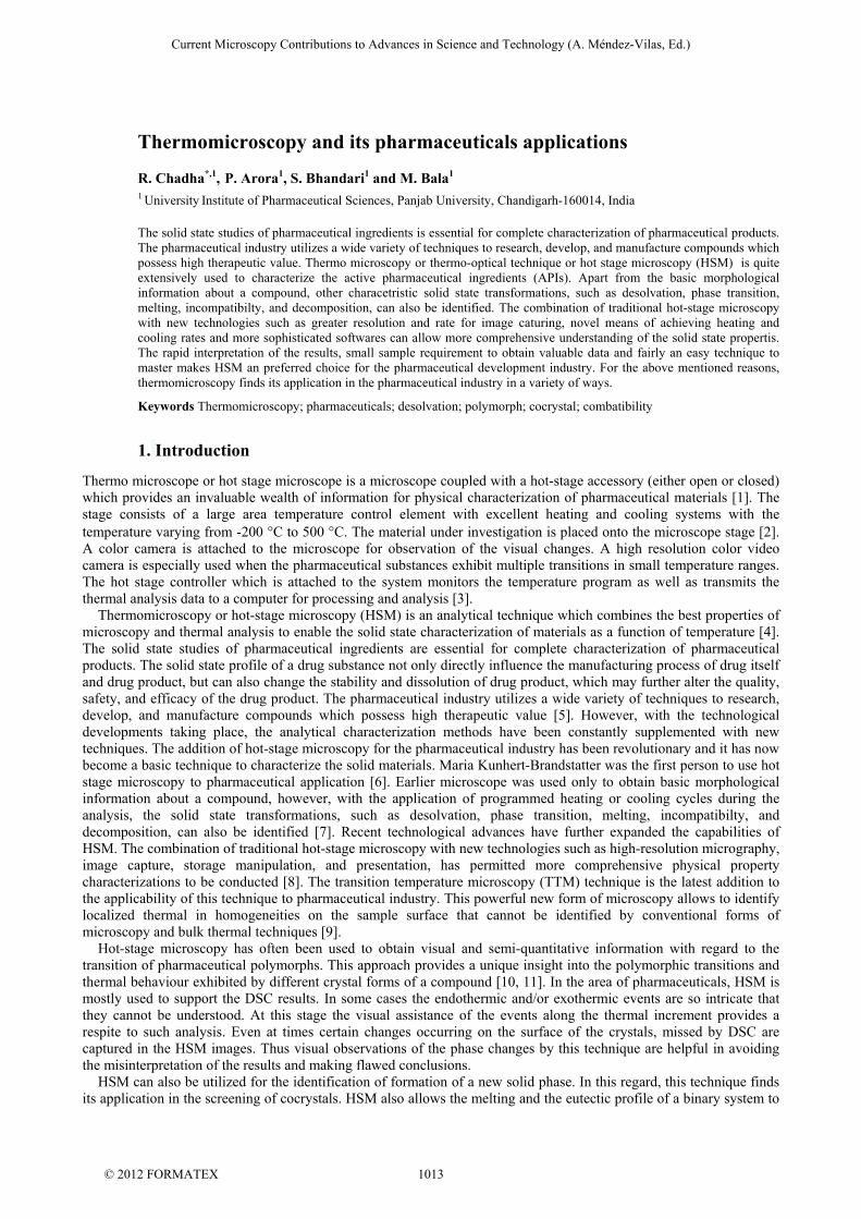
The significance of HSM to polymorph identification has been well demonstrated by Abu Bakar et al [16] in their study on sulfathiazole crystals. The authors have used the hot stage image analysis as well as the light intensity profiles obtained from the HSM images for verifying and describing the thermal events that take place during transformation of one polymorphic form to another. Five different methods which are already reported in literature for formation of pure polymorphs of sulfathiazole have been used by authors to generate four different phase pure polymorphs. In all the forms, the profile of HSM light intensity was fairly comparable to profile of DSC curves. However, the HSM results show that the sulfathiazole crystals obtained by method 1 are a mixture of Form I and Form II which is indicated by melting of Form II and recrystallization to Form I followed by complete melting. In method 2, the crystals of Form I, Form II and Form III have been observed (Figure 1). The conversion of Form III to Form II is accompanied by an endothermic event in DSC which was not melting, is confirmed by HSM image analysis. However, an optical property change has been observed in the light intensity profile verifying the conversion of Form III to Form II. In a similar manner, the method 3 resulted in formation of a mixture of Form II and Form III. Method 4 resulted in Form IV with traces of Form III. In method 5, mixture of crystals of Form III and Form II were observed. The above results indicate that with the help of HSM, the polymorphic impurities have been identified, although the established methods to generate pure forms have been utilized.
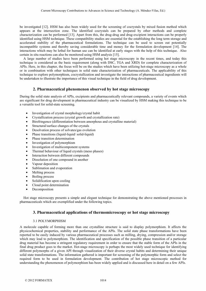
Figure 1: Images of sulfathiazole crystals obtained by method 2 during HSM analysis. Another example is of metoclopramide HCl monohydrate (MCP HCl H2O) where Lin and Cheng [17] have utilized HSM to depict different thermal events occurring during the thermal treatment (Figure 2). The three steps of phase transformation i.e. dehydration, recrystallization, and new crystal formation were markedly correlated with the endothermic and exothermic results of DSC study and the photovisual observations of HSM. The drug crystals of MCP HCl H2O maintained their morphology till 80 °C after which the crystalline lattices started to collapse and partially melt, which was similar to the DSC enotherm. A complete melting of crystals was seen until 97 °C. Once the heating temperature was reached to around 110 °C, several crystal nuclei appeared in a molten state were visibly observed. Upon further heating, the nucleation and resolidification continued to occur, and the formation of crystals attributable was finally culminated within the temperature range of 110–171 °C. After the temperature was beyond 176 °C, these new crystals began to melt but were completely melted around 184 °C, which was just the same as the melting point of MCP HCl. These HSM observations indicate that MCP HCl H2O crystals were first dehydrated and melted, then recrystallized and transformed to a new crystal form, and finally melted at higher temperature. It has been proposed by the authors that dehydration proceed via solid-state, solution, and occasionally via the melt mechanism. For solid-state mechanism of dehydration, the phase transformation occurred from solid to solid without intermediate liquid or vapor phases. In the present study, MCP HCl H2O crystals were previously dehydrated and melted, and then recrystallized, which belonged to solid-state dehydration process.
Current Microscopy Contributions to Advances in Science and Technology (A. Méndez-Vilas, Ed.)
© 2012 FORMATEX 1015
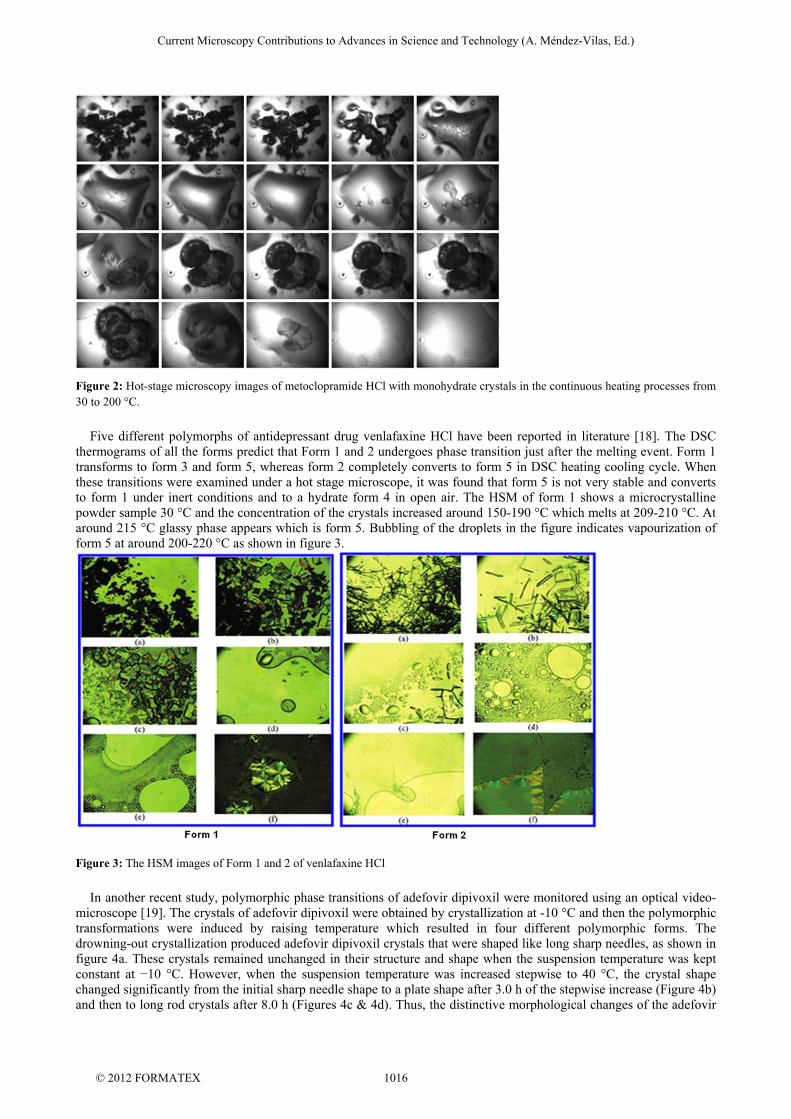
Figure 2: Hot-stage microscopy images of metoclopramide HCl with monohydrate crystals in the continuous heating processes from 30 to 200 °C.
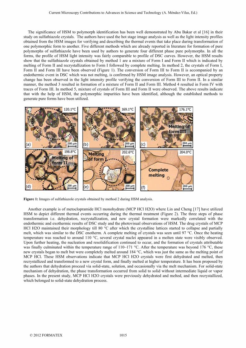
Five different polymorphs of antidepressant drug venlafaxine HCl have been reported in literature [18]. The DSC thermograms of all the forms predict that Form 1 and 2 undergoes phase transition just after the melting event. Form 1 transforms to form 3 and form 5, whereas form 2 completely converts to form 5 in DSC heating cooling cycle. When these transitions were examined under a hot stage microscope, it was found that form 5 is not very stable and converts to form 1 under inert conditions and to a hydrate form 4 in open air. The HSM of form 1 shows a microcrystalline powder sample 30 °C and the concentration of the crystals increased around 150-190 °C which melts at 209-210 °C. At around 215 °C glassy phase appears which is form 5. Bubbling of the droplets in the figure indicates vapourization of form 5 at around 200-220 °C as shown in figure 3.
Figure 3: The HSM images of Form 1 and 2 of venlafaxine HCl
In another recent study, polymorphic phase transitions of adefovir dipivoxil were monitored using an optical video-microscope [19]. The crystals of adefovir dipivoxil were obtained by crystallization at -10 °C and then the polymorphic transformations were induced by raising temperature which resulted in four different polymorphic forms. The drowning-out crystallization produced adefovir dipivoxil crystals that were shaped like long sharp needles, as shown in figure 4a. These crystals remained unchanged in their structure and shape when the suspension temperature was kept constant at −10 °C. However, when the suspension temperature was increased stepwise to 40 °C, the crystal shape changed significantly from the initial sharp needle shape to a plate shape after 3.0 h of the stepwise increase (Figure 4b) and then to long rod crystals after 8.0 h (Figures 4c & 4d). Thus, the distinctive morphological changes of the adefovir
Current Microscopy Contributions to Advances in Science and Technology (A. Méndez-Vilas, Ed.)
© 2012 FORMATEX 1016
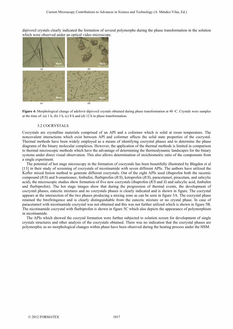
dipivoxil crystals clearly indicated the formation of several polymorphs during the phase transformation in the solution which were observed under an optical video microscope.
Figure 4: Morphological change of adefovir dipivoxil crystals obtained during phase transformation at 40 ◦C. Crystals were samples at the time of: (a) 1 h, (b) 3 h, (c) 8 h and (d) 12 h in phase transformation.
3.2 COCRYSTALS
Cocrystals are crystalline materials comprised of an API and a coformer which is solid at room temperature. The noncovalent interactions which exist between API and coformer affects the solid state properties of the cocrystal. Thermal methods have been widely employed as a means of identifying cocrystal phases and to determine the phase diagrams of the binary molecular complexes. However, the application of the thermal methods is limited in comparison to thermal microscopic methods which have the advantage of determining the thermodynamic landscapes for the binary systems under direct visual observation. This also allows determination of stoichiometric ratio of the components from a single experiment. The potential of hot stage microscopy in the formation of cocrystals has been beautifully illustrated by Blagden et al [13] in their study of screening of cocrystals of nicotinamide with seven different APIs. The authors have utilized the Kofler mixed fusion method to generate different cocrystals. Out of the eight APIs used (ibuprofen both the racemic compound (R/S) and S-enantiomer, fenbufen, flurbiprofen (R/S), ketoprofen (R/S), paracetamol, piracetam, and salicylic acid), the microscopic studies show formation of five new cocrystals (ibuprofen (R/S and S) and salicylic acid, fenbufen and flurbiprofen). The hot stage images show that during the progression of thermal events, the development of cocrystal phases, eutectic mixtures and no cocrystals phases is clearly indicated and is shown in figure. The cocrystal appears at the intersection of the two phases producing a mixing zone as can be seen in figure 5A. The cocrystal phase retained the birefiringence and is clearly distinguishable from the eutectic mixture or no crystal phase. In case of paracetamol with nicotinamide cocrystal was not obtained and this was not further utilized which is shown in figure 5B. The nicotinamide cocrystal with flurbiprofen is shown in figure 5C which also depicts the appearance of polymorphism in nicotinamide. The APIs which showed the cocrytal formation were further subjected to solution screen for development of single crystals structures and other analysis of the cocrystals obtained. There was no indication that the cocrystal phases are polymorphic as no morphological changes within phase have been observed during the heating process under the HSM.
Current Microscopy Contributions to Advances in Science and Technology (A. Méndez-Vilas, Ed.)
© 2012 FORMATEX 1017
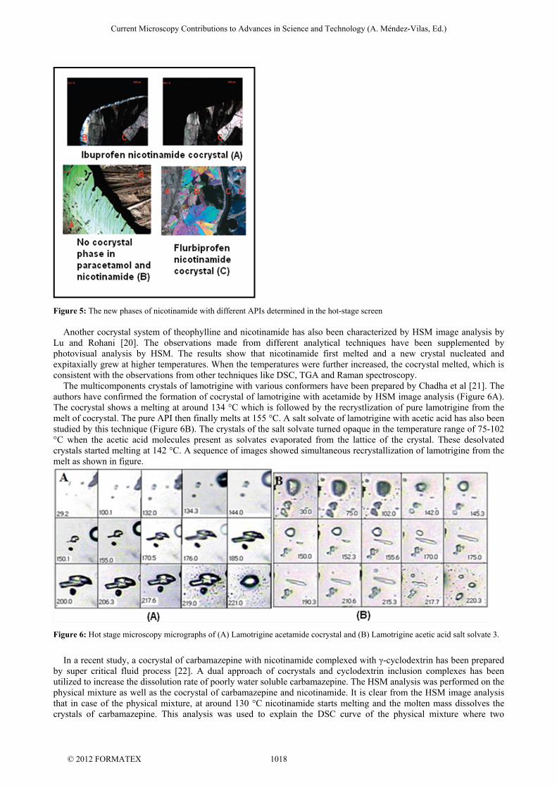
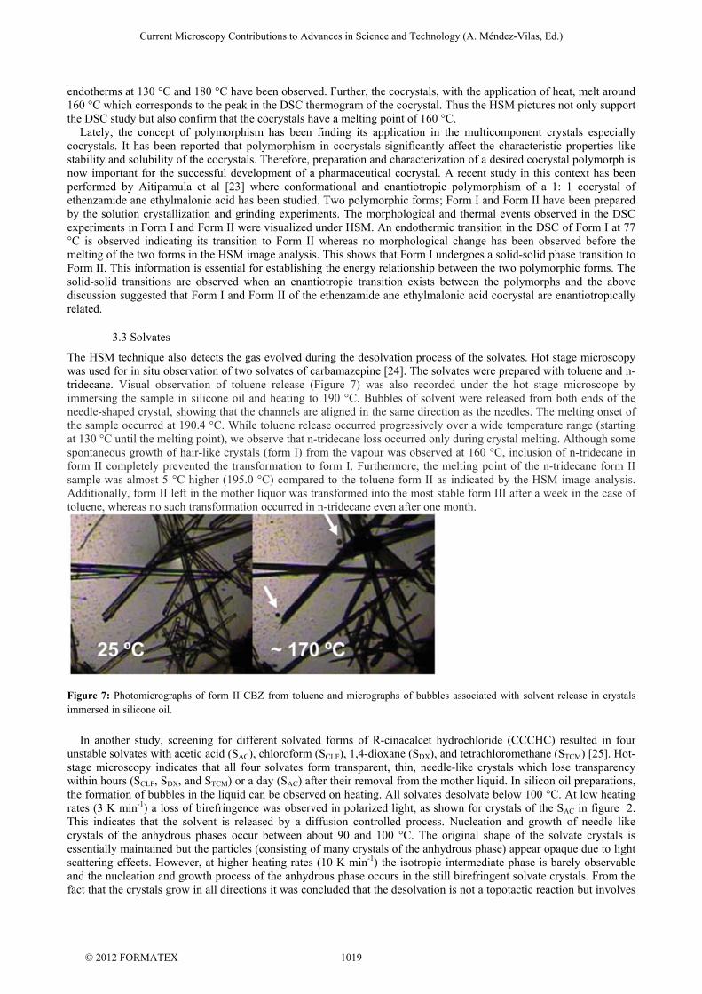
Figure 5: The new phases of nicotinamide with different APIs determined in the hot-stage screen Another cocrystal system of theophylline and nicotinamide has also been characterized by HSM image analysis by Lu and Rohani [20]. The observations made from different analytical techniques have been supplemented by photovisual analysis by HSM. The results show that nicotinamide first melted and a new crystal nucleated and expitaxially grew at higher temperatures. When the temperatures were further increased, the cocrystal melted, which is consistent with the observations from other techniques like DSC, TGA and Raman spectroscopy. The multicomponents crystals of lamotrigine with various conformers have been prepared by Chadha et al [21]. The authors have confirmed the formation of cocrystal of lamotrigine with acetamide by HSM image analysis (Figure 6A). The cocrystal shows a melting at around 134 °C which is followed by the recrystlization of pure lamotrigine from the melt of cocrystal. The pure API then finally melts at 155 °C. A salt solvate of lamotrigine with acetic acid has also been studied by this technique (Figure 6B). The crystals of the salt solvate turned opaque in the temperature range of 75-102 °C when the acetic acid molecules present as solvates evaporated from the lattice of the crystal. These desolvated crystals started melting at 142 °C. A sequence of images showed simultaneous recrystallization of lamotrigine from the melt as shown in figure.
Figure 6: Hot stage microscopy micrographs of (A) Lamotrigine acetamide cocrystal and (B) Lamotrigine acetic acid salt solvate 3.
In a recent study, a cocrystal of carbamazepine with nicotinamide complexed with γ-cyclodextrin has been prepared by super critical fluid process [22]. A dual approach of cocrystals and cyclodextrin inclusion complexes has been utilized to increase the dissolution rate of poorly water soluble carbamazepine. The HSM analysis was performed on the physical mixture as well as the cocrystal of carbamazepine and nicotinamide. It is clear from the HSM image analysis that in case of the physical mixture, at around 130 °C nicotinamide starts melting and the molten mass dissolves the crystals of carbamazepine. This analysis was used to explain the DSC curve of the physical mixture where two
Current Microscopy Contributions to Advances in Science and Technology (A. Méndez-Vilas, Ed.)
© 2012 FORMATEX 1018
endotherms at 130 °C and 180 °C have been observed. Further, the cocrystals, with the application of heat, melt around 160 °C which corresponds to the peak in the DSC thermogram of the cocrystal. Thus the HSM pictures not only support the DSC study but also confirm that the cocrystals have a melting point of 160 °C. Lately, the concept of polymorphism has been finding its application in the multicomponent crystals especially cocrystals. It has been reported that polymorphism in cocrystals significantly affect the characteristic properties like stability and solubility of the cocrystals. Therefore, preparation and characterization of a desired cocrystal polymorph is now important for the successful development of a pharmaceutical cocrystal. A recent study in this context has been performed by Aitipamula et al [23] where conformational and enantiotropic polymorphism of a 1: 1 cocrystal of ethenzamide ane ethylmalonic acid has been studied. Two polymorphic forms; Form I and Form II have been prepared by the solution crystallization and grinding experiments. The morphological and thermal events observed in the DSC experiments in Form I and Form II were visualized under HSM. An endothermic transition in the DSC of Form I at 77 °C is observed indicating its transition to Form II whereas no morphological change has been observed before the melting of the two forms in the HSM image analysis. This shows that Form I undergoes a solid-solid phase transition to Form II. This information is essential for establishing the energy relationship between the two polymorphic forms. The solid-solid transitions are observed when an enantiotropic transition exists between the polymorphs and the above discussion suggested that Form I and Form II of the ethenzamide ane ethylmalonic acid cocrystal are enantiotropically related.
3.3 Solvates
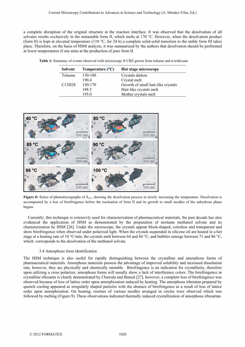
The HSM technique also detects the gas evolved during the desolvation process of the solvates. Hot stage microscopy was used for in situ observation of two solvates of carbamazepine [24]. The solvates were prepared with toluene and n-tridecane. Visual observation of toluene release (Figure 7) was also recorded under the hot stage microscope by immersing the sample in silicone oil and heating to 190 °C. Bubbles of solvent were released from both ends of the needle-shaped crystal, showing that the channels are aligned in the same direction as the needles. The melting onset of the sample occurred at 190.4 °C. While toluene release occurred progressively over a wide temperature range (starting at 130 °C until the melting point), we observe that n-tridecane loss occurred only during crystal melting. Although some spontaneous growth of hair-like crystals (form I) from the vapour was observed at 160 °C, inclusion of n-tridecane in form II completely prevented the transformation to form I. Furthermore, the melting point of the n-tridecane form II sample was almost 5 °C higher (195.0 °C) compared to the toluene form II as indicated by the HSM image analysis. Additionally, form II left in the mother liquor was transformed into the most stable form III after a week in the case of toluene, whereas no such transformation occurred in n-tridecane even after one month.
Figure 7: Photomicrographs of form II CBZ from toluene and micrographs of bubbles associated with solvent release in crystals immersed in silicone oil.
In another study, screening for different solvated forms of R-cinacalcet hydrochloride (CCCHC) resulted in four unstable solvates with acetic acid (SAC), chloroform (SCLF), 1,4-dioxane (SDX), and tetrachloromethane (STCM) [25]. Hot-stage microscopy indicates that all four solvates form transparent, thin, needle-like crystals which lose transparency within hours (SCLF, SDX, and STCM) or a day (SAC) after their removal from the mother liquid. In silicon oil preparations, the formation of bubbles in the liquid can be observed on heating. All solvates desolvate below 100 °C. At low heating rates (3 K min-1) a loss of birefringence was observed in polarized light, as shown for crystals of the SAC in figure 2. This indicates that the solvent is released by a diffusion controlled process. Nucleation and growth of needle like crystals of the anhydrous phases occur between about 90 and 100 °C. The original shape of the solvate crystals is essentially maintained but the particles (consisting of many crystals of the anhydrous phase) appear opaque due to light scattering effects. However, at higher heating rates (10 K min-1) the isotropic intermediate phase is barely observable and the nucleation and growth process of the anhydrous phase occurs in the still birefringent solvate crystals. From the fact that the crystals grow in all directions it was concluded that the desolvation is not a topotactic reaction but involves
Current Microscopy Contributions to Advances in Science and Technology (A. Méndez-Vilas, Ed.)
© 2012 FORMATEX 1019
a complete disruption of the original structure at the reaction interface. It was observed that the desolvation of all solvates results exclusively in the metastable form II, which melts at 170 °C. However, when the desolvation product (form II) is kept at elevated temperature (110 °C, for 24 h) a complete solid-solid transition to the stable form III takes place. Therefore, on the basis of HSM analysis, it was summarized by the authors that desolvation should be performed at lower temperatures if one aims at the production of pure form II.
Table 1: Summary of events observed with microscopy II CBZ grown from toluene and n-tridecane
Solvent Temperature (°C) Hot stage microscopy
Toluene 130-180 Crystals darken 190.4 Crystal melt C13H28 150-170 Growth of small hair-like crystals 188.5 Hair-like crystals melt 195.0 Mother crystals melt
Figure 8: Series of photomicrographs of SAC, showing the desolvation process at slowly increasing the temperature. Desolvation is accompanied by a loss of birefringence before the nucleation of form II and its growth to small needles of the anhydrous phase begins.
Currently, this technique is extensivly used for characterization of pharmaceutical materials, the past decade has also evidenced the application of HSM as demonstrated by the preparation of neotame methanol solvate and its characterization by HSM [26]. Under the microscope, the crystals appear block-shaped, colorless and transparent and show birefringence when observed under polarized light. When the crystals suspended in silicone oil are heated in a hot stage at a heating rate of 10 °C/min, the crystals melt between 64 and 84 °C, and bubbles emerge between 71 and 86 °C, which corresponds to the desolvation of the methanol solvate.
3.4 Amorphous form identification
The HSM technique is also useful for rapidly distinguishing between the crystalline and amorphous forms of pharmaceutical materials. Amorphous materials possess the advantage of improved solubility and increased dissolution rate, however, they are physically and chemically unstable. Birefringence is an indication for crystallinity, therefore upon utilizing a cross polarizer, amorphous forms will usually show a lack of interference colors. The birefringence in crystalline irbesartn is clearly demonstrated by Chawala and Bansal [27], however, a complete loss of birefringence was observed because of loss of lattice order upon amorphization induced by heating. The amorphous irbesatan prepared by quench cooling appeared as irregularly shaped particles with the absence of birefringence as a result of loss of lattice order upon amorphization. On heating, rosettes of various needles arranged in circles were observed which was followed by melting (Figure 9). These observations indicated thermally induced crystallization of amorphous irbesartan.
Current Microscopy Contributions to Advances in Science and Technology (A. Méndez-Vilas, Ed.)
© 2012 FORMATEX 1020
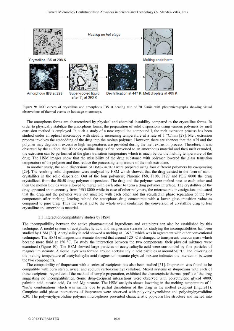
Figure 9: DSC curves of crystalline and amorphous IBS at heating rate of 20 K/min with photomicrographs showing visual observations of thermal events on hot stage microscope.
The amorphous forms are characterized by physical and chemical instability compared to the crystalline forms. In order to physically stabilize the amorphous forms, the preparation of solid dispersions using various polymers by melt extrusion method is employed. In such a study of a new crystalline compound I, the melt extrusion process has been studied under an optical microscope with steadily increasing temperature at a rate of 1 °C/min [28]. Melt extrusion process involves the embedding of the drug into the molten polymer. However, there are chances that the API and the polymer may degrade if excessive high temperatures are provided during the melt extrusion process. Therefore, it was observed by the authors that if the crystalline drug is first converted to an amorphous material and then melt extruded, the extrusion can be performed at the glass transition temperature which is much below the melting temperature of the drug. The HSM images show that the miscibility of the drug substance with polymer lowered the glass transition temperature of the polymer and thus reduce the processing temperature of the melt extrudate. In another study, the solid dispersions of BMS-347070 were prepared using four different polymers by co-spraying [29]. The resulting solid dispersions were analysed by HSM which showed that the drug existed in the form of nano-crystallites in the solid dispersion. Out of the four polymers; Pluronic F68, F108, F127 and PEG 8000 the drug crystallized from the 50/50 drug-polymer dispersions. The drug and the polymer were melted next to each other and then the molten liquids were allowed to merge with each other to form a drug polymer interface. The crystallites of the drug appeared spontaneously from PEG 8000 while in case of other polymers, the microscopic investigations indicated that the drug and the polymer were not miscible with each other and this resulted in phase separation of the two components after melting, leaving behind the amorphous drug concentrate with a lower glass transition value as compared to pure drug. Thus the visual aid to the whole event confirmed the conversion of crystalline drug to less crystalline and amorphous material.
3.5 Interaction/compatibility studies by HSM
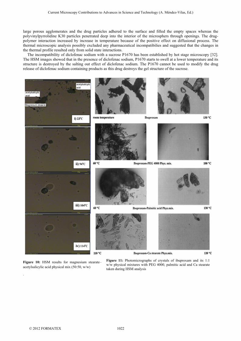
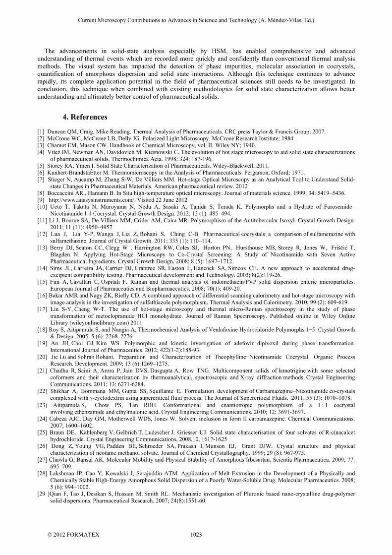
The incompatibility between the active pharmaceutical ingredients and excipients can also be established by this technique. A model system of acetylsalicylic acid and magnesium stearate for studying the incompatibilities has been studied by HSM [30]. Acetylsalicylic acid showed a melting at 136 °C which was in agreement with other conventional techniques. The HSM of magnesium stearate showed that around 120 °C it changed to transparent, viscous mass which became more fluid at 150 °C. To study the interaction between the two components, their physical mixtures were examined (Figure 10). The HSM showed large particles of acetylsalicylic acid were surrounded by fine particles of magnesium stearate. A liquid layer was formed around acetylsalicylic acid particles at around 90 °C. The lowering of the melting temperature of acetylsalicylic acid magnesium stearate physical mixture indicates the interaction between the two components. The compatibility of ibuproxam with a series of excipients has also been studied [31]. Ibuproxam was found to be compatible with corn starch, avicel and sodium carboxymethyl cellulose. Mixed systems of ibuproxam with each of these excipients, regardless of the method of sample preparation, exhibited the characteristic thermal profile of the drug suggesting no incompatibilities. Some drug-excipient interactions were observed with polyethylene glycol 4000, palmitic acid, stearic acid, Ca and Mg stearate. The HSM analysis shows lowering in the melting temperature of 1 %w/w combinations which was mainly due to partial dissolution of the drug in the melted excipient (Figure11). Complete solid phase interactions of ibuproxam were observed with polyvinylpyrrolidine and polyvinylpyrrolidine K30. The polyvinylpyrrolidine polymer microspheres presented characteristic pop-corn like structure and melted into
Current Microscopy Contributions to Advances in Science and Technology (A. Méndez-Vilas, Ed.)
© 2012 FORMATEX 1021
large porous agglomerates and the drug particles adhered to the surface and filled the empty spaces whereas the polyvinylpyrrolidine K30 particles penetrated deep into the interior of the microsphers through openings. The drug-polymer interaction increased by increase in temperature because of the positive effect on diffusional process. The thermal microscopic analysis possibly excluded any pharmaceutical incompatibilies and suggested that the changes in the thermal profile resulted only from solid state interactions. The incompatibility of diclofenac sodium with a sucrose P1670 has been established by hot stage microscopy [32]. The HSM images showed that in the presence of diclofenac sodium, P1670 starts to swell at a lower temperature and its structure is destroyed by the salting out effect of diclofenac sodium. The P1670 cannot be used to modify the drug release of diclofenac sodium containing products as this drug destroys the gel structure of the sucrose.
Figure 10: HSM results for magnesium stearate–acetylsalicylic acid physical mix (50:50, w/w)
.
Figure 11: Photomicrographs of crystals of ibuproxam and its 1:1 w/w physical mixtures with PEG 4000, palmitic acid and Ca stearate taken during HSM analysis
Current Microscopy Contributions to Advances in Science and Technology (A. Méndez-Vilas, Ed.)
© 2012 FORMATEX 1022
The advancements in solid-state analysis especially by HSM, has enabled comprehensive and advanced understanding of thermal events which are recorded more quickly and confidently than conventional thermal analysis methods. The visual system has impacted the detection of phase impurities, molecular association in cocrystals, quantification of amorphous dispersion and solid state interactions. Although this technique continues to advance rapidly, its complete application potential in the field of pharmaceutical sciences still needs to be investigated. In conclusion, this technique when combined with existing methodologies for solid state characterization allows better understanding and ultimately better control of pharmaceutical solids.
4. References
[1] Duncan QM, Craig, Mike Reading. Thermal Analysis of Pharmaceuticals. CRC press Taylor & Francis Group; 2007. [2] McCrone WC, McCrone LB, Delly JG. Polarized Light Microscopy. McCrone Research Institute; 1984. [3] Chamot EM, Mason CW. Handbook of Chemical Microscopy, vol. II, Wiley NY; 1940. [4] Vitez IM, Newman AN, Davidovich M, Kiesnowski C. The evolution of hot stage microscopy to aid solid state characterizations
of pharmaceutical solids. Thermochimica Acta. 1998: 324: 187-196. [5] Storey RA, Ymen I. Solid State Characterization of Pharmaceuticals. Wiley-Blackwell; 2011. [6] Kunhert-BrandstaÈtter M. Thermomicroscopy in the Analysis of Pharmaceuticals. Pergamon, Oxford; 1971. [7] Stieger N, Aucamp M, Zhang S-W, De Villiers MM. Hot-stage Optical Microscopy as an Analytical Tool to Understand Solid-
state Changes in Pharmaceutical Materials. American pharmaceutical review. 2012 [8] Boccaccini AR , Hamann B. In Situ high-temperature optical microscopy. Journal of materials science. 1999; 34: 5419–5436. [9] http://www.anasysinstruments.com/. Visited 22 June 2012 [10] Ueto T, Takata N, Muroyama N, Nedu A, Sasaki A, Tanida S, Terada K. Polymorphs and a Hydrate of Furosemide–
Nicotinamide 1:1 Cocrystal. Crystal Growth Design. 2012; 12 (1): 485–494. [11] Li J, Bourne SA, De Villiers MM, Crider AM, Caira MR. Polymorphism of the Antitubercular Isoxyl. Crystal Growth Design.
2011; 11 (11): 4950–4957 [12] Lua J, Lia Y-P, Wanga J, Lia Z, Rohani S, Ching C-B. Pharmaceutical cocrystals: a comparison of sulfamerazine with
sulfamethazine. Journal of Crystal Growth. 2011; 335 (1): 110–114. [13] Berry DJ, Seaton CC, Clegg W , Harrington RW, Coles SJ, Horton PN, Hursthouse MB, Storey R, Jones W, Friščić T,
Blagden N. Applying Hot-Stage Microscopy to Co-Crystal Screening: A Study of Nicotinamide with Seven Active Pharmaceutical Ingredients. Crystal Growth Design. 2008; 8 (5): 1697–1712.
[14] Sims JL, Carreira JA, Carrier DJ, Crabtree SR, Easton L, Hancock SA, Simcox CE. A new approach to accelerated drug-excipient compatibility testing. Pharmaceutical development and Technology. 2003; 8(2):119-26.
[15] Fini A, Cavallari C, Ospitali F. Raman and thermal analysis of indomethacin/PVP solid dispersion enteric microparticles. European Journal of Pharmaceutics and Biopharmaceutics. 2008; 70(1): 409-20.
[16] Bakar AMR and Nagy ZK, Rielly CD. A combined approach of differential scanning calorimetry and hot-stage microscopy with image analysis in the investigation of sulfathiazole polymorphism. Thermal Analysis and Calorimetry. 2010; 99 (2): 609-619.
[17] Lin S-Y, Cheng W-T. The use of hot-stage microscopy and thermal micro-Raman spectroscopy in the study of phase transformation of metoclopramide HCl monohydrate. Journal of Raman Spectroscopy. Published online in Wiley Online Library (wileyonlinelibrary.com) 2011
[18] Roy S, Aitipamula S, and Nangia A. Thermochemical Analysis of Venlafaxine Hydrochloride Polymorphs 1−5. Crystal Growth & Design. 2005; 5 (6): 2268–2276.
[19] An JH, Choi GJ, Kim WS. Polymorphic and kinetic investigation of adefovir dipivoxil during phase transformation. International Journal of Pharmaceutics. 2012; 422(1-2):185-93.
[20] Jie Lu and Sohrab Rohani. Preparation and Characterization of Theophylline−Nicotinamide Cocrystal. Organic Process Research. Development. 2009; 13 (6):1269–1275.
[21] Chadha R, Saini A, Arora P, Jain DVS, Dasgupta A, Row TNG. Multicomponent solids of lamotrigine with some selected coformers and their characterization by thermoanalytical, spectroscopic and X-ray diffraction methods. Crystal Engineering Communications. 2011; 13: 6271-6284.
[22] Shikhar A, Bommana MM, Gupta SS, Squillante E. Formulation development of Carbamazepine–Nicotinamide co-crystals complexed with γ-cyclodextrin using supercritical fluid process. The Journal of Supercritical Fluids. 2011; 55 (3): 1070–1078.
[23] Aitipamula S, Chow PS, Tan RBH. Conformational and enantiotropic polymorphism of a 1 : 1 cocrystal involving ethenzamide and ethylmalonic acid. Crystal Engineering Communications. 2010; 12: 3691-3697.
[24] Cabeza AJC, Day GM, Motherwell WDS, Jones W. Solvent inclusion in form II carbamazepine. Chemical Communications. 2007; 1600–1602.
[25] Braun DE, Kahlenberg V, Gelbrich T, Ludescher J, Griesser UJ. Solid state characterisation of four solvates of R-cinacalcet hydrochloride. Crystal Engineering Communications, 2008,10, 1617-1625
[26] Dong Z, Young VG, Padden BE, Schroeder SA, Prakash I, Munson EJ, Grant DJW. Crystal structure and physical characterization of neotame methanol solvate. Journal of Chemical Crystallography. 1999; 29 (8): 967-975.
[27] Chawla G, Bansal AK. Molecular Mobility and Physical Stability of Amorphous Irbesartan. Scientia Pharmaceutica. 2009; 77: 695–709.
[28] Lakshman JP, Cao Y, Kowalski J, Serajuddin ATM. Application of Melt Extrusion in the Development of a Physically and Chemically Stable High-Energy Amorphous Solid Dispersion of a Poorly Water-Soluble Drug. Molecular Pharmaceutics. 2008; 5 (6): 994–1002.
[29 ]Qian F, Tao J, Desikan S, Hussain M, Smith RL. Mechanistic investigation of Pluronic based nano-crystalline drug-polymer solid dispersions. Pharmaceutical Research. 2007; 24(8):1551-60.
Current Microscopy Contributions to Advances in Science and Technology (A. Méndez-Vilas, Ed.)
© 2012 FORMATEX 1023
[30] Harding L, Qi S, Hill G, Reading M, Craig DQM. The development of microthermalanalysis and photothermalmicrospectroscopy as novel approaches to drug–excipient compatibility studies. International Journal of Pharmaceutics. 2008; 354 (1–2): 149–157.
[31] Mura P, Faucci MT, Manderioli A, Bramanti G, Ceccarelli L. Compatibility study between ibuproxam and pharmaceutical excipients using differential scanning calorimetry, hot-stage microscopy and scanning electron microscopy. Journal of Pharmaceutical and Biomedical Analysis. 1998; 18(1-2):151-63.
[32] Angéla S, Mária B-S, Codruţa Ş, Cristina A. D, Piroska S-R.Evaluation of the interaction between a sucrose ester and Diclofenac sodium. Farmacia. 2010; 58 (2): 211-217.
Current Microscopy Contributions to Advances in Science and Technology (A. Méndez-Vilas, Ed.)
© 2012 FORMATEX 1024