therapeutic window in ischaemic stroke
TRANSCRIPT
PHARMACOLOGY AND PATHOPHYSIOLOGY eNS Drugs 1997 Dec; 8 (6): 474-491 1172-7047/97/0012-0474/$09.00/0
© Adis International Umlted. AU rights reserved.
Therapeutic Window in Ischaemic Stroke Experimental Concepts, N euroimaging Studies and Implications for Pharmacological Treatment
Wolf-Dieter Heiss and Rudolf Gra! Max-Planck-Institut fiir Neurologische Forschung and Neurologische Universitatsklinik, Cologne, Germany
Contents Summary ............................ . 1. Flow Thresholds for Functional and Morphological Integrity
1.1 Functional Damage . . . 1.2 Morphological Damage . . . . . .
2. The Ischaemic Penumbra . . . . . . . . 2.1 Mediators of Ischaemlc Damage 2.2 Potential Therapeutic Interventions
3. Clinical Evidence of an Ischaemic Penumbra 4. Techniques for Assessing Ischaemic Damage
4.1 Positron Emission Tomography: Correlation Between Animal Models and the Clinical Situation . . . . . . . . . . . . . . . . . . . . . . . . .
4.2 Magnetic Resonance Imaging: Use in Early Stroke. . . . . . . . . . . 5. Application of Therapeutic Windows to the Treatment of Ischaemic Stroke .
5.1 Therapeutic Window for the Re-Establishment of Perfusion . . . . . . . . 5.2 Therapeutic Window for the Inhibition of Biochemical/Molecular Changes
6. Conclusion ....................................... .
· 474 · 475 · 475 · 476 .477 .477 .479 · 479 .480
.480 · 483 .485 .485 · 486 · 487
Summary The development of ischaemic brain damage depends on the severity and duration of the focal disturbance in cerebral blood flow. Several flow thresholds exist; if flow is decreased to a certain threshold there can be functional but potentially reversible impairment, while a decrease to lower thresholds results in morphological and therefore permanent damage. Tissue perfused at a value between these thresholds is termed the 'penumbra'. This tissue has the potential for functional recovery if blood flow is re-established to a sufficient level within a limited time period, i.e. the 'therapeutic window'.
The existence of a therapeutic window for the restoration of blood flow is evident from experimental studies and clinical experience. It has formed the basis for the success of thrombolytic therapy (e.g. with alteplase) in ischaemic stroke if treatment is initiated within a short period (approximately 3 hours) after the onset of symptoms. Despite the success of thrombolytic therapy, additional strategies need to be developed for the treatment of ischaemic stroke. In particular, therapies that interrupt the molecular and biochemical alterations triggered during
Therapeutic Window in Ischaemic Stroke 475
the ischaemic period and that continue after reperfusion would be beneficial. These biochemical changes may propagate ischaemic damage and contribute to the enlargement of infarcts.
State-of-the-art imaging modalities, such as positron emission tomography and diffusion- and perfusion-weighted magnetic resonance imaging, demonstrate the presence of ischaemically compromised but viable tissue in patients who have had a stroke (and in animal models of focal ischaemia, in which the transient changes can be followed until the final state of infarction or recovery is reached). In selected groups of patients, therapeutic strategies, such as acute thrombolysis or the interruption of biochemical changes, can be evaluated using these imaging techniques, and the clinical course analysed in relation to effects on impaired perfusion and altered metabolism within ischaemic tissue.
Experimental studies in animal models and cell cultures have formed the basis of current concepts of the pathophysiology of ischaemic brain damage. Ischaemic cell death is hypothesised to be a consequence of progressive deleterious interactions between various circulatory, biochemical and molecular disturbances (for reviews, see Siesjo,D] Pulsinelli,[2] and Kogure et alP]), which are all, in principle, amenable to therapeutic interventions.[4] The transfer of many biochemical, and most molecular, data from the experimental to the clinical setting is still limited. However, insight into the dynamics of the development of ischaemic damage has an impact on the management of acute stroke, e.g. for the selection of patients who might benefit from early reperfusion, as achieved by thrombolytic therapy.
For brain tissue to function it must have an adequate supply of blood. Consequently, damage will occur if there is an impairment of blood supply, with the severity and extent of permanent neurological defects being dependent on the amount of tissue suffering an impairment of blood supply below certain flow thresholds for critical time periods.
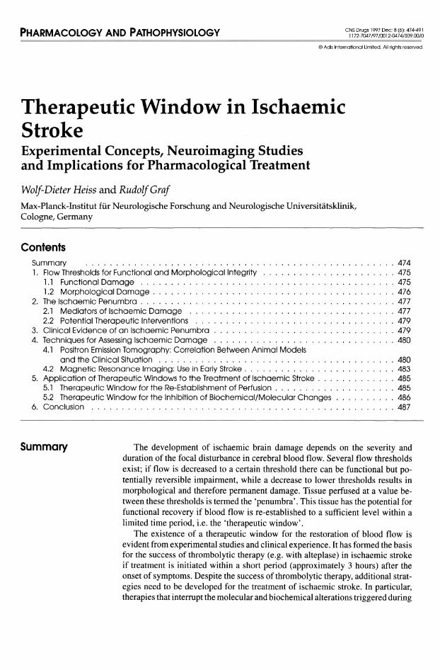
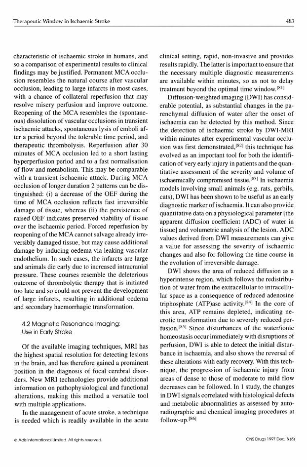
Experimental work on ischaemic flow thresholds has demonstrated the existence of 2 critical levels of decreased perfusion: (i) a level below which reversible neuronal failure occurs (functional threshold); and (ii) a lower threshold below which irreversible membrane failure and morphological damage occur (for a review see Heiss[5]) [fig. 1]. The range of perfusion values between these limits
© Adis Internalional Umited. All rights reserved.
is termed the 'ischaemic penumbra' .[6] Tissue exposed to blood flow rates within this range is characterised by the potential for functional recovery without morphological damage, provided that local blood flow can be re-established at a sufficient level and within a certain time. The duration of impairment in blood flow that can be tolerated is also dependent on the severity of associated metabolic disturbances. As a consequence, the theory of a 'therapeutic window' has been postulated - a duration of time during which effective restoration of blood flow and intervention to prevent the consequences of biochemical alterations can be successful in ensuring the viability of cerebral tissue.
This review will discuss the basic mechanisms of ischaemia and the hypothesis of the therapeutic window. The relevance of experimental results concerning these parameters to the clinical management of stroke is highlighted.
1. Flow Thresholds for Functional and Morphological Integrity
As mentioned in the introduction to this review, 2 ischaemic flow thresholds exist - one defines functional damage and the other morphological damage.
1 ,1 Functional Damage
The functional threshold was demonstrated in monkeys exposed to ischaemia. A reduction in blood flow led to the gradual development of a
eNS Drugs 1997 Dec; 8 (6)
476
50
40
c ~ 30
~ g ~ 20 ()
Normallunctlon
Functional Impairment • biochemical alterations • suppression 01 EEG and
evoked polentlals • cessation of Singte cell
activity
10 ---------Membrane lallure
Heiss & Graf
Viable lissu
~p~en~u~m=b=ra~ ____ ---------
Inlarctlon
Single cell necrosIs
0-' --L,------.-----,-----.---~/~/-----rl -----rI-----rI--1I,~/-., ----'I o 30 60 90 120 min 4 5 6 24 48h
Fig. 1. Diagram of cerebral blood flow (CBF) thresholds required for the preservation of function and morphology of brain tissue. The development of single cell necrosis and infarction is dependent on the duration of time for which CBF is impaired below a certain level. The solid line separates structurally damaged from functionally impaired but morphologically intact tissue (the 'penumbra'), and the dashed line distinguishes viable from functionally impaired tissue.
neurological deficit, progressing from mild pareses at a flow rate of 22 mIll ~Og/min to complete paralysis at a rate of 8 ml/100g/min.[7) Concurrently, the electrocorticogram and evoked potentials were abolished at a flow rate of 15 to 20 mIIl00g/min,IS.9) and the spontaneous activity of cortical neurons disappeared at approximately 18 mll100g/min.llO) There was a large variability in the functional threshold of individual neurons, indicating selective vulnerability. I I I)
Biochemical substrates and markers follow similar threshold dependencies. However, the pattern is more complex and the threshold values fall within a larger range, suggesting a specific role of individual metabolites in the development of ischaemic injury (for a review see Hossmannlt 2)) . With declining flow rates (from the normal cortical values of 60 to 100 mll100g/min, depending on species), protein synthesis is inhibited at about 55 mll100g/min,(13) followed by a stimulation of an-
© Adis International Umlted. All rights reserved.
aerobic glycolysis at 35 mll100g/min,(14) the release of neurotransmitters(15) and the disturbance of energy metabolism(16) at about 20 mll100g/min, and finally the terminal depolarisation and concomitant potassium efflux at 15 mIIl00g/min.(17)
1 .2 Morphological Damage
Whereas neuronal function is impaired immediately blood flow drops below the threshold, the development of irreversible morphological damage is time dependent. Once morphological damage becomes apparent, the initially reversible functional deficit develops into a persistent defect. Numerous studies have investigated the duration for which brain tissue or individual cells can tolerate ischaemia of a given intensity (reviewed by Heiss(5)).
The interaction of severity and duration of ischaemia in the development of irreversible cell damage can be studied by simultaneous recordings of cortical neuronal activity and local blood flow'! I I)
eNS Drugs 1997 Dec; 8 (6)
Therapeutic Window in Ischaemic Stroke
On the basis of a large number of neurons assessed during and after ischaemia of varying degree and duration, it is possible, despite the high variability of ischaemic tolerance, to construct a discriminant curve representing the worst possible constellations of residual blood flow and duration of ischaemia that still permit neuronal recovery. Typical points on this curve are blood flow rates of approximately 0, 10 or 15 ml/100g/min maintained for periods of 25, 40 and 80 minutes, respectively. Between 17 and 18 mlll00g/min, the duration oftolerated ischaemia tends to infinity, indicating that this flow state can lead to morphological damage but only when maintained for very long, as yet undefined, periods of time. These conditions of dense or prolonged ischaemia predict cell damage that may take the form of large infarcts or single cell necroses.
Interspecies differences must be taken into consideration when experimental results are compared. In monkeys and cats, large infarcts develop with residual flow rates of 12 mlll00g/min lasting for 2 to 3 hours'p,lS,19] and individual cells may become necrotic at lower flow values after shorter periods of time.[20,21] However, middle cerebral artery (MCA) occlusion in rats induced selective neuronal necrosis in the caudate/putamen after only 15 minutes, localised infarcts in the caudate/putamen and selective neuronal necrosis in the neocortex after 30 minutes, and cortical infarcts after 60 minutes. With an occlusion time of 120 to 180 minutes, infarct size increased and reached that found after permanent MCA occlusionP2]
Additionally, morphological tolerance to ischaemia is affected by various inherent or external factors: hypertensive rats are less tolerant than normotensive rats,[23] and tolerance is dependent on the number of ischaemic episodes[24] and body temperature.[25] It must also be kept in mind that delayed neuronal death after reperfusion may result from selective vulnerability of neurons.
2. The Ischaemic Penumbra
The term 'ischaemic penumbra' was originally applied to brain tissue perfused at values between
© Adls Interna~onal Umlted. All rights reserved.
477
the functional and morphological thresholdsJ6] However, the term has recently been extended to characterise ischaemically affected but still viable tissue that has an uncertain likelihood for infarction or recoveryP6] The hypothesis of the ischaemic penumbra has been developed from animal experiments, and so its relationship to the clinical situation is not always clear.
Penumbra tissue has had blood flow decreased to the point of causing electrophysiological silence and transient, recurrent losses of membrane ion gradients and energy metabolites.[27] In such tissue, blood flow is decreased below the metabolic demand, but energy metabolism is preserved at a level allowing morphological preservation of tissue ('misery perfusion')PS] However, continuing ischaemic stress and/or additional energy demanding episodes will exhaust this limited capacity and transform penumbra into necrotic tissue.
Results have accumulated supporting the concept of the ischaemic penumbra as a dynamic process of impaired perfusion and unstable energy metabolism, eventually propagating from the centre of ischaemia to the neighbouring tissue, with the potential of recovery, or progressive necrosis and growing infarction.
2.1 Mediators of Ischaemic Damage
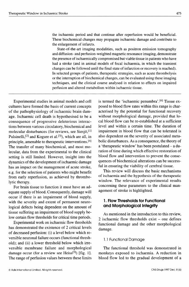
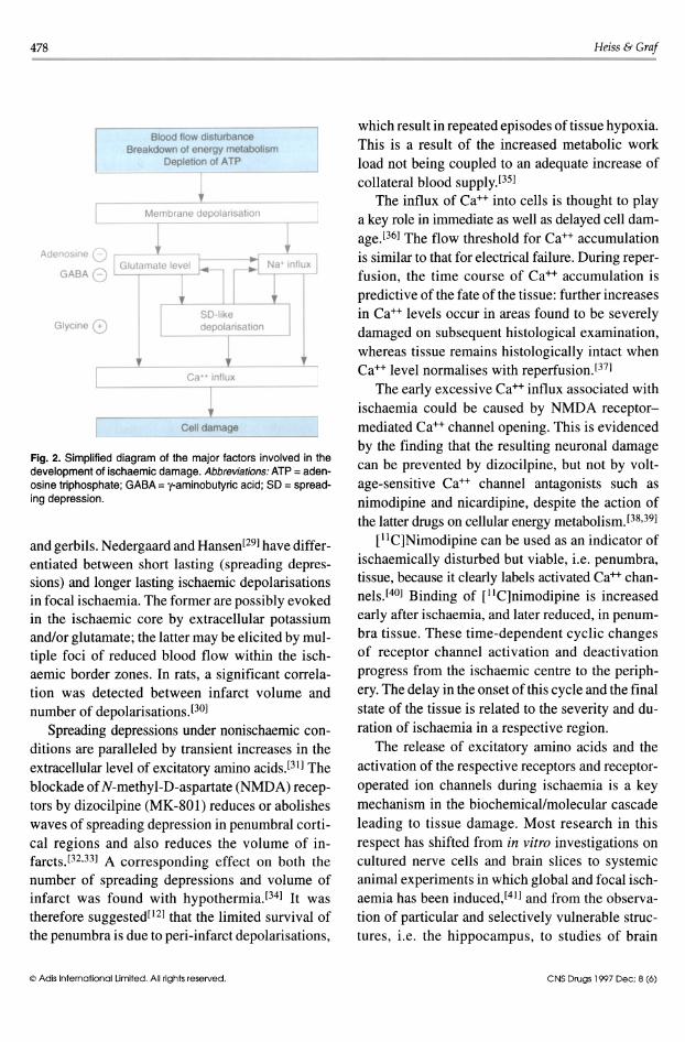
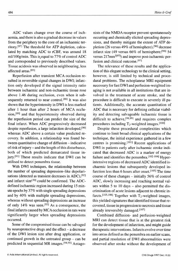
The processes involved in the propagation of ischaemic damage are outlined in figure 2. Various markers, mediators and modulators of ischaemic penumbra have been suggested, including: • waves of depolarisation • increases in the extracellular levels of excit
atory amino acids • activation of calcium (Ca++) channels • induction of immediate early genes and expres
sion of heat shock proteins. Some of these processes can be affected by ther
apeutic interventions. In the ischaemic core, the tissue depolarises
permanently within a few minutes. However, waves of transient neuronal depolarisation resembling spreading depression are regularly seen in periinfarct tissue, especially in small animals like rats
eNS Drugs 1997 Dec; 8 (6)
478
Ad noslne (.I
Blood liow dlslurbance Breakdown of energy metabolism
Deplebon of ATP
Membrane depolansatlon
GAB A <:) '----.---r-"
Ca" Influx
Cell damage
Fig. 2. Simplified diagram of the major factors involved in the development of ischaemic damage. Abbreviations: ATP = adenosine triphosphate; GABA = y-aminobutyric acid; SD = spreading depression.
and gerbils. Nedergaard and Hansen[29] have differentiated between short lasting (spreading depressions) and longer lasting ischaernic depolarisations in focal ischaemia. The former are possibly evoked in the ischaemic core by extracellular potassium and/or glutamate; the latter may be elicited by multiple foci of reduced blood flow within the ischaemic border zones. In rats, a significant correlation was detected between infarct volume and number of depolarisations.l30]
Spreading depressions under nonischaernic conditions are paralleled by transient increases in the extracellular level of excitatory amino acidsPIl The blockade of N-methyl-D-aspartate (NMDA) receptors by dizocilpine (MK-801) reduces or abolishes waves of spreading depression in penumbral cortical regions and also reduces the volume of infarctsJ32.33] A corresponding effect on both the number of spreading depressions and volume of infarct was found with hypothermia.[34] It was therefore suggested[l2] that the limited survival of the penumbra is due to peri-infarct depolarisations,
© Adis International Limited. All rights reserved.
Heiss & Graf
which result in repeated episodes of tissue hypoxia. This is a result of the increased metabolic work load not being coupled to an adequate increase of collateral blood supplyJ35]
The influx of Ca++ into cells is thought to play a key role in immediate as well as delayed cell damage.[36] The flow threshold for Ca++ accumulation is similar to that for electrical failure. During reperfusion, the time course of Ca++ accumulation is predictive of the fate of the tissue: further increases in Ca++ levels occur in areas found to be severely damaged on subsequent histological examjnation, whereas tissue remains histologically intact when Ca++ level normalises with reperfusion.l37]
The early excessive Ca++ influx associated with ischaemia could be caused by NMDA receptormediated Ca++ channel opening. This is evidenced by the finding that the resulting neuronal damage can be prevented by dizocilpine, but not by voltage-sensitive Ca++ channel antagonists such as nimodipine and nicardipine, despite the action of the latter drugs on cellular energy metabolism.l38•39]
[11C]Nimodipine can be used as an indicator of ischaemically disturbed but viable, i.e. penumbra, tissue, because it clearly labels activated Ca++ channels.[40] Binding of [11C]nimodipine is increased early after ischaemia, and later reduced, in penumbra tissue. These time-dependent cyclic changes of receptor channel activation and deactivation progress from the ischaemic centre to the periphery. The delay in the onset of this cycle and the final state of the tissue is related to the severity and duration of ischaemia in a respective region.
The release of excitatory amino acids and the activation of the respective receptors and receptoroperated ion channels during ischaemia is a key mechanism in the biochemical/molecular cascade leading to tissue damage. Most research in this respect has shifted from in vitro investigations on cultured nerve cells and brain slices to systemic animal experiments in which global and focal ischaemia has been induced,[41] and from the observation of particular and selectively vulnerable structures, i.e. the hippocampus, to studies of brain
eNS Drugs 1997 Dec; 8 (6)

Therapeutic Window in Ischaemic Stroke
structures relevant for clinical stroke, i.e. cortex and basal ganglia.
Increases in the levels of excitatory and inhibitory amino acid neurotransmitters in the cortex also follow a typical flow threshold pattern.142] This is not observed for nontransmitter amino acids. The magnitude of glutamate release during ischaemia was positively correlated with infarct volume.l43,44] During transient short ischaemic episodes, the extracellular glutamate levels reached are below a criticallevel,145] and reuptake systems for excitatory neurotransmitter amino acids or inhibitory neurotransmitters such as 'Y-aminobutyric acid (GAB A) and adenosinel46] may inhibit the deleterious excitotoxic action of excitatory transmitters. However, in prolonged ischaemia, excitotoxicity may be enhanced by a slowly increasing glycine level and the early reduction in the levels of adenosine.147] It must be considered, however, that increases in the levels of excitatory neurotransmitters in the periphery of ischaemia may not necessarily be a result of the generation of neuroactive amino acids within this region. Instead, it could be a result of leakage from the ischaemic core.l48] This effect has been demonstrated in the white matter and the CSF.l49]
The above findings might justify the speculation that a dynamic penumbra contributes to the progressive development of infarction: in prolonged ischaemia, an elevation of the levels of excitotoxic substances is sustained and the compounds might diffuse into the brain areas that are oligemic during the early phase of ischaemia. If not sufficiently inhibited, the deleterious cycle of ischaemic damage, including activation of Ca++ channels, is triggered, leading to a further increase of excitotoxic compounds that can diffuse into neighbouring areas.
2.2 Potential Therapeutic Interventions
Interference with the effect of excitatory amino acids and blockade of the respective receptors and ion channels appears to be a promising rationale for limiting ischaemic injury to the brain. Several strategies have been adopted for this purpose.150]
© Adis International Limited. All rights reserved.
479
For example, NMDAreceptors have been targeted, and several drugs that are antagonists of these receptors or sites on these receptors are currently in clinical trials for stroke, such as aptiganel, dextrorphan, selfotel, eliprodil and GV 150526A.151] In addition, the use of Ca++ antagonists as treatments for stroke has been investigated. Nimodipine is the agent for which the most data are available, although others such as nicardipine and flunarizine have also been studied.l51] However, the results with these agents are controversial and clinical data are scarce.
3. Clinical Evidence of an Ischaemic Penumbra
The assessment of pathophysiological changes leading to ischaemic cell damage in a clinical setting is extremely difficult, as most of the biochemical markers used in preclinical studies cannot be determined in patients. Furthermore, most techniques for clinical examination do not provide information about the viability of tissue, and routine techniques that do allow assessment of physiological variables [e.g. positron emission tomography (PET), single photon emission computed tomography (SPECT), x-ray computed tomography, magnetic resonance imaging (MRI) and magnetic resonance spectroscopy] are unsuitable because of their logistic complexity.
Magnetic resonance spectroscopy is still in the preclinical testing stage.l52] However, pertinent findings of pathophysiological changes occurring during the early period after ischaemic stroke have been obtained by multitracer PET. This provides quantitative 'maps' of several important physiological variables, including regional cerebral blood flow (rCBF), regional blood volume, regional cerebral metabolic rate of oxygen (rCMR02) and regional cerebral metabolic rate of glucose (rCMRgIc).
These studies conclude that tissue with rCMR02 below 65 Jlmol/lOOg/min and/or rCBF below 12 rnl/l00g/min definitely becomes necrotic.153,54] Regions with rCBF between 12 and 22 mlll00g/min have an unstable metabolic function, and are considered to be the penumbra zone, because infarc-
eNS Drugs 1997 Dec; 8 (6)
480
tion will occur in such a region if low flow values persist.[55] Patients with transitory ischaemic attacks have a minimum rCBF value of 22 mlli OOg/min. [56]
Uncoupled changes of flow and metabolism indicate the existence of viable but insufficiently supplied tissue in an ischaemic region: early perfusion failure manifests as a decrease in rCBF, with rCMR02 and rCMRglc remaining relatively preserved. This condition, termed 'misery perfusion' ,[28] implies that blood flow is inadequate relative to the metabolic energy demand for oxygen and other substrates for viable tissue. The observations in early stroke suggest that tissue affected by misery perfusion, but with rCMR02 still above the critical threshold, is viable and may recover.[57,58] However, the few published studies showed that most penumbra zones that had increased oxygen extraction fraction (OEF) turned into infarction in the period after a stroke.
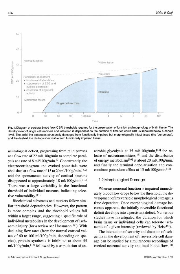
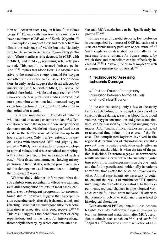
In a repeat multi tracer PET study of patients who had had an acute ischaemic stroke,[59] differentiation between the ischaemic core and periphery demonstrated that viable but misery perfused tissue exists in the border zone of ischaemia up to 48 hours after stroke. Only in a few regions, or in special cases with increased OEF and slightly impaired rCMR02, was metabolism preserved close to normal values, and tissue remained morphologically intact (see fig. 3 for an example of such a case). Most tissue compartments showing misery perfusion in the first day, suffered progressive metabolic derangement and became necrotic during the following 2 weeks.
Whereas the viable peri-infarct penumbra tissue exhibits some potential for effective treatment, available therapeutic options, in most cases, cannot prevent subsequent progression to necrosis. Some studies, however, suggest that hyperperfusion occurring early after the ischaemic attack and affecting tissue that has undergone little metabolic alteration is associated with good recovery.[60-62] This result suggests the beneficial effect of early reperfusion, and is the basis for interventional thrombolytic therapy, by which outcome after bas-
© Adis International Limited. All rights reserved.
Heiss & Graf
ilar and MCA occlusion can be significantly improved. [63-66]
In rare cases of carotid stenosis, low perfusion is accompanied by increased OEF indicative of a state of chronic misery perfusion or penumbra.[67,68] Such single cases described occasionally in the past may form a rationale for bypass surgery, by which flow and metabolism can be effectively increased.l69,70] However, the clinical impact of such a treatment remains controversial.[7I]
4. Techniques for Assessing Ischaemic Damage
4,1 Positron Emission Tomography: Correlation Between Animal Models and the Clinical Situation
In the clinical setting, only a few of the many factors contributing to the complex process of ischaemic tissue damage, such as blood flow, blood volume, oxygen consumption and glucose metabolism, can be assessed by functional imaging techniques. Additionally, clinical studies are restricted to anecdotal time points in the course of the disease. The complicated logistics involved in the regional determination of physiological variables prevent their repeated evaluation early after an ischaemic attack, which is when the fate of the patient is decided. Therefore, a gap exists between the results obtained at well defined but usually singular time points in animal experiments on the one hand, and the findings collected incidentally in patients at various times after the onset of stroke on the other. Animal experiments are necessary to better understand the results of multi tracer PET studies involving patients early after a stroke. In these experiments, regional changes in physiological variables can be followed, from the vascular attack to the permanent defective state, and then related to histological alterations.
With advanced PET equipment, it has become feasible to study pathophysiological changes in brain perfusion and metabolism after MCA occlusion in animals, such as baboons[72,73] and cats.[74,75] Tenjin et al. [72] observed a severe reduction of CBF
eNS Drugs 1997 Dec; 8 (6)
Therapeutic Window in Ischaemic Stroke 481 -------------------------------------
Fig. 3. Data from 2 positron emission tomography scans in a patient who recovered completely from hemiparalysis aiter an ischaemic stroke. The scans were taken 10 hours aiter the onset of hemiplegia (top line) and 10 days aiter the stroke (bottom line). A severe decrease in cerebral blood flow (CBF) in the first scan can be seen to have developed into hyperperiusion at the second scan. Regional cerebral metabolic rate of oxygen (CMR02) and regional cerebral metabolic rate of glucose (CMRg1c) remain above a critical level. A late scan (taken 10 days aiter the attack) showed that gross infarction had not developed. Abbreviation: CBV = cerebral blood volume.
(to below 18 ml/lOOg/min) and a significant increase of OEF 1 hour after occlusion in the core of ischaemia, and a decrease of OEF 9 hours after occlusion. However, Pappata et alp3l reported that the decrease of CBF in the territory of the occluded vessel in baboons was not so severe (21 % at 1 hour
and 31 % at 3 to 4 hours after occlusion). In these experiments, CMR02 was variable and
declined 3 to 4 hours after MCA occlusion, but was only moderately reduced in regions with maximal
OEF, suggesting prolonged viability. When the animals were followed after MCA occlusion,l76l
infarcts were observed in the deep MCA territory,
but not in the cortical regions. Changes of CMR02
occurred earlier and were more severe in the deep MCA territory than in the cortical regions. The in-
© Adis International Limited, All rights reserved.
farcts in this model increased in size over several days.[77l
In cats, CBF, CMR02, OEF and CMRglc were followed from control values before ischaemia to the endpoint of infarction 24 hours after MCA occlusionP4l CBF within the MCA territory fell im
mediately upon arterial occlusion to below 30% of
control, whereas CMR02 was less diminished and consequently OEF was increased, thus indicating
misery perfusion. This ischaemic penumbra spread with time from the centre to the borders of the
MCA territory. In most instances, the misery perfusion condition was followed by a marked decrease in OEF, reflecting progressive impairment of blood flow and metabolism and suggesting tran
sition to necrosis. The infarcts were more or less
eNS Drugs 1997 Dec: 8 (6)
482
complete 18 to 24 hours after MCA occlusion. Occasionally, spontaneous collateral reperfusion resolved the penumbra condition and morphological integrity of the cortex was preserved.
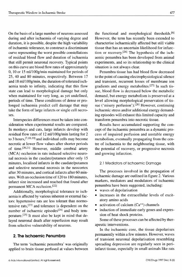
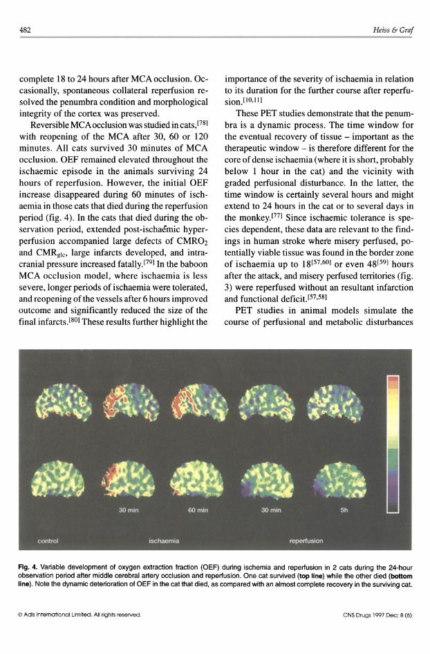
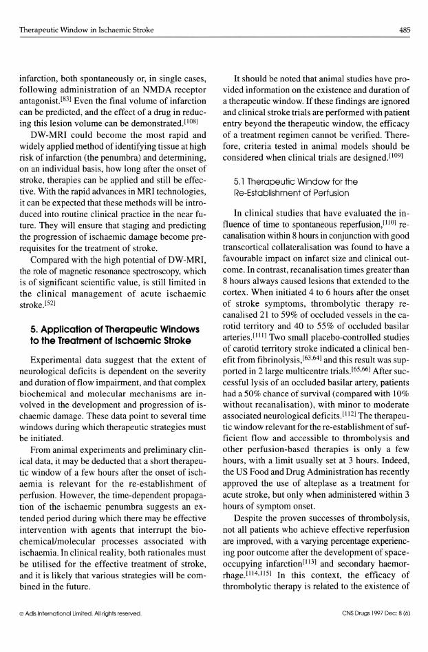
Reversible MCA occlusion was studied in cats, [78]
with reopening of the MCA after 30, 60 or 120 minutes. All cats survived 30 minutes of MCA occlusion. OEF remained elevated throughout the ischaemic episode in the animals surviving 24 hours of reperfusion. However, the initial OEF increase disappeared during 60 minutes of ischaemia in those cats that died during the reperfusion period (fig. 4). In the cats that died during the observation period, extended post-ischaemic hyperperfusion accompanied large defects of CMROz and CMRglc , large infarcts developed, and intracranial pressure increased fatally.[79] In the baboon MCA occlusion model, where ischaemia is less severe, longer periods of ischaemia were tolerated, and reopening of the vessels after 6 hours improved outcome and significantly reduced the size of the final infarcts.l80] These results further highlight the
Heiss &Graf
importance of the severity of ischaemia in relation to its duration for the further course after reperfusion.l 10,1I]
These PET studies demonstrate that the penumbra is a dynamic process. The time window for the eventual recovery of tissue - important as the therapeutic window - is therefore different for the core of dense ischaemia (where it is short, probably below 1 hour in the cat) and the vicinity with graded perfusional disturbance. In the latter, the time window is certainly several hours and might extend to 24 hours in the cat or to several days in the monkey.l77] Since ischaemic tolerance is species dependent, these data are relevant to the findings in human stroke where misery perfused, potentially viable tissue was found in the border zone of ischaemia up to 18[57,60] or even 48[59] hours after the attack, and misery perfused territories (fig. 3) were reperfused without an resultant infarction and functional deficit.[57,58]
PET studies in animal models simulate the course of perfusional and metabolic disturbances
Fig. 4. Variable development of oxygen extraction fraction (OEF) during ischemia and reperfusion in 2 cats during the 24-hour observation period after middle cerebral artery occlusion and reperfusion. One cat survived (top line) while the other died (bottom line). Note the dynamic deterioration of OEF in the cat that died, as compared with an almost complete recovery in the surviving cat.
© Adis International Umited . All rights reserved. eNS Drugs 1997 Dec; 8 (6)
Therapeutic Window in Ischaemic Stroke
characteristic of ischaemic stroke in humans, and so a comparison of experimental results to clinical findings may be justified. Permanent MCA occlusion resembles the natural course after vascular occlusion, leading to large infarcts in most cases, with a chance of collateral reperfusion that may resolve misery perfusion and improve outcome. Reopening of the MCA resembles the (spontaneous) dissolution of vascular occlusions in transient ischaemic attacks, spontaneous lysis of emboli after a period beyond the tolerable time period, and therapeutic thrombolysis. Reperfusion after 30 minutes of MCA occlusion led to a short lasting hyperperfusion period and to a fast normalisation of flow and metabolism. This may be comparable with a transient ischaemic attack. During MCA occlusion of longer duration 2 patterns can be distinguished: (i) a decrease of the OEF during the time of MCA occlusion reflects fast irreversible damage of tissue, whereas (ii) the persistence of raised OEF indicates preserved viability of tissue over the ischaemic period. Forced reperfusion by reopening of the MCA cannot salvage already irreversibly damaged tissue, but may cause additional damage by inducing oedema via leaking vascular endothelium. In such cases, the infarcts are large and animals die early due to increased intracranial pressure. These courses resemble the deleterious outcome of thrombolytic therapy that is initiated too late and so could not prevent the development of large infarcts, resulting in additional oedema and secondary haemorrhagic transformation.
4.2 Magnetic Resonance Imaging: Use in Early Stroke
Of the available imaging techniques, MRI has the highest spatial resolution for detecting lesions in the brain, and has therefore gained a prominent position in the diagnosis of focal cerebral disorders. New MRI technologies provide additional information on pathophysiological and functional alterations, making this method a versatile tool with multiple applications.
In the management of acute stroke, a technique is needed which is readily available in the acute
© Adls International Limited. All rights reserved.
483
clinical setting, rapid, non-invasive and provides results rapidly. The latter is important to ensure that the necessary multiple diagnostic measurements are available within minutes, so as not to delay treatment beyond the optimal time window.!81]
Diffusion-weighted imaging (DWI) has considerable potential, as substantial changes in the parenchymal diffusion of water after the onset of ischaemia can be detected by this method. Since the detection of ischaemic stroke by DWI-MRI within minutes after experimental vascular occlusion was first demonstrated,!82] this technique has evolved as an important tool for both the identification of very early injury in patients and the quantitative assessment of the severity and volume of ischaemically compromised tissue.[83] In ischaemia models involving small animals (e.g. rats, gerbils, cats), DWI has been shown to be useful as an early diagnostic marker of ischaemia. It can also provide quantitative data on a physiological parameter [the apparent diffusion coefficient (ADC) of water in tissue] and volumetric analysis of the lesion. ADC values derived from DWI measurements can give a value for assessing the severity of ischaemic changes and also for following the time course in the evolution of irreversible damage.
DWI shows the area of reduced diffusion as a hyperintense region, which follows the redistribution of water from the extracellular to intracellular space as a consequence of reduced adenosine triphosphate (ATP)ase activity.[84] In the core of this area, ATP remains depleted, indicating necrotic transformation due to severely reduced perfusion.!8S] Since disturbances of the water/ionic homeostasis occur immediately with disruptions of perfusion, DWI is able to detect the initial disturbance in ischaemia, and also shows the reversal of these alterations with early recovery. With this technique, the progression of ischaemic injury from areas of dense to those of moderate to mild flow decreases can be followed. In 1 study, the changes in DWI signals correlated with histological defects and metabolic abnormalities as assessed by autoradiographic and chemical imaging procedures at follow-up)86]
eNS Drugs 1997 Dec; 8 (6)
484
ADC values change over the course of ischaemia, and there is also a gradual decrease in values from the periphery to the core of an ischaemic territory.l87] The threshold for ATP depletion, calculated by matching ADC to rCBF, was around 18 mIll OOg/min. This is, equal to 77% of control ADC and corresponded to previously described values. Tissue acidosis was observed in neighbouring, less affected areas.[85]
Reperfusion after transient MCA occlusion resulted in reversible signal changes in DWI; infarction only developed if the signal intensity ratio between ischaemic and non-ischaemic tissue rose above 1.46 during occlusion, even when it subsequently returned to near contro1.l88] It was also shown that the hyperintensity in DWI is less marked after 1 hour than after 2 hours of MCA occlusion,[89] and that hyperintensity observed during the reperfusion period can predict the size of the final infarct. When ADC progressively decreased despite reperfusion, a large infarction developed,[90] whereas ADC above a certain value predicted recovery. In addition, a relationship was found between quantitative change of diffusion - indicative of risk of injury - and the length of this disturbance, both of which predicted demarcated tissue injury.[91] These results indicate that DWI can be utilised to detect penumbra tissue.
With DWI techniques, the relationship between the number of spreading depression-like depolarisations (detected as transient decreases in ADC),[92] and infarct size[30] could be confirmed. The ADCdefined ischaemic region increased during 15 minute epochs by 37% with single spreading depressions and by 40% with multiple spreading depressions, whereas without spreading depressions an increase of only 14% was seen.l93] As a consequence, the final infarcts caused by MCA occlusion in rats were significantly larger when spreading depressions occurred.
Some ofthe compromised tissue can be salvaged by neuroprotective drugs and the effect - a decrease of the DWI lesion size after drug application, or continued growth in the untreated group - can be predicted in sequential MR images)94,95] Antago-
© Adis International Limited. All rights reserved.
Heiss & Graf
nists of the NMDA receptor prevent spontaneously occurring and chemically elicited spreading depressions, and thereby mitigate the extent of ATP depletion (26 versus 49% ofhemisphere),[96] decrease infarct size (49 versus 66% of hemisphere;[96] 54 versus 217mm3[97]) and improve post-ischaemic perfusion and clinical outcome.l971
The relevance of these results and the application of this elegant technology to the clinical setting, however, is still limited by technical and procedural problems. The echoplanear MRI equipment necessary for fast DWI and perfusion-weighted imaging is not available in all institutions that are involved in the treatment of acute stroke, and the procedure is difficult to execute in severely ill patients. Additionally, the accurate quantitation of ADC that is necessary for defining cellular viability and detecting salvageable ischaemic tissue is difficult to achieve,[98,99] and requires complex procedures including T2-weighted images.[lOO]
Despite these procedural complexities which continue to limit broad clinical applications of the technique, the initial experience gathered in a few centres is promising.[lOI] Recent applications of DWI in patients early after ischaemic stroke indicated that decreased ADC is a marker of energy failure and identifies the penumbra.[102-104] Hyperintensive regions of decreased ADC identified ischaemic lesions that subsequently developed infarction less than 6 hours after onset.[105] The time course· of these changes - initially 56% of control ADC, slowly increasing and reaching normal values within 5 to 10 days - also permitted the discrimination of acute lesions adjacent to chronic infarcts.l 105, 106] Together with T 2-weighted images, this yielded signatures that identified tissue that recovered, tissue in progression to necrosis and tissue already irreversibly damaged.[107]
Combined diffusion- and perfusion-weighted MRI can detect tissue that is at the greatest risk for the development of infarction, and might guide therapeutic interventions. Infarcts evolve over time into areas defined as the penumbra on earlier scans, and partial resolution of DWI abnormalities were observed after stroke without the development of
eNS Drugs 1997 Dec: 8 (6)
Therapeutic Window in Ischaemic Stroke
infarction, both spontaneously or, in single cases, following administration of an NMDA receptor antagonist. l83] Even the final volume of infarction can be predicted, and the effect of a drug in reducing this lesion volume can be demonstrated.l 108]
DW-MRI could become the most rapid and widely applied method of identifying tissue at high risk of infarction (the penumbra) and determining, on an individual basis, how long after the onset of stroke, therapies can be applied and still be effective. With the rapid advances in MRI technologies, it can be expected that these methods will be introduced into routine clinical practice in the near future. They will ensure that staging and predicting the progression of ischaemic damage become prerequisites for the treatment of stroke.
Compared with the high potential of DW-MRI, the role of magnetic resonance spectroscopy, which is of significant scientific value, is still limited in the clinical management of acute ischaemic stroke.l52]
5. Application of Therapeutic Windows to the Treatment of Ischaemic Stroke
Experimental data suggest that the extent of neurological deficits is dependent on the severity and duration of flow impairment, and that complex biochemical and molecular mechanisms are involved in the development and progression of ischaemic damage. These data point to several time windows during which therapeutic strategies must be initiated.
From animal experiments and preliminary clinical data, it may be deducted that a short therapeutic window of a few hours after the onset of ischaemia is relevant for the re-establishment of perfusion. However, the time-dependent propagation of the ischaemic penumbra suggests an extended period during which there may be effective intervention with agents that interrupt the biochemical/molecular processes associated with ischaemia. In clinical reality, both rationales must be utilised for the effective treatment of stroke, and it is likely that various strategies will be combined in the future.
© Adis International Limited. All rights reserved.
485
It should be noted that animal studies have provided information on the existence and duration of a therapeutic window. If these findings are ignored and clinical stroke trials are performed with patient entry beyond the therapeutic window, the efficacy of a treatment regimen cannot be verified. Therefore, criteria tested in animal models should be considered when clinical trials are designed. l 109]
5.1 Therapeutic Window for the Re-Establishment of Perfusion
In clinical studies that have evaluated the influence of time to spontaneous reperfusion,lllO] recanalisation within 8 hours in conjunction with good transcortical collateralisation was found to have a favourable impact on infarct size and clinical outcome. In contrast, recanalisation times greater than 8 hours always caused lesions that extended to the cortex. When initiated 4 to 6 hours after the onset of stroke symptoms, thrombolytic therapy recanalised 21 to 59% of occluded vessels in the carotid territory and 40 to 55% of occluded basilar arteries. llll ] Two small placebo-controlled studies of carotid territory stroke indicated a clinical benefit from fibrinolysis,l63,64] and this result was supported in 2 large multicentre trials.l65,66] After successfullysis of an occluded basilar artery, patients had a 50% chance of survival (compared with 10% without recanalisation), with minor to moderate associated neurological deficits. [112] The therapeutic window relevant for the re-establishment of sufficient flow and accessible to thrombolysis and other perfusion-based therapies is only a few hours, with a limit usually set at 3 hours. Indeed, the US Food and Drug Administration has recently approved the use of alteplase as a treatment for acute stroke, but only when administered within 3 hours of symptom onset.
Despite the proven successes of thrombolysis, not all patients who achieve effective reperfusion are improved, with a varying percentage experiencing poor outcome after the development of spaceoccupying infarctionlll3] and secondary haemorrhage.l"4,15] In this context, the efficacy of thrombolytic therapy is related to the existence of
eNS Drugs 1997 Dec; 8 (6)
486
'penumbral' tissue perfused at rates below the functional threshold, yet above the level indicating or leading to irreversible morphological damage.
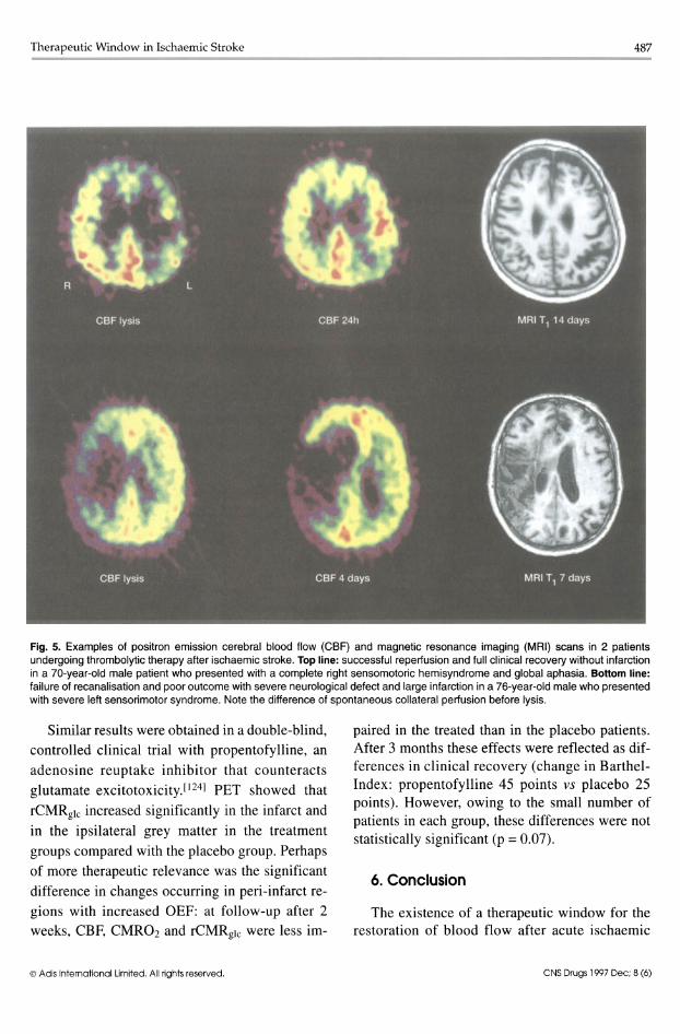
In order to further analyse the influence of residual perfusion on outcome, regional cerebral perfusion was studied at the start «3 hours after onset of symptoms) and repeatedly after thrombolytic therapy (alteplase 0.9 mg/kg according to a National Institute of Neurological Diseases and Stroke protocol)166] in patients with an acute hemispheric stroke.l1l6] After 3-dimensional alignment and normalisation on the mean of the contralateral hemisphere, the relative perfusion rates of first and second measurements were related to defects found in late T1-weighted MRI on a voxel-byvoxel basis. Before initiation of thrombolytic therapy, all patients showed marked hypoperfusion of their affected MCA territory (fig. 5). Within these hypoperfused areas, volumes of tissue at risk of ischaemic damage were operationally defined as <50% uptake and 50 to 70% uptake of the contralateral mean, representing tissue below or at the viability threshold and within the penumbra zone, respectively. Patients in whom large proportions of ischaemically compromised tissue were effectively reperfused, and not included in the final infarct identified on late MRI, recovered completely or experienced slight damage; those in whom recanalisation reached less than half the deficiently supplied territory had an unfavourable outcome with severe permanent defects. These findings indicate that residual flow in the given time interval is the crucial prognostic factor for thrombolytic therapy. However, it has to be further analysed whether higher residual blood flow values would justify an extended time window for thrombolysis.
5.2 Therapeutic Window for the Inhibition of Biochemical/Molecular Changes
The existence of a window of longer duration, during which agents could be given that interfere with the biochemical and molecular disturbances associated with ischaemia, is still controversial.11l7-120] Furthermore, the duration of the window cannot be easily deducted from a beneficial
© Adis International Umlted. All rights reserved.
Heiss & Graf
effect of therapeutic strategies. Further controlled studies need to be carried out to prove convincingly the value of therapies such as those that:[l21] • interfere with increased Ca++ entry into cells • inhibit amino acid excitotoxicity • scavenge free-radicals • affect nitric oxide synthetase.
There are a few examples indicating that interaction with such mechanisms may result in an improved outcome after a cerebrovascular attack. Furthermore, it appears that PET is able to identify early pathophysiological changes that occur after ischaemic stroke, and predict treatment effects and outcome. Therefore, PET and DW-MRI might be of value for the demonstration of viable tissue and of treatment effects in patients with stroke.
Ca++ antagonists were able to improve neurological outcome and reduce histological damage in animal experiments and therefore have been used in ischaemic stroke. The results of multicentre trials have been controversial and benefits were only indicated in a meta-analysis;1122] beneficial outcome was obtained only when treatment was started within the first 12 hours after the onset of symptoms. In multitracer PET studies,155] nimodipine was found to be effective in reversing the decline in CMR02 and increasing CBF in the densely ischaemic zone, whereas in the penumbra CMR02 was improved, but the difference compared with the control group was not statistically significant. In another study,1123] PET demonstrated that
rCMRglc in the cortex of the hemisphere outside the infarct improved significantly within the first 3 weeks after the stroke in patients treated with nimodipine. Furthermore, patients in the treatment group reached higher scores in the Barthel-Index after 6 months.
These data may help to explain the controversial conclusions from the multi centre trials of nimodipine. The drug can only be effective as long as viable tissue is present iIi a progressive ischaemic lesion; when given after that period (which might be 12 hours), a therapeutic effect is unlikely to be achieved.
eNS Drugs 1997 Dec; 8 (6)
Therapeutic Window in Ischaemic Stroke 487 ------------------------------------------
Fig. 5. Examples of positron emission cerebral blood flow (CBF) and magnetic resonance imaging (MRI) scans in 2 patients undergoing thrombolytic therapy after ischaemic stroke. Top line: successful reperiusion and full clinical recovery without infarction in a 70-year-old male patient who presented with a complete right sensomotoric hemisyndrome and global aphasia. Bottom line: failure of recanalisation and poor outcome with severe neurological defect and large infarction in a 76-year-old male who presented with severe left sensorimotor syndrome. Note the difference of spontaneous collateral periusion before lysis.
Similar results were obtained in a double-blind, controlled clinical trial with propentofylline, an
adenosine reuptake inhibitor that counteracts glutamate excitotoxicity.l124] PET showed that
rCMRglc increased significantly in the infarct and in the ipsilateral grey matter in the treatment
groups compared with the placebo group. Perhaps
of more therapeutic relevance was the significant difference in changes occurring in peri-infarct re
gions with increased OEF: at follow-up after 2 weeks, CBF, CMR02 and rCMRgic were less im-
© Adis International limited. All rights reserved.
paired in the treated than in the placebo patients. After 3 months these effects were reflected as differences in clinical recovery (change in BarthelIndex: propentofylline 45 points vs placebo 25 points). However, owing to the small number of patients in each group, these differences were not statistically significant (p = 0.07).
6. Conclusion
The existence of a therapeutic window for the restoration of blood flow after acute ischaemic
eNS Drugs 1997 Dec; 8 (6)
488
stroke is evident from experimental studies and clinical experience. This has formed the basis for the early thrombolytic treatment (within 3 hours of the onset of symptoms) of stroke and thus for the successful use of this therapy. State-of-the-art imaging modalities, such as PET and diffusion- or perfusion-weighted MRI, have helped to define the therapeutic window in animal models of focal ischaemia and in patients who have experienced a stroke, by demonstrating the presence of ischaemically compromised but viable tissue. These techniques are also used to evaluate the success of thrombolytics and other therapeutic strategies that aim to achieve early interruption of the devastating molecular and biological alterations that are triggered during the ischaemic period.
References I. Siesjo BK. Pathophysiology and treatment of focal cerebral isch
emia. Part I: Pathophysiology. J Neurosurg 1992; 77: 169-84 2. Pulsinelli W. Pathophysiology of acute ischaemic stroke. Lancet
1992; 339: 533-6 3. Kogure K, Hossmann K-A, Siesjo BK. Neurology of ischemic
brain damage. Amsterdam: Elsevier, 1993 4. Ginsberg MD. Emerging strategies for the treatment of isch
emic brain injury. In: Waxman SG, editor. Molecular and cellular approaches to the treatment of ischemic brain disease. New York: Raven Press, 1993: 207-37
5. Heiss WD. Experimental evidence for ischemic thresholds and functional recovery. Stroke 1992; 23: 1668-72
6. Astrup J, Siesjo BK, Symon L. Thresholds in cerebral ischemia - the ischemic penumbra. Stroke 1981; 12: 723-5
7. Jones TH, Morawetz RB, Crowell RM, et al. Thresholds offocal cerebral ischemia in awake monkeys. J Neurosurg 1981; 54: 773-82
8. Sharbrough FW, Messick JM, Sundt Jr TM. Correlation of continuous electroencephalograms with cerebral blood flow measurements during carotid endarterectomy. Stroke 1973; 4: 674-83
9. Branston NM, Symon L, Crockard HA, et aI. Relationship between the cortical evoked potential and local cortical blood flow following acute middle cerebral artery occlusion in the baboon. Exp Neuro11974; 45: 195-208
10. Heiss WD, Hayakawa T, Waltz AG. Cortical neuronal function during ischemia. Arch Neuro11976; 33: 813-20
II. Heiss WD, Rosner G. Functional recovery of cortical neurons as related to degree and duration of ischemia. Ann Neurol 1983; 14: 294-301
12. Hossmann KA. Viability thresholds and the penumbra of focal ischemia. Ann Neuro11994; 36: 557-65
13. Mies G, Ishimaru S, Xie Y, et al. Ischemic thresholds of cerebral protein synthesis and energy state following middle cerebral artery occlusion in rat. J Cereb Blood Flow Metab 1991; II: 753-61
14. Paschen W, Mies G, Hossmann KA. Threshold relationship between cerebral blood flow, glucose utilization, and energy
© Adis International limited. All rtghts reserved.
Heiss & Graf
metabolites during development of stroke in gerbils. Exp Neuro11992; 117: 325-33
15. Shimada N, Graf R, Rosner G, et al. Ischemic flow threshold for extracellular glutamate increase in cat cortex. J Cereb Blood Flow Metab 1989; 9: 603-6
16. Obrenovitch TP, Garofalo 0, Harris RJ, et al. Brain tissue concentrations of ATP, phosphocreatine, lactate, and tissue pH in relation to reduced cerebral blood flow following experimental acute middle cerebral artery occlusion. J Cereb Blood Flow Metab 1988; 8: 866-74
17. AstrupJ, Symon L, Branston NM, etal. Cortical evoked potential and extracellular K+ and H+ at critical levels of brain ischemia. Stroke 1977; 8: 51-7
18. Tamura A, Asano T, Sano K. Correlation between rCBF and histological changes following temporary middle cerebral artery occlusion. Stroke 1980; 11: 487-93
19. Marcoux FW, Morawetz RB, Crowell RM, et aI. Differential regional vulnerability in transient focal cerebral ischemia. Stroke 1982; 13: 339-46
20. Pulsinelli WA, Brierley JB, Plum F. Temporal profile of neuronal damage in a model of transient forebrain ischemia. Ann Neuro11982; 11: 491-8
21. Jenkins LW, Povlishock JT, Lewelt W, et al. The role of postischemic recirculation in the development of ischemic neuronal injury following complete cerebral ischemia. Acta Neuropathol 1981; 55: 205-20
22. Memezawa H, Smith ML, Siesjo BK. Penumbral tissues salvaged by reperfusion following middle cerebral artery occlusion in rats. Stroke 1992; 23: 552-9
23. Jacewicz M, Tanabe J, Pulsinelli WA. The CBF threshold and dynamics for focal cerebral infarction in spontaneously hypertensive rats. J Cereb Blood Flow Metab 1992; 12: 359-70
24. Tomida S, Nowak TS, Vass K, et al. Experimental model for repetitive ischemic attacks in the gerbil: the cumulative effect of repeated ischemic insults. J Cereb Blood Flow Metab 1987; 7: 773-82
25. Morikawa E, Ginsberg MD, Dietrich WD, et al. The significance of brain temperature in focal cerebral ischemia: histopathological consequences of middle cerebral artery occlusion in the rat. J Cereb Blood Flow Metab 1992; 12: 380-9
26. Heiss WD, Graf R. The ischemic penumbra. Curr Opin Neurol 1994; 7: 11-9
27. Ginsberg MD, Pulsinelli WA. The ischemic penumbra, injury thresholds, and the therapeutic window for acute stroke. Ann Neurol 1994; 36: 553-4
28. Baron JC, Bousser MG, Rey A, et al. Reversal of focal 'miseryperfusion syndrome' by extra-intracranial arterial bypass in hemodynamic cerebral ischemia. Stroke 1981; 12: 454-9
29. Nedergaard M, Hansen AJ. Characterization of cortical depolarizations evoked in focal cerebral ischemia. J Cereb Blood Flow Metab 1993; 13: 568-74
30. Mies G, Iijima T, Hossmann KA. Correlation between peri-infarct DC shifts and ischaemic neuronal damage in rat. Neuroreport 1993; 4: 709-11
31. Fabricius M, Jensen LH, Lauritzen M. Microdialysis of interstitial amino acids during spreading depression and anoxic depolarization in rat neocortex. Brain Res 1993; 612: 61-9
32. Gill R, Andine P, Hillered L, et al. The effect of MK-801 on cortical spreading depression in the penumbral zone following focal ischemia in the rat. J Cereb Blood Flow Metab 1992; 12: 371-9
33. Iijima T, Mies G, Hossmann KA. Repeated negative DC deflections in rat cortex following middle cerebral artery occlusion
eNS Drugs 1997 Dec; 8 (6)
Therapeutic Window in Ischaemic Stroke
are abolished by MK-801: effect on volume of ischemic injury. J Cereb Blood Flow Metab 1992; 12: 727-33
34. Chen Q, Chopp M, Bodzin G, et al. Temperature modulation of cerebral depolarization during focal cerebral ischemia in rats: correlation with ischemic injury. J Cereb Blood Flow Metab 1993; 13: 389-94
35. Back T, Htihn-Berlage M, Kohno K, et al. Diffusion NMR imaging in experimental stroke: correlation with cerebral metabolites. Stroke 1994; 25: 494-500
36. Siesjti BK, Bengtsson F. Calcium fluxes, calcium antagonists, and calcium-related pathology in brain ischemia, hypoglycemia, and spreading depression. J Cereb Blood Flow Metab 1989; 9: 127-40
37. Vematsu D, Araki N, Greenberg JH, et al. Combined therapy with MK-80 1 and nimodipine for protection of ischemic brain damage. Neurology 1991; 41: 88-94
38. Nakamura K, Hatakeyama T, Furuta S, et al. The role of early Ca++ influx in the pathogenesis of delayed neuronal death after briefforebrain ischemia in gerbils. Brain Res 1993; 613: 181-92
39. Lemons BV, Chehrazi BB, Kauten R, et al. The effect of nimodipine on high-energy phosphates and intracellular pH during cerebral ischemia. J Neurotrauma 1993; 10: 73-81
40. Hakim AM, Hogan MJ. In vivo binding of nimodipine in the brain: 1. The effect of focal cerebral ischemia. J Cereb Blood Flow Metab 1991; II: 762-70
41. Choi DW. NMDA receptor, AMP A receptors, and extracellular acidity in ischemic brain damage: a view from the dish. In: Krieglstein J, Oberpichler-Schwenk H, editors. Pharmacology of cerebral ischemia. Stuttgart: Wiss Veri Ges, 1992: 45-52
42. Shimada N, Graf R, Rosner G, et al. Differences in ischemiainduced accumulation of amino acids in the cat cortex. Stroke 1990; 21: 1445-51
43. Takagi K, Ginsberg MD, Globus MY-T, et al. Changes in amino acid neurotransmitters and cerebral blood flow in the ischemic penumbral region following middle cerebral artery occlusion in the rat: correlation with histopathology. J Cereb Blood Flow Metab 1993; 13: 575-85
44. Taguchi J, GrafR, Rosner G, et al. Prolonged transient ischemia results in impaired CBF recovery and secondary glutamate accumulation in cats. J Cereb Blood Flow Metab 1996; 16: 271-9
45. Veda Y, Obrenovitch TP, Lok SY, et al. Efflux of glutamate produced by short ischemia of varied severity in rat striatum. Stroke 1992; 23: 253-9
46. Matsumoto K, Graf R, Rosner G, et al. Flow thresholds for extracellular purine catabolite elevation in cat focal ischemia. Brain Res 1992; 579: 309-14
47. Matsumoto K, Graf R, Rosner G, et al. Elevation of neuroactive substances in the cortex of cats during prolonged focal ischemia. J Cereb Blood Flow Metab 1993; 13: 586-94
48. Obrenovitch TP, Vrenjak J, Richards DA, et al. Extracellular neuroactive amino acids in the rat striatum during ischemia: comparison between penumbral conditions and ischaemia with sustained anoxic depolarisation. J Neurochem 1993; 61: 178-86
49. Shimada N, GrafR, Rosner G, et al. Ischemia-induced accumulation of extracellular amino acids in cerebral cortex, white matter, and cerebrospinal fluid. J Neurochem 1993; 60: 66-71
50. Pulsinelli W, Sarokin A, Buchan A. Antagonism of the NMDA and non-NMDA receptors in global versus focal brain ischemia. In: Kogure K, Hossmann KA, Siesjti BK. editors. Prog-
© Adls Intemational Umited. All rights reserved.
489
ress in brain research. Vol. 96. Amsterdam: Elsevier Science Publishers. 1993: 125-35
51. Dorman PJ. Counsell CEo Sandercock PAG. Recently developed neuroprotective therapies for acute stroke: a quantitative systemic review of clinical trials. CNS Drugs 1996; 5 (6): 457-74
52. Prichard JW. The role of magnetic resonance spectroscopy in stroke. In: Moskowitz MA. Caplan LR, editors. Cerebrovascular diseases. Boston: Butterworth-Heinemann. 1995: 475-85
53. Baron JC. Positron tomography in cerebral ischemia. Neuroradiology 1985; 27: 509-16
54. Powers WJ, Grubb Jr RL, Darriet D. et al. Cerebral blood flow and cerebral metabolic rate of oxygen requirements for cerebral function and viability in humans. J Cereb Blood Flow Metab 1985; 5: 600-8
55. Hakim AM, Evans AC, Berger L, et al. The effect of nimodipine on the evolution of human cerebral infarction studied by PET. J Cereb Blood Flow Metab 1989; 9: 523-34
56. Powers WJ, Press GW, Grubb Jr RL, et al. The effect ofhemodynamically significant carotid artery disease on the hemodynamic status of the cerebral circulation. Ann Intern Med 1987; 106: 27-35
57. Furlan M. Marchal G, Viader F, et al. Spontaneous neurological recovery after stroke and the fate of the ischemic penumbra. Ann Neuro11996; 40: 216-26
58. Heiss W-D, Fink GR, Huber M, et al. Positron emission tomography imaging and the therapeutic window. Stroke 1993; 24 Suppl. I: 150-3
59. Heiss W-D, Huber M, Fink G, et al. Progressive derangement of peri infarct viable tissue in ischemic stroke. J Cereb Blood Flow Metab 1992; 12: 193-203
60. Marchal G, Serrati C, Rioux P, et al. PET imaging of cerebral perfusion and oxygen consumption in acute ischaemic stroke: relation to outcome. Lancet 1993; 341: 925-7
61. Fink GR, Herholz K, Pietrzyk V, et al. Peri-infarct perfusion in human ischemia: its relation to tissue metabolism, morphology, and clinical outcome. J Stroke Cerebrovasc Dis 1993; 3: 123-31
62. Marchal G, Furlan M, Beaudouin V, et al. Early spontaneous hyperperfusion after stroke - a marker of favourable tissue outcome? Brain 1996; 119: 409-19
63. Mori E, Yoneda Y, Tabuchi M. Intravenous recombinant tissue plasminogen activator in acute carotid artery territory stroke. Neurology 1992; 42: 976-82
64. Yamaguchi T, Hayakawa T, Kiuchi H. Intravenous tissue plasminogen activator ameliorates the outcome of hyperacute embolic stroke. Cerebrovasc Dis 1993; 3: 269-72
65. Hacke W, Kaste M, Fieschi C, et al. Intravenous thrombolysis with recombinant tissue plasminogen activator for acute hemispheric stroke. The European Cooperative ECASS Study Group. JAMA 1995; 274: 1017-25
66. The National Institute of Neurological Disorders and Stroke rt-PA Stroke Study Group. Tissue plasminogen activator for acute ischemic stroke. N Engl J Med 1995; 333: 1581-7
67. Yamauchi H, Fukuyama H, Fujimoto N, et al. Significance of low perfusion with increased oxygen extraction fraction in a case of internal carotid artery stenosis. Stroke 1992; 23: 431-2
68. Yamauchi H, Fukuyama H, Nagahama Y, et al. Evidence of misery perfusion and risk for recurrent stroke in major cerebral arterial occlusive diseases from PET. J Neurol Neurosurg Psychiatry 1996; 61: 18-25
69. Ishikawa T, Yasui N, Suzuki A, et al. STA-MCA bypass surgery for internal carotid artery occlusion - comparative follow-up study. Neurol Med Chir (Tokyo) 1992; 32: 5-9
eNS Drugs 1997 Dec; 8 (6)
490
70. Ogawa A, Kameyama M, Muraishi K, et aI. Cerebral blood flow and metabolism following superficial temporal artery to superior cerebellar artery bypass for vertebrobasilar occlusive disease. J Neurosurg 1992; 76: 955-60
71. Powers WJ, Grubb Jr RL, Raichle ME. Clinical results of extracranial-intracranial bypass surgery in patients with hemodynamic cerebrovascular disease. J Neurosurg 1989; 70: 61-7
72. Tenjin H, Veda S, Mizukawa N, et aI. Positron emission tomographic measurement of acute hemodynamic changes in primate middle cerebral artery occlusion. Neurol Med Chir (Tokyo) 1992;32: 805-10
73. Pappata S, Fiorelli M, Rommel T, et al. PET study of changes in local brain hemodynamics and oxygen metabolism after unilateral middle cerebral artery occlusion in baboons. J Cereb Blood Flow Metab 1993; 13: 416-24
74. Heiss W-D, Graf R, Wienhard K, et al. Dynamic penumbra demonstrated by sequential multi tracer PET after middle cerebral artery occlusion in cats. J Cereb Blood Flow Metab 1994; 14: 892-902
75. Heiss W-D, Wienhard K, Graf R, et al. High-resolution PET in cats: application of a clinical camera to experimental studies. J Nucl Med 1995; 36: 493-8
76. Young AR, Touzani 0, Baron J-C, et al. To reperfuse or not to reperfuse?: Quantitative infarct volumes after middle cerebral artery occlusion in the baboon [abstract]. J Cereb Blood Flow Metab 1995; 15 Suppl. 1: S72
77. Touzani 0, Young AR, Derlon J-M, et al. Sequential studies of severely hypometabolic tissue volumes after permanent middle cerebral artery occlusion: a positron emission tomographic investigation in anesthetized baboons. Stroke 1995; 26: 2112-9
78. Heiss W-D, Graf R, Uittgen J, et al. Repeat positron emission tomographic studies in transient middle cerebral artery occlusion in cats: residual perfusion and efficacy of postischemic reperfusion. J Cereb Blood Flow Metab 1997; 17: 388-400
79. Hayakawa T, Waltz AG. Changes of epidural pressures after experimental occlusion of middle cerebral artery in cats: relationship to severity of neurologic deficits, sizes of infarcts, and anesthesia. J Neurol Sci 1975; 26: 319-33
80. Young AR, Sette G, Touzani 0, et al. Relationships between high oxygen extraction fraction in the acute stage and final infarction in reversible middle cerebral artery occlusion: an investigation in anesthetized baboons with positron emission tomography. J Cereb Blood Flow Metab 1996; 16: 1176-88
81. Warach S, Boska M, Welch KMA. Pitfalls and potential of clinical diffusion-weighted MR imaging in acute stroke. Stroke 1997; 28: 481-2
82. Moseley ME, Cohen Y, Mintorovitch J, et al. Early detection of regional cerebral ischemia in cats: comparison of diffusionand T2-weighted MRI and spectroscopy. Magn Reson Med 1990; 14: 330-46
83. Warach S. Diffusion-weighted imaging. In: Moskowitz MA, Caplan LR, editors. Cerebrovascular diseases. Boston: Butterworth-Heinemann, 1995: 451-62
84. Mintorovitch J, Moseley ME, Chileuitt L, et al. Comparison of diffusion- and T2-weighted MRl for the early detection of cerebral ischemia and reperfusion in rats. Magn Reson Med 1991; 18: 39-50
85. Hoehn-BerJage M, Norris DG, Kohno K, et al. Evolution of regional changes in apparent diffusion coefficient during focal ischemia of rat brain: the relationship of quantitative diffusion NMR imaging to reduction in cerebral blood flow and metabolic disturbances. J Cereb Blood Flow Metab 1995; 15: 1002-11
© Adls Intema~onal Umlted. All rights reserved.
Heiss & Graf
86. Hossmann K-A, Hoehn-BerJage M. Diffusion and perfusion MR imaging of cerebral ischemia. Cerebrovasc Brain Metab Rev 1995; 7: 187-217
87. Rother J, de Crespigny AJ, D'Arceuil H, et al. Recovery of apparent diffusion coefficient after ischemia-induced spreading depression relates to cerebral perfusion gradient. Stroke 1996; 27: 980-6
88. Mintorovitch J, Chen J, Tsuura M, et al. Delayed neuronal death predicted by early diffusion-weighted hyperintensity in temporary focal ischemia in a rat model [abstract]. 11th Annual Meeting of the Society of Magnetic Resonance in Medicine; 1992 Aug 8-14; Berlin, 1805
89. Minematsu K, Li L, Fisher M, et al. Diffusion-weighted magnetic resonance imaging: rapid and quantitative detection of focal brain ischemia. Neurology 1992; 42: 235-40
90. Hasegawa Y, Fisher M, Latour LL, et aI. MRI diffusion mapping of reversible and irreversible ischemic injury in focal brain ischemia. Neurology 1994; 44: 1484-90
91. Miyabe M, Mori S, van Zijl PC, et al. Correlation of the average water diffusion constant with cerebral blood flow and ischemic damage after transient middle cerebral artery occlusion in cats. J Cereb Blood Flow Metab 1996; 16: 881-91
92. Hasegawa Y, Latour LL, Formato JE, et aI. Spreading waves of a reduced diffusion coefficient of water in normal and ischemic rat brain. J Cereb Blood Flow Metab 1995; 15: 179-87
93. Takano K, Latour LL, Formato JE, et al. The role of spreading depression in focal ischemia evaluated by diffusion mapping. Ann Neuro11996; 39: 308-18
94. Lo EH, Matsumoto K, Pierce AR, et al. Pharmacologic reversal of acute changes in diffusion-weighted magnetic resonance imaging in focal cerebral ischemia. J Cereb Blood Flow Metab 1994; 14: 597-603
95. Hall WL, Benveniste H, Hedlund LW, et aI. A new in vivo method for quantitative analysis of stroke lesions using diffusionweighted magnetic resonance microscopy. Neuroimage 1996; 3: 158-66
96. Busch E, Gyngell ML, Eis M, et al. Potassium-induced cortical spreading depressions during focal cerebral ischemia in rats: contribution to lesion growth assessed by diffusion-weighted NMR and biochemical imaging. J Cereb Blood Flow Metab 1996; 16: 1090-9
97. Minematsu K, Fisher M, Davis MA, et al. Effects of a novel NMDA antagonist on experimental stroke rapidly and quantitatively assessed by diffusion-weighted MRI. Neurology 1993;43:397-403
98. Warach S, Moseley M, Sorensen AG, et al. Time course of diffusion imaging abnormalities in human stroke. Stroke 1996; 27: 1254-5
99. Welch KMA, Levine SR, Chopp M, et al. Time course of diffusion imaging abnormalities in human stroke. Stroke 1996; 27: 1255-6
100. Ulug AM, Beauchamp N, Bryan RN, et al. Absolute quantitation of diffusion constants in human stroke. Stroke 1997; 28: 483-90
101. Edelman RR, Warach S. Magnetic resonance imaging. N Engl J Med 1993; 328: 785-91
102. Warach S, Chien D, Li W, et al. Fast magnetic resonance diffusion-weighted imaging of acute human stroke. Neurology 1992; 42: 1717-23
103. Le Bihan D, Turner R, Douek P, et al. Diffusion MR imaging: clinical applications. Am J Roentgenol1992; 159: 591-9
104. Chien D, Kwong KK, Gress DR, et aI. MR diffusion imaging of cerebral infarction in humans. AJNR Am J Neuroradiol 1992; 13: 1097-102
eNS Drugs 1997 Dec; 8 (6)
Therapeutic Window in Ischaemic Stroke
105. Warach S, Gaa J, Siewert B, et al. Acute human stroke studied by whole brain echo planar diffusion-weighted magnetic resonance imaging. Ann Neuro11995; 37: 231-41
106. Marks MP, de Crespigny A, Lentz D, et al. Acute and chronic stroke: navigated spin-echo diffusion-weighted MR imaging. Radiology 1996; 199: 403-8
107. Welch KM, Windham J, Knight RA, et al. A model to predict the histopathology of human stroke using diffusion and T2-weighted magnetic resonance imaging. Stroke 1995; 26: 1983-9
108. Warach S, Benfield A, Schlaug G, et al. Reduction of lesion volume in human stroke by citicoline detected by diffusionweighted magnetic resonance imaging: pilot study [abstract]. Ann Neurol 1996; 40: 527
109. Hsu CY. Criteria for valid preclinical trials using animal stroke models. Stroke 1993; 24: 633-6
110. Ringelstein EB, Biniek R, Weiller C, et al. Type and extent of hemispheric brain infarctions and clinical outcome in early and delayed middle cerebral artery recanalization. Neurology 1992; 42: 289-98
111. Del Zoppo GJ. Thrombolytic therapy in acute stroke: recent experience. Cerebrovasc Dis 1993; 3: 256-63
112. Miiller-Kiippers M, Brandt T, von Kummer R, et al. Late clinical outcome of survivors of basilar artery occlusion and thrombolytic therapy [abstract]. Stroke 1994; 25: 256
113. Koudstaal PJ, Stibbe J, Vermeulen M. Fatal ischaemic brain oedema after early thrombolysis with tissue plasminogen activator in acute stroke. BMJ 1988; 297: 1571-4
114. Wolpert SM, Bruckmann H, Greenlee R, et al. Neuroradiologic evaluation of patients with acute stroke treated with recombinant tissue plasminogen activator. AJNR Am J Neuroradiol 1993; 14: 3-13
© Adis Intemational Limited. All rights reserved.
491
115. Hacke W, Stingele R, Steiner T, et al. Critical care of acute ischemic stroke. Intensive Care Med 1995; 21: 856-62
116. Heiss W-D, Grond M, Rudolf J, et al. Is pretreatment residual blood flow ofthe ischemic zone critical to the clinical benefit from thrombolytic therapy [abstract]? Stroke 1997; 28: 252
117. Ginsberg MD. The concept of the therapeutic window: a synthesis of critical issues. In: Moskowitz MA, Caplan LR, editors. Cerebrovascular diseases. Boston: ButterworthHeinemann, 1995: 331-51
118. Baron JC, von Kummer R, del Zoppo GJ. Treatment of acute ischemic stroke: challenging the concept of a rigid and universal time window. Stroke 1995; 26: 2219-21
119. Garcia JH, Lassen NA, Weiller C, et al. Ischemic stroke and incomplete infarction. Stroke 1996; 27: 761-5
120. Fisher M, Garcia JH. Evolving stroke and the ischemic penumbra. Neurology 1996; 47: 884-8
121. Major ongoing stroke trials. Stroke 1994; 25: 541-5 122. Mohr JP, Orgogozo JM, Harrison MJG, et al. Meta-analysis of
oral nimodipine trials in acute ischemic stroke. Cerebrovasc Dis 1994; 4: 197-203
123. Heiss W-D, HolthoffV, Hartrnann-Klosterkotter U, et al. Nimodipine improves glucose metabolism after stroke. Stroke 1990; 21 Suppl. 1: 1\58-9
124. Huber M, Kittner B, Hojer C, et al. Effect of propentofylline on regional cerebral glucose metabolism in acute ischemic stroke. J Cereb Blood Flow Metab 1993; 13: 526-30
Correspondence and reprints: Prof. Wolf-Dieter Heiss, MaxPlanck-Institut fUr neurologische Forschung, Gleueler Strasse 50, D-50931 Cologne, Germany.
eNS Drugs 1997 Dec; 8 (6)