therapeutic effect of bee venom formulation in the ... › research-paper-0616 ›...
TRANSCRIPT
International Journal of Scientific and Research Publications, Volume 6, Issue 6, June 2016 711 ISSN 2250-3153
www.ijsrp.org
Therapeutic effect of bee venom formulation in the
treatment of FMD viral infection: Preclinical and clinical
evaluation
Amira M. Mansour1, Abir A. Elfiky2, Aly Fahmy3, Ayman Diab1
1Faculty of Biotechnology, October University for Modern Sciences and Arts, Egypt.
2Director of ANDI Coe in antivenom research, (VACSERA), Egypt. 3Head R&D Sector, (VACSERA), Egypt.
Abstract- Foot-and-Mouth disease (FMD) is one of the world’s
most important infectious animal diseases especially cloven-
hooved livestock caused by picornavirus and responsible for
huge global losses of livestock production and trade, as well as
frequent and highly disruptive large-scale epidemics. Bee (Apis
mellifera) venom therapy is an alternative form of healing to treat
various diseases. Bee venom contains at least active components
which have some pharmaceutical properties; the most significant
components of bee venom which have antiviral properties like
mellitin, phospholipase A2 (PLA2) and Protease inhibitor.
Melittin is main active component and the powerful stimulator of
phospholipase A2 that destroys phospholipids and dissolves the
cell membrane of the agent. Protease inhibitor inhibits the
activity of different proteases like trypsin, chymotprypsin,
plasmin, thrombin, thus decreasing inflammation. In this work,
Egyptian bee venom (Apis mellifera lamarckii venom) was
obtained from VACSERA used as a treatment against FMDV-O
in Egypt. There are improvements in hematological, biochemical
and histopathological tests, the clinical signs are decreased in the
treatment period and the virus load is more significance
decreases. So this study proved that the bee venom used as
natural medicine has been developed to fulfill recent medical
requirements without noticed side effects. Bees have been
appreciated for their medicinal purposes as a treatment for many
diseases and restoring the vitality of the body.
Index Terms- Foot-and-Mouth Disease, FMDV-O, Bee venom,
Apis mellifera lamarckii, hematological, biochemical and
histopathological.
I. INTRODUCTION
oot-and-Mouth Disease (FMD) is a disease listed in the
World Organization for Animal Health (OIE) Terrestrial
Animal Health Code. FMD is a highly contagious viral disease of
cloven-hooved animals with significant economic impact, in
cattle and swine as well as sheep, camels and goats as well as
more than 70 species of wild animals. All species of deer and
antelope as well as elephant, and giraffe are susceptible to FMD.
It is caused by apicornavirus of the genus Aphthovirus Foot-and-
mouth disease, there are seven immunologically distinct types of
FMD viruses, A, O, C, SAT1, SAT2, SAT3 (South African
Territories), and Asia1 (Grubman and Baxt, 2004), It causes
damage to the epithelial cells around the mouth and feet, and the
mucous membrane lining the mouth and gut system. FMD virus
enters the body through inhalation. FMD morbidity is high, but
mortality is usually low in adult animals. In young animal's
mortality can be high because the disease damages heart muscle
cells. FMD also causes painful sores and is characterized by
fever and blister-like sores on the tongue and lips, in the mouth,
on the teats and between the hooves. The disease causes severe
production losses and while the majority of affected animals
recover, the disease often leaves them weakened and debilitated
(Aftosa, F. (2014).
Bee venom is therapy which utilizes the application of bee
venom to treat many different diseases. Bee venom contains at
least 18 pharmacologically active components including various
enzymes, peptides and amines. It is a complex mixture of
proteins, peptides and enzymes low molecular components.
Using these components which have antiviral and antibacterial
properties such as phospholipase A2 (PLA2), melittin, apamin,
mast cell degranulating peptide, histamine, and dopamine (Ali,
2012). It has long been used for the treatments of chronic
inflammation (rheumatoid arthritis), skin disease and for pain
relief in traditional medicine for thousands of years. The aim of
the present study was to determine the possible alterations in
haematological and biochemical and histopathological
parameters in large and small animals with FMD and under the
effect of bee venom.
II. MATERIALS & METHODS
Materials: Venom: Apis mellifera lamarckii (Egyptian) venom.
Provided from Honey Bee Keeping Department Agriculture
Research Center – Egypt. Virus: Foot and Mouth Disease Virus
(FMDV-O) Provided from Animal Health Institute –Egypt.
Animals: Fifteen guinea Pigs (Duncken hurtly) and Twelve
goats provided from Helwan Farm, VACERA – Egypt. Blood
samples: From each animal (at time of bleeding) about 2 ml
bloods were taken in vacutainers, containing EDTA. These
samples were used for determining the hematological picture
(erythron, leukon, and thrombon), and prothrombin time
respectively. Serum samples: About 20 ml blood was taken
simultaneously as before in vacutainers but without addition of
any anticoagulant. The obtained serum was kept in a deep-freezer
(at - 20°C) until used.it was used for determination of different
blood chemistry, and immunological parameters.
F
International Journal of Scientific and Research Publications, Volume 6, Issue 6, June 2016 712
ISSN 2250-3153
www.ijsrp.org
Methods: -
Infection of tested animals by FMD virus: The test group
were injected subcutaneously in the foot bad by FMDV
(1×10⁵dose TCID50%)in guinea pig and (1×106.5 dose
TCID50%) in goats, both were kept under supervision for seven
days till observed the clinical signs of FMD on animals which
manifested by high temperature and vesicles formation in the
foot bad and between the toes.
Injection of bee venom solution as treatment in guinea
pigs and goats: Guinea pigs were divided to two groups (test
and control) each consist of 15 animals. The control group will
be kept far from the tested animals without any infection,
Application of treatment will be done just appear the vesicles by
injection of bee venom 0.2 ml/animal daily intradermal by
recommended dose according to the animal weight for five days.
Goats were divided to two groups (test and control) each consist
of 6 animals. The control group will be kept far from the tested
animals without any infection. Application of treatment will be
done just appear the vesicles by injection of bee venom 4
ml/animal daily intradermal by recommended dose according to
the animal weight for seven days.
Collection of whole blood samples: -
Blood samples were collected from each tested animal into
EDTA vacutainers. The samples were gently mixed immediately
and put on ice bag till time of examination, and were used for
Hematological examination (RBCs Count, Hematocrit value
(PCV), Hemoglobin content, blood indices (MCV, MCH,
MCHC, WBCs count, and platelets.
Preparation of serum samples: -
Part of the blood samples was taken simultaneously into empty
clean vacutainers without anticoagulant. Each blood sample was
left to coagulant at room temperature till the clot retracted. The
serum was removed by automated pipette and clarified by
centrifugation at 3000 r.p.m. for 15 min. And clean non
hemolysed serum samples were kept at -20 °C till time of
analysis to be done directly after separation. And were used for
Biochemical investigations (ALT, lactate dehydrogenase (LDH),
and creatinine level)
Histopathological examination: Specimens like liver,
kidney, and heart from guinea Pigs and goats in all experimental
groups were collected and fixed in neutral buffered formalin
10%, washed in tap water overnight and exposed to ascending
concentration of ethanol (70%, 80, 90 and 100%), cleared in
xylene and embedded in paraffin. Sections of the tissues (4-5 µ
thick) were prepared and stained with hematoxylin and Eosin
(Bancroft et al, 1996). Statistical analysis: The obtained results were processed
statistically according to Snedecor and Cochran (1980), where
minimum, maximum, mean value, standard deviation, standard
error, and range were represented. Comparison between groups
for significance was done by using T-test.
III. RESULTS & DISCUSSION
Results: -
Preclinical evaluation of bee venom treatment against
FMDV–O in guinea pigs: - Clinical signs observation: The
severity of the lesion and the heels condition of the animals were
noticed and monitored during the treatment period. At the
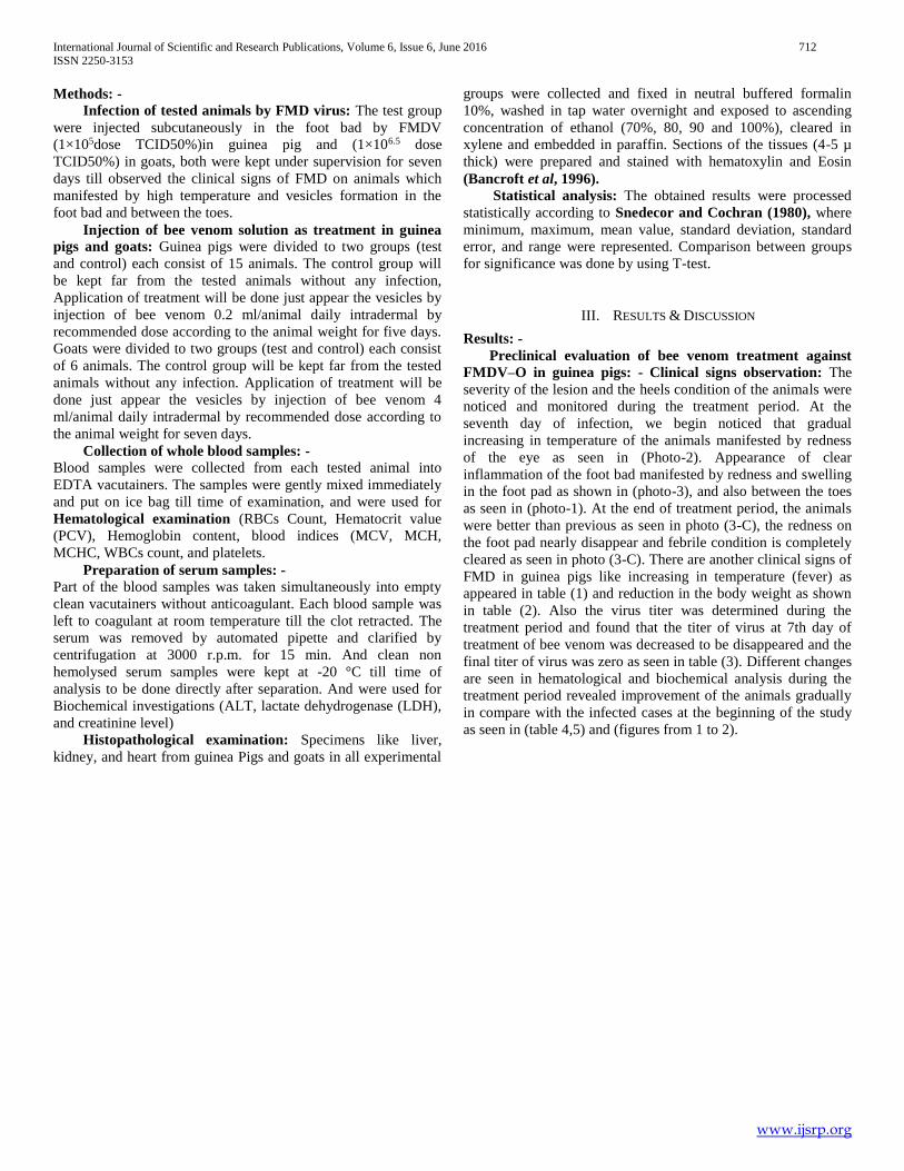
seventh day of infection, we begin noticed that gradual
increasing in temperature of the animals manifested by redness
of the eye as seen in (Photo-2). Appearance of clear
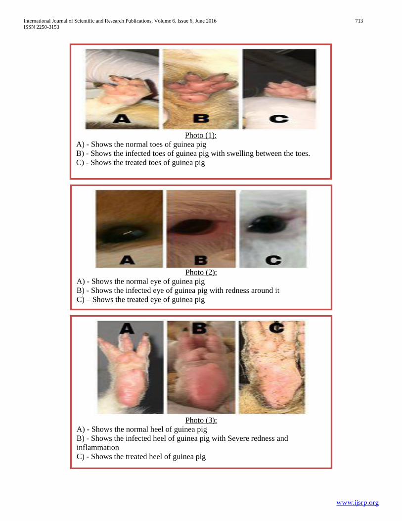
inflammation of the foot bad manifested by redness and swelling
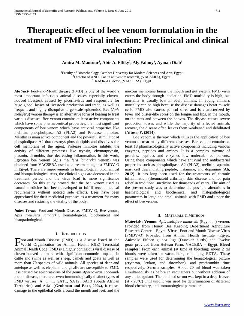
in the foot pad as shown in (photo-3), and also between the toes
as seen in (photo-1). At the end of treatment period, the animals
were better than previous as seen in photo (3-C), the redness on
the foot pad nearly disappear and febrile condition is completely
cleared as seen in photo (3-C). There are another clinical signs of
FMD in guinea pigs like increasing in temperature (fever) as
appeared in table (1) and reduction in the body weight as shown
in table (2). Also the virus titer was determined during the
treatment period and found that the titer of virus at 7th day of
treatment of bee venom was decreased to be disappeared and the
final titer of virus was zero as seen in table (3). Different changes
are seen in hematological and biochemical analysis during the
treatment period revealed improvement of the animals gradually
in compare with the infected cases at the beginning of the study
as seen in (table 4,5) and (figures from 1 to 2).
International Journal of Scientific and Research Publications, Volume 6, Issue 6, June 2016 713
ISSN 2250-3153
www.ijsrp.org
Photo (1):
A) - Shows the normal toes of guinea pig
B) - Shows the infected toes of guinea pig with swelling between the toes.
C) - Shows the treated toes of guinea pig
Photo (2):
A) - Shows the normal eye of guinea pig
B) - Shows the infected eye of guinea pig with redness around it
C) – Shows the treated eye of guinea pig
Photo (3):
A) - Shows the normal heel of guinea pig
B) - Shows the infected heel of guinea pig with Severe redness and
inflammation
C) - Shows the treated heel of guinea pig
International Journal of Scientific and Research Publications, Volume 6, Issue 6, June 2016 714
ISSN 2250-3153
www.ijsrp.org
Table (1): The temperature of Guinea pigs during infection of FMDV-O and treatment of bee venom.
Normal temperature
of animals
Temperature at the 7th day
after infection (1st day of
treatment)
Temperature of
treated animals at 3th
day
Temperature of
treated animals at 7th
day
35.9 ± 2.3 °C
38.8* ± 0.8 °C
37.5 ± 0.7°C
36 ± 0.6°C
Table (2): The body weight of Guinea pigs during infection of FMDV-O and treatment of bee venom.
Normal body weight of
animals
Body weight of infected
animals at 7th day
Body weight of treated animals at
7th day of treatment period
500 ± 50 gm.
460* ± 45 gm.
490 ± 49 gm.
Table (3): The titer of FMDV-O in infected and treated guinea pigs:
FMDV-O titer (infected)
FMDV-O titer (treated)
1st day of
Infection titer
At the 7th day after
infection (1st day of
treatment)
At 3rd day
At 7th day
1×10⁵
1×10⁶
1×102.7**
After 24 hrs After 48
hrs
After 72 hrs
1×101.7***
1×101.2***
0***
International Journal of Scientific and Research Publications, Volume 6, Issue 6, June 2016 715
ISSN 2250-3153
www.ijsrp.org
Hematological examination of guinea pigs: -
Table (4): Hematological values of guinea pigs: -
Parameters
Control
Test 1
(after 7 days from
infection)
Test 2
(at 3rd day of
Treatment)
Test 3
(7th day of
treatment)
Heamoglubin (g/dI)
16.0 ±1.6
12.2 *±1.22
15.8 ±1.58
15.1 ± 1.51
Red cell count
(RBCS)
(millions/cmm)
5.5± 0.55
4.7±0.47
4.8±0.48
5.66± 0.566
Haematocrit (PCV)
(%)
50.0 ±5
36.3*±3.63
45.0±4.3
43.0 ±4.3
MCV (fl)
90.9 ±9.09
77.2* ±7.72
89.6±8.96
85.5 ± 85.5
MCH (pg)
29.1 ±2.91
26.0*±2.6
29.2±2.9
30.0 ±3.3
MCHC (%)
32.0 ±3.2
33.8 ±3.38
32.6 ±3.26
33.7± 3.37
Platelets count
(T/cmm)
333 ±33.3
310±33
316 ±31.6
313.8 ±31.38
White cell count
(WBCS)
(Thausands/cmm)
8.0 ±0.8
8.8 ±0.88
11.8 *±1.18
10.0**± 0.1
Staff (%)
3 ±0.3
3 ±0.3
2 ±0.2
2 ±0.2
Segmented
(Neutrophils)
(%)
60 ± 6
60 ±6
60 ±6
45* ±4.5
Lymphocytes (%)
30 ±3
30 ±3
30 ±3
45* ±4.5
Monocytes (%)
5 ±0.5
4 ±0.4
5 ±0.5
5 ±0.5
Eosinophils (%)
2 ±0.2
3 ±0.3
3 ±0.3
3 ±0.3
Basophils (%)
0 ±0
0 ±0
0 ±0
0 ±0
International Journal of Scientific and Research Publications, Volume 6, Issue 6, June 2016 716
ISSN 2250-3153
www.ijsrp.org
A B
C D
Figure (1): Shows the hematological examination of guinea pigs. (A): The values of
heamoglubin, RBCs and haematocrit (PCV) in control, infected and treated guinea pigs.
(B): The values of MCH, MCV AND MCHC in control infected and treated guinea pigs.
(C): The values of platelets in control infected and treated guinea pigs. (D): The values of
WBCs and its derivatives in control infected and treated guinea pigs.
International Journal of Scientific and Research Publications, Volume 6, Issue 6, June 2016 717
ISSN 2250-3153
www.ijsrp.org
Biochemical examination of guinea pigs: -
Table (5): Enzyme activities in serum of guinea pigs: -
Parameters
Control
Test 1
(after 7 days from
infection)
Test 2
(at 3rd day of
Treatment)
Test 3
(7th day of
treatment)
Alanine
Transaminase
(ALT)(U/L)
19 ±1.9
30** ±3
26* ±2.6
21 ±2.1
S. Creatinine (mg/dl)
0.8± 0.08
1.0 ±0.1
0.7±0.07
0.7±0.07
Lactic
Dehydrogenase
(LDH)(U/L)
922 ±92.2
1531**± 153.1
810* ±81
683** ±68.3
A B C
Figure (2): The biochemical values of guinea pigs.
(A): Shows ALT (AST) enzyme activity in control infected and treated guinea pigs.
(B): Shows S. creatinine enzyme activity in control, infected and treated guinea pigs.
(C): Shows LDH enzyme activity in control, infected and treated guinea pigs.
International Journal of Scientific and Research Publications, Volume 6, Issue 6, June 2016 718
ISSN 2250-3153
www.ijsrp.org
Histopathological examination of guinea pigs: -
A B C
Figure (4): Sections of kidney tissue from control, infected with FMDV-O and treated
guinea pigs. (A): Kidney of control guinea pig showing the normal histological structure
of renal parenchyma (H & E X 400). (B): Kidney of infected guinea pig showing
cytoplasmic vacuolization of epithelial lining renal tubules, atrophy of glomerular tufts
and interstitial nephritis (H & E X 400). (C): Kidney of treated guinea pig showing
cytoplasmic vacuolization of epithelial lining renal tubules (H & E X 400).
A B C
Figure (3): Represents sections of liver tissues from control, infected with FMDV-O and
treated guinea pigs. (A): Shows the liver tissue of control guinea pig revealed the
normal histological structure of hepatic lobule, Meanwhile, (B): Shows liver of infected
guinea pig showed cytoplasmic vacuolization of centrilobular hepatocytes, while (C):
Represents treated geania pig showing normalhistopathological structureof hepatic
lobule (H & E X 400).
International Journal of Scientific and Research Publications, Volume 6, Issue 6, June 2016 719
ISSN 2250-3153
www.ijsrp.org
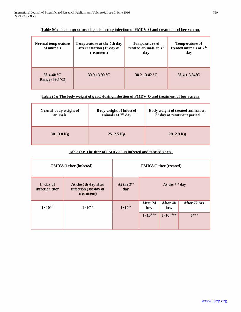
Clinical evaluation of bee venom treatment against FMDV–O in goats: -
Clinical signs observation: The severity of the lesion, temperature and the heels condition of the animals were noticed and
monitored during the treatment period. (Photo-4) represent lesions on gum of goat, the lesions on gum nearly disappear and febrile
condition is completely cleared at the end of treatment period. There are another clinical signs of FMD in goats like increasing in
temperature (fever) as appeared in table (6) and reduction in the body weight as shown in table (7). Also virus titer was determined
before and during the treatment period and showed that the titer of virus at 7th day of treatment of bee venom was decreased till
disappeared finally as seen in table (8). Different changes are seen in hematological and biochemical analysis during the treatment
period revealed improvement of the animals gradually in compare with the infected cases at the beginning of the study as seen in
(table 9,10) and figures from 6 to 7.
A B C
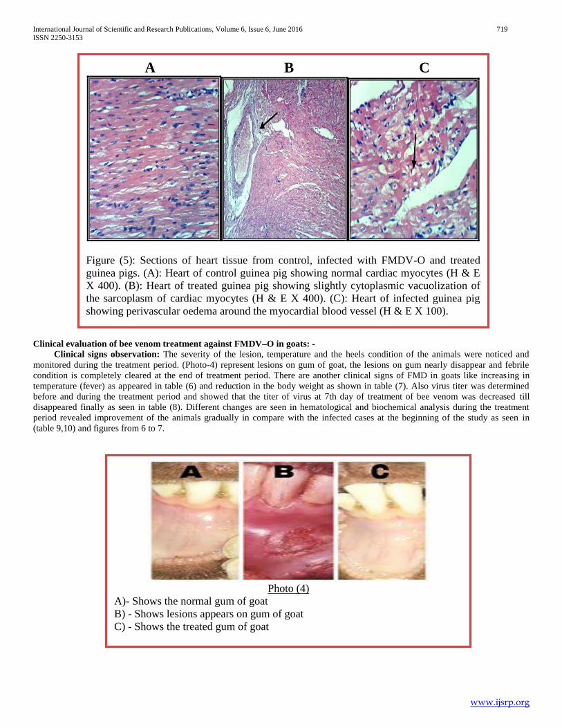
Figure (5): Sections of heart tissue from control, infected with FMDV-O and treated
guinea pigs. (A): Heart of control guinea pig showing normal cardiac myocytes (H & E
X 400). (B): Heart of treated guinea pig showing slightly cytoplasmic vacuolization of
the sarcoplasm of cardiac myocytes (H & E X 400). (C): Heart of infected guinea pig
showing perivascular oedema around the myocardial blood vessel (H & E X 100).
Photo (4)
A)- Shows the normal gum of goat
B) - Shows lesions appears on gum of goat
C) - Shows the treated gum of goat
International Journal of Scientific and Research Publications, Volume 6, Issue 6, June 2016 720
ISSN 2250-3153
www.ijsrp.org
Table (6): The temperature of goats during infection of FMDV-O and treatment of bee venom.
Normal temperature
of animals
Temperature at the 7th day
after infection (1st day of
treatment)
Temperature of
treated animals at 3th
day
Temperature of
treated animals at 7th
day
38.4-40 °C
Range (39.4°C)
39.9 ±3.99 °C
38.2 ±3.82 °C
38.4 ± 3.84°C
Table (7): The body weight of goats during infection of FMDV-O and treatment of bee venom.
Normal body weight of
animals
Body weight of infected
animals at 7th day
Body weight of treated animals at
7th day of treatment period
30 ±3.0 Kg
25±2.5 Kg
29±2.9 Kg
Table (8): The titer of FMDV-O in infected and treated goats:
FMDV-O titer (infected)
FMDV-O titer (treated)
1st day of
Infection titer
At the 7th day after
infection (1st day of
treatment)
At the 3rd
day
At the 7th day
1×106.5
1×106.5
1×10⁵*
After 24
hrs.
After 48
hrs.
After 72 hrs.
1×104.1* 1×102.4** 0***
International Journal of Scientific and Research Publications, Volume 6, Issue 6, June 2016 721
ISSN 2250-3153
www.ijsrp.org
Hematological examination of goats: -
Table (9): Hematological values of goats: -
Parameters
Control
Test 1 (7th day of
Infection Period)
Test 2
(3rd day of
Treatment)
Test 3
(7th day of
treatment)
Heamoglubin
(g/dl)
11.5 ±1.15
11.4 ±1.14
9.2* ±0.92
11.3 ±1.13
Red cell count
(RBCS)
(millions/cmm)
5.0 ±0.1
4.4±0.4
4.56± 0.056
4.4 ±0.44
Haematocrit (PCV)
(%)
33.0 ±0.3
33.9±3.39
32.2±3.22
37.6 *±3.76
MCV (fl)
30.0±3
77.0** ±7.7
79.3±3.93
76.4± 7.64
MCH (pg)
25.3±11.53
25.9±2.5
24.3±2.43
25.7 ±2.57
MCHC (%)
38.0 ±38.3
33.6 ±3.36
41.2 ±41.82
33.7±3.37
Platelets count
(T/cmm)
400 ±4
979**±97.9
512* ±51.2
400.0 ±40
White cell count
(WBCS)
(T/cmm)
22.0 ±2.2
14.0*±1.4
13.0**±1.3
14.5* ±1.45
Staff (%)
2±0.2
2 ±0.2
1 ±0.1
2 ±0.2
Segmented
(Neutrophils)
(%)
45 ±4.5
55* ±5.5
50 ±5
45 ±4.5
Lymphocytes (%)
45 ±4.5
65** ±6.5
70** ±7
50 ±5
Monocytes (%)
5±0.5
5±0.5
6± 0.6
3 ±0.3
Eosinophils (%)
3 ±0.3
3 ±0.3
2 ±0.2
1 ±0.1
Basophils (%)
0 ±0
1 ±0
0 ±0
0 ±0
International Journal of Scientific and Research Publications, Volume 6, Issue 6, June 2016 722
ISSN 2250-3153
www.ijsrp.org
A B
C D
Figure (6): The hematological values of goats. (A): The values of heamoglubin, RBCs and
haematocrit (pcv) in control, infected and treated goats. (B): The values of MCH, MCV AND
MCHC in control infected and treated goats. (C): The values of platelets in control infected and
treated goats. (D): The values of WBCs and its derivatives in control infected and treated goats.
International Journal of Scientific and Research Publications, Volume 6, Issue 6, June 2016 723
ISSN 2250-3153
www.ijsrp.org
Biochemical examination of goats: -
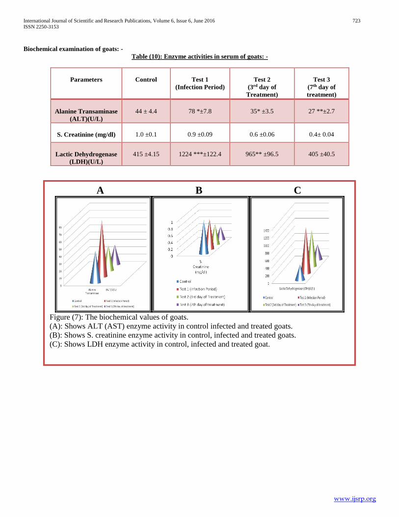
Table (10): Enzyme activities in serum of goats: -
Parameters
Control
Test 1
(Infection Period)
Test 2
(3rd day of
Treatment)
Test 3
(7th day of
treatment)
Alanine Transaminase
(ALT)(U/L)
44 ± 4.4
78 *±7.8
35* ±3.5
27 **±2.7
S. Creatinine (mg/dl)
1.0 ±0.1
0.9 ±0.09
0.6 ±0.06
0.4± 0.04
Lactic Dehydrogenase
(LDH)(U/L)
415 ±4.15
1224 ***±122.4
965** ±96.5
405 ±40.5
A B C
Figure (7): The biochemical values of goats.
(A): Shows ALT (AST) enzyme activity in control infected and treated goats.
(B): Shows S. creatinine enzyme activity in control, infected and treated goats.
(C): Shows LDH enzyme activity in control, infected and treated goat.
International Journal of Scientific and Research Publications, Volume 6, Issue 6, June 2016 724
ISSN 2250-3153
www.ijsrp.org
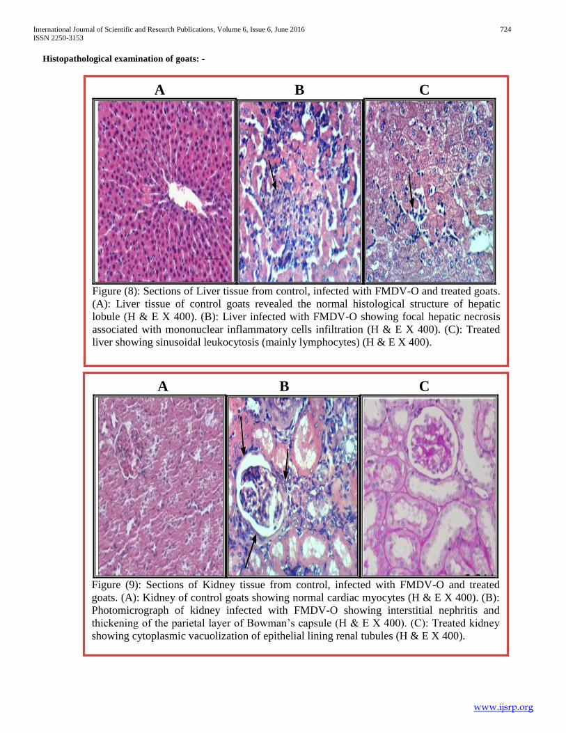
Histopathological examination of goats: -
A B C
Figure (9): Sections of Kidney tissue from control, infected with FMDV-O and treated
goats. (A): Kidney of control goats showing normal cardiac myocytes (H & E X 400). (B):
Photomicrograph of kidney infected with FMDV-O showing interstitial nephritis and
thickening of the parietal layer of Bowman’s capsule (H & E X 400). (C): Treated kidney
showing cytoplasmic vacuolization of epithelial lining renal tubules (H & E X 400).
A A B C
Figure (8): Sections of Liver tissue from control, infected with FMDV-O and treated goats.
(A): Liver tissue of control goats revealed the normal histological structure of hepatic
lobule (H & E X 400). (B): Liver infected with FMDV-O showing focal hepatic necrosis
associated with mononuclear inflammatory cells infiltration (H & E X 400). (C): Treated
liver showing sinusoidal leukocytosis (mainly lymphocytes) (H & E X 400).

International Journal of Scientific and Research Publications, Volume 6, Issue 6, June 2016 725
ISSN 2250-3153
www.ijsrp.org
Discussion: -
Regarding to the previous results, Infection of Guinea pigs
by FMDV-O resulted in redness and swelling in the foot-pad and
eye and between the toes and febrile /condition, these previous
results due to the effect of virulent virus on guinea pig as agreed
by Chen and Liu (2013), Lohse, et al (2012), Wang et al, 2011,
Núñez et al, 2007, and Knudsen et al, 1979. Infection of goats
by FMDV-O resulted in appearance of lesions in gum and febrile
condition of the animals manifested by shivering in all tested
animals, these previous results due to the effect of virulent virus
on goats and guinea pigs as agreed by Gakuya et al (2011), and
Wernery & Kinne (2012), while rectal temperatures was
determined and we found significant decrease in temperature
after treatment by bee venom injection as shown in (table 1,6),
this result was agreed with (Kittelberger, et al, 2015). Regarding
the hematological results, total number of RBCs in the infected
group was significantly lower than that in the treated group (P
<0.05), while MCV values were significantly higher in the
treated group. There were no significant differences in the other
haematological parameters between the groups. The significant
reduction in RBCs and elevation in MCV may be attributed to
endocrinopathy as reported previously by (Gokce et al., 2004).
Regarding the biochemical results as shown in (5,10), there were
significant increase in ALT, Creatinine and LDH in infected
group comparing with the control group as reported previously
by (Gokce et al., 2004)., these biochemical and haemotological
alterations may indicate the development of anaemia and
pancreatic dysfunction in animal suffering from FMD. This study
also indicated that Aphtovirus induces the production of nitric
oxide, in vivo. These findings provided information on our
understanding of the clinical pathology and pathogenesis of
FMD.
Treatment of diseased guinea pigs and goats by bee venom
injection daily for 7 days resulted in disappearance of the
previous clinical signs at the end of treatment period.
Regarding the RBCs count, hemoglobin content, and PCV
value of infected and test guinea pigs and goats as shown in
(tables 4, 9 and figures 1 and 6), there was non-significant
increase in RBCs count, and hemoglobin content after 7 days of
bee venom injection with concomitant gradual significant
increase in Hematocrit percent within normal range along the
period of the study. Similar results obtained by (Husseien, et al,
2001). While MCV, MCH and MCHC values showed no
significant increase along the testing period between the infected
and tested group. This result was in agreement with that
previously reported by (Ishay et al, 1975). The overall results of
the erythron indicated that injection of bee venom in guinea pigs
for 7 days resulted in improving blood status which explained by
improving integrity of RBCs leading to increase in erythron and
stimulatory effect on the heart that may result in increasing blood
flow as reported by (Vick et al, 1974), (Eliezer kaplinsky et al,
1976) and Husseien et al (2001). Concerning leukons, there was
significant increase in total leukocytes with concomitant increase
in lymphocytes count although this increase was within the
normal range after 7 days of bee venom injection compared with
the control group. This may be due to enhancement of
lymphocytosis under the effect of bee venom components as
PLA2 which discussed by (Prinz et al, 1987), or due to
lymphocytes proliferation by stimulation of immune system as
result of bee venom injection, this was assessed by (Lomnitzer
and Rabson, 1986). Monocytes showed non-significant
decreased after 7 days of bee venom injection although this was
within the normal range. This decrease can clarify that increase
in lymphocytes was due to its proliferation and not due to allergy
A B C
Figure (10): Sections of heart tissue from control, infected with FMDV-O and treated goats.
(A): Heart of control goats showing normal cardiac myocytes (H & E X 400). (B):
Photomicrograph of heart infected with FMDV-O showing focal Zenker’s necrosis of
myocytes associated with mononuclear cells infiltration (H & E X 400). (C): Treated heart
showing intermuscular mononuclear inflammatory cells infiltration (H & E X 100).
International Journal of Scientific and Research Publications, Volume 6, Issue 6, June 2016 726
ISSN 2250-3153
www.ijsrp.org
otherwise monocytes was also increased in response to allergy
(Moneret -Vautrin et al, 1987). Neutrophiles showed non-
significant decreased while esinophiles and basophiles count
cause non-significant changes along the test period, these results
were in agreement with (Somerfield et al, 1984) and may be due
to the injection of nontoxic dose as mentioned by (Kosnik and
Wraber, 2000). The platelets count as shown in (table 4, 9 and figures 1,6),
there was non- significant increase after 7 days of bee venom
injection, while the observed increament in the values were
within the normal level. This platelets activity was due to bee
venom hypersensitivity (Tsicopolous et al, 1989) but the use of
bee venom in special schedule for injection may cause
desensitization which give rise to adjustment of platelets level to
be in the normal range. Also may be due to inhibition in platelets
aggregation under the stimulatory effect for production of
prostaglandin as mentioned by (Ribardo et al, 2001).
Regarding the biochemical activities, AST activity showed
moderate significant decrease (P ≥ 0.01) in its levels in treated
group along the test period as shown in (table 5,10 and figure 2,
8). These give us more confidence that bee venom has
improvement action on the liver. The serum creatinine level
(table 5, 10 and figure 2, 8) of treated group resulted in
significant decreases after 7 days with no-significant decrease
along the test period compared with infected group. This means
that bee venom has benefit effect on the kidney. As shown in
(table 5, 10 and figure 2, 8), there was moderate significant
decrease in the LDH levels in compared with infected group
along the test period. These previous biochemical changes
attributed to the benefit effect of bee venom on the blood which
increase the tissue tolerance to lack of oxygen (hypoxia) with
increase in arterial blood flow and vascular permeability so the
exchange between blood and tissue is increased (Foster, 1969,
Vick et al, 1974, and Chkenderow and Kobourova, 1981). This explained that bee venom didn't induce cellular toxicity
determined by decrease in release of lactic dehydrogenase
(LDH).
Regarding the pathological examination of guinea pigs and
goats, as shown in (figure 3,4,5,8,9,10), Microscopically, In
Figure (3-B) shows liver of infected guinea pig showed
cytoplasmic vacuolization of centrilobular hepatocytes, while
figure (3-C) represents treated geania pig showing
normalhistopathological structureof hepatic lobule. In Figure (4-
B) shows sections of kidney tissues of infected guinea pig
revealed cytoplasmic vacuolization of epithelial lining renal
tubules. Meanwhile, In figure (4-C) shows treated geania pig
revealed slightly vacuolization. In figure (5-B) shows heart of
infected geania pig showing perivascular oedema around the
myocardial blood vessel. Meanwhile, In figure (5-C) represents
heart of treated guinea pig showing slightly cytoplasmic
vacuolization of the sarcoplasm of cardiac myocytes. In figure
(8-B) shows liver infected with FMDV-O showing focal hepatic
necrosis associated with mononuclear inflammatory cells
infiltration. In figure (8-C) shows treated liver showing
sinusoidal leukocytosis. (9-B) represents Photomicrograph of
kidney infected with FMDV-O showing interstitial nephritis and
thickening of the parietal layer of Bowman’s capsule and in
figure (9-C) shows treated kidney showing cytoplasmic
vacuolization of epithelial lining renal tubules. In figure (10-B)
shows heart infected with FMDV-O showing focal Zenker’s
necrosis of myocytes associated with mononuclear cells
infiltration. (10-C) shows treated heart of goat showing
intermuscular mononuclear inflammatory cells infiltration.
Finally, there was improvement in histopatholgical case
according to the previous results comparing between treated and
infected organs.
By determination of the virus load before and after the
treatment period we found that there is significant decrease in
virus load along the period of treatment in compare with the
beginning virus load. These results revealed that there is antiviral
activity of Apis mellifera lamarckii venom may be due to the
high level of meliittin and These results were agree with De-
Clercq et al, (2000) proved that bee venom melittin inactivates
HIV and also aborts cell-to-cell fusion and transmission of HIV,
due to its high-affinity interaction with gp120 and Masuda et al,
(2005) explained that the antiviral effect of the bee venom
phospholipase A2 depends essentially on its ability to hydrolyze
phospholipids in host cell membranes Also proved that Bee
venom melittin inactivates HIV and also aborts cell-to-cell fusion
and transmission of HIV, due to its high-affinity interaction. As
shown in Moreno and Giralt (2015), The antiviral activities
described for melittin and its analogs are caused by specific
intracellular events, with the selective reduction of the
biosynthesis of some viral proteins, as reported for the melittin
analog Hecate on herpes virus-1 and for melittin itself on HIV-1-
infected lymphoma cells. In the 90s, active melittin was
presented to provide an improved composition complementary to
azidothymidine (AZT) to inhibit the reverse transcriptase and
growth of HIV-infected cells. Another study of Bose and
Acharya (2015) shown that although melittin destroys the
infectivity of HIV particles, the utility of this toxin is limited by
its nonspecific cytotoxic effects: melittin kills cells by disrupting
membrane structure and function. Lawrence and Skalka (1980)
stated that the melittin compound can make the viral envelope
permeable, that make it loss of its infectivity and Mitsuishi et al,
(2006) indicated that the direct addition of bee venom
phospholipase A2 to 293A cells suppressed adenovirus plaque
formation in both number and size. Pharney (1996) described
the beneficial effect of bee venom active components on the
human body as follow: Apamin and melittin were more potent
than penicillin against certain bacteria, while mast cell
degranulating peptide and adolapin were non-steroidal anti-
inflammatory. Matsuzaki (1997) said that honeybee venom
contains at least 18 active substances. Melittin, is the most
prevalent and it is one most potent anti-inflammatory agents
known (100 times more potent than hydrocortisol). Adolapin is
another strong anti-inflammatory substance and inhibit
cyclooxidase. Apamine inhibits complement C3 activity and
blocks calcium-dependent potassium channels, thus enhancing
nerve transmission. Other substances such as compound X,
hyaluronidase, phospholipase A2, histamine and mast cell
degeneration protein are involved in the inflammatory response
of the venom in during a softening of tissue and facilitation of
fluid flow.
These results due to antiviral effect of bee venom as
reported byDe-Clercq et al, (2000), Masuda et al, (2005),
Moreno & Giralt (2015), Bose and Acharya (2015), Lawrence
and Skalka (1980), Mitsuishi et al, (2006), Ramadan et al,
International Journal of Scientific and Research Publications, Volume 6, Issue 6, June 2016 727
ISSN 2250-3153
www.ijsrp.org
(2009), (Fenard et al, 1999), (Yong et al, 1990) and
(Yunginger et al, 1978) whose mentioned that this antiviral
activity due to melittin and or phospholipase A2 enzyme activity
also may be due to protease inhibitor which involved in bee
venom (Bogdanov, 2015). Also bee venom has anti-
inflammatory effect which manifested by decrease foot bad
inflammation during the treatment period by induction of COX2
expression and stimulation of prostaglandin generation which
have biological activities resulting in revealed viremia and
inflammation as reported by Ribardo et al, (2002), Park et al
(2004), kim (1992) and lee et al, (2005).
Also in Vick et al, (1972) observed that bee venom or one of its
components (melittin) when injected S/C in Rhesus monkeys
stimulated the production of cortisol from the adrenal gland
through its action on the pituitary gland without any gross or
microscopic tissue changes. These observations may explain bee
venom benefits in variety of disease conditions that also respond
to adrenal steroid therapy. In Lee et al, (2004) study reported that
acupuncture therapy with bee venom suppressed the
development of arthritis and caused inhibition of the immune
responses in type-II collagen-induced arthritis. The incidence of
arthritis, the mean arthritis index, and the number of arthritic
limbs were significantly lower in the bee venom treatment
compared to the control group. Among the serum pro-
inflammatory cytokines, examination of the histopathology of the
joints of murine CIA showed decreased inflammation signs and
less lymphocyte infiltration after bee venom acupuncture
therapy. Hyoung et al, (2005) stated that bee venom has been
used traditionally for the control of pain and inflammation in
various chronic inflammatory diseases. They also suggested that
bee venom inhibits the proliferation of rheumatoid synovial
fibroblast cells through induction of apoptosis by caspase-3
activation. Kwon et al, 2003 reported that subcutaneous bee
venom injection produced a marked suppression of leucocyte
migration and tumor necrosis factor (TNF)-alpha concentration.
The anti-inflammatory effect bee venom administration was
mediated in part by the release of catecholamines from the
adrenal medulla. Also the previous results may be attributed to
effect of bee venom as immunomodulator which it stimulates
immune system to protect the body against infection by its
stimulation of prostaglandin generation which have biological
activities resulting in revealed of viremia due to stimulation of
IL-10, TNF alpha, and CD8 which result in regulation in IgE
which responsible for histamine release (Rekka et al, 1990).
McHugh et al, (1995) examined changes in cytokine secretion in
patients before and during both rush and conventional venom
immunotherapy (VIT) in bee venom allergen patients and
showed that immunotherapy shifted cytokine responses to
allergen from a TH-2 to a TH-1 dominant pattern, suggesting
direct effects on T cells. So it is possible to use whole venom in
treatment of various medical disorders/diseases. Kammerer et
al, (1997) said that venom immunotherapy (VIT) lead to
transient increase in T-cell proliferation followed by T-cell hypo
responsiveness and modulation of cytokine secretion from T
(H0)-type to a T(H1) type pattern. Akdis et al, (1998) found that
bee venom phospholipase A2 (PLA) induced high IL-4, IL-5 and
IL-13 production in peripheral blood mononuclear cell cultures.
These features may enable new applications for safer
immunotherapy.
IV. RECOMMENDATION
From the previous project we recommend to use bee venom
as injection as alternative medicine in veterinary practice as it is
safe and effective medication against virus infection specially
RNA viruses as seen in the previous results on FMDV-O. We
succeeded to treat laboratory and large animals by bee venom.
Using of bee venom by intradermal injection is useful to
maintain the health condition through its immune-stimulant
effect.
ACKNOWLEDGMENT
The authors have taken efforts in this project; however, it
would not have been possible without the kind support and help
of many individuals. The authors extremely thankful and
indebted to Dr. Gehan Safwat for sharing expertise, and sincere
and valuable guidance and encouragement extended to me.
Various members of VACSERA team also provided considerable
assistance during the project. Finally, I want to thank Dr. Samia
Kamal for providing the Foot-and-mouth disease virus Type-O
from Animal Health Institute –Egypt.
REFERENCES
[1] Aftosa, F. (2014). Foot and Mouth Disease. P.1-9.
[2] Akdis C, A; Blesken T; Akdis M; Wuthrich B; Blaser K. (1998a). Differential regulation of human T cell cytokine patterns and IgE and IgG4 responses by conformational antigen variants. Eur. J. Immunol. 1998 Mar., 28 (3): 914-25.
[3] Akdis C, A; Blesken T; Akdis M; Wuthrich B; Blaser K. (1998b). Role of interleukin 10 in specific immunotherapy. J, Clin. Invest. 1998 Hul 1., 102 (1): 98-106.
[4] Ali, M. (2012). Studies on Bee Venom and Its Medical Uses, International Journal of Advancements in Research & Technology. Faculty of Agriculture, Ain Shams University, Cairo, Egypt.1(2). Pp.1-15.
[5] Barkakati J.; Sarma S; and Kalita D.J. (2015). Effect of foot and mouth disease on haematological and biochemical profile of cattle, Indian J. Anim. Res. 49 (5): 713-716. DOI: 10.18805/ijar.5588.
[6] Bartles H; Bohmer M; Heirili C. (1972). Serum creatinine determination with out protein precipitation. Clin. Chem Acta 37, 193-7.
[7] Bashandy, M.A.(2003). Guide for the care and use of laboratory animals in medical research. Publishers of scientific books for publication and distribution, Egypt. 977-287-318-4.
[8] Bogdanov, S. (2015).Bee Venom:Composition, Health, Medicine: A Review. Bee Product Science, www.bee-hexagon.net
[9] Bancroft J. D; Stevans A; and Tuner D.R. (1996). Theory and practice of histological techniques. 4th Ed. Churchill Livinigstone, Edinburgh, London, Melbourne, New York.
[10] Chen H-t, Liu Y-s (2013). Immunity of Foot-and-Mouth Disease Serotype Asia 1 by Sublingual Vaccination. PLoS ONE 8(5): e63839. doi:10.1371/journal.pone.0063839.
[11] Chkenderov S; and Kabourova K. (1981). Mecanisme de l'action antiinflamatoire du venin d'ebeille, in the XXVIII-Th. Apimondia Congress, Acapulco, Mexico, P.446.
[12] De Clercq . (2000). Current lead natural products for the chemotherapy of human immunodeficiency virus (HIV) infection, E. Medicinal research reviews.
[13] El-Fiky, A. (2006). Clinical studies on the use of animal venoms as alternative therapy in the veterinary practices. PHD. Faculty of veterinary medicine, Cairo University. 1-237.
International Journal of Scientific and Research Publications, Volume 6, Issue 6, June 2016 728
ISSN 2250-3153
www.ijsrp.org
[14] Eliezer kaplinsky; Jacob Ishay, David Ben-Schcher, and Simon Gitter. (1976). Effect of bee (Apis Mellifera) venom on the electrocardiogram and blood pressure. Toxicon. Vol. 17, 251-256.
[15] Fenard D; Lambeau G; Valentin E; Lefebvre J. G; Lazdunski M.; Doglio A. (1999). Secreted phospholipases A(2), a new class of HIV inhibitors that block virus entry into host cells. J. Clin. Invest. 1999 Sep; 104 (5), 611-8.
[16] Forster K. A. (1969). Chemically, pharmacology, and therapeutic effectiveness of bee venom in the XXII-Nd. Apimondia congress, Munich, Germany.
[17] Gakuya D. W; Mulei C.M.1; Wekesa S. B. (2011). Use of ethnoveterinary remedies in the management of foot and mouth disease lesions in a diary herd, Afr J Tradit Complement Altern Med. 8(2):165-169.
[18] Gokce G. et al. (2004). Alterations in Some Haematological and Biochemical Parameters in Cattle Suffering from Foot- and -Mouth Disease, Turk J Vet Anim Sci, 28 (2004), 723-727.
[19] Grubman, M ; and Baxt, B. (2004). Foot-and-Mouth Disease, clinical microbiology reviews.17 (2). 465–493. Agricultural Research Service; North Atlantic Area, Greenport, New York 11944. DOI: 10.1128/CMR.17.2.
[20] Habiela M, et al. (2014). Laboratory animal models to study foot-and-mouth disease: a review with emphasis on natural and vaccine-induced immunity, J Gen Virol. 95(Pt 11): 2329–2345. doi: 10.1099/vir.0.068270-0.
[21] Hugh M. Hyre and Randall A.Smith (1986). Immunological effects of honey bee (Apis mellifera) venom using BALB/c mice. Toxicon, 24 (2), 435-440.
[22] Hussein A. A; Nabil Z. I; Zalat S. M; Rakha M. K. (2001). Comparative study of the venoms from three species of bees: effects on heart activity and blood. J Nat Toxins. 2001 Nov; 10 (4): 343-57.
[23] Hyoung-Seok Jang; Sun Kwang Kim; Jae-Bok Han; Hyun-Jong Ahn; Hyunsu Bae; and Byung-II Min. (2005). Effects of bee venom on the pro-inflammatory responses in RAW 264.7 macrophage cell line. Journal of Ethnopharmacology, 99 (1), 13 May 2005, 157-160.
[24] Hyre H. M and Smith R. A. (1986). Immunological effects of honey bee (Apis mellifera) venom using BALB/c mice. Toxicon, 24 (5), 435-40.
[25] International Federation of clinical chemistry. (1980). Scientific Committee. J. Clin. Chem. Clin. Biochem. 1980; 18:521-534.
[26] Ishay J; Ben-Shacher D; Elazer Z; and Kaplinsky E. (1975): Effects of melittin on the central nervous system. Toxicon, 13 (4). 227-283.
[27] Kammerer R; Chvatchko Y; Kettner A; Dufour N; Corradin G; Spertini F. (1997). Modulation of T-CELL response to phospholipase A2 and phospholpase A2-derived peptides by conventional bee venom immunotherapy. J. Allergy Clin Immunol. 1997 Jun; 100 (1): 96-103.
[28] Kim C. M. H. (1992). Bee Venom Therapy and Bee Acupuncture Therapy. Korea Educational pub; 1992. 515pp.
[29] Kittelberger R, et al. (2015). Foot-and-Mouth Disease in Red Deer – Experimental Infection and Test Methods Performance, Transboundary and Emerging Diseases. 3 13.doi:10.1111/tbed.12363.
[30] Kosnik M; and Wraber B. (2000). Shift from Th2 to Th1 response in immunotherapy with venoms. Pflugers Arch, 440 (5 suppl): R70-1.
[31] Kwon Y. B; Kim H. W; Ham T.W; Yoon S.Y; Roh D. H; Han H.J; Beitz A. J; Yang I.S; Lee J.H. (2003). The anti-inflammatory effect of bee venom stimulation in a mouse air pouch model is mediated by adrenal medullary activity. J. Neuroendocrinol. 2003 Jan; 15 (1): 93-6.
[32] Lawrence R. B. and Skalka, A. (1980). Two species of full-length cDNA are synthesized in high yield by melittin-treated avian retrovirus particles. Proc. Nati. Acad. Sci. USA 77(2): 847-851.
[33] Lee J. D; Kim S. Y; Kim T.Wl Lee S. H; Yang H. I; Lee D. I; Lee Y. H. (2004). Anti-inflammatory effect of bee venom on type II collagen-induced arthritis. Am J Chin Med. 2004; 32 (3): 361-7.
[34] Lee J. Y; Kang S.S; Kim J.H; Bae C.S; Choi S. H. (2005). Inhibitory effect of whole bee venom in adjuvant-induced arthritis. In-vivo. 2005 Jul-Aug; 19(4):801-5.
[35] Lohse L; Jackson T; Bøtner A; Belsham G. J. (2012). Capsid coding sequences of foot-and-mouth disease viruses are determinants of pathogenicity in pigs, Vet Res. 43(1): 46. doi: 10.1186/1297-9716-43-46.
[36] Lomnitzer R; Rabson A. R. (1986). Lack of responsiveness of beekeeper mononuclear cells to in vitro stimulation with pure bee venom. J Allergy Clin Immunol. 78 (1 pt 1): 25-30.
[37] Masuda, S.; Murakami, M.; Takanezawa, Y.; Aoki, J.; Arai, H.; Ishikawa, Y.; Ishii, T.; Arioka, M. and Kudo, I.. (2005). Neuronal expression and neuritogenic action of group X secreted phospholipaseA2. J. Biol. Chem. 280: 23203- 23214.
[38] McHugh S. M; Deighton J; Stewart A. G; Lachmann P. J; Ewan P. W. (1995). Bee venom immunotherapy induces a shift in cytokine responses from a TH-2 to a TH-1 dominat pattern: comparison of rush and conventional immunotherapy. Clin Exp Allergy. 1995 Sep, 25 (9): 828-38.
[39] Mitsuishi M.; Masuda S.; Kudo I. and Murakami, M. (2006). Group V and X secretory phospholipase A2 prevents adenoviral infection in mammalian cells. Biochem J.; 393(Pt 1):97-106.
[40] Moneret-Vautrin D. A; Bene M. C; Grave P; Faure G; and Maria Y. (1987). Margueurs membranaires des lymphocytes sanguins avant et 28 heures après immunotherapy acceleree. Revue Francaise d' Allergologie et d'immunologie Clinique, 27 (4), 175-180.
[41] Moreno, I and Giralt, E. (2015). Three Valuable Peptides from Bee and Wasp Venoms for Therapeutic and Biotechnological Use: Melittin, Apamin and Mastoparan, Toxins. 7, 1126-1150; doi:10.3390/toxins7041126.
[42] Park H. J; Lee S. H; Son D.J; Oh K.H; Song H. S; Kim G.T; Yoon do Y; Hong J.T. (2004). Antiarthritic effect of bee venom: inhibition of inflammation mediator generating by suppression of NF-kappaB through interaction with the p50 subunit. Arthritis Rheum. 2004 Nov; 50 (11): 3504-15.
[43] Pharney, J. (1996). Bee venom: how does it achieve its therapeutic effects? Bee Informed 3(2): 4-5, 10-11, 15-17.
[44] Prinz J. C; Endres N; Rank G; Ring J; Rieber E.P. (1987). Expression of Fc epsilon receptors on activated human T lymphocytes. Eur. J. Immunol. 17 (6), 757-61.
[45] Ramadan1, H; Mohamed, A; Abd El- Daim, M. (2009). Evaluation of antiviral activity of honeybee venom on DNA and RNA virus models. Egypt. Acad. J. biolog. Sci., 2 (1): 247- 258.
[46] Rekka E; Kourounakis L; Kourounakis P. (1990). Antioxidant activity of and interleukin production affected by honey bee venom. Arzneimittelforschung. 1990 Aug; 40 (8): 912-3.
[47] Ribardo D. A; Crowe S. E; Kuhl K. R; Peterson J. W; Chopra A. K. (2001). Prostaglandin levels in stimulated macrophages are controlled by phospholipase A2-activating protein and by activation of phospholipase C and D. J. Biol. Chem. 276(8): 5467-75.
[48] Ribardo D. A; Kuhl K. R; Peterson J. W; Chopra A. K. (2002). Role of melittin- like region within phospholipase- A(2)-activating protein in biological function. Toxicon. 2002 May; 40 (5): 519-26.
[49] Richard C. Knudsen; Christopher M. Groocock; and Arthur A. Andersen. (1979). Immunity to Foot-and-Mouth Disease Virus in Guinea Pigs: Clinical and Immune Responses, Infection and immunity. 787-792 24 (3).
[50] Schalm, O. W., Jain, N.C and Carrol, E.J. (1975). Veterinary Haematology, 3rd Edn., Lea and Febiger, Philadelphia, pp. 197-199.
[51] Simics, Mihály. (1994). Bee Venom: Exploring the Healing Power - Age Old Remedies for Arthritis, Rheumatism and Other Ailments. Apitronic Publishing, book, paperback, ISBN 0-9697654-0-1, 80 pp.
[52] Smith H. A; Jones, T.C; and Hunt R. D. (1972). Veterinary pathology. 4th Ed. Lea & Febiger, Philadelphia, pp. 145-154.
[53] Snedecor G.W; and Cochran W.G. (1980). Statistical methods. 7th Ed. Ames:Lowa state university press.
[54] Somerfield S. D; Stach J. L; Mraz C; Gervais F; and Skamene E. (1984). Bee venom inhibits superoxide production by human neutrophils. Inflammation. 8 (4): 385-91.
[55] Tsicopoulos A; Tannel A.B; Wallaert B; Joseph M; Ramon P; and Capron A. (1989). A circulating suppressive factor of platelet cytotoxic functions after rush immunotherapy in Hymenoptera venom hypersensitivity. J Immunol. 15, 142 (8): 2683-8.
[56] Van der heiden C; B. Ais; Gerh Ardt W; and Rosallsis. (1994). Approved recommendation on IFCC methods for the measurement of catalytic concentration of enzymes. Part 8. IFCC method for LDH. Eur. J. Clinical chem.. Clin Biochem; 1994; 32-639-655.
[57] Vick J. A; Mehlman B; Brooks R; Phillips S. J; Shipman W. (1972). Effect of bee venom and melittin on plasma cortisol in the unanesthetized monkey, Toxicon 10, 581-586.
[58] Vick J.A; Shipman W. H; and Brooks Jr. R. (1974). Beta adrenergic and anti-arrhythmic effects of cardiopep, a newly isolated substance from whole bee venom. Toxicon , 12(2), 139-142
International Journal of Scientific and Research Publications, Volume 6, Issue 6, June 2016 729
ISSN 2250-3153
www.ijsrp.org
[59] Wang G, Pan L, Zhang YG, Wang YL, Zhang ZW, Lu JL, et al. (2011). Intranasal Delivery of Cationic PLGA Nano/Microparticles- Loaded FMDV DNA Vaccine Encoding IL-6 Elicited Protective Immunity against FMDV Challenge. Plos One. 6(11). ARTN e27605 doi: 10.1371/journal.pone.
[60] Wernery U; Kinne J. (2012). Foot and mouth disease and similar virus infections in camelids: a review, Rev. sci. tech. 31 (3), 907-918.
[61] Young A W; D E Haan EHFand Newcombe F. (1990). Unawareness of impaired face recognition. Brain and Cognition, 14, 1 -18.
AUTHORS
First Author – Amira M. Mansour, Graduation project student,
Faculty of Biotechnology, October university for modern
sciences and Arts, Egypt, Email: [email protected].
Second Author – Abir A. Elfiky, Director of ANDI Coe in
antivenom research, (VACSERA), Egypt.
Third Author – Aly Fahmy, Head Research &Development
sector, (VACSERA), Egypt.
Fourth Author– Ayman Diab, Dean, Faculty of biotechnology,
October university for modern sciences and Arts, Egypt.