the volgograd state medical university chair for human anatomy lecture: general arthrology...
TRANSCRIPT

The Volgograd State Medical University
Chair for Human Anatomy
Lecture:
General arthrologyGeneral arthrology
Particular anatomy of Particular anatomy of
jointsjoints
is delivered by: Dr. Elena Vladimirovna Gorelik, PhD

Types of junctions:Types of junctions:
1.1. Continuous junctionsContinuous junctions in in which there is a layer of which there is a layer of connective tissue or cartilage connective tissue or cartilage between bones.between bones.
A fissure or a cavity between A fissure or a cavity between joining bones is absent.joining bones is absent.

2.2. Interrupted junctionsInterrupted junctions or joints (synovial or joints (synovial junctions) junctions)
are characterized by the are characterized by the presence of cavitypresence of cavity and and synovial membrane covering synovial membrane covering the inside part of the inside part of joint joint capsulecapsule between the bones.between the bones.

3.3. SymphysesSymphyses (or semijoints) (or semijoints) have a little fissure in the have a little fissure in the cartilaginouscartilaginous or connective-tissued layer or connective-tissued layer between the joining bones between the joining bones
(a transitional form from (a transitional form from continuous junctions to continuous junctions to
interrupted ones). interrupted ones).

Continuous junctions of bones Continuous junctions of bones
Continuous junctions are more Continuous junctions are more elastic, solid and, as a rule, they elastic, solid and, as a rule, they have limited mobility.have limited mobility.
There areThere are: : a)a) fibrous junctionsfibrous junctions, , b)b) synchondroses (cartilaginous synchondroses (cartilaginous junctionsjunctions),), c)c) bony junctionsbony junctions..

a) a) Fibrous junctionsFibrous junctions,, articulationes fibrosaearticulationes fibrosae, , are are solid junctions of bones by solid junctions of bones by means of thick fibrous means of thick fibrous connective tissueconnective tissue. . There are three types of fibrous There are three types of fibrous junctions junctions: : -- syndesmoses syndesmoses, , - - suturaesuturae,, -- impaction impaction..

SyndesmosisSyndesmosis is formed by is formed by the connective tissue, its the connective tissue, its collagenic fibers knit with collagenic fibers knit with periosteum and turn into it periosteum and turn into it without a definite border. without a definite border. SyndesmosisSyndesmosis include include ligamentsligaments and and interosseal interosseal membranesmembranes..

LigamentsLigaments,, ligamentaligamenta,, are thick bundles of thick fibrous are thick bundles of thick fibrous connective tissue.connective tissue.Ligaments are between the bones Ligaments are between the bones and they are obstacles that limit and they are obstacles that limit movements.movements. In the vertebral column there In the vertebral column there are ligaments formed by elastic are ligaments formed by elastic connective tissue of yellowish connective tissue of yellowish colour colour ((yellow ligaments,yellow ligaments, ligamenta flaualigamenta flaua).). They are stretched in flexion of They are stretched in flexion of the vertebral column forward the vertebral column forward (flexion of the vertebral column) (flexion of the vertebral column) and then they become shorter and then they become shorter again to promote extension of the again to promote extension of the vertebral columnvertebral column..

Interosseal membranesInterosseal membranes,,
membranae interosseaemembranae interosseae,,
are stretched between the are stretched between the
dyaphyses of long tubular bonesdyaphyses of long tubular bones..
Interosseal membranes, ligaments Interosseal membranes, ligaments
areare often served as the basis of often served as the basis of
muscles. muscles.

SutureSuture,, suturasutura,, is a variety of fibrous is a variety of fibrous junction in which there is junction in which there is a narrow connective-a narrow connective-tissued layer between the tissued layer between the edges of joining bonesedges of joining bones. .
There is a sutura serrata,There is a sutura serrata, a sutura squamosa anda sutura squamosa and a sutura plana a sutura plana depending on depending on configuration of joining configuration of joining bones edgesbones edges..
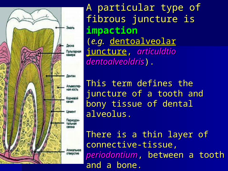
A particular type of A particular type of fibrous juncture is fibrous juncture is impactionimpaction ((e.g.e.g. dentoalveolar juncturedentoalveolar juncture, , articuldtio dentoalveoldrisarticuldtio dentoalveoldris). ).
This term defines the This term defines the juncture of a tooth and bony juncture of a tooth and bony tissue of dental alveolustissue of dental alveolus..
There is a thin layer of There is a thin layer of connective-tissue, connective-tissue, periodontiumperiodontium, between a , between a tooth and a bonetooth and a bone..

b)b) SynchondrosesSynchondroses are are junctions of bones by means of junctions of bones by means of cartilaginous cartilaginous tissuetissue..They are firm and little movable They are firm and little movable that depends on the thickness that depends on the thickness and structure of cartilaginous and structure of cartilaginous layer between the bones.layer between the bones. ** If a cartilage between the If a cartilage between the joining bones exists during the joining bones exists during the whole life, such synchondroses whole life, such synchondroses are constant.are constant. ** If a cartilaginous layer If a cartilaginous layer between the bones is kept till between the bones is kept till the definite agethe definite age ( (fonticulifonticuli),), this this temporal juncture, a cartilage, is temporal juncture, a cartilage, is substituted with osseal tissue substituted with osseal tissue – – c)c) synostosissynostosis..

Interrupted or Interrupted or synovial junctionssynovial junctions of bones (of bones (jointsjoints))
They are notable for more mobility, They are notable for more mobility, variety of movements.variety of movements. Every joint includes Every joint includes articular articular surfacessurfaces of bones covered with of bones covered with cartilagecartilage,,articular capsulearticular capsule, , articular cavityarticular cavity filled filled wiyh wiyh synovial fluidsynovial fluid.. Some joints also include lacertus Some joints also include lacertus formations such as formations such as articular disksarticular disks, , meniscusmeniscus and and glenoid lieglenoid lie..

In majority of cases in joining
bones articular surfaces correspond to each other. They are congruent (lat. congruens means fitting, corresponding). * If one articular surface is convex (articular head), the other one which joins the first one is equally concave (articular cavity). In some joints these surfaces are not either the same shape or size, they are incongruent.

Articular cartilage, cartilage articularis, smoothes roughness of articular surfaces of bones, in motion it amortizes jolts. The bigger exertion the joint has under the action of gravity, the more thickness of articular cartilages becomes. As a rule, articular cartilage is flat, it is constantly moist with synovial fluid which facilitates movements of joints. There are no blood and lymphatic vessels in articular cartilage, its nourishment is implemented by synovial fluid.

Articular capsule, capsula articulаris, is fastened to the joining bones at the edges of articular surfaces; it firmly knits with periosteum, forming isolated articular cavity. * Capsule has two layers: external layer is fibrous membrane, membrana fibrosa (stratum fibrosum), and internal layer is synovial membrane, membrana synouialis (stratum synovidle). In some parts fibrous membrane forms thickenings – capsular ligaments: extracapsular ligaments, intracapsular ligaments.

Synovial membrane covers fibrous membrane inside. Synovial membrane has not big outgrowths turned to the joint cavity - synovial villi – which are rich in blood vessels. They extend its surface. Internal surface of articular capsule (synovial membrane) is always moist with synovial fluid, which removes rubbing of articular surfaces.

Articular cavityArticular cavity,, is a fissural space is a fissural space between between articular surfacesarticular surfaces.. It is limited by synovial It is limited by synovial membrane of articular membrane of articular capsule, it keeps some capsule, it keeps some quantaty ofquantaty of synovialsynovial fluid.fluid. An articular cavity form An articular cavity form depends on a form of joining depends on a form of joining surfaces, presence or surfaces, presence or absence of lacertus absence of lacertus formations inside the jointformations inside the joint ((disk, meniscus, disk, meniscus, intracapsular ligamentsintracapsular ligaments).).

Articular disks and menisci are cartilaginous plates of various forms which are located between not fully corresponded to each other (incogruent) articular surfaces. Disk is an entire plate which divides the articular cavity into two chambers (two levels). Menisci are not entire cartilaginous or connective-tissued plates. Disks and menisci are able to be dislocated in motion.

Glenoid lie is at the edge of concave articular surface, it supplements it and makes it deeper. Synovial bursae are diverticula of synovial membrane in thin parts of joint fibrous membrane. Bursae remove rubbing of contiguous tendons and bones.

Biomechanics of joints
There are frontal,
sagittal and longitudinal
(along the joining
bones)
rotation axes.

On frontal axis flexion (flexio) and extension (extensio) are implemented. In flexion the angle between the joining bones becomes less (e.g. in flexion in the elbow joint the angle between an arm and a forearm becomes less). In extension the angle becomes bigger (up to 180°)
and straightening of an extremity or a body occurs.

On sagittal axis adduction (adductio), and
abduction (abductio) are implemented.
In adduction one of the joining bones approches to the median cavity.
In abduction one of the joining bones moves off away from the median cavity.

In rotation (rotаtio) a bone rotates in both directions on its longitudinal axis.
Circular motion (circumductio) is a consecutive motion on their axis in which a free end of a moving bone or an extremity (a hand) rotates a round.

Joint classification Simple joint, articulatio simplex,is formed only by two articular surfaces. Compound joint, articulatio composita,is formed by three or more articular surfaces.

Complex joint is characterized by the presence of an articular disk or a meniscus between joining surfaces. Combined joint is introduced by two anatomical isolated joints acting together.

According to the form of articular surfaces there are trochoid, ellipsoidal and spheroid joints.
Variations of these joint forms are the following: hinge joint, calix joint and plane joint.


Form of articular surfaces determines number of axes on which there is motion in the given joint: 1) joints with one axis of motion -uniaxial; 2) joints with two axes of motion -biaxial; 3) joints with many axes of motion, three of which are basic -multiaxial, or triaxial.

Uniaxial joints
Trochoid joint, articulatio trocholdea. (joining of an atlas with a dens, proximal and distal radioelbow joints). Hinge joint, ginglymus. On the articular surface of cylindrical form there is a bony crest, and on the correspondent articular cavity there is a directing sulcus (interphalangeal joints of a foot and a hand).

Biaxial joints
Ellipsoidal joint, articulatio ellipsoidea is a radiocarpal joint which has two axes: frontal axis and sagittal axis. Saddle joint, articulatio sellaris is a joint between a metacarpal bone of the first finger of the hand and the bone-trapezium of the wrist (art. carpometacdrpea pollicis). Condylar articulation, articulatio bicondylaris is motions on two axes (knee joint).

Multiaxial jointsSpheroid joint, articulatio spheroidea (humeral articulation).Calex joint, articulatio cotylica (hip joint).Plane joint, articulatio plana Movements in joints are able to rotate on three axes, but their size is limited because of nonconsiderable difference in curvature and sizes of articular surfaces.

Symphysis
are fibrous or cartilaginous junctures in the thickness of which there is a small cavity as a narrow fissure. This juncture is not covered with a capsule outside and the inner surface of a fissure is not covered with synovial membrane.
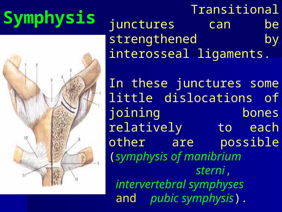
Symphysis
Transitional junctures can be strengthened by interosseal ligaments. In these junctures some little dislocations of joining bones relatively to each other are possible (symphysis of manibrium sterni, intervertebral symphyses and pubic symphysis).

Thank you for attention