the temporal region and temporo-mandibular joint (tmj) head & neck unit – lecture 6 د....
TRANSCRIPT
The Temporal Region AndTemporo-Mandibular Joint (TMJ)
Head & Neck Unit – Lecture 6األعسم. جليل حيدر د
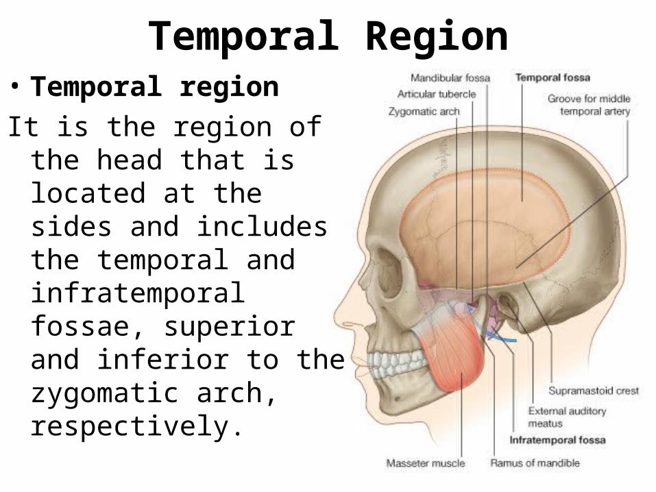
• Temporal region It is the region of the head
that is located at the sides and includes the temporal and infratemporal fossae, superior and inferior to the zygomatic arch, respectively.
Temporal Region
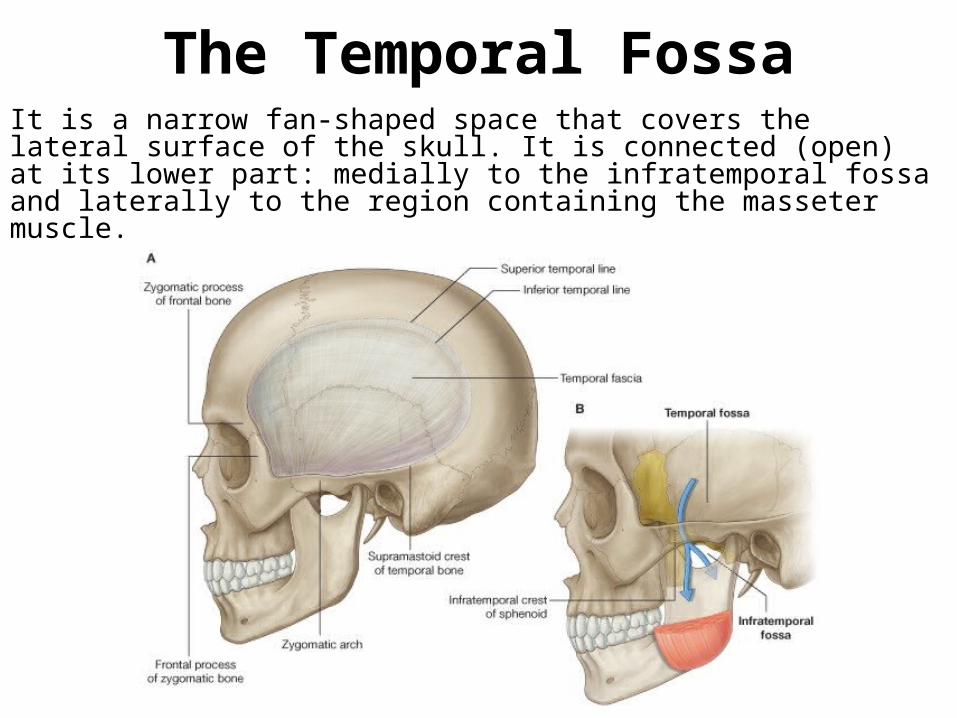
The Temporal FossaIt is a narrow fan-shaped space that covers the lateral surface of the skull. It is connected (open) at its lower part: medially to the infratemporal fossa and laterally to the region containing the masseter muscle.
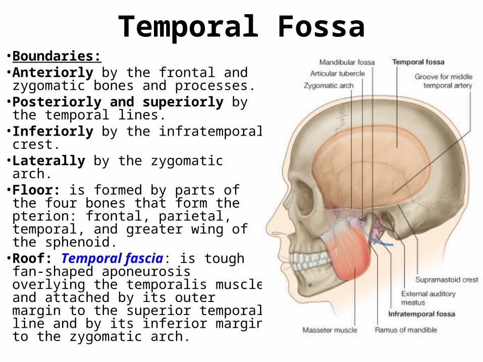
• Boundaries:• Anteriorly by the frontal and
zygomatic bones and processes.• Posteriorly and superiorly by the
temporal lines.• Inferiorly by the infratemporal crest.• Laterally by the zygomatic arch.• Floor: is formed by parts of the four
bones that form the pterion: frontal, parietal, temporal, and greater wing of the sphenoid. • Roof: Temporal fascia: is tough fan-
shaped aponeurosis overlying the temporalis muscle and attached by its outer margin to the superior temporal line and by its inferior margin to the zygomatic arch.
Temporal Fossa
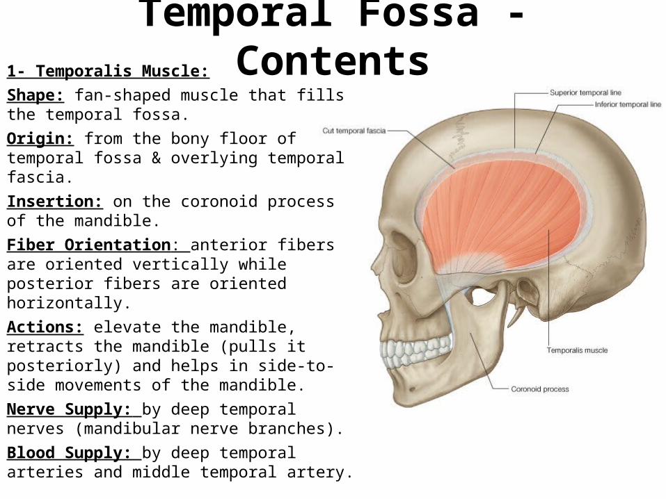
Temporal Fossa - Contents1- Temporalis Muscle:Shape: fan-shaped muscle that fills the temporal fossa.Origin: from the bony floor of temporal fossa & overlying temporal fascia.Insertion: on the coronoid process of the mandible. Fiber Orientation: anterior fibers are oriented vertically while posterior fibers are oriented horizontally. Actions: elevate the mandible, retracts the mandible (pulls it posteriorly) and helps in side-to-side movements of the mandible. Nerve Supply: by deep temporal nerves (mandibular nerve branches).Blood Supply: by deep temporal arteries and middle temporal artery.
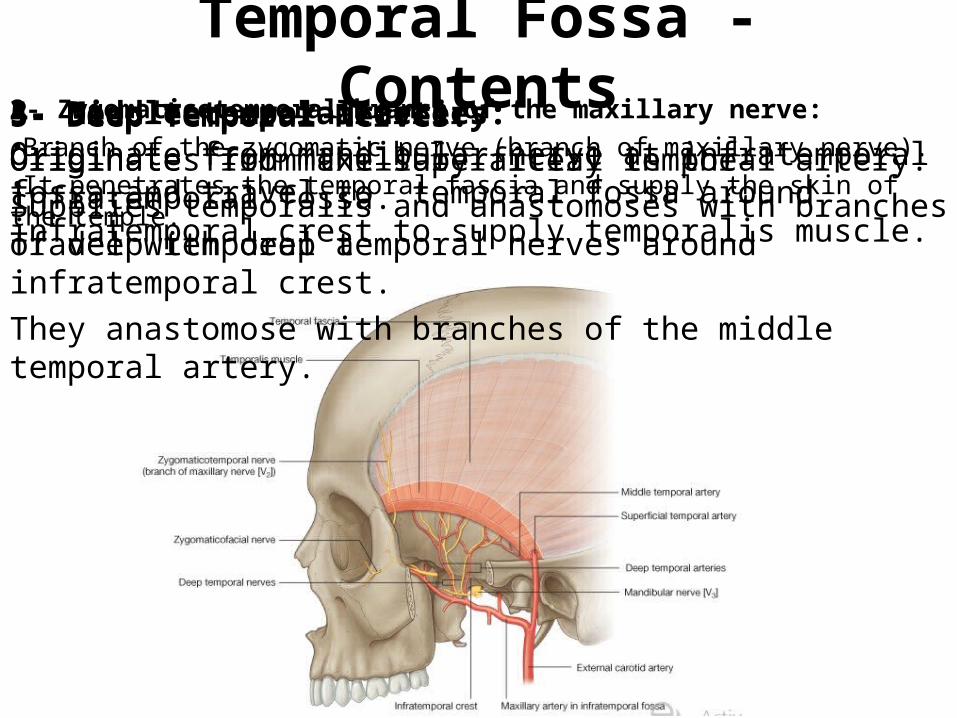
Temporal Fossa - Contents2- Zygomaticotemporal branch of the maxillary nerve: •Branch of the zygomatic nerve (branch of maxillary nerve). •It penetrates the temporal fascia and supply the skin of the temple.
3- Deep temporal arteries: Originate from maxillary artery in the infratemporal fossa.Travel with deep temporal nerves around infratemporal crest.They anastomose with branches of the middle temporal artery.
4- Middle temporal artery: Originates from the superficial temporal artery. Supplies temporalis and anastomoses with branches of deep temporal a
5- Deep Temporal Nerves: Originate from mandibular nerve at infratemporal fossa and travel to temporal fossa around infratemporal crest to supply temporalis muscle.
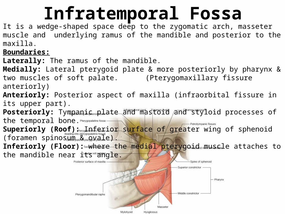
Infratemporal FossaIt is a wedge-shaped space deep to the zygomatic arch, masseter muscle and underlying ramus of the mandible and posterior to the maxilla. Boundaries:Laterally: The ramus of the mandible.Medially: Lateral pterygoid plate & more posteriorly by pharynx & two muscles of soft palate.
(Pterygomaxillary fissure anteriorly)Anteriorly: Posterior aspect of maxilla (infraorbital fissure in its upper part). Posteriorly: Tympanic plate and mastoid and styloid processes of the temporal bone.Superiorly (Roof): Inferior surface of greater wing of sphenoid (foramen spinosum & ovale).Inferiorly (Floor): where the medial pterygoid muscle attaches to the mandible near its angle.
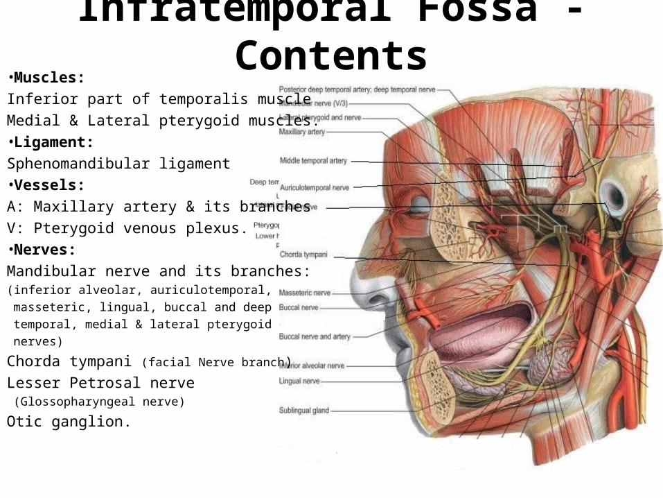
Infratemporal Fossa - Contents•Muscles: Inferior part of temporalis muscleMedial & Lateral pterygoid muscles.•Ligament: Sphenomandibular ligament•Vessels: A: Maxillary artery & its branches V: Pterygoid venous plexus.•Nerves: Mandibular nerve and its branches:(inferior alveolar, auriculotemporal, masseteric,
lingual, buccal and deep temporal, medial & lateral pterygoid nerves)
Chorda tympani (facial Nerve branch)
Lesser Petrosal nerve (Glossopharyngeal nerve)
Otic ganglion.
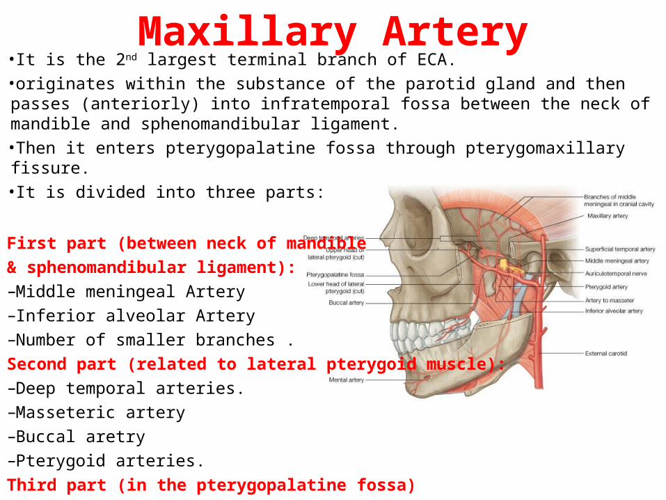
Maxillary Artery•It is the 2nd largest terminal branch of ECA.•originates within the substance of the parotid gland and then passes (anteriorly) into infratemporal fossa between the neck of mandible and sphenomandibular ligament. •Then it enters pterygopalatine fossa through pterygomaxillary fissure.•It is divided into three parts:
First part (between neck of mandible & sphenomandibular ligament): –Middle meningeal Artery–Inferior alveolar Artery–Number of smaller branches .Second part (related to lateral pterygoid muscle):–Deep temporal arteries.–Masseteric artery –Buccal aretry–Pterygoid arteries. Third part (in the pterygopalatine fossa)
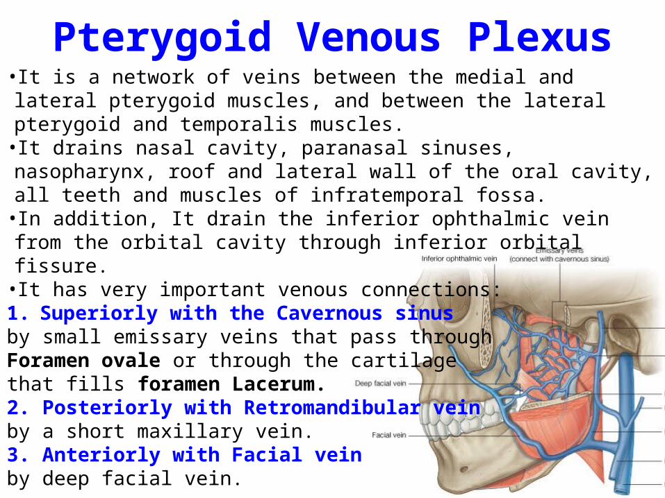
Pterygoid Venous Plexus•It is a network of veins between the medial and lateral pterygoid muscles, and between the lateral pterygoid and temporalis muscles. •It drains nasal cavity, paranasal sinuses, nasopharynx, roof and lateral wall of the oral cavity, all teeth and muscles of infratemporal fossa. •In addition, It drain the inferior ophthalmic vein from the orbital cavity through inferior orbital fissure. •It has very important venous connections:1. Superiorly with the Cavernous sinusby small emissary veins that pass through Foramen ovale or through the cartilage that fills foramen Lacerum.2. Posteriorly with Retromandibular veinby a short maxillary vein.3. Anteriorly with Facial veinby deep facial vein.
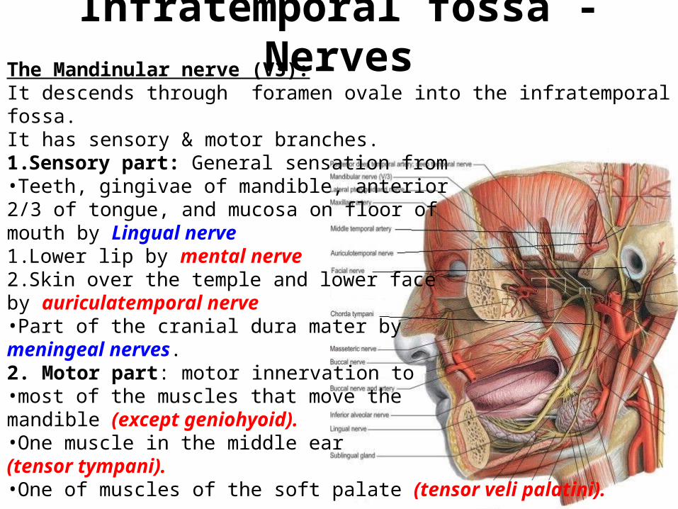
Infratemporal fossa - NervesThe Mandinular nerve (V3):It descends through foramen ovale into the infratemporal fossa.It has sensory & motor branches. 1.Sensory part: General sensation from •Teeth, gingivae of mandible, anterior 2/3 of tongue, and mucosa on floor ofmouth by Lingual nerve1.Lower lip by mental nerve2.Skin over the temple and lower face by auriculatemporal nerve•Part of the cranial dura mater by meningeal nerves.2. Motor part: motor innervation to •most of the muscles that move the mandible (except geniohyoid).•One muscle in the middle ear (tensor tympani).•One of muscles of the soft palate (tensor veli palatini).
Infratemporal fossa - NervesInferior alveolar nerve:It is a sensory branch of mandibular nerveIt enters the mandibular canal after giving the only motor branch nerve to mylohyoid.
The lingual nerve:It lies anterior to inferior alveolar nerve (important). It enters the mouth between medial pterygoid muscle & ramus of mandible.It is sensory to anterior two thirds of tongue, floor of mouth, and lingual gingivae. It distributes taste fibers from chorda tympani to anterior 2/3 of tongue.
Auriculotemporal nerve:It carries general sensation from skin over a large area of the temple Contributes to sensory innervation of external ear, external auditory meatus, tympanic membrane, & temporomandibular joint. It delivers postganglionic parasympathetic nerves from glossopharyngeal nerve to the parotid gland.
The Otic Ganglion: It is parasympathetic ganglion located in infratemporal fossa, just inferior to the foramen ovale, medial to mandibular nerve and posterior to the medial pterygoid muscle. The Presynaptic parasympathetic fibers, derived mainly from glossopharyngeal nerve, synapse in the otic ganglion. The Postsynaptic parasympathetic fibers pass from otic ganglion to parotid gland through the auriculotemporal nerve. Lesser petrosal nerve:It is a branch from the tympanic plexus which is formed mainly by tympanic nerve (a branch from glossopharangyeal nerve).The glossopharyngeal nerve exit the skull through jugular foramen and gives a tympanic nerve that re-enters the skull to temporal bone forms tympanic plexus over the promontory of the middle ear. The lesser petrosal nerve leaves the temporal bone & enter middle cranial fossa. Then, it descend through foramen ovale into infratemporal fossa.
Infratemporal fossa - Nerves
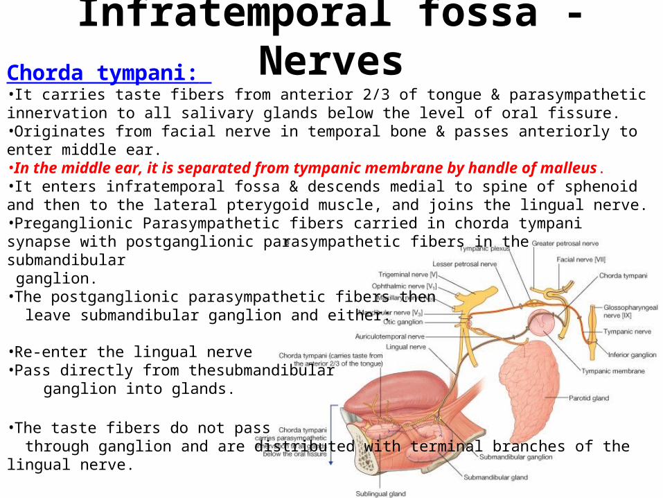
Chorda tympani: •It carries taste fibers from anterior 2/3 of tongue & parasympathetic innervation to all salivary glands below the level of oral fissure. •Originates from facial nerve in temporal bone & passes anteriorly to enter middle ear. •In the middle ear, it is separated from tympanic membrane by handle of malleus. •It enters infratemporal fossa & descends medial to spine of sphenoid and then to the lateral pterygoid muscle, and joins the lingual nerve. •Preganglionic Parasympathetic fibers carried in chorda tympani synapse with postganglionic parasympathetic fibers in the submandibular ganglion. •The postganglionic parasympathetic fibers then leave submandibular ganglion and either:
•Re-enter the lingual nerve•Pass directly from thesubmandibular ganglion into glands.
•The taste fibers do not pass through ganglion and are distributed with terminal branches of the lingual nerve.
Infratemporal fossa - Nerves
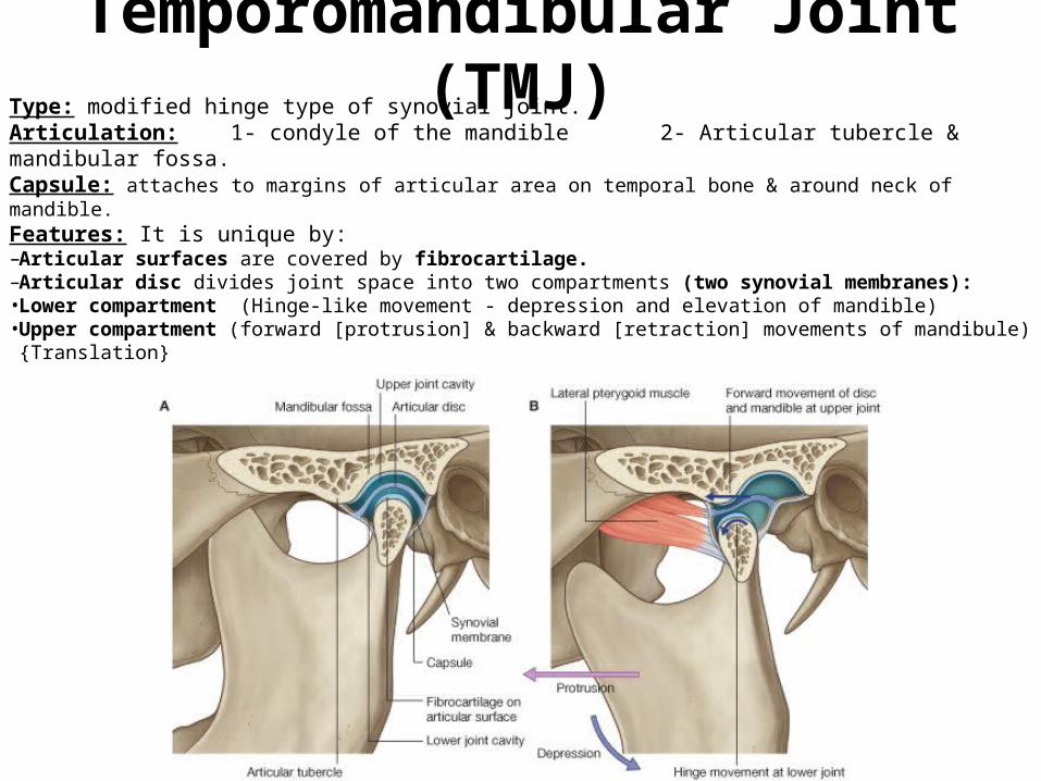
Temporomandibular Joint (TMJ)Type: modified hinge type of synovial joint. Articulation: 1- condyle of the mandible 2- Articular tubercle & mandibular fossa.Capsule: attaches to margins of articular area on temporal bone & around neck of mandible. Features: It is unique by:–Articular surfaces are covered by fibrocartilage.–Articular disc divides joint space into two compartments (two synovial membranes): • Lower compartment (Hinge-like movement - depression and elevation of mandible)•Upper compartment (forward [protrusion] & backward [retraction] movements of mandibule)
{Translation}
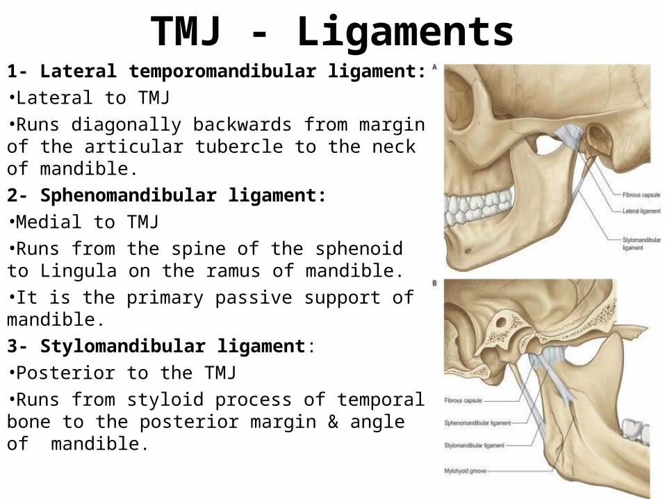
TMJ - Ligaments1- Lateral temporomandibular ligament:•Lateral to TMJ•Runs diagonally backwards from margin of the articular tubercle to the neck of mandible. 2- Sphenomandibular ligament:•Medial to TMJ•Runs from the spine of the sphenoid to Lingula on the ramus of mandible.•It is the primary passive support of mandible.3- Stylomandibular ligament:•Posterior to the TMJ•Runs from styloid process of temporal bone to the posterior margin & angle of mandible.
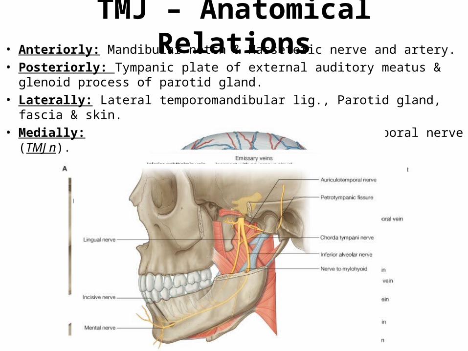
TMJ – Anatomical Relations• Anteriorly: Mandibular notch & Masseteric nerve and artery.• Posteriorly: Tympanic plate of external auditory meatus & glenoid
process of parotid gland.• Laterally: Lateral temporomandibular lig., Parotid gland, fascia & skin.• Medially: Maxillary artery & vein and Auriculotemporal nerve (TMJ n).
Muscles of Mastication1- Masseter Muscle:2- Medial Pterygoid Muscle:3- Lateral Pterygoid Muscle:4- Temporalis Muscle:
• Quadrangular in shape• Overlies the lateral surface of the ramus of mandible. • Has superficial part and deep part. • Innervated by masseteric nerve and supplied by masseteric artery
from the maxillary artery.
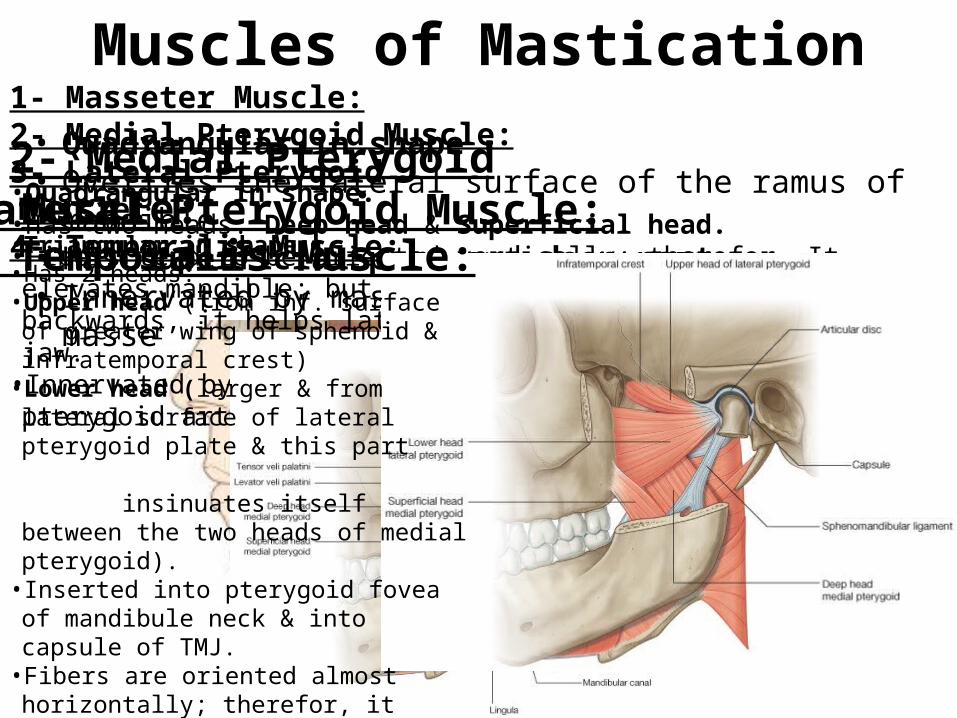
2- Medial Pterygoid Muscle:•Quadrangular in shape.•Has two heads: Deep head & Superficial head.•Fibers tend to be oriented vertically; therefor, It elevates mandible; but because it passes also obliquely backwards, it helps lateral pterygoid in protruding lower jaw.• Innervated by nerve to medial pterygoid & supplied by pterygoid arteries.
3- Lateral Pterygoid Muscle:• Triangular in shape.•Has 2 heads: •Upper head (from inf. surface of greater wing
of sphenoid & infratemporal crest)• Lower head (larger & from lateral surface of
lateral pterygoid plate & this part insinuates itself between the two heads of medial pterygoid).• Inserted into pterygoid fovea of mandibule
neck & into capsule of TMJ. • Fibers are oriented almost horizontally;
therefor, it pulls the articular disc & head of mandible forward & is major protruder of the lower jaw.• Innervated by nerve to lateral pterygoid and
supplied by pterygoid arteries.
4- Temporalis Muscle:
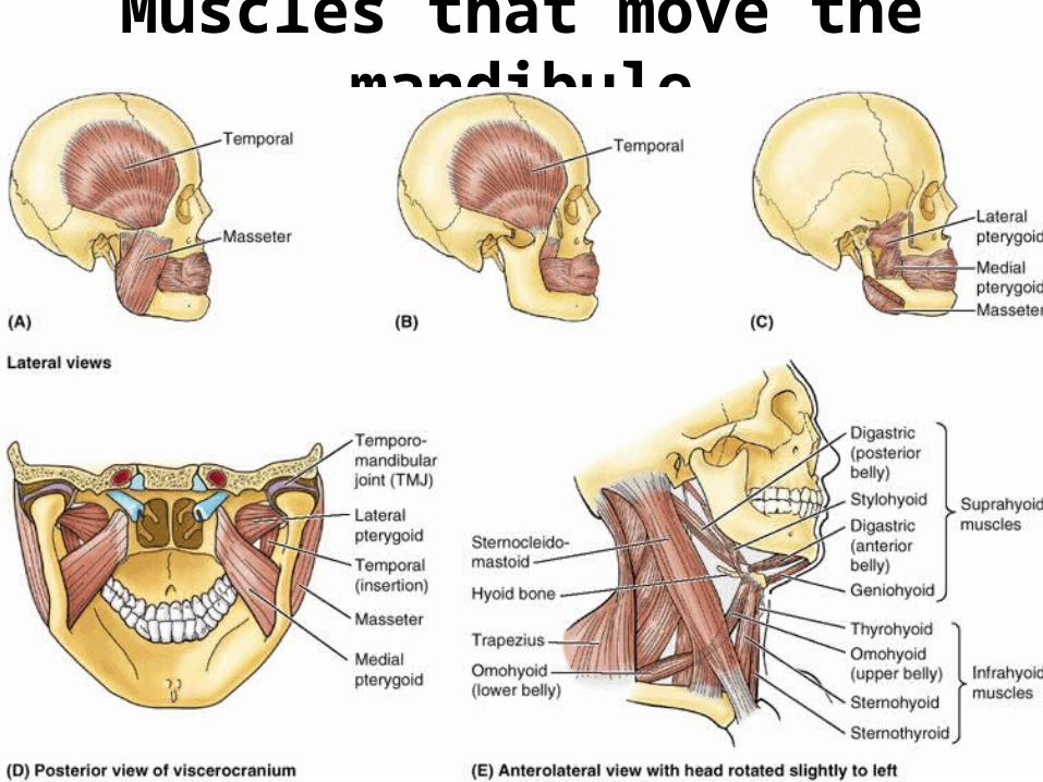
Muscles that move the mandibule
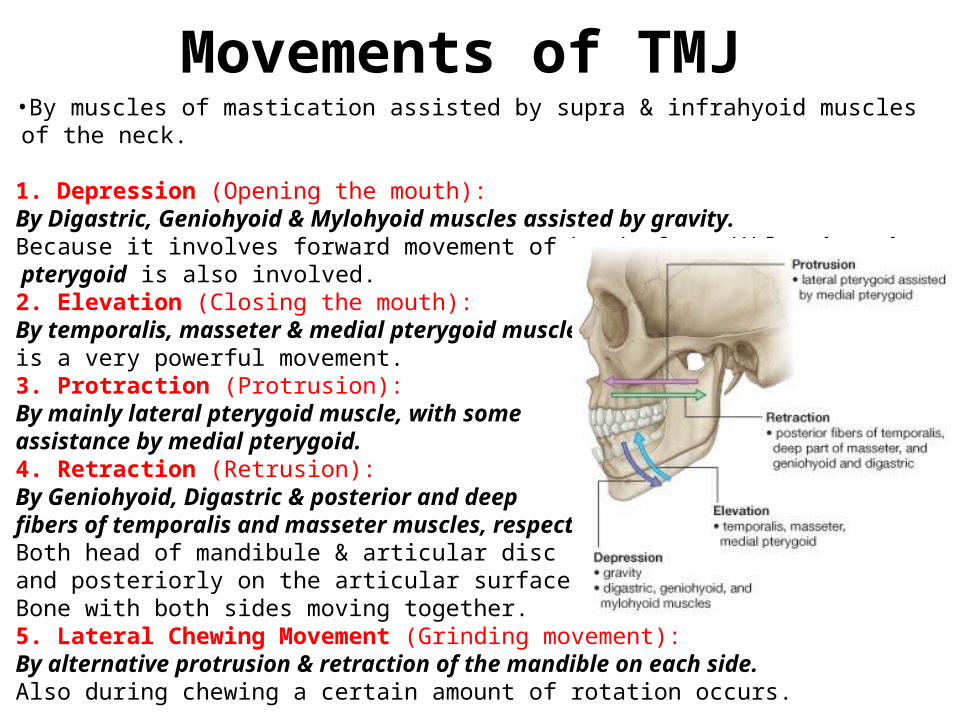
Movements of TMJ •By muscles of mastication assisted by supra & infrahyoid muscles of the neck.
1. Depression (Opening the mouth): By Digastric, Geniohyoid & Mylohyoid muscles assisted by gravity.Because it involves forward movement of head of mandible, lateral pterygoid is also involved.2. Elevation (Closing the mouth): By temporalis, masseter & medial pterygoid muscles.is a very powerful movement.3. Protraction (Protrusion):By mainly lateral pterygoid muscle, with some assistance by medial pterygoid.4. Retraction (Retrusion):By Geniohyoid, Digastric & posterior and deep fibers of temporalis and masseter muscles, respectively. Both head of mandibule & articular disc slide anteriorly and posteriorly on the articular surface of the temporal Bone with both sides moving together.5. Lateral Chewing Movement (Grinding movement): By alternative protrusion & retraction of the mandible on each side.Also during chewing a certain amount of rotation occurs.

End of the Lecture
GOOD LUCK