the stanford medical student clinical journal · the stanford medical student clinical journal...
TRANSCRIPT

�H&P Autumn 2006
H&PThe Stanford Medical Student Clinical Journal
Innocents Abroad Practicing Western Medicine in Resource-Poor Settings
Volume ��, Number � Autumn 2006

H&P Autumn 20062
Contents
Editors’ Note Early Interstitial Lung Disease in an Adolescent with Mixed Connective Tissue DiseaseJessica Yasnovsky
Scalp Lesion: What Lies BeneathJoanna Chan
Sex, Friendship, and Confidentiality Steven Lin
Innocents Abroad: Practicing Western Medicine in Resource-Poor SettingsNavigating Health in NicaraguaJames Colbert
500 Grams: The Gap Between Russian and Western MedicineAsya Agulnik
Reflections: The State of HIV/AIDS in TanzaniaLena Winestone
Papua New Guinea: The Last Frontier?Naresh Ramarajan
Days of AweHetty Beth Eisenberg
The Mailbox: An Interview with Audrey ShaferChantal Forfota
From Bedside to BonesawsWilliam Slikker
A Cormorant Drying its WingsChantal Forfota
Stanford 2006 Nobel LaureatesManeesh Singh
clinical case reports
ethicalcase report
H&PThe Stanford Medical Student Clinical Journal
Editors-in-ChiefJames ColbertThomas Tsai
Associate Editors
ClinicalJoanna WredeJessica Yasnovsky
EthicsSteven Lin
ForumMalavika Prabhu
Literary ArtsChantal ForfotaWilliam Slikker
ScienceManeesh Singh
LayoutWei Gu
Faculty AdvisorsPatricia Cross, PhDAudrey Shafer, MDElliott Wolfe, MD
H&P, the Stanford Medical Student Clinical Journal, is published by students of the Stanford University School of Medicine. All articles are written or co-authored by students. H&P is not an official publication of the Stanford University School of Medicine or its faculty. Expressed written opinions are solely those of the authors and do not necessarily represent those of Stanford University or the School of Medicine.
H&P is published quarterly. Submissions are accepted on CD or by email.
Please submit inquiries to Thomas Tsai ([email protected]). Correspondence may be addressed to OSA, Stanford University School of Medicine, Stanford, CA 94305.
3
forum
literary arts
science
4
�4
�8
�2
��
8
2�
24
26
28
29
30
Cover/Rear Photos: Little Corn Island, Nicaragua by James Colbert

3H&P Autumn 2006
In 1893 Lane Hospital was erected on the corner of Sac-ramento and Webster streets in San Francisco to provide facilities for the burgeoning Cooper Medical College, the precursor to the Stanford University School of Medicine. In the vestibule of the hospital, a marble inscription read:
This hospital erected in the year 1893 by Levi Cooper Lane, physician and surgeon, with money earned by himself in his profession, is given by him to suffering humanity and to the healing art in the hope that the former may here find refuge and relief; the latter exercise of the human skill and intelligent sympathy.
This year marks the 100th anniversary of Lane Medical Library, formally created on August 29, 1906. This occasion has emboldened us as the new editors-in-chief of the Stanford Medical Student Clinical Journal to find inspiration in the roots of Stanford Medical School in redefining the mission of this journal to reflect our vision of the future of medicine.
We have renamed this journal H&P: The Stan-ford Medical Student Clinical Journal. The title H&P reflects the importance of the basic history and physical examination in clinical medicine in every corner of the world. It also represents Hygeia and Panacea, two daughters of Asclepius. In Greek mythol-ogy, Hygeia is the goddess of welfare and the prevention of sickness, while Panacea is the goddess of healing and cures. We believe that these figures represent the two facets of our medical education—to treat and cure illnesses while promoting the welfare of our patients by preventing disease. H&P also reflects the new mission of our journal—as the daughters of Asclepius, Hygeia and Panacea represent a new generation.
The title H&P embodies the journal’s commitment to clinical medical education while reflecting the chal-lenges of modern medicine through the lenses of both prevention and treatment. Therefore, we see the oppor-
Editors’ Note
tunity for H&P to become a student forum for the “ex-ercise of the human skill and intelligent sympathy” that inspired the construction of Lane Hospital a century ago.
A new addition to H&P is the creation of a case re-ports section. The clinical case reports aim to provide a venue for Stanford medical students to share clinical expe-riences with their peers, while the ethical case reports will allow students to share the ethical dilemmas they have encountered on the wards. It is our hope that the ensuing dialogue will awaken the restlessness of reason that will guide us here at Stanford and in our future medical careers.
Each issue will also feature a forum, a collection of pieces written around a central theme. The theme for this issue is “Innocents Abroad: Practicing Western Medicine in Resource-Poor Settings.” As medical students here at Stanford, we have the luxury of having the most modern and technologi-
cally advanced diagnostic tools at our disposal – yet the world is not Stanford. In the following pages we present the encounters of four current students who spent their summers in Nicaragua, Tanzania, Papua New Guinea, and Russia. In the process, many discovered that their most power-ful tools were their empathy and clinical acumen.
And because medicine is art as well as science, we have continued the tradition of the journal in
presenting the creative works of the Stanford medi-cal community. In this issue we have paintings, photo-
graphs, and poetry from our student colleagues as well as an interview with one of our professors, a physician-author who has embraced the healing art. We have also chosen to de-fine art and science in a broad sense, envisioning medicine as the nexus of multiple disciplines. As the journal grows, you will see columns reflecting the interdisciplinary nature of medicine, addressing the human condition from myriad perspectives.
We hope you enjoy this issue of H&P. For the fac-ulty and administration of Stanford Medical School, this journal will provide the pulse of the educational growth of your students. For the students, this journal is you.
James Colbert and Thomas TsaiEditors-in-Chief
introEditors

H&P Autumn 20064
CLINICAL CASE REPORT
Early Interstitial Lung Disease in an Adolescent with Mixed Connective Tissue Disease Jessica Yasnovsky
Mixed connective tissue disease (MCTD) is an illness characterized by overlapping features of systemic lupus erythematosus, systemic sclerosis, and polymyositis-dermatomyositis. Almost one quarter of cases of this disease occur in children. Presentations of MCTD are variable. Pulmonary involvement, including pulmonary arterial hypertension, accounts for most disease-associated mortality. This report describes an adolescent with MCTD and early lung disease, which progressed despite aggressive immunosuppressive and cytotoxic treatment. The possibility for rapid disease progression in MCTD, as in other connective tissue diseases, makes early consideration of this diagnosis important. Close monitoring for development of pulmonary arterial hypertension is recommended in patients with MCTD, especially in those with pulmonary involvement.
Abstract
lived in a community without a pediatric rheumatologist, she was initially managed by an internist rheumatologist.
Physical examination at that time was notable for thick-ening and tightening of the skin of the fingers (sclerodactyly) and proximal forearms, hypopigmented patches and pos-sible increased tightness of the skin on the face, along with decreased mobility of the left wrist. The patient was found to have an elevated ANA titer at 1:2560 (reference: <1:80), with a speckled pattern, and positive anti-RNP antibody. She also had moderately positive anti-Smith antibody and moderately positive anti-Ro antibody. Anti-La and anti-double stranded DNA antibodies were within normal limits. Creatinine kinase was elevated to 105 U/L (reference: 10-80 U/L). Pulmonary function testing (PFT) revealed early restrictive lung disease (figure 1). High resolution computed tomography (CT) imag-ing of the chest demonstrated minimal subpleural reticular findings consistent with possible early pulmonary fibrosis.
The patient then developed difficulty swallowing solids. For this reason she ate less and began losing weight. Treat-ment with low-dose prednisone was initiated, and omepra-zole was given for esophageal symptoms and to reduce the likelihood of steroid-induced gastrointestinal changes. The patient immediately noted improvements in her joint symp-toms, myalgias, and swallowing. However, she developed a dry cough and xerophthalmia. Upon referral to a pediatric rheumatologist, she had a physical examination notable for visible and palpable bilateral parotid enlargement, a dry oropharynx, and significant joint findings. There was 1+ swelling of all metacarpophalangeal joints, tenderness of some of these joints, mild flexion contractures of all proximal
Mixed connective tissue disease (MCTD) is an illness characterized by overlapping features of systemic lupus erythematosus (SLE), systemic sclerosis, and polymyositis-dermatomyositis.1 MCTD is associated with positive anti-nuclear antibodies (ANA), specifically anti-ribonuclear protein (anti-RNP) antibodies. First recognized as a distinct rheuma-tologic entity in 1972, MCTD has been described in children since 1973. Although MCTD is rare in all age groups, almost one-quarter of presentations of this disease are in children.2
Presentations of MCTD are highly variable in both adults and children.1,3 The most common features of this disease include arthralgia or arthritis, Raynaud’s phenomenon, esophageal dysmotility, fatigue, swollen hands, inflammatory muscle disease, and impaired pul-monary diffusing capacity for carbon monoxide (DLCO).1,2
Pulmonary involvement affects approximately 67% of adults and 24 - 42% of children with MCTD and is an impor-tant cause of morbidity and mortality associated with this disease.1,2,4 This report documents an adolescent with MCTD who developed early, progressive interstitial lung disease.
A previously healthy fifteen year-old Mexican-American young woman noticed increasing arthralgias and morning stiffness in her knees, ankles, back, elbows, wrists, and in multiple joints of her hands. Due to hand cramping and stiffness, she needed to take breaks during standard writing exercises at her school. Ibuprofen provided incomplete relief. She also noticed myalgias and proximal and distal weak-ness in her arms and legs. Within two weeks, progressive difficulty moving and walking caused her to stop attending school. At the same time, the patient noticed an unusual soft-tissue swelling below her mandible. She had noted no recent illnesses or fevers. There were no known toxin expo-sures for the patient or any family members. Past medical history was unremarkable, and birth and developmental histories were without abnormalities. Immunizations were up to date. Family history was negative for rheumatologic, immunologic, infectious, neurologic, and neoplastic disease.
The patient presented to her pediatrician, who ordered laboratory tests, including an erythrocyte sedimentation rate, elevated at 44 mm/hr (reference: 4-20 mm/hr) and a total protein, elevated at 9.2 g/dL (reference: 6-7.9 g/dL). A complete blood count was within normal limits. Concerned about a possible autoimmune disease, the pediatrician referred the patient for rheumatologic evaluation. As the patient
Introduction
Case Report
Yasnovskycase report

�H&P Autumn 2006
interphalangeal joints of the hands and feet, and 1+ swelling at the medial patellar borders bilaterally. Pulmonary exami-nation was normal, and there was no clubbing of fingers or toes. Cardiac auscultation was notable for a loud second heart sound. Further laboratory testing revealed that rheumatoid factor was positive at 127 U/mL (reference: <30 U/mL) and that lupus anticoagulant was not detected. In addition, muscle inflammation was suggested by the following laboratory test results: aspartate aminotransferase of 162 U/L (reference: 15-45 U/L), alanine aminotransferase of 125 (reference: 7-35 U/L), creatinine phosphokinase of 365 U/L (reference: 10-80 U/L), lactate dehydrogenase of 437 U/L (reference: 100-190 U/L), and aldolase of 24.7 U/L (reference: 1.2-8.8 U/L).
The patient was diagnosed with MCTD. Studies were undertaken to evaluate the extent of pulmonary and cardiac involvement, as this would determine the treatment regimen. While continuing on prednisone and awaiting the results of these tests, the patient experienced progression of her fatigue, weak-ness, and myalgia. She also developed dyspnea, which affected her even at rest. In addition, there was new onset of bother-some dry mouth and of symptoms of Raynaud’s phenomenon, including fingertip numbness and color changes (pallor and cy-anosis on exposure to cold, followed by rubor on rewarming).
The patient’s echocardiogram and chest x-ray were within normal limits. A second read of her chest CT revealed extensive intralobular septal thickening involving all lung lobes, consistent with pulmonary fibrosis, and interstitial and ground-glass opacities at bilateral posterior costo-phrenic angles, indicating possible inflammation or alveolitis.
Prednisone dosing was increased to 1 mg/kg/day and treatment with mycophenolate mofetil was initiated and increased to the maximum pediatric dose of 2000 mg/day. Care had been taken to ensure a negative PPD and immunity against vaccine-preventable illnesses prior to starting this medication regimen. Calcium supplementation was also given. Regular physical therapy was begun. The patient had im-provements in her symptoms and was able to return to her school and activities, including dancing.
One month later, a repeat echocardiogram demonstrated normal pressures but mild flat-tening of the interventricular sep-tum. Due to concern for possible early signs of pulmonary arterial hypertension (PAH), mycophe-nolate mofetil was then replaced with monthly intravenous infu-sions of cyclophosphamide. In order to protect the patient’s ovaries from this medication, monthly leuprolide acetate injec-tions were begun. Medication for pneumocystis carinii pneumonia prophylaxis was also given. A calcium channel blocker was used to relieve continued symp-toms of Raynaud’s phenom-
enon, and hydroxychloroquine was used to treat joint and skin symptoms. Her course was complicated by an out-patient pneumonia and a brief hospitalization for febrile neutropenia, which resolved. For unclear reasons, she has also had intermittent elevations in serum transaminases.
The patient’s fatigue and muscle weakness gradually worsened, and she was given several intravenous infusions of methylprednisolone at 20 mg/kg. There was also continued dyspnea on exertion, and oxygen saturation dropped from 97% at rest to 92% after brief (less than five minutes) walk. This was mirrored by worsening of restrictive lung disease on PFTs (Figure 1). A repeat high resolution chest CT (Figure 2) fifteen months after initial presentation demonstrated continued pro-gression of intralobular septal thickening and ground-glass, reticular opacities. Peripheral fibrosis and traction bronchi-ectasis were seen. However, echocardiograms suggested reversal of the septal flattening and did not demonstrate other signs of PAH. An electrocardiogram, although not a sensitive screening tool for PAH, showed no signs of right axis deviation or right ventricular hypertrophy, common findings in PAH.5
The patient will continue to be treated with immu-nosuppressive and cytotoxic medications. A six-minute exercise PFT and overnight oxygen saturation study are planned. The patient will be followed with frequent rheu-matology and pulmonology clinic appointments and se-rial echocardiograms and PFTs occurring every six months.
This patient’s presentation illustrates several signs and symptoms of MCTD, including fatigue, arthritis, myositis, Raynaud’s phenomenon, sclerodactyly, dysphagia, dyspnea, and dry cough. Xerostomia, xerophthalmia, and enlargement of salivary glands suggest secondary Sjogren’s syndrome. Although this patient exhibits primarily features of systemic sclerosis, a diagnosis of MCTD was given for several reasons.
At Presentation
3 Months after Presentation
12 Months after Presentation
FVC (% Pred) NA 73 63 FEV1 (% Pred) 86 75 67 FEV1/FVC (%) NA 96 100 FRC (% Pred) NA
NA
70
RV (% Pred)
46
128
145 TLC (% Pred)
75
83
88
DLCO (% Pred) 73
74
50
Figure 1: Pulmonary Function Testing showing restrictive lung disease, including decreased forced vital capacity (FVC), forced expiratory volume in one second (FEV�), functional residual capacity (FRC), total lung capacity (TLC) and diffusing capacity for carbon monoxide (DLCO) and increased ratio of FEV�/FVC. The elevations in residual volume (RV) were attributed to weakness and inability to fully exhale. The 3 and �2-month tests were done at the same laboratory. (% Pred = % Predicted; NA= Not available.)
Discussion
Yasnovsky case report

H&P Autumn 20066
continued in spite of aggressive management with corticoste-roids, mycophenolate mofetil, and cyclophosphamide. ILD is a common complication of MCTD and may be associ-ated with active alveolitis, sug-gested by ground-glass opacity on high resolution CT, a high neutrophil or eosinophil count on bronchoalveolar lavage, or reduced DLCO on PFTs .1,2,6 Other respiratory diseases as-sociated with MCTD include pleural effusion, obstructive airways disease, pulmonary in-fections and pulmonary vascu-litis.4 Aside from dyspnea and cough, another common initial manifestation of pulmonary involvement in MCTD is chest pain, which may be persistent or pleuritic. Bibasilar rales are also sometimes heard on pul-monary auscultation.6 Asymp-tomatic lung disease is often detected on imaging, which was the case for this patient during her initial chest CT scan.
While the CT scan and PFTs showed abnormali-ties, her pulmonary involve-ment was not detectable on
lung auscultation or chest x-rays. This pattern has been seen in many patients with MCTD.1 Pulmonary symptoms in patients with this disease thus warrant a thorough in-vestigation, and a chest CT and set of PFTs are part of the recommended initial work-up for all patients with MCTD.6
ILD may result not only in severe pulmonary symptoms but also in the dangerous complication of PAH, in children as in adults.7 This is because parenchymal lung destruction causes hypoxia and therefore pulmonary vasoconstriction. PAH is indicated by a mean pulmonary artery pressure of greater than 25 mm Hg at rest and greater than 30 mm Hg with exercise, in the setting of a normal pulmonary capillary wedge pressure.4
Patients with connective tissue diseases may be at higher risk for PAH than other patients with equivalent ILD for several reasons. Thromboemboli associated with the hypercoagulable state found in some patients with connective tissue diseases can lead to PAH. It is also thought that inflammatory cell infiltrates and deposits of antibodies and complement fractions affect the vascular endothelium, contributing to risk for pulmonary vasoconstriction, even in patients without lung disease.4,8
It is unknown what percentage of patients with MCTD and ILD generally develop PAH. According to two small studies of patients with MCTD and any pulmonary dysfunction or pa-thology, rates for confirmed pulmonary hypertension ranged from 35 to 60%.4 Other studies document the incidence of PAH in all patients with MCTD to be as high as 20 to 30%.9 PAH
While arthritis and myositis can be present in systemic scle-rosis, they are also common features of SLE and polymyosi-tis-dermatomyositis respectively. Her presentation includes the presence of anti-Smith and anti-Ro antibodies, which can be seen in SLE, and the laboratory findings described above as being consistent with myositis. In addition, this patient’s presentation demonstrates a laboratory profile having higher specificity for MCTD, including a high titer (>1:1000) of speckled ANA and confirmed anti-RNP antibodies.2,4
There are conflicting sets of diagnostic criteria for MCTD, and its existence as a distinct clinical entity has recently been called into question. However, many clinicians and research-ers still believe it is important to distinguish this illness from other rheumatologic conditions.1 One reason to make this distinction is to prepare patients and physicians for a heterogenous and changing pattern of clinical involvement. This pattern includes the possibility for future evolution into either SLE or systemic sclerosis.1 Another important reason to distinguish MCTD is that, compared with SLE or systemic sclerosis, it carries a higher risk for erosive arthritis as well as the potentially lethal complications of pulmonary hypertension, fibrosing alveolitis, and severe myositis.1
Pulmonary involvement in MCTD can be quite striking, as in this patient, who experienced dyspnea and persistent dry cough within two months of onset of symptoms of MCTD. Pul-monary fibrosis and progressive interstitial lung disease (ILD)
Figure 2: Non-contrast high resolution CT chest, lower lung lobes, pulmonary window: bronchiec-tasis (dilated, thick-walled bronchioles) and adjacent ground glass opacities are seen.
Yasnovskycase report

�H&P Autumn 2006
can result in right ventricular failure and is the most common cause of death that is directly related to MCTD.1,4 In studies of adults with connective tissue disease-associated PAH, one year survival ranged from 45 to 69%.10 Although there is some evi-dence that PAH may be less severe in children with MCTD,2,11 long-term follow-up studies are not yet available. Right ven-tricular impairment from PAH in patients with MCTD was not reversible in a recent study.12 However, outcomes in PAH are more favorable if the condition is diagnosed and treated early.6 Therefore, this patient will be followed closely with serial echo-cardiograms and PFTs occurring every six months. Although her ILD, history of a loud second heart sound, and temporary interventricular septal flattening on echocardiogram are con-cerning for risk of progression to PAH, the severity of this risk is unknown.13 It is likely that her aggressive medical manage-ment has decreased her risk for PAH or delayed its onset.
Clinical trials of medications for MCTD are lacking.6 Treat-ment regimens are based on the specific organs involved and the severity of disease activity. Medications used to treat this disease include corticosteroids, antimalarials, methotrexate, cytotoxic medications (usually cyclophosphamide), TNF-alpha inhibitors, cyclosporine, and vasodilators. Patients with MCTD and ILD accompanied by active alveolitis are often managed with high-dose corticosteroids, along with mycophenolate mofetil or cyclophosphamide, both used in this patient, or azathioprine. As these medications do not adequately control ILD in many patients with MCTD, further research on treatments for this disease is needed.6
There is a misconception based on earlier studies which could not be replicated that extensive response is usually at-tainable in MCTD using low-dose corticosteroids.1 In addition, many physicians have concerns about beginning high-dose (ap-proximately 1 mg/kg per day) corticosteroids and other immu-nosuppressive or cytotoxic agents in children. Although these medications have accompanying side effects and risks, such as the potential for infection associated with immunosuppres-sion, in many patients their benefits may outweigh their risks. Where possible, children with MCTD should be seen by a pedi-atric rheumatologist, who will have the most experience man-aging these medications in children with autoimmune disease.
Because there is a scarcity of pediatric rheumatolo-gists, internist rheumatologists are often called upon to see pediatric patients, as initially occurred for this patient.14 Children with MCTD may also present to dermatologists, pulmonologists, gastroenterologists, and other specialists. It is therefore important for physicians in a wide variety of spe-cialties to be familiar with presentations of pediatric MCTD.
I would l ike to thank Dr. Peter Chira, Pedi -atric Rheumatologist and Instructor at Stanford Uni-versity School of Medicine, for reviewing this report.
Yasnovsky case report
References
Acknowledgement
1. Venables P. Mixed connective tissue disease. Lupus 2006;15:132-7.
2. Mier R, Shishov M, Higgins G, Rennebohm R, Wortmann D, Jerath R, et al. Pediatric-onset mixed connective tissue disease. Rheum Dis Clin N Am 2005;31:483-96.
3. Mier R, Ansell B, Hall M, Hasson N, Levinson J, Lovell D, et al. Long term follow-up of children with mixed connective tissue disease. Lupus 1996;5(3):221-6.
4. Bull T, Fagan K, Badesch D. Pulmonary vascular manifestations of mixed connective tissue disease. Rheum Dis Clin N Am 2005;31:451-64.
5. Galie N, Manes A, Branzi A. Evaluation of pulmonary arterial hypertension. Curr Opin Cardiol 2004;19:575-81.
6. Kim P, Grossman J. Treatment of mixed connective tissue disease. Rheum Dis Clin N Am 2005;31:549-65.
7. Roy R, Couriel J. Secondary pulmonary hypertension. Paediatr Respir Rev 2006;7(1):36-44.
8. Sanchez O, Sitbon O, Jais X, Simonneau G, Humbert M. Immunosuppressive therapy in connective tissue diseases-associated pulmonary arterial hypertension. Chest 2006;130:182-9.
9. Vegh J, Szodoray P, Kappelmayer J, Csipo I, Udvardy M, Lakos G, et al. Clinical and immunoserological characteristics of mixed connective tissue disease associated with pulmonary arterial hypertension. Scand J Immunol 2006; 64:69-76.
10. Coghlan J, Handler C. Connective tissue associated pulmonary arterial hypertension. Lupus 2006;15:138-42.
11. Friedman D, Mitnick H, Danilowicz D. Recovery from pulmonary hypertension in an adolescent with mixed connective tissue disease. 1992;51(8):1001-4.
12. Vegh J, Hegedus I, Szegedi G, Zeher M, et al. Diastolic function of the heart in mixed connective tissue disease. Clin Rheumatol 2006; epub ahead of print.
13. Pilatis N, Jacobs L, Rerkpattanapipat P, Kotler M, Owen A, Manzarbeitia C, et al. Clinical predictors of pulmonary hypertension in patients undergoing liver transplant evaluation. Liver Transpl 2000;6(1):85-91.
14. Mayer M, Sandborg, C, Mellins E. Role of pediatric and internist rheumatologists in treating children with rheumatic diseases. 2004;113(3 pt 1):e173-81.
H&P Autumn 20068
CLINICAL CASE REPORT
Scalp Lesion: What Lies BeneathJoanna L. Chan
Disorders of the skin account for a large percentage of chief complaints and can cause significant distress for patients in both outpatient and hospital settings. This clinical vignette describes a seventy-five year-old gentleman who presented to a dermatology clinic with a scalp lesion that was diagnosed as a cylindroma. Pathological diagnosis and clinical features of this lesion are highlighted. In addition, the potential for malignant transformation and innovative treatment options for cylindroma are discussed.
Abstract
given his previous history. A shave biopsy was performed to confirm the benign nature of the lesion and to address therapeutically the bothersome nature of its location.
Pathology:
In Figure 2, the scanning low power view demonstrates that the tumor masses are fairly well-circumscribed nests of basaloid cells assembled in a complex mosaic pattern to form larger nodular aggregates, or the so-called jigsaw puzzle-like pattern. No smaller, micronodular subunits are seen
infiltrating the periphery. Further magnification with medium power (Figure 3), shows the small subunits separated by glassy, eosinophilic base-ment membrane material. This peri-odic acid-Schiff (PAS)-positive hyaline sheath material is composed of type IV collagen bands and fragments of Type VII collagen anchoring fibrils. The hyaline material envelops and penetrates the tumor islands, some of which have angulated contours. High power magnification (Figure 4) clearly shows the two characteristic cell vari-ants. The basaloid, basophilic (darker)
cells are palisading at the periphery of the tumor nests com-pared to the central and larger pale-staining cells. Although not present in this specimen, rare foci of tubular differentiation may occasionally be seen with careful observation. These small tubal lumina are usually few in number or absent. The base-ment membrane zones of these tumor cells exhibit multiple molecular defects, which include abnormalities in collagen IV alpha-1 and alpha-5 chain expression, defective laminin 5 processing, and the absence of mature hemidesmosomes.1
Based on the pathologic evidence obtained from the skin biopsy, the patient was diagnosed with a scalp cylindroma.
Dermatologic manifestations of disease account for a large percentage of chief complaints and can cause significant dis-tress for patients in both the outpatient and hospital settings. Patients frequently note disorders of the skin to interfere with their activities of daily living, often accounting for significant preoccupation with the symptomatology. Certainly, skin can-cer screening, diagnosis, and treatment among patient popu-lations play a significant role in health care maintenance and expenditures. In the following clinical vignette, a variation of the simple scalp lesion is presented, followed by a discussion of the pathological diagnosis and a novel treatment option guided by the basic molecular pathophysiology of the disorder.
The patient, a 75 year-old Caucasian gentleman with a his-tory of junctional atypical melanocytic proliferation, status post excision, presented at the Palo Alto Veterans Affairs dermatol-ogy clinic for the evaluation of a scalp lesion. Although his past medical history was notable for having multiple actinic kerato-ses, he had no internal malignancies and was otherwise healthy. By his-torical report, he noted that the skin growth on his scalp had been pres-ent and stable in size for multiple years. Notably, he denied bleeding, pruritis, and pain, though intermit-tent irritation with combing had occurred. Although he reported intermittent sunscreen application, he never wore hats for photo-pro-tection. In addition, he denied any family history of skin cancer or vas-cular conditions, and he was using no dermatologic medications.
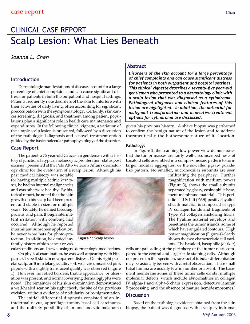
On physical examination, he was well-appearing with Fitz-patrick Type II skin, in no apparent distress. On his right pari-etal scalp, an 8 mm telangiectatic, soft, well-circumscribed pink papule with a slightly translucent quality was observed (Figure 1). However, no rolled borders, friable appearance, or ulcer-ation was present, and normal overlying dermatoglyphs were noted. The remainder of his skin examination demonstrated a well-healed scar on his right cheek, the site of the previous excision, without evidence of nodularity or re-pigmentation.
The initial differential diagnosis consisted of an in-tradermal nevus, appendage tumor, basal cell carcinoma, and the unlikely possibility of an amelanocytic melanoma
Introduction
Case Report
Discussion
Figure 1: Scalp lesion
case report Chan

9H&P Autumn 2006
Cylindromas are uncommon but not rare adnexal neoplasms that have eccrine, apocrine, and follicular differentiation. The exact incidence is unknown, and they tend to occur in mid to old age, reportedly affecting women up to nine times more often than their male counterparts. No racial predilec-tion has been noted in the literature. Although generally dermal lesions without epidermal attachment, cylindromas may penetrate subcutaneous fat if extensive invasion occurs.
The clinical presentation can vary, presenting as skin-colored, erythematous or bluish and as papules, nodules, or tumors depending on size. They may be asymptom-atic or painful but all tend to be slowly-growing, firm, rubbery, and only a few millimeters to centimeters in size. Exceptions to this size description do occur, and a uni-corn-like cylindroma tumor has been recently presented.2
The pathophysiology is debated by dermatopathologists, but the general consensus over the years points to a multipotent primitive sweat gland that differentiates into a cylindroma. Mixed variants occur, in association with spiradenomas of ec-crine differentiation or trichoepitheliomas of apocrine and fol-licular origin, as apocrine glands and hair follicles derive from a common precursor. Recent investigations into the origin of cylindromas using histochemical analysis lend support to this folliculosebaceous apocrine unit theory. Cylindromas exhibit markers of both eccrine & apocrine origin, including cytokera-tins & IKH-4 (coiled duct region) of eccrine differentiation. Multiple apocrine markers have been found, in addition to car-cinoembryonic antigen (CEA) & epithelial membrane antigen.3
Solitary cylindromas are the most common, occuring in sporadic fashion and most commonly located on the scalp or head and neck region. The variant with multiple cylindromas is inherited, generally in an autosomal dominant fashion and may resemble grapes or small tomatoes in this so-called turban tumor syndrome. Though typically occurring on the head and neck, this variant may also appear on the trunk and extremities, though they are rarely widespread and resemble a neurofibromatosis pattern. Some reports have described cylindromas to be confluent or arising in a linear arrangement.4
Patients with the inherited familial cylindromatosis, or Brooke-Spiegler syndrome, have multiple cylindromas and trichoepitheliomas. Occasionally, these cylindromas have oc-curred in association with multiple basal cell adenomas of the parotid glands, milia, organoid nevi, basal cell carcinomas, and spiradenomas. The genetic mutation is well-described, map-ping to the CYLD-1 tumor suppressor gene on chromosome 16.5
CYLD acts as a de-ubiquitinating enzyme in the TNF-alpha pathway. Ubiquitination functions in normal cell ho-meostasis to target proteins for cellular degradation. CYLD de-ubiquitinates TRAF-2, which associates with the TNF-alpha receptor. When TRAF-2 is ubiquitinated, it is cytoplasmically activated and brings about downstream activation of NF-κB, an antiapoptotic transcription factor that leads to cellular proliferation.6 CYLD usually “puts the brakes” on NF-κB and thus the CYLD deficiency results in proliferation, such as that seen in patients with cylindromas. In terms of therapies, aspirin may compensate for the CYLD deficiency as it acts downstream, inhibiting both the release and the activation of NF-κB. Phase 1 trials with topical aspirin for cylindroma-
Figure 2: Pathology, low power (~ 40x) magnification
Figure 3: Pathology, medium power (~ 200x) magnification
Figure 4: Pathology, high power (~ 400x) magnification
case reportChan

H&P Autumn 2006�0
tosis are already underway. Of twelve cylindroma lesions treated with topical salicylic acid in one small study, eight exhibited partial response and two resolved completely.7
Malignant transformation is rare but will generally occur in the setting of multiple cylindromas, preferentially affecting women and patients over 50 years of age.8 These malignant le-sions can occur on the scalp and are associated with ulceration, rapid growth, bleeding, and a blue to pink hue. Increased Ki67 expression has been described, but other markers tend to be unhelpful. The literature reports 11 cases of metastases with 9 deaths, with spread to lymph nodes, stomach, thyroid, liver, lung & bones.9 Histological examination would reveal loss of the typical jigsaw pattern. In addition, polymorphous clear cells with prominent pleomorphic nucleoli and increased or abnormal mitoses have been described. Generally, the striking vascular component of cylindrocarcinoma can be appreciated.10
Treatment choices include surgical excision or electrosur-gery for a solitary tumor, though reports suggest that carbon dioxide laser may be used for smaller lesions.11 Multiple cylindromas may require extensive plastic surgery or even progressive excision. The technique of Mohs micrographic surgery has been reported in the treatment of cylindroma for the tissue-sparing benefits in cosmetically sensitive lo-cations.12 Close follow-up is recommended to monitor for new lesions or malignant transformation, and radiotherapy can be used if the lesions are inoperable. The morbidity and mortality is low for cylindromas but is associated with occasional erosion through the skull,9 which may result in hemorrhage or meningitis. Transformation to malignancy confers poor survival as visceral metastases frequently follow.9
For this patient, surgical excision was performed with adequate margins although no current clinical guidelines exist regarding the exact margins required. Sutures were removed during a subsequent visit, and he remained disease-free and without regrowth or nodularity six months following the procedure. An annual physical examination was scheduled for continued surveillance.
1. Massoumi R, Podda M, Fassler R, Paus R. Cylindro-ma as tumor of hair follicle origin. J Invest Dermatol 2006;126(5):1182-4.
2. Chaer RA, Lipnick S. Images in clinical medicine. Cylindroma. N Engl J Med 2004;351(24):2530.
3. Bumgardner AC, Hsu S, Nunez-Gussman JK, Schwartz MR. Trichoepitheliomas and eccrine spi-radenomas with spiradenoma/cylindroma overlap. Int J Dermatol 2005;44(5):415-7.
4. Nerad JA, Folberg R. Multiple cylindromas. The “turban tumor.” Arch Ophthalmol 1987;105:1137.
5. Young AL, Kellermayer R, Szigeti R, Teszas A, Azmi S, Celebi JT. CYLD mutations underlie Brooke-Spiegler, familial cylindromatosis, and mul-tiple familial trichoepithelioma syndromes. Clin Genet 2006;70(3):246-9.
6. Lakhani SR. Putting the brakes on cylindromatosis? N Engl J Med 2004;350(2):187-8.
7. Oosterkamp HM, Neering H, Nijman SM, Dirac AM, Mooi WJ, Bernards R, Brummelkamp TR. An evaluation of the efficacy of topical application of salicylic acid for the treatment of familial cylindro-matosis. Br J Dermatol 2006;155(1):182-5.
8. De Francesco V, Frattasio A, Pillon B, Stinco G, Scott CA, Trotter D, Patrone P. Carcinosarcoma arising in a patient with multiple cylindromas. Am J Dermato-pathol 2005;27(1):21-6.
9. Durani BK, Kurzen H, Jaeckel A, Kuner N, Nae-her H, Hartschuh W. Malignant transformation of multiple dermal cylindromas. Br J Dermatol 2001;145(4):653-6.
10. Weedon D, Strutton G. Skin Pathology. 2nd ed. London: Churchill Livingstone; 2002.
11. Sajben FP, Ross EV. The use of the 1.0 mm hand-piece in high energy, pulsed CO2 laser destruction of facial adnexal tumors. Dermatol Surg 1999;25:41–4.
12. Behroozan DS, Goldberg LH, Glaich AS, Kaplan B, Kaye VN. Mohs micrographic surgery for deeply penetrating, expanding benign cutaneous neo-plasms. Dermatol Surg 2006;32(7):958-65.
References
Nephron by Margie Teng
case report Chan
��H&P Autumn 2006 ��
ETHICAL CASE REPORT
Sex, Friendship, and Confidentiality
Eric is a 23 year old Chinese male studying econom-ics at a prestigious college. For two months, Eric has been suffering from nausea, jaundice, and stomach pains. When he finally goes to see his doctor, Eric is devastated to find out that he has chronic hepatitis B and the early stages of liver cirrhosis. His doctor tells him that his infection is both lifelong and incurable, and that he is at high risk of develop-ing liver cancer before the age of forty. Fearing for his life, Eric drops out of college and returns home to his parents, who are both poorly-educated, non-English speaking im-
migrants and knew nothing about hepatitis B or liver cancer. Looking for help, Eric contacts his best friend Jay, who
is a medical student doing research on hepatitis B and liver cancer. Jay works hard to educate Eric about his ill-ness and explains that the hepatitis B virus is spread exactly like HIV, that is, through blood and unprotected sex. Jay advises Eric to protect his loved ones by making sure that his family is tested and vaccinated. Eric asks Jay to help keep his condition a secret, because in Chinese culture, having hepatitis B is a devastating social stigma that, if re-vealed, will bring much shame and disgrace to his family.
Meanwhile, Eric succumbs to major depression, partly due to the emotional trauma of his illness, and partly
The following case describes an actual situation that happened to a medical student at Stanford. The case has been developed only up to a crucial “decision point” and then left to your ethical consideration. Names and details have been modified to protect the individuals involved.
due to the severe psychiatric effects of interferon treat-ment. Afraid that their son will soon die, Eric’s parents become convinced that the best thing to do is to find Eric a wife. Merely six months after his diagnosis of chronic hepatitis B, Eric marries Kim in an arranged marriage.
Although he is happy for his friend, Jay is startled to learn that Eric intends to keep his sexually transmitted disease a secret from Kim. As close friends of both Eric and Kim, and as a doctor in training, Jay feels obligated to protect Kim by informing her of Eric’s condition. However, he is also acutely
aware that his actions can destroy a marriage between two friends. Ethically torn, Jay decides to warn Eric that he will be exposing Kim to a very high risk of infection, and that if Kim is infected, she can pass the virus down to their children. Eric ac-knowledges the warning, so Jay stops short of informing Kim.
Ten months later, Jay receives a phone call from a very dis-traught Kim, who is now six months pregnant. In tears, Kim tells Jay that she has just been tested positive for the hepatitis B virus.
What ethical principles are in play here? Did Jay have a duty to warn Kim that overrides his obligation to maintain Eric’s confidentiality? What if Eric was Jay’s pa-tient, instead of a friend asking for a consult? Did friend-ship interfere with Jay’s judgment? What should Jay do?
Steven Lin
AutonomyJustice AANon-Maleficence
Beneficencemymy
ethicsLin

H&P Autumn 2006�2
Navigating Health in NicaraguaJames Colbert
Three-year-old Carlos had cold feet. Dengue virus had altered the endothelial cells in his body, causing his capillaries to become overly porous. By the third day of his illness, his cap-illaries were leaking so much fluid that he didn’t have enough blood left in circulation to adequately perfuse his body tissues. His blood pressure dropped, his heart rate went up, and his ex-tremities were cool to the touch. The doctors gave him fluids by IV, but his blood pressure failed to respond. The only option left was to put a catheter into one of his large veins (i.e.,. a “central line”) in order to both increase his blood volume and give him medicine that would increase blood flow to his vital organs.
Obtaining consent from the parents was a struggle, as they were wholly uncomfortable with the idea of inserting a tube into their son’s heart. When the resident informed the parents of the gravity of the situation, Carlos’ mother burst into tears and stormed out of the room. The resident continued to ex-plain to the father the necessity of the procedure, but he seemed quite skeptical and repeatedly questioned why a regular IV wasn’t sufficient. Finally, af-ter 20 critical minutes had al-ready passed, the father gave his consent for the procedure.
The official name of the hos-pital in the Nicaraguan capital of Managua—where Carlos was being treated—is “Hospital In-fantil Manuel de Jesus Riveras.” But that’s a mouthful to say, and so it is instead referred to as “La Mascota”—The Pet. The story goes that during the 1970s when Nicaragua was ruled by the ruth-less dictator Somoza, a young boy was murdered by Somoza’s National Guard police force. During his lifetime, this boy had been called “the Pet” by his family members, so when a chil-dren’s hospital was built near the site of his murder, people be-gan using the deceased boy’s nickname to refer to the hospital.
La Mascota is one of the best public children’s hospitals in Nicaragua, and children from all over the country come there for treatment. The doctors are knowledgeable and well-trained. It is a teaching hospital and serves as a major training center for the medical students who attend the public university in Mana-gua. On the Infectious Disease Unit, where I worked, there are two senior residents, four interns, and six medical students, all of whom work under the supervision of the attending physicians.
The medical hierarchy functions much as it does in the United States. Each morning, the entire medical team visits
every patient on the ward. Each is presented to the group by a medical student or resident, who summarizes the patient’s history and physical exam. Then, the attending questions the child or parent to fill in any gaps in the history and, afterwards, briefly examines the patient. The final step before leaving each patient is devising a plan of what needs to be done for the patient. The entire visita usually lasts about an hour, and writing up the orders for tests, external consults, discharge summaries, etc., usually keeps the interns and residents busy until 3 p.m. or so. Patients do not have electronic medical records, so residents spend a great deal of time handwriting everything on top of carbon paper to ensure that copies can
be saved for recordkeeping. In many ways, La Mascota
is similar to an academic medi-cal center that could exist in a developed country such as the United States. However, there are some important distinc-tions: La Mascota is a public hospital and is owned by the government of Nicaragua. Physicians are employees of the state and earn meager salaries of less than $500 per month. Care is completely free, and all hospitalization costs are covered by the state. Nica-ragua is the poorest country in Latin America, and, without a national healthcare system, the majority of patients would be unable to pay for their care; however, the downside of Nicaragua’s system is that funding is severely limited. Patient rooms are basic and contain nothing more than
beds, chairs, and sinks. There are no electronic gadgets. IVs are hung on poles by patient bedsides and flow is controlled only by gravity. Without automatic sensors to monitor vital signs, nurses have an important responsibility to frequently check blood pressure, temperature, pulse and other vitals. Know-ing how to perform a thorough physical exam is essential.
Because government salaries for physicians are so low, most doctors supplement their incomes by seeing patients in private clinics in the afternoon. In the Infectious Disease ward, the attending physicians left the hospital by 1 or 2 p.m. so they could spend the rest of the afternoon seeing patients in the pri-vate setting, earning $25 per consult. For many of these doctors, the income earned in the private setting is double or triple their state salaries. The downfall of this system is that doctors have very little incentive to devote extra time to their inpatients at
forum Colbert

�3H&P Autumn 2006
the public hospitals. Many Nicaraguans I spoke with who had been treated in public hospitals expressed frustration at the lack of personal attention they had received from their doctors.
Despite these pitfalls, the medical attention that Carlos received was quite excellent. Within minutes of obtaining his father’s consent, a surgeon arrived in the ward to begin the procedure. I observed from the back of the room as a resident and two nurses held the boy firmly in place during the procedure, which was done under local anesthesia with the boy awake for much of it. After injecting the anesthesia, the surgeon made an incision to the right of the groin and then set about to locate the femoral vein (which leads to the inferior vena cava—the route by which all blood returns to the heart). However, the procedure did not go smoothly, as the first catheter did not go in correctly and had to be removed. A second surgeon then arrived on the scene, his task being made more difficult by Carlos’s kicking and flailing arms if ever the nurses relaxed their hold for a brief moment. The entire ordeal lasted over an hour, and there was a collective sigh of relief when the second surgeon finally succeeded after much effort.
After less than 24 hours in the intensive care unit, Carlos’s blood pressure rose to a normal level. The fluid he received through the central line allowed his body to overcome the critical shock phase of dengue hemorrhagic fever. Two days later Carlos had left the ICU and was back in his hospital room, full of smiles. After reviewing his chart, the attending physician had decided that Carlos was well enough to go home. His endothelial cells were function-ing again, and his capillaries were no longer leaking fluid.
Carlos was lucky. First, his mother had recognized his dengue symptoms and had brought him to the hos-pital before he went into hypovolemic shock. Second, his mother brought him to a hospital that is participating in
a multi-site collaborative clinical study of dengue infec-tion funded by the World Health Organization Program in Tropical Disease Research. Consequently, the doctors treating Carlos are perhaps the most knowledgeable pe-diatric infectious disease specialists in all of Nicaragua.
As I watched him make faces at his younger sister, I couldn’t help noticing the sharp contrast between Carlos’s life of poverty and mine of relative comfort. Yet, despite our different backgrounds, together we were both participants in a timeless tradition that involved hope, courage, and healing. The Nicaraguan health system may be under-funded and resource-poor, but the doctors and nurses who cared for Carlos were able to overcome these obstacles in their pursuit of a more important goal—for Carlos to walk out of the hospital on feet that were no longer cold.
The Hospital Infantil Manuel de Jesus Rivera—“La Mascota”
forumColbert

H&P Autumn 2006�4
�00 Grams: The Gap between Russian and U.S. Medicine
The neonatal ward of Moscow City Hospital No. 13 is lo-cated in a three-story cement building that was built sometime between Stalin and Brezhnev and appears as if it hasn’t been touched since. The ward consists of two long hallways lined with small, whitewashed rooms and studded with windows. Each room cradles eight infants, although when other hos-pitals close for the summer, there can be ten or even twelve cribs clustered against each narrow wall. In the intermediate nursery, the rooms are always crammed—with nurses, doc-tors, cleaning staff, and even mothers who come to take care of their newborns because they know how short-staffed the hospital is. Outsiders and fathers, however, are strictly pro-hibited from entering what Russian neonatologists consider a sterile space. Deeper into the hospital, the area set aside for the Neonatal Intensive Care Unit (NICU), or Reanimation in Russian, is eerily calmer. Mothers are rarely allowed to enter, and doctors prefer to do their paperwork elsewhere. All that remains is the haunting beeping of heart rate moni-tors and the occasional clicking of heels on the tiled floor.
Sometimes, as I watched the physicians minister to their patients using techniques vastly different from those I have become accustomed to in the Stanford NICU, I wondered what exactly I was doing, eleven time zones away. Even before enter-ing medical school, I knew that I wanted to spend this summer doing research in the country where I was born. Despite emi-grating from Russia as a refugee in 1991, a part of me wanted to come back, to make myself useful to the place where I came from. I had always had a hunch that Russia’s medical system was disintegrating with its government and was falling far be-hind its Western counterparts, but the reality of the situation was both shocking and yet, to be cynically honest, quite expected.
Along with my research conducting a chart review on
the management of neonatal jaundice, I was able to shadow a number of neonatologists and observe their typical pa-tients. One of the first patients I saw was a 1000 gram premie, probably 30 weeks gestation. He was a week or two old and still tiny. Although he was not intubated, he needed supple-mental oxygen, and it was clear that he was still struggling to breathe. His legs were limp, the muscle tone present in a healthy newborn being almost completely absent. Using an ultrasound machine, the doctors determined that the baby had lost more than a third of its white matter, an injury that was serious enough to be fatal. If he survived, it was un-likely that he would ever be able to have higher brain func-tion—it was difficult to say if he would ever have a real life.
This situation, unfortunately, is not rare in Russia. In Mos-cow, 1000 grams or 28 weeks gestation is considered the limit for resuscitation. If an infant is born at an earlier gestational age, life saving measures will still be attempted, but these babies are not expected to survive and doctors are not held responsible for their death. Outside of Moscow, the situation only worsens. In Peno, a medium-sized village five hours outside of Moscow, I shadowed an elderly pediatrician, one of two in a three-hour radius, who was ready to retire from a profession that had worn her out. Being old enough to have practiced during the Soviet era, she remembered better days when her small clinic had enough supplies to function. It was difficult for her to practice medicine knowing that, due to lack of medicine, technology, and staff, she was not able to provide the best possible care to her patients. When I asked about neonatal resuscitation, she sighed. Without personnel capable of providing infants airway support, the limit for survival in Peno was 1750 grams or about 33 weeks—a shocking differ-ence from the threshold in the country’s capital. In compari-
Asya Agulnik
forum Agulnik

��H&P Autumn 2006
in English and don’t read international medical journals, the Russian system results in a medical culture rooted in a num-ber of outdated beliefs and convictions that have long been pushed aside in American and Western European medicine.
As an American, former national, and medical student, I found myself in a strange position. Like hundreds of physicians
who came to Russia after the fall of communism, I chose my project with the hope of providing the neonatologist a tool that he could use in order to improve the care of his patients. Yet the doctors
I spoke with often had a conflicting response to receiving such foreign help. I felt at times that, for being a student, my opinion was given far too much weight because of my foreign educa-tion, while on other occasions, the same doctors would become defensive about their own system for managing patients and cite examples of how Western medicine was inferior. From my conversations with the physicians, I think my experience was similar to that of most foreign doctors who visit Russia. Like a team of American neonatologists who came to Moscow a decade before me to teach new methods of resuscitation, the
information was heard, the techniques learned, but after the visitors left, the knowl-edge was applied only limit-edly, and exclusively within the original medical system that had yet to be healed.
After my summer in Rus-sia, my impression about the outlook for their health care system is a complex one. The problems facing medicine are strongly rooted in a culture of government dictatorship and corruption that don’t seem to change despite Russia’s current attempts at democ-racy. Yet my time spent with the younger doctors in our
clinic has given me some reason to be optimistic. Physicians in their twenties and early thirties are generally fluent in English, and many of them make an effort to keep abreast of current research in their fields. Some who have been abroad and have a sense for the disparity faced by Russian medicine work with public health programs to help change the way that medicine is addressed nationally. Still, I am painfully aware that these physicians are in the minority, and it’s far from certain that their efforts will be enough to pull Russia into the 21st century—in essence, to close the 500 gram gap between Russian and Western medicine. It is for this reason that, despite the difficulties facing foreign doctors in Russia, it is important for us to continue visiting and lending our help.
son, most major hospitals in America resuscitate newborns, often successfully, above 500 grams or 23 weeks. This gap, representing more than 30 years of research, is substantial.
In practice, it is difficult to explain why Russia, a country brimming with oil money and a strong education system, isn’t able to keep up with Europe and the United States in the field of medical develop-ment. What’s more surprising is that the disparity within Russia is almost as great as the dispar-ity between it and other Western na-tions. Russian medicine, much like its government and society, is focused in Moscow, with St. Petersburg a distant second. Everything else—funds, money, people, and doctors—is thinly distributed throughout the vast land that, before the fall of the Soviet Republic in 1991, was the largest country in the world. Considering the limitations of this system, it is less inconceivable that a village driving distance away from the capital does not have neonatal respirators or anyone capable of aiding an infant born with difficulty breathing.
The gap between Moscow and the West may be repre-sented by 500 grams of life, but, in truth, it pervades all areas of health care. Soon after the revo-lution in 1917, Lenin declared free education and health care for all, a notion that remained in place even after communism fell apart. Unfortunately, the government’s support for na-tional doctors dissolved with communism’s fall. In a city that was recently deemed the most expensive in the world, doctors who work for Moscow city clinics are paid about 100 U.S. Dollars a month. It’s not surprising that top graduates from medical school go into private (paid) practice, leave medicine, or move to America. Of those who stay, some become indoctrinated into the hierarchy of a transparently corrupt hospital bureaucracy, while others, like my mentor in Peno, serve their calling until frustration leads to retirement.
In addition, medical research in Russia lags, primarily due to the severe lack of funding. Large national grants and power-ful medical academic institutions simply don’t exist. Like the doctors in the NICU where I worked, some go on to complete an Ordinatura—the Russian version of a medical fellowship—which requires them to publish some original research. But even these physicians rarely continue to participate in research after receiving their degree. This isn’t surprising—doctors are paid for their clinical work, and, except for the few fellowship directors, have no incentive, monetary or otherwise, to conduct research. Since most doctors do not have a strong competency
In Moscow, 1000 grams or 28 weeks gestation is considered the limit for resuscitation...In comparison, most major
hospitals in America resuscitate newborns, often successfully, above 500 grams or 23 weeks.
forumAgulnik
H&P Autumn 2006�6
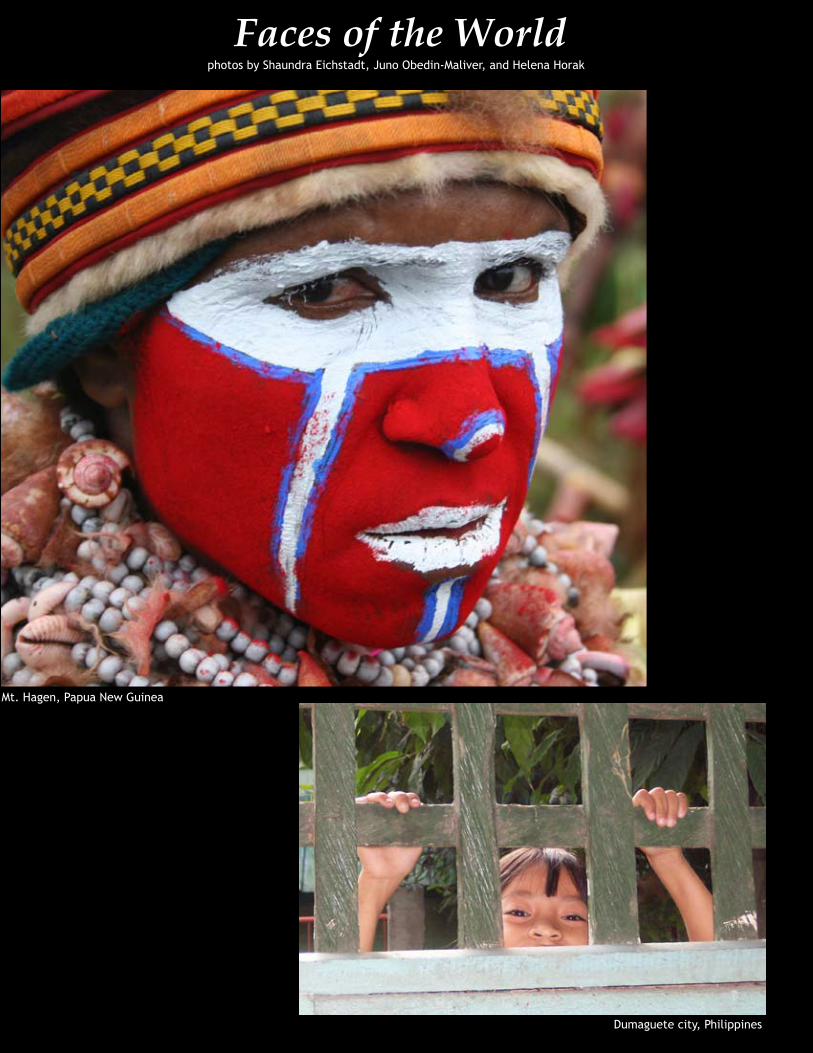
Faces of the Worldphotos by Shaundra Eichstadt, Juno Obedin-Maliver, and Helena Horak
Dumaguete city, Philippines
Mt. Hagen, Papua New Guinea

��H&P Autumn 2006
Qinghai, China Pushkar, India
Bagamoyo, Tanzania
Pushkar, India

H&P Autumn 2006�8�8
on the dirt road off a main highway in Northern Tanzania. We learned that the orphanage only houses children under the age of two because they require the most attention and are the most difficult to care for. Unlike America, in Tanzania it is easier to find a home for children after the age of two be-cause they are more independent and more likely to survive.
It was a long time before I finally decided to go back to Cradle of Love. I eventually realized that the orphanage was not a place of sadness any more than a hospital was—it was a place of healing. Children frequently die there but are not alone in the world, as many orphans are. They have people who love and care for them; in fact, the reality is that they live in better conditions than many children whose parents are still alive.
Upon my return to the orphanage several weeks later, someone immediately handed me a bowl filled with uji—porridge—and set me to work. I was amazed at the speed with which these babies ate. Before I was even able to fill the next spoonful, their mouths were open and ready
Reflections:The State of HIV/AIDS in TanzaniaLena Winestone
“That one’s mother died during childbirth, too much blood; that one’s mother died during a car crash; that one’s mother simply abandoned him; and that one’s mother died of HIV/AIDS.” Recounting each orphan’s story of how he arrived at the Cradle of Love Baby Home, one of the care-givers introduced us to the orphans she had handed us moments before, practically as we had walked in the door. When asked if many of the children there were infected, she proceeded to point at those infected, about 13 of the 23 babies in the room, including the one that I was holding.
Although I was aware that more than 15 million children have been orphaned by HIV/AIDS worldwide, this was the first time I had held an HIV-positive child in my arms. All of a sudden, staring into the face of this innocent child who had lightened my day only moments ago, my good humor faded. I heard one of my classmates begin to weep. I don’t think any of us fully comprehended that we would be entering a place of so much grief when we bumped into a staff member outside
forum Winestone

�9H&P Autumn 2006
for the next bite. It was as though they believed that if they didn’t eat fast enough, they wouldn’t be fed enough.
After I finished feeding the first baby, I began to play with one of the others, telling him to ‘nipe tano,’ or give me five. He got really into it and began repeatedly slapping both my hands. After dinner, one of the workers who took him to change him into his pajamas promptly brought him back out, as he had apparently been asking for me. She told me that his name was Cory. As we played for the next hour, I realized how much Cory resembled a completely normal, happy child. He was very good at repeating, so we talked about all sorts of things, with me saying phrases and him repeating them.
I later discovered his story. Cory tested positive for HIV when he was only a week old. When Cory arrived at Cradle of Love, he had pustules all over his body, caused by a staph infection. He was put on an antibiotic, which soon cleared up the sores, but while on the antibiotic, he got thrush. To look at Cory, one wouldn’t know that he had gone through any of this intensive therapy several months ago. Cory is among the extremely lucky five percent of HIV-positive children who receives medical attention and among an even slimmer per-cent to improve so drastically.
In Tanzania, when a baby ‘tests positive for HIV,’ it means that antibodies against HIV have been detected in the ba-by’s bloodstream. However, these antibodies could reflect a variety of situations: in some instances, they could be the result of passive transfer of antibodies from the mother to the child, rather than the result of the virus itself in the child’s bloodstream. Therefore, currently available serologic testing equipment in Tanzania cannot definitively determine infants’ HIV status before 18 months, at which time ma-ternal antibodies dissipate.
In contrast, the standard of care in the United States is to immediately confirm a positive antibody test in a newborn using polymerase chain reaction to detect viral DNA. According to Dr. Naftal Ole King’ori, the Arusha Regional Medical Officer, an international NGO is in the process of setting up a modern molecular biology labora-tory in Usa River, Tanzania, which would be capable of measuring viral load.1 It will serve the entire Arusha region with a population of almost 1.3 million and will be the first center of its kind in Tanzania serving the general public.
By chance, I had the privilege of visiting the dispensary where this laboratory will be set up. A dispensary is somewhat similar to a walk-in outpatient clinic where medications are
also supplied. I observed one of the doctors there as he conduct-ed his interviews in Kiswahili. We also talked at length about what the most common illnesses were; he had a handwritten chart on the wall of the disease burden from 2003. Malaria, acute respiratory infections, and minor surgeries such as tonsil-lectomies were the most common diseases seen in Usa River. I noticed that neither HIV nor tuberculosis was represented
on the chart, although I would have expected the prevalence of both to be relatively high.
Without any expla-nation about why they
were not included on the charts, he opened a log on his desk and responded that they had documented 84 cases of HIV in 2003. When I asked what kinds of services are provided for HIV-positive patients, he seemed to think it obvious that they provided treatment with antiretroviral therapy (ART) and home-based care, including nutritional support as well volun-tary counseling and testing, a session I witnessed on a later visit.
Detecting my interest in the HIV epidemic, he offered to take me down to the center where they refer their patients who test positive. Elated, I agreed, and he led me down a dirt path to Drug Resource Enhancement against AIDS and Malnutrition (DREAM). DREAM is an AIDS therapy program, funded by the
Community of St. Egidio, designed to give access to free treatment with generic highly-active antiretroviral therapy to the poor in Africa on a large scale. So far, 5,000 people are receiving ART through this program.
The program began in Mozam-bique but has recently expanded to Malawi, Guinea, and Tanzania, among other countries. Despite be-ing free, the program aims at excel-lence in treatment, providing the best existing range of drugs and regular blood testing. It is linked with a nutrition program as well as guidance and sanitary educa-tion by volunteers who themselves are HIV-positive. These measures encourage new patients to adhere to their treatment and come to their
appointments. The compliance rate is very high—94 percent —and the annual cost of the program per person is $800.2
I talked to the physician who runs the clinic and was amazed at how closely treatment regimens in Tanzania resembled those offered in back in U.S. When I asked ques-tions about whether they check patients’ CD4+ counts or use cocktail therapy, the answers were invariably, “Of course, how else could we treat them appropriately?”
As I reflect on what seemed like a surprising response, I am reminded of meeting Dr. Joia Mukherjee, the medi-cal director of Partners in Health, a highly successful non-profit healthcare organization that strives to bring the best of western medicine to the poorest of the poor. She had
I realized that, as doctors, we have an obligation to provide the care our patients need and to find ways to avoid
compromising clinical standards, literally at all costs.
forumWinestone

H&P Autumn 200620
used the same matter-of-fact tone to describe a paradigm shift in which individuals, and physicians in particular, stop thinking about what we can do and instead think about what we need to do. Then, we should use the available re-sources appropriately to do what we have deemed necessary.
At the t ime , I hadn’t believed it re-alistic or practical to think in this manner, but seeing it genuinely put into practice made it make sense. I real-ized that, as doctors, we have an obligation to provide the care our patients need and to find ways to avoid compromis-ing clinical standards, literally at all costs. Although many challenges regarding implementation of this principle come immediately to the fore, namely the sustainability of such an approach, the DREAM case reveals that the humanitarian spirit combined with a flexible health model working on a micro-scale to address the specific needs of communities can bring the successes of Western medicine to a resource-limited setting.
Despite the promise and success of DREAM in its select lo-cations, there are an estimated 1.8 million HIV-positive people in Tanzania, and in 2003, 160,000 deaths occurred as result of the epidemic, or 438 per day. Putting this staggering statistic in perspective reveals that this is equivalent to the entire student body of Stanford Medical School dropping off the face of the planet every day. Although I was very impressed with the ser-
vices provided at DREAM, I discovered that it is by no means typical of HIV/AIDS treatment in Tanzania. In fact, a study conducted by the United States Agency for International De-velopment found the levels of ART use in Tanzania to be one of the lowest in the region. Apparently, half a million people are currently awaiting treatment, while only 17,000 patients have been placed on treatment this year. More disturbing still is that “there is a very big gap in ensuring that [antiretroviral drugs] collectively reached those targeted… large consignments of ARVs meant for country districts have been lying idle, some-times expiring in government stores due to non-utilization.”3
Considering that 70 percent of the Tanzanian population lives in rural areas, better ways to dispense medications to those who need them must be developed. Despite the existence and successes of programs such as the Cradle of Love Baby Home and DREAM, there is clearly still significant progress to be achieved in Tanzania so that its citizens living with HIV/AIDS receive the treatment they need and deserve. Although
health infrastructure poses a significant barrier, the tangible institution of a molecular biology lab to measure viral loads in the near future represents a step in the desired direc-
tion. We can only hope that the HIV pandemic will serve as an impetus for creating primary healthcare systems equipped to treat chronic illness and ultimately improve the health status of the populations afflicted with HIV/AIDS across sub-Saharan Africa. Perhaps the Corys of sub-Saharan Africa—and across the world—will be given the opportunity to live long, healthy lives such as those we take for granted here in the developed world.
References:1. “Arusha Pioneers Mother-to-Child HIV Prevention” Arusha Times Reporter 19 August 2006. http://www.arushatimes.co.tz/2006/33/local_news_6.htm.2. http://dream.santegidio.org/ 3. Athumani, Rose & Tom Mosoba. “ARVS pile up as thou-sands suffer.” The Citizen 26 July 2006: 2.
The DREAM case reveals that the humanitarian spirit combined with a flexible health model ...can bring the
successes of Western medicine to a resource-limited setting.
forum Winestone

2�H&P Autumn 2006
The Papua New Guinea (PNG) Sculpture Garden in Stan-ford portrays a vibrant yet frightening culture, particularly by night. Distinctive images, such as an impassive child in the mouth of a crocodile and a snake or kookaburra burst-ing forth from the mouth of a woman particularly stand out. Such carvings only added to my initial expectations of PNG, framed by my pre-departure readings depicting it as an island comprised of many cultures, discovered as recently as fifty years ago, and beset by headhunters, pests, and saltwater crocodiles. Amid lore of killer cockroaches and mosquitoes, a group of California medical students and undergraduates set out for the remote Middle Sepik river valley in northern PNG for a medical service project.
Dr. Kelly Murphy and Dr. Peter Lu, or “Keli” and “Pitalu” to our hosts, have been leading the Stanford PNG Medi-cal Project since 1996. Each year, they venture into the East Sepik rainforest with Stanford medical and undergraduate students to provide medical care as well as train local medics to take care of their patients. As a member of this team last summer, I traveled from village to village helping to set up medical camps for the sick. In the short span of participating in the care for these individuals, I encountered a few indi-viduals whose stories I will carry for the remainder of my life.
B was a young woman, possibly in her mid-twenties, who had boated in from the village of Saronapi in the dry season to our base camp in Siot, a journey of at least a day and of in-ordinate expense and difficulty. She had heard that a medical
team was coming and hoped for a cure. When Dr. Murphy, Gideon, a second-year med student at UCLA, and I went to see her, she could barely get down from her hut. Supported by three people, she took about fifteen minutes to cover the ten meters to the examination table. Upon initial expection, we im-mediately noticed her enlarged right wrist and edematous feet.
Almost as soon as I put my stethoscope to her heart, I could hear the characteristic high-pitched diastolic mur-mur over its apex. Her mitral stenosis murmur was so loud that I missed the regurgitation murmur Dr. Murphy also detected in the background. A patchy, twice-translated history revealed what was most probably a post-partum infection followed by either infectious endocarditis or rheu-matic heart disease. Such calcification and insufficiency of the mitral valve is a problem routinely corrected in neigh-boring Indonesia or India, where both the disease and the resources to fight them exist. Here, it was a death sentence.
In the United States, a woman such as B would most probably not have had a fourth child in her early twen-ties lead to uterine prolapse, as was reported in B’s his-tory. B’s American counterpart most likely would not have contracted a post-partum infection, but, if so, would have been treated before it progressed and damaged her heart valves. If her illness was of rheumatic etiology, she might have taken the penicillin prophylaxis appropriately.
We could do nothing for B now. We had no furosemide or digoxin, drugs that would have helped ease her suffering
Naresh Ramarajan
Papua New Guinea: The Last Frontier? A Two-Week Medical Safari in the Sepik
forumRamarajan

H&P Autumn 200622
another day or two, as we were not carrying medications for chronic conditions, and our inventories were based on donations. We wrote down her presumable diagnosis, with-out the necessary diagnostics, and the drugs she needed and told her that a medic at the Hauna clinic, two days by boat, or Mapoti, one day by boat, might be able to help. We also took down her information in case we could send her the requisite medications upon our return to the U.S.
The next day, we saw an elderly woman with chronic tuber-culosis (TB) and liver failure. She was completely wasted up to her diaphragm, and her abdomen was grossly distended with fluid from her liver failure. Dr. Murphy didn’t give her too long to live in such a condition but asked her to come to our camp if possible. We could drain a few liters of fluid to give her some comfort in her last days. While her lungs were scarred by her TB, her as-cites was pushing up her diaphragm, making it incred-ibly laborious to draw every breath. It was as though someone had taken a medical picture from the 1930s on extreme complica-tions of TB, brought it back to color, and infused a terrible life back into it. What the woman needed was a liver transplant and aggressive multiple drug regimens for her TB, if she even had a chance of living. She deserved at least comfort and care. She had access to neither, and our diagnosis was the only paltry contribu-tion we could make.
Two days later, I was shopping in the village for small shields when I heard wail-ing at the waterfront. A villager immediately told me, “She has died.” Thinking that the woman with TB must have died from the strain of travel en route to her paracentesis, I rushed to the waterfront.
The wailing had a haunting musical quality to it. The village gathered as the women carrying on a lament and the deceased’s relatives lay over the tarpaulin-wrapped body. Upon inquiring, I found out that it was B who had passed
away while on the search for the medicines at the outpost in Mapoti. There, she had gone into respiratory distress and ar-rest for which she was given two antibiotic injections. Despite these efforts, B had not survived the night. I later wondered if she would have had a few more days to live if she hadn’t exerted herself to see us and then trek to the outpost hoping for help. Her four children, all somewhat in grief, were also too young to comprehend exactly what had happened. One might say B was too young herself, merely in her mid-twenties.
D was too young to die as well. Only four, his mother brought him in un-conscious and run-ning a fever of 42°C for several days. The boy was clinically de-hydrated from the fever and diarrhea and was responsive only to painful stimu-lation. The medics tried to start an IV line under Dr. Murphy’s supervision, but all his veins were col-lapsed. Dr. Murphy palpated the femo-ral artery, placed a make-shift central line in his left femo-ral vein, and a saline bolus was initiated.
Unfortunately for the child, neither the medics nor we had any IV medications available. Although we tried both oral and rectal administrations of Tylenol and Motrin to reduce the fever, it persisted, and we
tried our last-ditch, creative solution: move him out of the clinic onto the balcony and set up the generator and the one fan that we had to blow over him. Another student and I took turns wetting the child down while the others conducted
clinic for sick patients in the village. Although Dr. Murphy was su-pervising the others, he was preoccupied with our patient and con-
tinually checked in on his progress. The fever slowly would come down to 38°C with aggressive wetting, only to climb up minutes after we stopped wiping him down.
The medics treated D with artemesinin for malaria and chloramphenicol for anything else, both strong drugs with strong toxicities. Chloramphenicol in particular has been discontinued for use across the developed world because of
It was as though someone had taken a medical picture from the 1930s on extreme complications of TB, brought it
back to color, and infused a terrible life back into it.
forum Ramarajan

23H&P Autumn 2006
its toxicities, especially irreversible aplastic anemia in chil-dren. Nevertheless, it is cheap and potent, so the stores of the drug find their ways to places such as the Middle Sepik. We had started D earlier on an oral dose of Azithromycin suspension as well, in the hopes of successfully targeting a spectrum of bugs between the three drugs. In the mean-time, D had begun having seizures lasting a few minutes, a not-uncommon occurrence in children with fevers of 42°C.
Without ice baths or cold rooms, our diligence with the wet towel still yielded success, slowly bringing down the fever to 37.6°C. Despite this success, D continued to seize at least once an hour even when afebrile. Our differential diagnosis included both cerebral malaria and bacterial meningitis; both foreboding grim prognoses, given the conditions we faced. Dr. Murphy was willing to try one dose of dexamethasone to reduce any menin-geal inflammation and irritation. Too much and it would sup-press his immune response, clearly an undesirable side effect.
Anticipating the daily mosquito attack around 5:30 p.m., we made a place for D in our hut by setting up a mosquito net, tube light, fan and generator, along with a bag mask, toilet pa-per, thermometer, hand sanitizer, and other medical supplies. D was shifted to our hut just before the mosquitoes set upon us.
From there on, it was a long night. The entire team took turns wetting D down and ensuring that his tem-perature hovered around 37.5°C. He slowly began return-ing to consciousness and by 1 a.m. was alert and, in fact, irritated. He rocked back and forth, obviously in pain, screaming, grasping anything in his reach, and kicking over a bucket of water and whatever else was available.
Despite this progress, D continued to seize even while conscious, and we were all afraid for any brain damage post-recovery. The Ambu bag was used to stimulate his breathing after his sei-zures stopped and he was coming back each time, but it was a continuous challenge.
We gave him a dose of lorazepam to sedate him, po-tentially protecting him from further seizures. Unfortunately it didn’t seem to have much effect for what seemed like an eternity. Just as D calmed down, the medics gave him a second dose of chlorampheni-col, which woke him right up. Eventually, bit by bit, he fought back and we were able to start him on oral Tylenol, which controlled the fever from then on. Around 4 a.m., he began taking some oral rehydration solution, and by 6 a.m., he was eating navy crack-ers and bland beef biscuits.
The locals had formed a crowd around D from 1 p.m. the day before and had waited below our hut until 1 a.m. They had seen him unconscious and near death, and now had returned to see him smiling in his mother’s arms and voraciously eating his crackers and asking for salt.
I don’t think there has been a more dramatic testament to modern medicine in that village. We had come in, braving malaria—indeed a member of our team has since contracted it this year—as well as saltwater crocodiles lazing on the bank, eyeing our canoes, and local “rascals” in the cities. The locals had given us their trust and their last hopes, which we were not able to reward on a few occasions, especially in the case of B and the many women with TB. Yet D’s story exemplified the goodwill and trust that was generated from the thousands of people who have benefited over the years from the medi-cal projects led out of Stanford, such as the PNG experience. D’s story proved that we could work and show the power of modern medicine to change life dramatically even in the most resource-starved areas, despite concerns of trust, culture, funding, supplies, and personal risk. In the words of the chief of one of the villages where we were hosted, “We don’t know where you come from, or what drives you to come here. You are the first ‘white people’ to enter our village. We welcome your knowledge and medicines and are very grateful that you have traveled so far and hope you will keep coming back.”
forumRamarajan

H&P Autumn 200624
I would not want to die hereTubed and cathedSterile-gloved and gowned procedure-ladenIf this were my lifeMy soul would cry itself to sleep each nightMy pity melting sterile treatmentsMy mother’s love an overwhelming lifeline
The gates are shutMy heart is cased in metalPresumed dead and hardI am, too, a rock at the mercy of boundariesEmprisoned by a bodily selfInstructed by this institutionalized machineA culture closed to genuine survival
I hold my breast against this knowledgeTear flesh from an experience that achesDisallusion I feel is two-dimensionalAnd I see the depth behind your eyes, my friendEach craves The truth that trumpsThe fire that lasts forever
Will you shofar sound for usBeat clots from callous heartsFill atria with warm sunlightNatal essence we wait in needWith our friendships begging presenceWith our emptiness foretelling revelationWe are at the verge of release
This evening we say goodbyeFor memories I am gratefulFor weeks have I awaited the momentThe sound you make You summon my sparkling eyes With tenderness deepeningAnd again I fall into the unseen of liberated truth
Gates burst My heart reveals its birthMy soul embraces deathAnd I am here to share the spaceAssert a quality we cannot knowPersevering from inspirationWhatever the form we change this world
Days of AweHetty Beth Eisenberg
literary arts Eisenberg

2�H&P Autumn 2006
Mount Fuji
literary artsChao
I would not want to die hereTubed and cathedSterile-gloved and gowned procedure-ladenIf this were my lifeMy soul would cry itself to sleep each nightMy pity melting sterile treatmentsMy mother’s love an overwhelming lifeline
The gates are shutMy heart is cased in metalPresumed dead and hardI am, too, a rock at the mercy of boundariesEmprisoned by a bodily selfInstructed by this institutionalized machineA culture closed to genuine survival
I hold my breast against this knowledgeTear flesh from an experience that achesDisallusion I feel is two-dimensionalAnd I see the depth behind your eyes, my friendEach craves The truth that trumpsThe fire that lasts forever
Will you shofar sound for usBeat clots from callous heartsFill atria with warm sunlightNatal essence we wait in needWith our friendships begging presenceWith our emptiness foretelling revelationWe are at the verge of release
This evening we say goodbyeFor memories I am gratefulFor weeks have I awaited the momentThe sound you make You summon my sparkling eyes With tenderness deepeningAnd again I fall into the unseen of liberated truth
Gates burst My heart reveals its birthMy soul embraces deathAnd I am here to share the spaceAssert a quality we cannot knowPersevering from inspirationWhatever the form we change this world
Christina Chao

H&P Autumn 200626
Chantal Forfota
Audrey Shafer is an anesthesiologist at the Palo Alto Veteran’s Affairs Hospital as well as the co-director of the Medical Humanities Program at Stanford Medical School. Her new book, The Mailbox, is a novel for young adults. Chantal Forfota recently spoke with the author about her book.
Can you tell me a little bit about your background and how you arrived at the twin professions of writing and medicine?
I grew up in Philadelphia with my sister and my mother, who was a costume designer. My father was a playwright. We attended poetry readings and other cultural events with him. It was a culturally rich child-hood, but in other ways, not rich. My sister and I both wound up being physicians, and I think part of the reason was just to have the security of a steady job.
The idea of becoming a writer would have been very foreign to me as a kid, because I really wanted to have some stabil-ity. Nonetheless, perhaps because of this exposure to the arts, I took a poetry-writing workshop when I was a medi-cal student at Stanford with Diane Middlebrook. One of the wonderful things about Stanford Medical School has always been the ability to do other things and enrich your life while being a medical student. That was the first time I had taken creative writing. After finishing fellowship, back at Stanford, I took another poetry writing workshop with Denise Levertov. This was the turning point for me. The first class opened the door—this opened a whole world.
I began to understand that when I view the world it is in a sense as a writer. I think about words a lot. I trans-late what I’m seeing into words. They may not all hit the paper, but that’s how I go through my life. So I begin writ-ing and I always wrote poetry. It just came out that way. I like finishing something, and a poem can be complete on one page. It was a long time before I decided to try fiction.
In terms of my academic career, I absolutely love anes-thesiology and encourage every medical student to enter that specialty. There is a great amount of compassion and care that goes on in the practice of anesthesiology and a true connection with patients. Those opportunities suited my personality. But for my academic work, I thought perhaps I could combine this
The Mailbox: An Interview with Audrey Shafer, M.D.
interest in words and language with my career at Stanford and began looking into the field of medical humanities. That’s how it came about—investigating how to meld those two worlds.
What inspired you to write The Mailbox, and did you originally intend to write it as a children’s novel?
The Mailbox was written in response to my feelings during the build-up to the Iraq war. Here at the Palo Alto Veteran’s Hospital, our patients were becoming quite agitated prior to be-ing brought into the OR. They seemed to express a need to talk about their war experiences. As an attending anesthesiologist, I meet the patient about fifteen minutes before we bring him or her into the OR. It’s not really a time when we encourage reminiscing about being on the field and yet there was a need. I had a very profound witnessing experience time and again. I was moved by how close to the surface these experiences were that had occurred decades ago. It wasn’t just Vietnam veterans. There were also World War II veterans who were expressing a desire to talk about their war experiences and an
incredible empathy for the soldiers who were about to be deployed.
That was the impetus for writing the story so perhaps I was writing toward that population. I didn’t envision a nine year old read-ing it although as I wrote it, I read it to my daughter, who was a 6th grad-er at the time, and was my resident expert on what a 6th grader would say. She was very helpful with the initial shape of story, the characters and type of dialogue that kids use.
Did the veterans actually share their stories with you, fifteen minutes before they went into surgery?
Yes, they shared details of losing their buddies right next to them at a certain place during a certain battle. An anesthetic and an operation can be turning points in a person’s life. There’s a sense of uncertainty involved even with minor procedures. They seemed to feel the need to talk about these memories, to disburden themselves. It’s true that as someone who works routinely in the OR, I get used to it, but that is not necessarily true for the patient. It can be a dramatic experience for them. Patients feel perhaps they won’t survive. That may be why they chose to speak to me.
This seems to be related to the difficulty of communication in the book, and the power of the written word. How is the written word distinct from other kinds of communication? Does it, in your view, have a special capacity to draw people out of isolation?
It wasn’t a conscious choice when I was writing it, but books play an important role in my life and they have since I was a kid. The idea that Gabe is a reader, enjoys reading, and is almost compelled to read—that’s been my experi-ence. I do think that writing is a way of communication that
Synopsis: Twelve-year-old Gabe is terrified of going back into the foster care system—so when his crusty Vietnam veteran uncle dies, he tells no one. The next day, he discovers a strange note in his mailbox: I have a secret. Do not be afraid. And his uncle’s body is gone. Thus begins a unique exchange between Gabe and a secret correspondent. THE MAILBOX is a story about connections—and about how two people in need can save each other.
literary arts Forfota

2�H&P Autumn 2006
gives you a chance to put down what you truly want to say. Smitty in particular is a very isolated character and has fil-tered the world through words as a way to protect himself.
The Mailbox is also a story about secrets. Can you talk about the double nature of secrets in the book?
Yes, the keeping and sharing of secrets is one of the threads in the book. Opening up to someone else, communi-cating about something you’ve held close inside can be part of the healing of that secret. Keeping secrets can be playful, a way to form an identity, but it can also be harmful. Shame and self-criticism can fester and become toxic when they are held inside. Many veterans have symptoms of post-traumatic stress disorder—re-living, thinking about, dwelling on an experience, and perhaps feeling like they cannot really share it, that people will be tired of hearing about it. In 2003, I had the sense that everyone here [at the Palo Alto VA] had a wealth of experiences that they were keeping within and only very rarely letting other people know about them.
T e l l m e a b o u t t h e p a t i e n t s w h o i n s p i r e d V e r n o n ’ s c h a r a c t e r .
There was no particular veteran whom I had in mind, but he might be a composite in some ways in terms of being self-reliant, having had a hard life, having had substance abuse. In that sense he’s a composite of many veterans. His diction is an attempt to make him unique and forceful but not have x-rated language, which I think would be more natural in some circumstances in the book. I wanted to keep him human and real, somebody you could believe was this boy’s uncle.
Many of the relationships in this book are predicated on loss. Why did you choose to explore this kind of relationship?
I think that must be a combination of my thinking about the veterans’ experiences and what they lost in their youth by having to go to war, particularly wars where there wasn’t a great welcome for them coming home. They experienced the loss of who they were before they left coupled with an inability to return to a normal life. The other loss I was thinking about is parental loss. Perhaps my own experi-ence of loss as an adolescent when I lost my father plays into it as well, knowing there can be healing after loss.
Can you say more about the role of dreaming and wishing in the novel? Smitty writes, ‘Dreams are a gift and a curse. Wishes make us human.’ What is particularly human and revealing about dreams and wishes?
Sometimes I look around the place where I am, a conference room filled with people, for instance, and wonder what’s going on in everyone’s head. I think about the difference between our public persona and our private life. Wishing can be a very personal, very private type of thinking, never to be revealed. It
is somewhat uncontrollable, in the way dreams are uncontrol-lable. Wishes are a reflection of how different people are. Even in the most homogenous-looking group, everyone is unique.
What do doctoring and writing have in common?
Curiosity, and a real interest in people. It may sounds odd that writing requires an interest in people, but much of what writing is about is trying to find meaning in what we do, and that meaning includes curiosity about what makes people tick. You need that kind of curiosity in doctoring as well. You need to have a real interest in who this person is, why they’ve come to see you. I think that’s why a lot of people
go into medicine—because they are interested in people. There is that interest in investigat-ing what makes people do what they do, and with that, a curiosity about the world at large.
There are a number of differences though, between the experience of doctoring and the experience of writing. Writing is an individual activity; doctoring is not like that. The creative aspect of writing is different than that of doctoring. There is a “letting go” in writing—just allowing oneself to put words on the page and not worry initially about whether it is good or bad. That is very difficult to do, particularly for a physician, because we’ve gone through so much education and evaluation externally and internally through the years. In writing creativity needs to be
allowed to blossom in an uncritical way. You can be sur-prised by what turns up on the page. Whereas the creativity in doctoring, I think, is more people-oriented. A doctor can be creative in finding ways to communicate with a difficult patient or someone from a different culture, or in the devising of an anesthetic plan. There is creativity in both writing and doctoring, but they have a different flavor and different goals.
How is your writing influenced by your work as a physician?
This book was greatly influenced by my work as a physician. There is quite a bit about post-traumatic stress disorder, which affects many of our patients at the Palo Alto Veteran’s Hospital. Some of my poetry has been re-lated to my physician experiences – the emotional aspects of being an anesthesiologist, being with a patient who has given you his or her life to be held in your hands, the pro-fundity of that responsibility. Some of my poems are an attempt to explore and deal with that huge responsibility.
Even with things that aren’t directly medically related, the whole experience of being a physician is dealing with people from all walks of life who tell you their story. You have entry into people’s lives. In that sense, being a phy-sician is a wonderful profession to have if you want to be a writer, because you get to hear the wealth of experience out there. Each patient encounter is a gift. A patient’s trust is a remarkable gift, which I try to honor as best I can.
literary artsForfota

H&P Autumn 200628
William Slikker
“Welcome to medical school.” Hearing the voice over the microphone, I looked up from my plate to see Dean Pizzo, who not only was the dean of one of the most renowned medical schools in the world, but who also got up every morning at 4:00 a.m. to put in his usual 20 mile jog around campus. Even though I eagerly listened to his speech as he initiated us into the world of medicine, I remember most clearly the moment when he called my name to receive my stethoscope. As he shook my hand and gave me the box containing my stethoscope, I knew it was a moment that I would always remember. I opened the lid and there it was—the sleek black and silver object that symbolized so much of what I aspired to do with my life. In return for endless nights of studying and countless sacrifices along the road to medical school, I had in my hands this small, simple tool of my future profes-sion. I looked around the room to see a hundred beaming faces. We had arrived. We were officially medical students.
Laughter and smiles filled the room as my classmates posed in pictures pretending to listen to heartbeat with all the gravitas of an experienced cardiologist. Having this simple tool around my neck made us feel like doctors. This masquerade would continue as we hit the wards a few days later for Dr. Wolfe’s first session of physical findings rounds. My roommates and I sauntered through the halls of Stanford Hospital decked in our white coats, glowing from head to toe with pride, and sporting silly grins on our faces. We didn’t know what we were doing, but it was amusing to see that other people seemed to think we did.
That first week was filled with many new experiences. There was the usual meeting of classmates and excitement of starting new classes, but there were also unexpected experiences such as finding out just how fast Dr. Theriot can speak, learning to do stealthy reconnaissance missions in the surgery locker rooms to steal scrubs, standing on chairs for sake bombs at Miyake’s, and watching classmates fly off the local mechanical bull at Old Pro.
It only took a few days of class to realize that there was work involved in medical school, which jerked me out of my SWEAT-induced reverie. With all of the free lunches and late nights of hanging out during orientation, I almost forgot that I was about to embark on the most difficult course load of my life. I’ll never forget that Tuesday when I stepped into anatomy lab for the first time. Even though I had seen cadavers before, I had no idea how I was going to react when we first unzipped that body bag, not to men-
From Bedside to Bone Saws:A Week in the Life of an Incoming Stan-ford Medical Student
tion when actually started making incisions into the body before us. I felt lucky to have remained calm unfazed and conscious upon seeing the pale, leathery cadaver, completely devoid of life, looking stiffer than the table that he was lay-ing on. I was surprised by how unusual the person looked. He looked fake and nothing like I would have predicted.
In the end, I finished that first dissection without passing out or vomiting, but I think I can say pretty confidently that my classmates and I left that day with a slightly different percep-tion of what it was like to be around a dead person. It’s amazing how fast someone can be desensitized. The first day of anatomy I was worried about losing my lunch, but by the next session, I was happily cutting open my cadaver’s chest with a bone saw. In those first two sessions my level hesitation regarding the use of a scalpel changed as well. Initially, I was reluctant about even making the first cut, but I was soon snapping ribs with bone shears and digging into the mediastinum with my bare hands. The first time I held the heart in the palm of my hand I began to feel a little heady. It is hard to believe the things we are allowed to do with just a few weeks of medical school under our belts.
I found all my classes to be fascinating. Even histol-ogy became interesting. With the help of Dr. Cross and her straightforward explanations given in her distinct, soft-spoken manner, I even came to like searching for polychromatophilic (although I am not fond of those elusive basophils). Even molecular biology, a class that I had a terrible background
photo by Joanna Wrede
literary arts Slikker
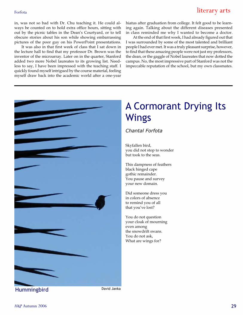
29H&P Autumn 2006
in, was not so bad with Dr. Chu teaching it. He could al-ways be counted on to hold extra office hours, sitting with out by the picnic tables in the Dean’s Courtyard, or to tell obscure stories about his son while showing embarrassing pictures of the poor guy on his PowerPoint presentations.
It was also in that first week of class that I sat down in the lecture hall to find that my professor Dr. Brown was the inventor of the microarray. Later on in the quarter, Stanford added two more Nobel laureates to its growing list. Need-less to say, I have been impressed with the teaching staff. I quickly found myself intrigued by the course material, feeling myself draw back into the academic world after a one-year
hiatus after graduation from college. It felt good to be learn-ing again. Talking about the different diseases presented in class reminded me why I wanted to become a doctor.
At the end of that first week, I had already figured out that I was surrounded by some of the most talented and brilliant people I had ever met. It was a truly pleasant surprise, however, to find that these amazing people were not just my professors, the dean, or the gaggle of Nobel laureates that now dotted the campus. No, the most impressive part of Stanford was not the impeccable reputation of the school, but my own classmates.
A Cormorant Drying Its WingsChantal Forfota
Skyfallen bird,you did not stop to wonderbut took to the seas.
This dampness of feathersblack hinged capegothic remainder.You pause and surveyyour new domain.
Did someone dress youin colors of absenceto remind you of all that you’ve lost?
You do not questionyour cloak of mourningeven among the snowdrift swans.You do not ask,What are wings for?
Hummingbird
literary artsForfota
David Janka

H&P Autumn 200630
A. Fire, S.Q. Xu, M.K. Montgomery, S.A. Kostas, S. E. Driv-er, C.C. Mello: Potent and specific genetic interference by double-stranded RNA in Caenorhabditis elegans, Nature 1998; 391: 806-811.
Using the nematode Caenorhabditis elegans as an animal model, Andrew Fire and Craig Mello of the Carnegie Institu-tion in Washington, D.C., tested the experimental introduction of RNA fragments on gene expression. Making use of genes known to generate unambiguous phenotypes when silenced, they introduced fragments of single and double-stranded RNA encoding segments of these genes. Single-stranded RNA proved unpredictable in its effects on gene expression. Combining the strands of RNA into a double-stranded helix, however, obtained predictable results and uncovered a means of robustly and specifically dampening gene expression.
This seminal paper, cited over three thousand times since its publication, laid the groundwork for subsequent RNAi findings, including the rapid identification of the cellular machinery responsible for the RNAi-induced gene silencing. Specifically, RNAi is an RNA-dependent process that utilizes several endogenous proteins to silence a targeted gene. The ribonuclease protein Dicer binds and cleaves double-stranded RNA molecules, leaving short RNA fragments with overhang-ing, unpaired bases on each end. These fragments, called small interfering RNAs (siRNAs) are bound by a protein complex known as the RNA-induced silencing complex (RISC). Using inserted RNA as a guide, RISC degrades corresponding mRNA prior to its translation to protein, thus silencing target genes.
In recent years, the discovery of hundreds of genes encod-ing small RNA molecules called microRNAs has provided this gene-silencing mechanism an unexpected role as a natural and essential element of human growth and development.
MicroRNA molecules form a double-stranded RNA frag-ment (much like that inserted into C. elegans by Fire and Mello) that activates RNA interference machinery to block mRNA translation, thus silencing expression of a particular gene. The influence these new molecules hold over gene expression is staggering, with recent studies estimating that they regulate expression of approximately 25% of our genes.
In the realm of genetic therapy, the ability of RNAi to dampen specific gene expression holds great therapeutic potential. Treatment of macular degeneration and respira-tory syncytial virus using RNAi has recently reached clinical trials. Animal studies show RNAi capable of silencing a gene known to cause high blood cholesterol levels in animals, while in vitro investigation has effectively used RNAi to inhibit genes of hepatitis A and B, influenza, measles, and cancer cells. Potential treatments for neurodegenerative diseases have also been proposed, with particular attention focusing on trinucleotide repeat disorders such as Huntington’s disease.
However, with all of the promise of RNAi research, there are several challenges that need to be addressed before RNAi can earn a position in our therapeautic armamentarium. First, there has been some concern raised regarding the safety of RNA interference and its potential for “off-target” gene dampening effects. A computational genomics study estimated that the error rate of off-target interactions by RNAi-based drugs is about 10%. In addition, there is the question of the best mode of delivery as well as the need for techniques to improve cell type specificity. Despite these concerns, there is no doubt that Fire and Mello have not only identified a fundamental mechanism of gene regulation but have also come upon a potentially powerful therapeutic agent.
In the fall of 2006, two faculty members of Stanford University School of Medicine received No-bel Prizes in recognition of their exemplary research. Andrew Fire received the prize in Physiology or Medicine for his work on RNA interference (RNAi), while
Maneesh Singh
science Singh

3�H&P Autumn 2006
P. Cramer, D.A. Bushnell, R.D. Kornberg. Structural Basis of Transcription: RNA Polymerase II at 2.8 Ångstrom Resolution, Science 2001; 292: 1863.
A.L. Gnatt, P. Cramer, J. Fu, D.A. Bushnell, R.D. Kornberg. Structural Basis of Transcription: An RNA Polymerase II Elongation Complex at 3.3 Å Resolution, Science 2001; 292: 1876.
For many years the complexity, low cell concentra-tion, and activity of RNA polymerase made its structure particularly elusive to x-ray crystallography and electron microscopy. In 2001 Dr. Roger Kornberg and colleagues published two papers in Science that finally captured the structure of this important molecule in amazing detail. By displaying RNA polymerase in functionally relevant com-plexes, these papers have enabled a dynamic understanding of the role of RNA polymerase in the transcription process.
To fully appreciate the complexity behind developing these images, it is important to understand a little about conventional x-ray crystallography. First, the complex of interest must be crystallized to create an object sizeable enough to scatter x-rays and thus produce an image. This step required Kornberg to develop novel techniques includ-ing a method to form two-dimensional protein crystals on a lipid surface. Once prepared, the crystals are harvested and mounted on a diffractometer for x-ray analysis. The x-rays are diffracted by interactions with electrons in the crystal, and this diffraction pattern is recorded by detectors. Successive images are recorded as the crystal is rotated to produce a computer-generated three-dimensional image.
Kornberg’s first prize-winning Science paper describes crystal forms of yeast RNA polymerase II in both an opened
and partially closed state. These images, produced at high resolution (3.1 and 2.8 angstroms, respectively), provided enough information for Kornberg and colleagues to propose a convincing, detailed model for many steps of transcription. To this end they identified numerous key functional regions of the enzyme and named them with respect to their role in tran-scription: lid, wall, zipper, clamp, rudder, funnel, and bridge.
The second paper displays the structure of RNA poly-merase while actively transcribing DNA. With remarkable clarity, this image depicts RNA polymerase enclosing the constructed DNA-RNA hybrid, the synthesized RNA, and the unwound region of DNA template. This was accomplished by pausing transcribing polymerase molecules at a predetermined template site by withholding one of the four ribonucleases. This structure also displays the active site where the nucleoside triphosphate ester bond is broken prior to mRNA elongation.
Since the publication of these prize-winning papers in 2001, Kornberg has reported several new structures of RNA polymerase with transcription factor TFIIB, general transcrip-tion factors, and Mediator - an essential component of gene regulation and transcription. Importantly, RNA polymerase II in yeast is remarkably similar to the corresponding polymerase in humans. Thus Kornberg’s discoveries are actually quite rel-evant to our understanding of human pathologies. His insights into the RNA machine may soon allow for us to develop thera-pies for disturbances in transcriptional regulation that lead to cancer, inflammation, heart disease, and metabolic disease.
Roger Kornberg received the prize in Chemistry for his re-search on the molecular basis of eukaryotic transcription. H&P returns to the seminal research papers of Fire and Kornberg to explain the significance of their key discoveries.
scienceSingh

H&PVolume ��, Number � Autumn 2006
The Stanford Medical Student Clinical Journal