the prevalence of retinopathy and associated medical risk factors in type i (insulin-dependent)...
TRANSCRIPT

journal o$ Internal Medicine 1989: 226: 47-52
The prevalence of retinopathy and associated medical risk factors in type I (insulin-dependent) diabetes mellitus
E. AGARDH. 0. TORFFVIT* & C.-D. AGARDH* From the Lkpartmenfs OJ Ophthalmology and 'Internal Medicine. University Hospital. Lund. Sweden
Abstract. Agardh E. Torffvit 0. Agardh C-D (Departments of Ophthalmology and Internal Medicine, University Hospital, Lund. Sweden). The prevalence of retinopathy and associated medical risk factors in type I (insulin-dependent) diabetes mellitus. journal o$ lnternal Medicine 1989: 226: 47-52.
The prevalence of diabetic retinopathy and the associated medical risk factors. such as age at onset and duration of diabetes, metabolic control. blood pressure, albumin clearance and serum creatinine. were studied in 501 patients with type I diabetes mellitus. The prevalence of retinopathy. characterized as simplex, maculopathy. preproliferative. and proliferative, was 60.5 %. Patients with retinopathy were younger at the onset of diabetes, and had a longer duration of disease. In patients with more than 10 years of diabetes, proliferative retinopathy was more frequent if onset was before they were 1 5 years old. despite the fact that the duration of diabetes did not differ. Patients with severe retinopathy had worse metabolic control, and were more frequently treated for hypertension. In addition. the systolic blood pressure was elevated in all groups of patients with any type of retinopathy. whereas the diastolic blood pressure was elevated only in patients with more severe forms. Patients with severe retinopathy also had higher levels of albumin clearance.
Keywords: albumin clearance, diabetes mellitus, duration, hypertension, metabolic control, retinopathy. type 1.
Introduction
Diabetic retinopathy is the major cause of severe vision disability among young and middle-aged people [l]. In most patients, the severe progress of retinopathy can be prevented with adequate treat- ment [2]. Since the cause of diabetic retinopathy is unknown, it is of prime importance to identify the patients with a higher risk.
The aim of the present study was to characterize the level of retinopathy in a homogenous population of patients with type I diabetes, and to correlate this to various possible medical risk factors, such as age, age at onset, duration of diabetes, metabolic control, blood pressure, and clinical signs of diabetic nephro- pathy.
Abbreviations: N = normal, S = simplex. M = maculopathy, PP = preproliferative. IRMA = intraretinal abnormalities. P = prolifera- tive, SBP = systolic blood pressure. DBP = diastolic blood pressure. UK = unknown retinopathy.
Study population and methods
Patients
All adult patients ( n = 501) with type I diabetes (onset before 30 years of age) regularly attending the Department of Internal Medicine during a 2-year period were included in the study. Their age. age at onset, duration of diabetes, insulin dosage and body weight were registered. The duration of diabetes was calculated from the year of diagnosis until the actual visit. The metabolic control was measured with HbAl c. Clinical signs of diabetic nephropathy were examined with albumin clearance, urinary albumin and serum creatinine.
Most patients were also regularly examined at the Department of Ophthalmology, at least once a year. The retinal examination was performed through a dilated pupil. either by ophthalmoscopy or fundus photography. The fundus photography was per-
47

48 E. AGARDH et a l .
formed with a fundus camera angle of 50' visualizing three fields in each eye. The patients were examined within 1 year of the other measurements. In patients with an inveterated proliferative retinopathy, an examination older than 1 year was accepted as adequate. In 92.2 % of the patients it was possible to categorize the stage of retinopathy. The remaining 7.8% were not examined because they had moved, died, were ophthalmologically controlled elsewhere, or did not want to be examined.
Definition of retinopathy
The patients were classified into five groups depend- ing on the degree of retinopathy. The most seriously affected eye was used for the evaluation.
When there were no signs of retinopathy, the eyes were classified as normal (N). Eyes with single or multiple microaneurysms. haemorrhages, hard exudates outside the macular region, or with single ischaemic zones, were classified as simplex (S). Leakage in the macular region, which threat- ened or had caused vision disability, classified the retinopathy as maculopathy (M). In 82.4%. the retinopathy was verified with fluorescein angiography. The retinopathy was classified as preprolifer- ative (PP) when multiple ischaemic zones and intraretinal abnormalities (IRMA) were pre- sent. In 85.2%. the degree of retinopathy was verified with fluorescein angiography. When elevated retinal vessels were evident, the retinopathy was classified as proliferative (P).
Analytical techniques
Glycosylated haemoglobin (HbAlc) levels were ana- lysed by ion-exchange chromatography using com- mercially available microcolumns (BIO-RAD, Richmond, CA, USA). The normal value is less than 6 %. The urinary albumin was measured with electro- immunoassay using human albumin (Kabi Vitrum, Stockholm. Sweden) as the standard. The detection limit was 12.5 mg I-'. Urine and serum creatinine levels were analysed by routine techniques. Albumin clearance was calculated as the ratio between the urine albumin clearance and the urine creatinine clearance x The normal value is less than 0.01 x 10⁻³.
Statistical methods
Student's t-test (two tailed) and the X2-test were used to test the significance.
Results The proportion of patients with different levels of retinopathy is presented in Table 1 , where the group of patients with unknown retinopathy (UK) is in- cluded as well. In 31.8% of the patients there were no signs of retinopathy. whereas 60.5% had retino- pathy at different grades. The remaining 7.8% were not examined.
Patients with simplex, preproliferative. and pro- liferative retinopathy were younger at the onset of diabetes than those without (Fig. 1) . Patients with diabetes for more than 10 years were divided into two groups: (a) age at onset was less than 1 5 years
Table 1. Patients with different levels of retinopathy and duration of diabetes ( Q 10 years or > 10 years)
Degree of retinopathy.
N s M PP P UK
Total % n
31.8 28.1 3.4 5.4 23.6 7.8 159 141 17 27 118 39
Duration < 10 years % 74.8 13.4 0 2.4 0 9.4 n 95 17 0 3 0 12
17.2 33.3 4.6 6.5 31.5 7.2 % n 64 124 17 24 118 27
Duration > 10 years
N = normal, S = simplex, M = maculopathy. PP = preproliferative. P = proliferative, UK = unknown.

RETINOPATHY A N D RISK FACTORS 49
20 1 T - e 0 >I - e 01
c 0 - 10
l3 a
N S M PP P Degree of retinopathy
Fig. 1. Age at the onset of diabetes (meankSEM) for various degrees of retinopathy: N = no retinopathy. S = simplex, M =
maculopathy. PP = preproliferative retinopathy. P =
proliferative retinopathy. For P vs. N. S and M. and also PP vs. N P < 0 .001 : for S vs. N P < 0.01 : and for PP vs. S and M P < 0.05.
30
- 20 e 0 - c ._ - e 0
10
T T
N S M PP P
Degree of retinopathy
Fig. 2. Duration of diabetes (rneankSEM) for various degrees of retinopathy (abbreviations are shown in Fig. 1 ). For P vs. N and S. and also PP. M and S vs. N P < 0 .001 : for P us. PP P < 0.01 : and for M vs. S P < 0 . 0 5 .
I I / I / / /
\ \
\ \
1 1 I N S M PP P
Degree of retinopathy
Fig. 3. Haemoglobin Alc (HbAlc) levels (mean+SEM) for various degrees of retinopathy (abbreviations as shown in Fig. 1 ). For P vs. N P < 0.001 : and for PP and M vs. N P < 0 . 0 5 .
(n = 213), and (b) at or greater than (n = 159) 15 years of age. Proliferative retinopathy was more frequent when age at onset was before 15 years (85 out of 213 patients: 39.4%) than after (33 out of 159 patients: 20.7%; P < 0 . 0 0 1 , ~ ~ ) . This difference was seen despite the duration of disease being the same (2 7.6 f 0.9 compared to 29.0 f 1.5 years : mean f SEM).
Patients with any type of retinopathy had a longer duration of diabetes (Fig. 2) than patients without. The metabolic control. as assessed by HbAlc levels, was poorer in patients with maculopathy, preprolifer- ative, and proliferative retinopathy than in patients without retinopathy (Fig. 3).
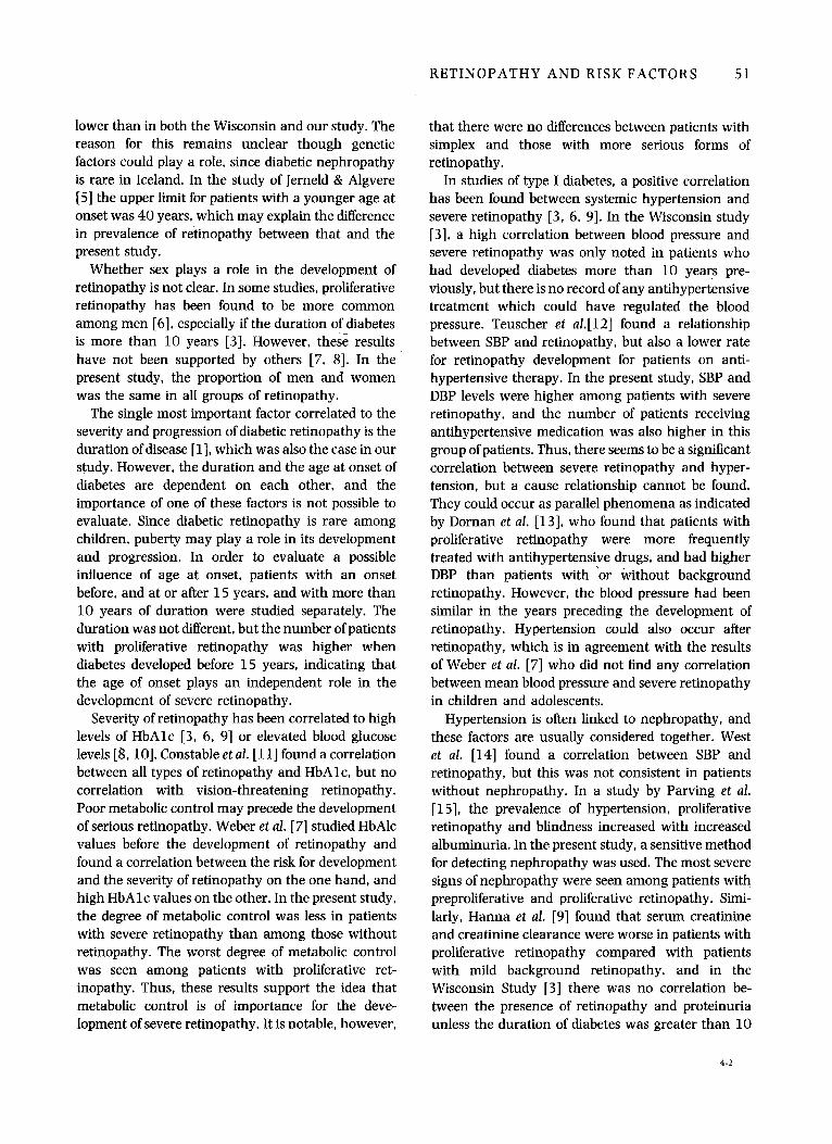
In comparison to patients with no retinopathy. those with proliferative and preproliferative retino- pathy had antihypertensive treatment more fre- quently. The number of patients with antihyper- tensive treatment was highest in the group with proliferative retinopathy (50 out of 11 8 patients: 43 %). In the other groups the corresponding figures were: for patients without retinopathy 7 out of 159 patients (4%). simplex 12 out of 141 patients (9%). maculopathy 3 out of 1 7 patients ( 18 %), preprolifer- ative retinopathy 7 out of 27 patients (26%):
4 I M H 126

50 E. A G A R D H e t a / .
N S M PP P Degree o f retinopathy
Fig. 4. Systolic blood pressure (SBP) and diastolic blood pressure (DBP. mean k SEM) for various degrees of retinopathy (abbreviations as shown in Fig. 1 ) . Top: for P vs. N and S. and also M and S vs. N P < 0.001 : for PP vs. N and M vs. S P < 0.01. Bottom: for P vs. N and S P < 0.001: and for PP and M vs. N P < 0.05.
P < 0.001 for P vs N and S. P < 0.01 for PP vs. N, and P < 0.05 for PP vs. S. xz. In addition, patients with any type of retinopathy had a higher systolic blood pressure (SBP), and patients with maculopathy, preproliferative. and proliferative retinopathy had higher diastolic blood pressure (DBP) levels than patients without retinopathy (Fig. 4).
There were no clinical signs of diabetic nephro- pathy in patients with no and simplex retinopathy, whereas the patients with maculopathy. preprolifer- ative. and proliferative retinopathy had elevated albumin clearance levels (Fig. 5). Patients with preproliferative and proliferative retinopathy had elevated levels of serum creatinine (mean f SEM : no retinopathy = 76.4 f 0.9 pmol I-'. simplex = 76.7 f 1.2 pmol I-'. maculopathy = 95.7f l l .7pmoll~ ' , preproliferative = 87.7 f 5.4pnol I-', proliferative = 97.2f4.8pmol I-').
The proportion of men and women was the same in all groups of retinopathy.
N S M PP P Degree of retinopathy
Fig. 5. The albumin clearance ratio (meankSEM) (abbreviations as shown in Fig. 1). For P vs. N and S P < 0.001: and for PP vs. N and S P < 0.01.
Patients with unclassified degree of retinopathy
As for patients with an unclassified degree of retino- pathy, the figures for the different parameters are given for comparison (mean fSEM). The age at onset of diabetes = 17.4 f 1.2 years, duration =
16.5f1.8 years, HbAlc = 8.2f0.3%. SBP = 130.1 +2.6mmHg, DBP = 78.7f 1.2mmHg, albu- min clearance 0.03 f O . 0 1 x u-albumin =
94f4.2 mg I-'. serum creatinine = 79.8f3.2 pmol I-'.
Discussion
The University Hospital of Lund serves a population of 150000 people. With few exceptions, all adult patients with type I diabetes are regularly controlled at the Department of Internal Medicine, and are included in the present study on the prevalence of diabetic retinopathy and associated medical risk factors. The prevalence of diabetic retinopathy was 60.5%. which is slightly less than in the Wisconsin Study [3]. Different grading protocols and the ex- clusion of non-participants (7.8%) in the present study could explain the different results. In a study of type I diabetes on Iceland, Danielsen et al. [4] describe retinal lesions in 3 3.5 %. This figure is considerably
RETINOPATHY AND RISK FACTORS 51
lower than in both the Wisconsin and our study. The reason for this remains unclear though genetic factors could play a role, since diabetic nephropathy is rare in Iceland. In the study of Jerneld & Algvere [5] the upper limit for patients with a younger age at onset was 40 years, which may explain the difference in prevalence of retinopathy between that and the present study.
Whether sex plays a role in the development of retinopathy is not clear. In some studies, proliferative retinopathy has been found to be more common among men [6], especially if the duration of diabetes is more than 10 years [3]. However, these results have not been supported by others [7, 81. In the present study, the proportion of men and women was the same in all groups of retinopathy.
The single most important factor correlated to the severity and progression of diabetic retinopathy is the duration of disease [l], which was also the case in our study. However, the duration and the age at onset of diabetes are dependent on each other, and the importance of one of these factors is not possible to evaluate. Since diabetic retinopathy is rare among children, puberty may play a role in its development and progression. In order to evaluate a possible influence of age at onset, patients with an onset before, and at or after 15 years, and with more than 10 years of duration were studied separately. The duration was not different, but the number of patients with proliferative retinopathy was higher when diabetes developed before 15 years, indicating that the age of onset plays an independent role in the development of severe retinopathy.
Severity of retinopathy has been correlated to high levels of HbAlc [3, 6, 91 or elevated blood glucose levels [8, 101. Constable et al. [ l l ] found a correlation between all types of retinopathy and HbAlc, but no correlation with vision-threatening retinopathy. Poor metabolic control may precede the development of serious retinopathy. Weber et al. [7] studied HbAlc values before the development of retinopathy and found a correlation between the risk for development and the severity of retinopathy on the one hand, and high HbAlc values on the other. In the present study, the degree of metabolic control was less in patients with severe retinopathy than among those without retinopathy. The worst degree of metabolic control was seen among patients with proliferative ret- inopathy. Thus, these results support the idea that metabolic control is of importance for the deve- lopment of severe retinopathy. It is notable, however,
that there were no differences between patients with simplex and those with more serious forms of retinopathy.
In studies of type I diabetes, a positive correlation has been found between systemic hypertension and severe retinopathy [3, 6, 91. In the Wisconsin study [3], a high correlation between blood pressure and severe retinopathy was only noted in patients who had developed diabetes more than 10 years pre- viously, but there is no record of any antihypertensive treatment which could have regulated the blood pressure. Teuscher et al.[12] found a relationship between SBP and retinopathy, but also a lower rate for retinopathy development for patients on anti- hypertensive therapy. In the present study, SBP and DBP levels were higher among patients with severe retinopathy, and the number of patients receiving antihypertensive medication was also higher in this group of patients. Thus, there seems to be a significant correlation between severe retinopathy and hyper- tension, but a cause relationship cannot be found. They could occur as parallel phenomena as indicated by Dornan et al. [13], who found that patients with proliferative retinopathy were more frequently treated with antihypertensive drugs, and had higher DBP than patients with ‘or without background retinopathy. However, the blood pressure had been similar in the years preceding the development of retinopathy. Hypertension could also occur after retinopathy, which is in agreement with the results of Weber et al. [7] who did not find any correlation between mean blood pressure and severe retinopathy in children and adolescents.
Hypertension is often linked to nephropathy, and these factors are usually considered together. West et al. [14] found a correlation between SBP and retinopathy, but this was not consistent in patients without nephropathy. In a study by Parving et al. [ 1 51, the prevalence of hypertension, proliferative retinopathy and blindness increased with increased albuminuria. In the present study, a sensitive method for detecting nephropathy was used. The most severe signs of nephropathy were seen among patients with preproliferative and proliferative retinopathy. Simi- larly, Hanna et al. [9] found that serum creatinine and creatinine clearance were worse in patients with proliferative retinopathy compared with patients with mild background retinopathy, and in the Wisconsin Study [3] there was no correlation be- tween the presence of retinopathy and proteinuria unless the duration of diabetes was greater than 10
4-2

52 E. AGARDH e t a l .
years. In the present study, patients with no or simplex retinopathy had a normal albumin clear- ance, indicating that nephropathy does not precede retinopathy. This is in contrast to the results of Vigstrup & Mogensen [ 161, who found microalbumin- uria to be a strong predictor for later development of proliferative retinopathy. However, these results are not supported by a different study [17] where 3 5 % of patients with preproliferative or proliferative retino- pathy had normal values of albumin clearance. Similarly, Feldman et al. [18] found that only 33 % of diabetic patients with proliferative retinopathy had evidence of renal disease.
In summary, with the exception of maculopathy, patients with retinopathy had an earlier onset of diabetes than those without. Proliferative retino- pathy was more frequent when the age at the onset of diabetes was younger than 15 years, despite the fact that the duration of disease was the same. In all groups with retinopathy, the duration of diabetes was longer. The metabolic control was poorer in patients with more severe forms of retinopathy. In addition to the increased number of patients treated for systemic hypertension, the SBP was elevated in patients with any type of retinopathy, whereas the DBP was only elevated in patients with the more severe forms. There were no clinical signs of nephro- pathy in patients with no or simplex retinopathy, whereas all other groups had an increased albumin clearance. Thus, diabetic patients with more severe forms of retinopathy also often have other diabetic Fomplications. However, the time course for the development of the different complications is at present impossible to evaluate. To that end, patients with milder forms of diabetic retinopathy will have to be studied prospectively.
Acknowledgements This study was supported by grants from the Swedish Diabetes Association and the Malmo Diabetes Association.
References 1 Kohner EM. Recent advances in diabetic retinopathy. In:
Alberti KGMM, Krall LP, eds. The Diabetes Annual, Vol. 1. Amsterdam: Elsevier Science Publishers BV, 1985 : 257-87.
2 Early Treatment Diabetic Retinopathy Study Research Group. Photocoagulation for diabetic macular edema. Arch Ophthalmol 1985; 103: 1796-806.
Klein R, Klein BEK, Scot EM. Davis MD, DeMets DL. The Wisconsin epidemiologic study of diabetic retinopathy 11. Prevalence and risk of diabetic retinopathy when age at diagnosis less that 30 years. Arch Ophthalmol 1984; 102:
Danielsen R. Jonasson F, Helgason T. Prevalence of retino- pathy and proteinuria in type I diabetics in Iceland. Acta Med
Jerneld B, Algvere P. Relationship of duration and onset of diabetes to prevalence of diabetic retinopathy. Am J Ophthalmol
Bodansky HJ, Cudworth AG, Drury PL, Kohner EM. Risk factors associated with severe proliferative retinopathy in insulin-dependent diabetes mellitus. Diabetes Care 1982 : 5:
Weber B, Burger W. Hartmann R. Hovener G, Malchus R. Oberdisse U. Risk factors for the development of retinopathy in children and adolescents with type I (insulin-dependent) diabetes mellitus. Diabetologia 1986: 29: 23-9. Krolewski AS, Warram JH, Rand LI. Christlieb AR. Busik EJ, Kahn CR. Risk of proliferative diabetic retinopathy in juvenile- onset type I diabetes: a 40-year follow-up study. Diabetes Care 1986; 9: 443-52. Hanna AM, Roy M, Zinman B et al. An evaluation of factors associated with proliferative diabetic retinopathy. Clinical and Investigative Medicine 1985 8: 109-16.
520-6.
Stand 1982; 212: 277-80.
1986; 102: 431-7.
97-100.
10 Rand k, Krolewski AS, Aiello LM. Warram JH, Baker RS, Maki T. Multiple factors in the prediction of risk of proliferative diabetic retinopathy. N Engl J Med 1985; 313: 1433-8.
11 Constable IJ, Knuiman MW, Welborn TA et al. Assessing the risk of diabetic retinopathy. Am J Ophthalmol 1984; 97:
12 Teuscher A, Schnell H, Wilson PWF. Incidence of diabetic retinopathy and relationship to baseline plasma glucose and blood pressure. Diabetes Care 1988: 11: 246-51.
13 Dornan T. Mann JI, Turner R. Factors protective against retinopathy in insulin-dependent diabetics free of retinopathy for 30 years. Br Med J 1982; 285: 1073-7.
14 West KM, Ahuja MMS, Bennett PH et al. Interrelationships of microangiopathy. plasma glucose and other risk factors in 3 583 diabetic patients : a multinational study. Diabetologia
15 Parving H-H, Hommel E, Mathiesen E ef al. Prevalence of microalbuminuria. arterial hypertension, retinopathy. and nephropathy in patients with insulin dependent diabetes. Br Med J 1988; 296: 156-60.
16 Vigstrup J, Mogensen CE. Proliferathe diabetic retinopathy: at risk patients identiied by early detection of microalbuminuria. Acta Ophthalmol 1985; 63: 530-4.
17 Agardh E. Tallroth G, Bauer B, Cavallin-Sjoberg U, Agardh C-D. Retinopathy and nephropathy in insulin-dependent diabetics : an inconsistent relationship? Diabetic Med 1987: 4: 248-50.
18 Feldman JN, Hirsch SR, Beyer BS, James WA, L’Esperance FA, Friedman FA. Prevalence of diabetic nephropathy at time of treatment for diabetic retinopathy. In: Friedman EH. L’hperance FA, eds. Diabetic Renal-Retinal Syndrome. London : Grune & Stratton Ltd. 1982; pp 9-22.
53-61.
1982; 22: 412-20.
Received 21 December 1988, accepted 24 January 1989.
Correspondence : Carl-David Agardh, MD, Department of Internal Medicine, University Hospital, S-22 1 85 Lund, Sweden.