the pathophysiologic role of monocytes and ...while-science-sleeps.com/pdf/519.pdfthe...
TRANSCRIPT
Mfi
*i
M
M
1
●●●
0d
ARTICLE IN PRESS
The Pathophysiologic Role of Monocytes andMacrophages in Systemic Lupus Erythematosus: A Reappraisal
Christina G. Katsiari, MD, PhD,* Stamatis-Nick C. Liossis,† andPetros P. Sfikakis‡
Objectives: To review current developments, regarding the pathophysiologic role of monocytesand macrophages in systemic lupus erythematosus (SLE).Methods: We searched Medline for articles written in the English language using the followingterms: monocyte(s) or macrophage(s) and lupus. Although our search spanned the years 1971 to2008, the majority of the short-listed articles belonged to the period 2000 to 2008. Publishedliterature on phenotypic and functional properties of monocytes/macrophages (Mo/M�) in SLEwas reviewed. References from identified articles were also selected. Currently available experi-mental data and their relevance to the pathogenesis of SLE are critically discussed.Results: It has traditionally been held that impaired phagocytosis by monocytes and macrophagesin SLE allows for the accumulation of apoptotic debris leading to a sequel of autoimmunephenomena. Recent paradigms derived from animal models of systemic autoimmunity, however,has broadened our understanding regarding the possible pathophysiologic roles of Mo/M� inSLE. Data derived from studies in patients with SLE show multiple aberrations in activation statusand secretory functions of circulating and tissue-infiltrating Mo/M�. Such aberrations may beassociated with dysregulation of T-cell function and autoantibody production in SLE. Moreover,emerging evidence suggests that phagocytic capacity and antigen-presenting properties of Mo/M�are enhanced in some patients with SLE.Conclusions: While defective phagocytosis represents a distinctive feature of monocyte function insome patients with SLE, aberrant activation of the Mo/M� system may be a more appropriateconcept to encompass the broad spectrum of Mo/M� disorders in SLE. Aberrant function of lupusMo/M� appears to play a dynamic role in the initiation and perpetuation of the systemic auto-immune response and organ damage. Delineation of the altered biology of lupus Mo/M� couldprovide possible future therapeutic targets for patients with SLE.© 2009 Elsevier Inc. All rights reserved. Semin Arthritis Rheum xx:xxxKeywords: monocytes, macrophages, systemic lupus erythematosus, pathogenesis, phagocytosis, antigenpresentation
emstsmca(e
p
onocytes/macrophages (Mo/M�) are versatilecells aiming to defend, regulate inflammation,and induce immunity. Monocytes develop
rom pluripotent stem cells in the bone marrow under thenfluence of specific growth factors. Following the differ-
Clinical and Research Associate, 1st Department of Propedeutic and Internal Med-cine, University of Athens Medical School, Athens, Greece.
†Assistant Professor of Internal Medicine/Rheumatology, University of Patrasedical School, Athens, Greece.‡Associate Professor of Internal Medicine/Rheumatology University of Athensedical School, Athens, Greece.
MAddress reprint requests to Christina Katsiari, MD, PhD, 102 Agias Lavras Str,
5773, Athens, Greece. E-mail: [email protected].
049-0172/09/$-see front matter © 2009 Elsevier Inc. All rights reserved.oi:10.1016/j.semarthrit.2008.11.002
ntiation process that lasts less than 24 hours, matureonocytes leave the bone marrow and enter the blood-
tream as quiescent cells. Circulating monocytes differen-iate further into resident tissue macrophages and acquirepecialized phenotypes and functions depending on the localicroenvironment. Furthermore, blood monocytes are re-
ruited to sites of inflammation where they become activatednd evolve into cells that express the macrophage phenotype1). Experimental evidence suggests that monocytes differ-ntiate also into dendritic cells (DCs) in vivo (2,3).
The hallmark function of the Mo/M� system ishagocytosis and subsequent antigen presentation (1).
o/M� recognize and remove pathogens as well as1
sattgvfrMatafi
pr
ospodaseris
FC(echmwpecABlirmacT
2 Pathophysiologic role of monocytes and macrophages in SLEARTICLE IN PRESS
enescent, dead, or damaged host cells. Phagocytosis istriggered process requiring the activation of recep-
ors, which in turn transmit signals to the cell interioro initiate the response. Following digestion of the tar-et, Mo/M� present target-derived antigen(s) to rele-ant T-cells, resulting in helper and/or effector T-cellunctions. Consequently, Mo/M� possess a centralole in initiating the immune response. In addition,
o/M� have well-developed secretory functions and aren important source for a variety of cytokines, whereas morehan 100 biologically active substances have been describeds Mo/M� products. Cytokine receptors found on the sur-ace membrane of Mo/M� allow them to become involvedn indirect complex interactions with different cell type
PHAGOCYTOSISPHAGOCYTOSIS
ANTIGEN ANTIGEN
ACTIVATIONACTIVATION
T cell
M
surface molecules secretory productsIL1, IL6, IL10, IL12, IL23 IL1, IL6, IL10, IL12, IL23
neopterinneopterinnitric oxidenitric oxide
sCD14sCD14↑↑ MHCMHC--IIII
AC
CD14
“phagocyticsynapse”
CD16-~90%
CCR2CD11b
TNFTNFαα
BLySBLyS
↑↑ CD68CD68
↑↑ CD80/86CD80/86
↑↑ SinglecSinglec--11
MCPMCP--11
↑↑ ICAMICAM--11 ↑↑ CD40CD40
↑↑ CD44CD44
TGFbTGFb
Mer
gas-6PS
PS-R
FcγR
CRP
Ox-PL
CD14
ACAMPs
CD91
calreticulin C1q
CD44
αvβ3/5
CD36TSP1
TSP1-bs
TFTF
TCR
CD40
CD28
PHAGOCYTOSISPHAGOCYTOSIS
ANTIGEN ANTIGEN
ACTIVATIONACTIVATION
T cell
MM
surface molecules secretory productsIL1, IL6, IL10, IL12, IL23 IL1, IL6, IL10, IL12, IL23
neopterinneopterinnitric oxidenitric oxide
sCD14sCD14↑↑ MHCMHC--IIII
ACAC
CD14
“phagocyticsynapse”
“phagocyticsynapse”
CD16-CD16-~90%
CCR2CD11b
TNFTNFαα
BLySBLyS
↑↑ CD68CD68
↑↑ CD80/86CD80/86
↑↑ SinglecSinglec--11
MCPMCP--11
↑↑ ICAMICAM--11 ↑↑ CD40CD40
↑↑ CD44CD44
TGFbTGFb
Mer
gas-6PS
PS-R
FcγR
CRP
Ox-PL
CD14
ACAMPs
CD91
calreticulin C1q
CD44
αvβ3/5
CD36TSP1
TSP1-bs
TFTF
TCR
CD40
CD28
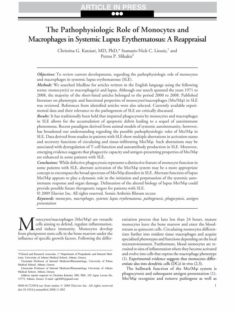
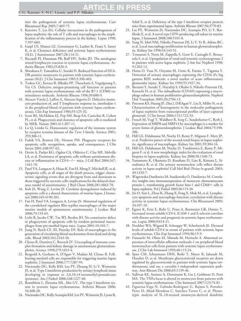
igure 1 Synopsis of monocytes characteristics. CirculatingD14 (LPS receptor) and CD16 (Fc�R-III). Approximately
CD142�/CD16�) and the remaining 10% express CD16xpressed on monocytes present a preferential expression beells indicating distinct functional properties. Because, duringigh levels of TNF� and low levels of IL-10, they are also conocytes results in the upregulation of several surface molehich participate in various functions such as chemotaxis (hagocytosis (lower left panel), antigen presentation andffector functions (lower right panel), and differentiation to mytokines enhance (in red) or inhibit (in green) the depictedbbreviations: �v�3/5, vitronectin receptor integrins; AC, apolyS, B-lymphocyte stimulator; C1q, 1st component of comp
igand; CRP, C-reactive protein; DC, dendritic cell; Fc�R, Fcntercellular adhesion molecule 1; IL, interleukin; MCP-1,eproductive tyrosine kinase; mGCR, membrane glucocorticoonocyte(s); M�, macrophage(s); Ox-PL, oxidized phosph
cid-binding Ig-like lectin 1; sCD14, soluble CD14; TCR, T-cell; TF, tissue factor; TNF�, tumor necrosis factor �; Treg, T-
SP1-bs, TSP-1 binding site. (Color version of figure is available onroducts. The main characteristics of monocytes are summa-ized in Figure 1.
Numerous studies have improved our understandingf the diverse components of immune dysregulation inystemic lupus erythematosus (SLE); however, the patho-hysiologic role(s) of Mo/M� remains unclear (4). The-ries formulated in the 1980s propose that lupus Mo/M�isplay defective phagocytic function, thus enabling theberrant accumulation of apoptotic debris leading to aequel of autoimmune phenomena. Recent studies, how-ver, exploring mainly lupus nephritis suggest an activeole of Mo/M� in mediating tissue inflammation andnjury (5), broadening our understanding regarding thepectrum of Mo/M� aberrant function in SLE.
T CELL MODULATIONT CELL MODULATION
ENTATIONENTATION
DIFFERENTIATION IN TISSUESDIFFERENTIATION IN TISSUES
Mφ DC
IL-12IL-10
Th1Th1
Th2Th2
Th17Th17TregTreg
TGF-b
TregTreg--ppIL- 6 IL- 23
CD16+14+
~10%
cγR-I, IIMHC-II
CD86
IL-12
I
T CELL MODULATIONT CELL MODULATION
ENTATIONENTATION
DIFFERENTIATION IN TISSUESDIFFERENTIATION IN TISSUES
Mφ DC
IL-12IL-10
Th1Th1
Th2Th2
Th17Th17TregTreg
TGF-b
TregTreg--ppIL- 6 IL- 23
CD16+14+
~10%
cγR-I, IIMHC-II
CD86
IL-12
I
cytes are divided into 2 subsets based on the expression off monocytes express high levels of CD14 but not CD16
ower levels of CD14 (CD14�/CD16�). Surface moleculesCD142�/CD16� (red color) and CD14�/CD16� (blue color)
tions, CD14�/CD16� monocytes are increased and producepro-inflammatory monocytes (central panel). Activation ofand the secretion of multiple monokines (upper left panel),R2, MCP-1), adhesion (eg, ICAM-I), coagulation (eg, TF),ulation (lower central panel), modulation of lymphocyte
hages and DCs (upper right panel). In the lower right panel,ization of Th cells.cell; ACAMPs, apoptotic cell-associated molecular patterns;t; CCR2, chemokine (C-C motif) receptor 2; CD40L, CD40a receptor; gas-6, growth arrest-specific gene-6; ICAM-1,
cyte chemoattractant protein 1; Mer, myeloid epithelialptor; MHC-II, major histocompatibility complex class II; Mo,; PS, phosphatidylserine; PS-R, PS-receptor; Siglec-1, sialiceptor; TGFb, transforming growth factor beta; Th, T-helpertory cell; Treg-p, Treg-precursor; TSP-1, thrombospondin-1;
PRESPRES
o
++ CD
F
MCH-I
L CD40
CD86
PRESPRES
oo
++ CD
F
MCH-I
L CD40
CD86
mono90% oand ltweeninfecalledcules
eg, CCcostimacroppolar
ptoticlemengammmono
id receolipidsell recregula
line.)

ogasatppMt
M
Wlrrwtst2pap
R
TM
DocusaolwcS
ti
cda(koamt
LIL
NreMtDlalMracioatditfsMspo
nt
C.G. Katsiari, S.-N.C. Liossis, and P.P. Sfikakis 3ARTICLE IN PRESS
As recent advances have improved our understandingf the role of innate immunity in the expression and pro-ression of SLE, we present, in brief, data derived fromnimal studies regarding the central role of Mo/M� inystemic autoimmunity and we discuss several functionalspects of Mo/M� in patients with SLE. In interpretinghese data we propose that the role of Mo/M� in theathogenesis of SLE should reach beyond the “defectivehagocytosis” model and that aberrant activation ofo/M� may be a more appropriate concept underlying
he pathophysiologic role of these cells in SLE.
ETHODS
e searched Medline for articles written in the Englishanguage using the following terms: monocyte(s) or mac-ophage(s) and lupus. The abstracts were screened forelevance and the publications relating to Mo/M� in SLEere obtained. Additional references were identified from
he bibliographies of the retrieved reports. Although ourearch spanned the years 1971 to 2008, the majority ofhe short-listed articles belonged to the period 2000 to008. Published literature on phenotypic and functionalroperties of Mo/M� in SLE was reviewed. Currentlyvailable experimental data and their relevance to theathogenesis of SLE are critically discussed.
ESULTS
he Model of “Defective”onocyte Function in SLE
efective clearance represents the hallmark of the modelf “defective” monocyte function in SLE (6). Reducedlearance of apoptotic cells led to the hypothesis thatningested apoptotic cells might represent an importantource of autoantigens with the potential to trigger anutoimmune process such as the one seen in SLE. More-ver, defective clearance of immune complexes (IC) al-owing tissue deposition of IC represents a mechanism byhich tissue damage may occur in SLE. Data on phago-
ytosis and clearance by monocytes from patients withLE will be discussed in detail below.
Other studies have shown that monocytes from pa-ients with SLE when cultured with autologous T-cells
Table 1 Major Aspects of the “Defective Model” of Monoc
Defect Selecte
Decreased autologous MLR (7)Decreased accessory function for T-cell activation (8)Decreased of MCH II surface expression (8)Decreased expression of CD80 (9)Decreased production of IL-1b (10)Decreased phagocytosis (6,77,8
MLR: mixed lymphocyte reaction.
nduce a defective proliferative T-cell response (7). De- t
reased accessory functions for T-cell activation were alsoescribed and linked to decreased expression of HLA-DRntigens. Low levels of surface HLA-DR (8) and CD809) expression along with reduced production of interleu-in-1 (IL-1) (10) have been linked with a decreased abilityf lupus monocytes to process and present antigen as wells to provide appropriate costimulation to T-cells. Theajor aspects of the model of “defective” monocyte func-
ion in SLE are summarized in Table 1.
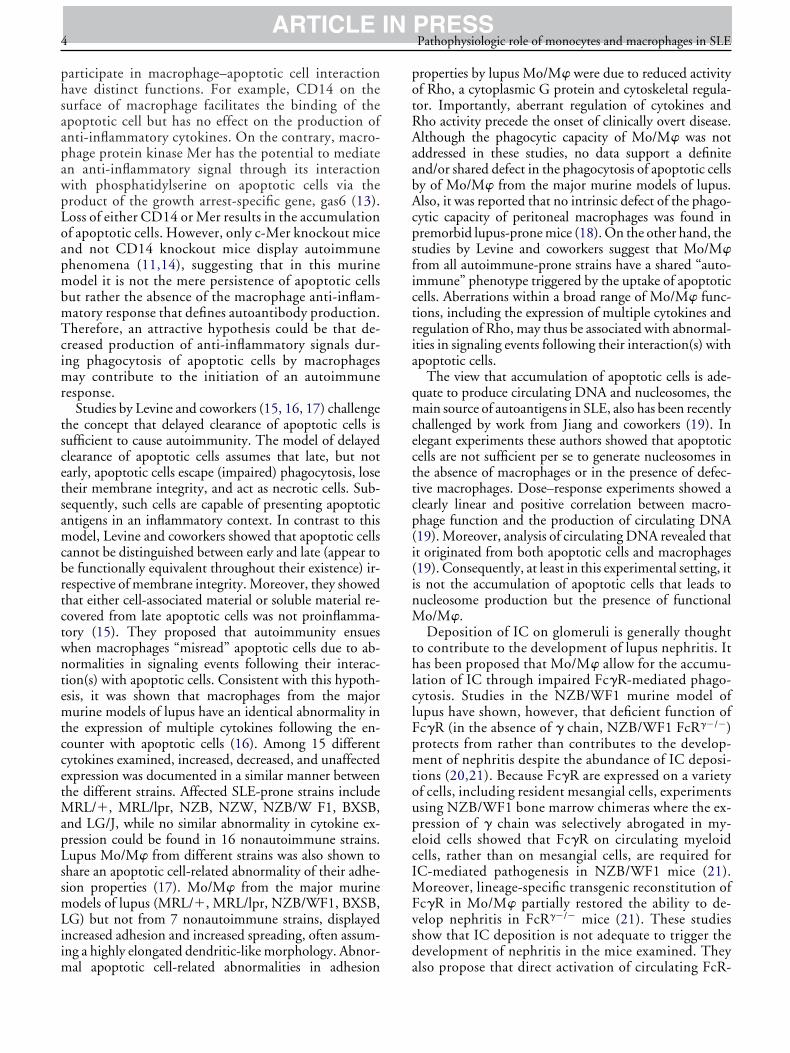
upus Mo/M� Beyond thempaired Phagocytosis Model:essons From Animal Models
ew insights on the possible mechanisms underlying theole of monocytes in the autoimmune process havemerged from studies in animal models. Tyro3, Axl, and
er comprise the TAM receptor family of receptor pro-ein tyrosine kinases and are expressed on Mo/M� andC, but not on lymphocytes or granulocytes. The bio-
ogic role of these receptors has been recently explored andppears to be essential in immunoregulation. In particu-ar, macrophages from mice lacking the cytoplasmic tail of
er (c-mer) are characterized by phagocytic deficiencyestricted to apoptotic cells. This abnormality leads tonti-dsDNA autoantibody production but without con-omitant significant polyclonal B-cell expansion or signif-cant renal pathology (11). Triple Tyro3/Axl/Mer knock-ut animals developed a lupus-like disease with overtrthritis, skin lesions, and nephritis along with produc-ion of multiple autoantibodies; their Mo/M� (and DC)isplay phenotypic and functional features that character-
ze activated cells (12). Although the expression and func-ion of the Tyro 3 receptor molecules in Mo/M� derivedrom patients with SLE has not been examined, thesetudies provide paradigms where aberrant activation of
o/M� (triple knockouts) promotes the expression ofystemic autoimmune disease, while isolated Mo/M�hagocytic deficiency of apoptotic cells (c-mer knock-uts) is not adequate to lead to overt disease.
Clearance of apoptotic cells is usually described asoninflammatory phagocytosis where macrophages ac-ively produce anti-inflammatory cytokines such as
nction in SLE
Functional Consequence
Impaired ability to activate T-cells
Inability to process and present antigen and to provideappropriate co-stimulation to lymphocytes
Impaired clearance leading to accumulation of apoptoticcells and immune complexes
yte Fu
d Ref.
2,92)
ransforming growth factor-beta (13). Molecules that
phsaapawpLoapmbmTcimr
tscetsamcbrtctwntemtccetMapLssmLiim
potRAaabAcpsfictria
qmcecttcp(i(inM
thlclFpmtoupecIMFvsd
4 Pathophysiologic role of monocytes and macrophages in SLEARTICLE IN PRESS
articipate in macrophage–apoptotic cell interactionave distinct functions. For example, CD14 on theurface of macrophage facilitates the binding of thepoptotic cell but has no effect on the production ofnti-inflammatory cytokines. On the contrary, macro-hage protein kinase Mer has the potential to mediaten anti-inflammatory signal through its interactionith phosphatidylserine on apoptotic cells via theroduct of the growth arrest-specific gene, gas6 (13).oss of either CD14 or Mer results in the accumulationf apoptotic cells. However, only c-Mer knockout micend not CD14 knockout mice display autoimmunehenomena (11,14), suggesting that in this murineodel it is not the mere persistence of apoptotic cells
ut rather the absence of the macrophage anti-inflam-atory response that defines autoantibody production.herefore, an attractive hypothesis could be that de-
reased production of anti-inflammatory signals dur-ng phagocytosis of apoptotic cells by macrophages
ay contribute to the initiation of an autoimmuneesponse.
Studies by Levine and coworkers (15, 16, 17) challengehe concept that delayed clearance of apoptotic cells isufficient to cause autoimmunity. The model of delayedlearance of apoptotic cells assumes that late, but notarly, apoptotic cells escape (impaired) phagocytosis, loseheir membrane integrity, and act as necrotic cells. Sub-equently, such cells are capable of presenting apoptoticntigens in an inflammatory context. In contrast to thisodel, Levine and coworkers showed that apoptotic cells
annot be distinguished between early and late (appear toe functionally equivalent throughout their existence) ir-espective of membrane integrity. Moreover, they showedhat either cell-associated material or soluble material re-overed from late apoptotic cells was not proinflamma-ory (15). They proposed that autoimmunity ensueshen macrophages “misread” apoptotic cells due to ab-ormalities in signaling events following their interac-ion(s) with apoptotic cells. Consistent with this hypoth-sis, it was shown that macrophages from the majorurine models of lupus have an identical abnormality in
he expression of multiple cytokines following the en-ounter with apoptotic cells (16). Among 15 differentytokines examined, increased, decreased, and unaffectedxpression was documented in a similar manner betweenhe different strains. Affected SLE-prone strains include
RL/�, MRL/lpr, NZB, NZW, NZB/W F1, BXSB,nd LG/J, while no similar abnormality in cytokine ex-ression could be found in 16 nonautoimmune strains.upus Mo/M� from different strains was also shown tohare an apoptotic cell-related abnormality of their adhe-ion properties (17). Mo/M� from the major murineodels of lupus (MRL/�, MRL/lpr, NZB/WF1, BXSB,G) but not from 7 nonautoimmune strains, displayed
ncreased adhesion and increased spreading, often assum-ng a highly elongated dendritic-like morphology. Abnor-
al apoptotic cell-related abnormalities in adhesion a
roperties by lupus Mo/M� were due to reduced activityf Rho, a cytoplasmic G protein and cytoskeletal regula-or. Importantly, aberrant regulation of cytokines andho activity precede the onset of clinically overt disease.lthough the phagocytic capacity of Mo/M� was notddressed in these studies, no data support a definitend/or shared defect in the phagocytosis of apoptotic cellsy of Mo/M� from the major murine models of lupus.lso, it was reported that no intrinsic defect of the phago-ytic capacity of peritoneal macrophages was found inremorbid lupus-prone mice (18). On the other hand, thetudies by Levine and coworkers suggest that Mo/M�rom all autoimmune-prone strains have a shared “auto-mmune” phenotype triggered by the uptake of apoptoticells. Aberrations within a broad range of Mo/M� func-ions, including the expression of multiple cytokines andegulation of Rho, may thus be associated with abnormal-ties in signaling events following their interaction(s) withpoptotic cells.
The view that accumulation of apoptotic cells is ade-uate to produce circulating DNA and nucleosomes, theain source of autoantigens in SLE, also has been recently
hallenged by work from Jiang and coworkers (19). Inlegant experiments these authors showed that apoptoticells are not sufficient per se to generate nucleosomes inhe absence of macrophages or in the presence of defec-ive macrophages. Dose–response experiments showed alearly linear and positive correlation between macro-hage function and the production of circulating DNA19). Moreover, analysis of circulating DNA revealed thatt originated from both apoptotic cells and macrophages19). Consequently, at least in this experimental setting, its not the accumulation of apoptotic cells that leads toucleosome production but the presence of functionalo/M�.Deposition of IC on glomeruli is generally thought
o contribute to the development of lupus nephritis. Itas been proposed that Mo/M� allow for the accumu-
ation of IC through impaired Fc�R-mediated phago-ytosis. Studies in the NZB/WF1 murine model ofupus have shown, however, that deficient function ofc�R (in the absence of � chain, NZB/WF1 FcR��/�)rotects from rather than contributes to the develop-ent of nephritis despite the abundance of IC deposi-
ions (20,21). Because Fc�R are expressed on a varietyf cells, including resident mesangial cells, experimentssing NZB/WF1 bone marrow chimeras where the ex-ression of � chain was selectively abrogated in my-loid cells showed that Fc�R on circulating myeloidells, rather than on mesangial cells, are required forC-mediated pathogenesis in NZB/WF1 mice (21).
oreover, lineage-specific transgenic reconstitution ofc�R in Mo/M� partially restored the ability to de-elop nephritis in FcR��/� mice (21). These studieshow that IC deposition is not adequate to trigger theevelopment of nephritis in the mice examined. They
lso propose that direct activation of circulating FcR-
bl
id(ltmnpra�osrcalcutbImo(
AMHpptM2CellloMmpthmngc((cf
C.G. Katsiari, S.-N.C. Liossis, and P.P. Sfikakis 5ARTICLE IN PRESS
earing myeloid cells, including Mo/M�, by glomeru-ar IC deposits is required for lupus nephritis.
Studies by Reeves and coworkers showed that exper-mental lupus induced by 2,6,10,14-tetramethylpenta-ecane (TMPD) is associated with excess interferonIFN)-I production and upregulation of INF-I–stimu-ated genes (ISGs), a phenomenon termed “IFN signa-ure” (22). TMPD-induced lupus is at present the onlyurine model of lupus reported to have the “IFN sig-
ature,” a molecular phenomenon frequently found inatients with SLE (23). IFN-I signaling is criticallyequired in this model as shown by the absence of lupusutoantibodies and kidney disease in IFN-I receptor-chain-deficient (IFNAR–/–) mice (24). The sourcef INF-I in the TMPD murine model of lupus was aubset of monocytes (Ly6Chigh monocytes) (25). Up-egulation of IFN-I and ISGs occurred long before thelinical manifestations of lupus and coincided with anccumulation of Ly6Chigh monocytes, which expressedarge amounts of IFN-I. Depletion of Ly6Chigh mono-ytes abolished the increased levels of INF-I and thepregulation of ISGs. On the contrary, systemic deple-ion of DCs did not alter IFN-I production triggeredy TMPD. Although other factors such as IL-12 andNF� may be important as well, INF-I production byonocytes appear to be the critical event for the devel-
pment of the disease in the TMPD model of lupus25) (Table 2).
Table 2 Mo/M� Abnormalities and Clinical Outcome in An
Mice
Monocytes/Macroph
Molecular Abnormality Func
c-mer�/� Absence of c-Merkinase only in M�
2 Phagocells on
Tyro/Axl/c-mer triplemutants
Inactive Tyro/Axl/c-Mer kinases in M�
Activated
MRL/lprMRL�NZBWF1LGBXSB
Reduced activity of thecytoplasmic proteinRho
“Misreadcells disdysreguadhesivmorpho
NZB/WF1FcR��/�
Absence of � chain N/A
TMPD model N/A IncreasedI INF
Mo, monocyte(s); M�, macrophage(s); TMPD, tetramethylpentadecane;
ctivation Status ofo/M� From Patients with SLEistological data deriving from patients with lupus ne-
hritis demonstrate that activated Mo/M� vigorouslyarticipate in renal inflammation and injury. Renal pa-hology in lupus nephritis is characterized by infiltrating
o/M� bearing the activation markers CD68 (26,7), CD16 (Fc�RIII) (28), and sialoadhesin (Siglec-1,D169) (29). Also, cDNA microarray analysis of gene
xpression in glomeruli from patients with proliferativeupus nephritis revealed increased expression of myeloidineage transcripts, characteristic of those found in iso-ated activated macrophages (30). Expression of myel-id-related protein-8 (MRP-8), MRP-14, and MRP-8/RP-14 complexes, which indicates pro-inflammatoryacrophage activity, is increased in infiltrating macro-
hages in the glomeruli in lupus nephritis (31). In addi-ion, macrophage infiltrates in lupus nephritis containigh numbers of proliferating cells. Local proliferation ofacrophages has been proposed as an important mecha-
ism for amplifying renal injury (26), while the extent oflomerular and tubular infiltration by activated Mo/M�ritically influences the outcome of lupus nephritis32,33). Accordingly, monocyte numbers in the urine28,34) and urinary monocyte chemoattractant protein 1oncentrations (35) have been proposed as useful markersor monitoring the activity of lupus nephritis. Increased
odels of Autoimmunity
Associated Outcome Select Ref.Properties
s of apoptotic Autoantibody productionNo polyclonal B-cell
activationNo clinical disease
(11)
Production of multipleautoantibodies
(12)
Marked lymphoproliferationMarked T- and B-cell
activationArthritisSkin lesionsIgG deposits in glomerulusThrombosis/hemorrhages
f apoptoticd as cytokine, increasedand DC-like
Lupus-like disease (16,17)
Abundance of IC kidneydepositions
(20,21)
No nephritis
uction of type Lupus-like disease (25)
imal M
ages
tional
cytosily
M�
ing” oplayelationeness,logy
prod
N/A, not available.

efiwacSatap
bsacpabh(osmicfvCemtrflom
IM
Ippo(tt(tT
aaiMTt
acprmfcl
API
Nfppph(glvnpdda1rc(SdpS
1carfdstisofdSdwb1d
6 Pathophysiologic role of monocytes and macrophages in SLEARTICLE IN PRESS
xpression of cyclogenase-2 (COX-2) in macrophages in-ltrating the kidneys has been documented in patientsith active lupus nephritis but not in nonlupus nephrop-
thies, suggesting a disease-specific abnormality (27). Be-ause monocytes derived form the peripheral blood ofLE patients with active nephritis also express increasedmounts of COX-2 (27), COX-2 upregulation in infil-rating macrophages may represent a primary rather thanlocal secondary event during renal inflammation in lu-us nephritis.Enhanced activation status of lupus Mo/M� may not
e restricted only to inflammatory lesions in afflicted tis-ues such as the kidney. A validated marker of monocytectivation in humans is the serum and/or urinary con-entration of neopterin. Serum levels of neopterin, ayrazino-pyrimidine compound produced in largemounts by macrophages following stimulation, haveeen found increased in patients with SLE compared withealthy individuals and correlate with disease activity36). Soluble CD14, another surrogate activation markerf the Mo/M� system, is also persistently elevated in theera of patients with SLE (37,38). Furthermore, surfaceolecules upregulated on activated monocytes such as
ntercellular adhesion molecule 1 and membrane glu-ocorticoid receptor are overexpressed on monocytesrom patients with SLE (39,40), whereas subsets of acti-ated monocytes defined by the CD14�CD16� and theD14�CD11b� phenotypes are expanded in the periph-
ry (41,42). Tissue factor upregulation, another feature ofonocyte activation, is enhanced in monocytes from pa-
ients with SLE (43). The activation marker Siglec-1 wasecently found increased in peripheral blood monocytesrom patients with SLE (44). Furthermore, unmanipu-ated peripheral blood monocytes from patients with SLEverproduce nitric oxide (NO), an additional typicalarker of monocyte activation (45).
ncreased Production of NO by Lupusonocytes: Implications for T-cell Function
n contrast to lupus monocytes, which spontaneouslyroduce increased amounts of NO, lupus T and B lym-hocytes, as well as cells defined by the absence of CD14r CD3 surface markers, produce normal levels of NO45). NO is a key signal of mitochondrial hyperpolariza-ion (MHP) that initiates mitochondrial biogenesishrough enlargement and proliferation of mitochondria46). Furthermore, NO mediates CD3/CD28 costimula-ion-induced MHP and cytosolic Ca2� concentration in-cells.T-cells from patients with SLE display persistent MHP
nd increased mitochondrial mass, as well as increasednd sustained concentrations of intracellular Ca2� follow-ng stimulation (45,47). Interestingly, both increased
HP and aberrant Ca2� kinetics encountered in SLE-cells were reproduced in normal T-cells when cocul-
ured with monocytes from patients with SLE. Although a
dditional factors, such as soluble mediators and/or cell–ell contact could account for this effect, it was shown thatretreatment of purified normal T-cells with NO alte-ed CD3/CD28 costimulation-induced Ca2� signaling,imicking the pattern observed in lupus T-cells. There-
ore, excess production of NO by activated lupus mono-ytes may contribute to abnormalities that characterizeupus T-cells (48).
berrant Cytokineroduction by Lupus Mo/M�:mplications for Autoantibody Production
ormal Mo/M� produce increased amounts of IL-12p70ollowing activation, along with the induction of NOroduction. It is of interest that although monocytes fromatients with SLE secrete large quantities of NO (45),roduction of IL-12p70 is decreased (49). On the otherand, these same cells produce excessive amounts of IL-1050,51), a cytokine that has been implicated in the patho-enesis of SLE, and IL-12p70 concentrations in stimu-ated lupus monocytes culture supernatants correlate in-ersely with IL-10 concentrations. The addition ofeutralizing anti-IL-10 antibodies increases the in vitroroduction of IL-12p70, suggesting that impaired pro-uction of IL-12p70 may result from the increased pro-uction of IL-10 (52). In control subjects, however,nti-IL-10 antibodies had no effect on monocytes IL-2p70 production, indicating an altered/defectiveesponsiveness of lupus monocytes to IL-10 (52). Mono-ytes also contribute to the increased production of IL-653) found in the sera and tissues from patients with SLE.ince the production of IL-6 by normal monocytes isownregulated in the presence of IL-10, this furtheroints to an altered secretory function of monocytes inLE (54).
Indirect evidence suggests that deregulation of IL-0, IL-6, and IL-12p70 production by lupus mono-ytes contributes to the development of pathogenicutoantibodies. Addition of recombinant IL-10 in pe-ipheral blood mononuclear cells (PBMC) culturesrom patients with SLE induces immunoglobulin pro-uction. Moreover, anti-IL-10 monoclonal antibodiestrongly inhibits the production of anti-dsDNA autoan-ibodies in mice with severe combined immunodeficiencynjected with PBMC from patients with SLE (55). Ithould be noted that B-cells from patients with SLE alsoverproduce IL-10. On the other hand, IL-10 producedrom monocytes but not from B-cells contributes in estra-iol-2-mediated anti-dsDNA Ab production in humanLE (56). Testosterone on the contrary reduces anti-sDNA production by PBMCs from patients with SLE,hich is partly due to the inhibition of IL-6 productiony lupus monocytes (57). Addition of exogenous IL-2p70 to lymphocytes derived from lupus patients re-uced spontaneous polyclonal IgG production by B-cells,
s well as the number of anti-dsDNA-secreting cells. This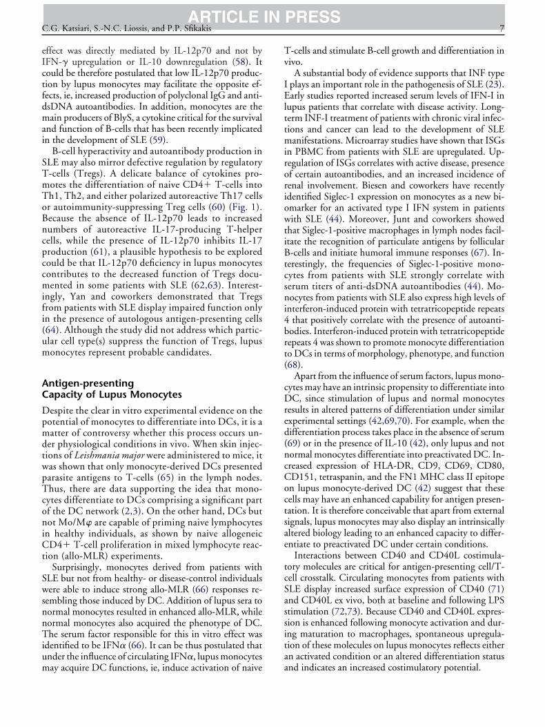
eIctfdmai
STmToBncpccmifi(um
AC
DpmdtwpTconiCt
SwsnnTium
Tv
IElttmiroriowtiBtcsni4brt(
cDred(ncCoctsae
tcSassita
C.G. Katsiari, S.-N.C. Liossis, and P.P. Sfikakis 7ARTICLE IN PRESS
ffect was directly mediated by IL-12p70 and not byFN-� upregulation or IL-10 downregulation (58). Itould be therefore postulated that low IL-12p70 produc-ion by lupus monocytes may facilitate the opposite ef-ects, ie, increased production of polyclonal IgG and anti-sDNA autoantibodies. In addition, monocytes are theain producers of BlyS, a cytokine critical for the survival
nd function of B-cells that has been recently implicatedn the development of SLE (59).
B-cell hyperactivity and autoantibody production inLE may also mirror defective regulation by regulatory-cells (Tregs). A delicate balance of cytokines pro-otes the differentiation of naive CD4� T-cells intoh1, Th2, and either polarized autoreactive Th17 cellsr autoimmunity-suppressing Treg cells (60) (Fig. 1).ecause the absence of IL-12p70 leads to increasedumbers of autoreactive IL-17-producing T-helperells, while the presence of IL-12p70 inhibits IL-17roduction (61), a plausible hypothesis to be exploredould be that IL-12p70 deficiency in lupus monocytesontributes to the decreased function of Tregs docu-ented in some patients with SLE (62,63). Interest-
ngly, Yan and coworkers demonstrated that Tregsrom patients with SLE display impaired function onlyn the presence of autologous antigen-presenting cells64). Although the study did not address which partic-lar cell type(s) suppress the function of Tregs, lupusonocytes represent probable candidates.
ntigen-presentingapacity of Lupus Monocytes
espite the clear in vitro experimental evidence on theotential of monocytes to differentiate into DCs, it is aatter of controversy whether this process occurs un-
er physiological conditions in vivo. When skin injec-ions of Leishmania major were administered to mice, itas shown that only monocyte-derived DCs presentedarasite antigens to T-cells (65) in the lymph nodes.hus, there are data supporting the idea that mono-
ytes differentiate to DCs comprising a significant partf the DC network (2,3). On the other hand, DCs butot Mo/M� are capable of priming naive lymphocytes
n healthy individuals, as shown by naive allogeneicD4� T-cell proliferation in mixed lymphocyte reac-
ion (allo-MLR) experiments.Surprisingly, monocytes derived from patients with
LE but not from healthy- or disease-control individualsere able to induce strong allo-MLR (66) responses re-
embling those induced by DC. Addition of lupus sera toormal monocytes resulted in enhanced allo-MLR, whileormal monocytes also acquired the phenotype of DC.he serum factor responsible for this in vitro effect was
dentified to be IFN� (66). It can be thus postulated thatnder the influence of circulating IFN�, lupus monocytes
ay acquire DC functions, ie, induce activation of naive a-cells and stimulate B-cell growth and differentiation inivo.
A substantial body of evidence supports that INF typeplays an important role in the pathogenesis of SLE (23).arly studies reported increased serum levels of IFN-I in
upus patients that correlate with disease activity. Long-erm INF-I treatment of patients with chronic viral infec-ions and cancer can lead to the development of SLEanifestations. Microarray studies have shown that ISGs
n PBMC from patients with SLE are upregulated. Up-egulation of ISGs correlates with active disease, presencef certain autoantibodies, and an increased incidence ofenal involvement. Biesen and coworkers have recentlydentified Siglec-1 expression on monocytes as a new bi-marker for an activated type I IFN system in patientsith SLE (44). Moreover, Junt and coworkers showed
hat Siglec-1-positive macrophages in lymph nodes facil-tate the recognition of particulate antigens by follicular-cells and initiate humoral immune responses (67). In-
erestingly, the frequencies of Siglec-1-positive mono-ytes from patients with SLE strongly correlate witherum titers of anti-dsDNA autoantibodies (44). Mo-ocytes from patients with SLE also express high levels of
nterferon-induced protein with tetratricopeptide repeatsthat positively correlate with the presence of autoanti-
odies. Interferon-induced protein with tetratricopeptideepeats 4 was shown to promote monocyte differentiationo DCs in terms of morphology, phenotype, and function68).
Apart from the influence of serum factors, lupus mono-ytes may have an intrinsic propensity to differentiate intoC, since stimulation of lupus and normal monocytes
esults in altered patterns of differentiation under similarxperimental settings (42,69,70). For example, when theifferentiation process takes place in the absence of serum69) or in the presence of IL-10 (42), only lupus and notormal monocytes differentiate into preactivated DC. In-reased expression of HLA-DR, CD9, CD69, CD80,D151, tetraspanin, and the FN1 MHC class II epitopen lupus monocyte-derived DC (42) suggest that theseells may have an enhanced capability for antigen presen-ation. It is therefore conceivable that apart from externalignals, lupus monocytes may also display an intrinsicallyltered biology leading to an enhanced capacity to differ-ntiate to preactivated DC under certain conditions.
Interactions between CD40 and CD40L costimula-ory molecules are critical for antigen-presenting cell/T-ell crosstalk. Circulating monocytes from patients withLE display increased surface expression of CD40 (71)nd CD40L ex vivo, both at baseline and following LPStimulation (72,73). Because CD40 and CD40L expres-ion is enhanced following monocyte activation and dur-ng maturation to macrophages, spontaneous upregula-ion of these molecules on lupus monocytes reflects eithern activated condition or an altered differentiation status
nd indicates an increased costimulatory potential.
P
T
DtctCtasptsiitft1c
I
Shctysmatmwlcvf
wtltrifpwosar
vwp
pst(b(malticittarwaC
dhatopStowdpShppmoiapStp
II
Dacdtp
8 Pathophysiologic role of monocytes and macrophages in SLEARTICLE IN PRESS
hagocytic Capacity of Lupus Monocytes
he “Phagocytic Synapse”
uring phagocytosis, phagocyte membrane receptors,arget cell-associated ligands, and intermediate moleculesooperate forming the “phagocytic synapse” (74). Recep-ors recognized on phagocytes include the moleculesD14 (lipopolysaccharide receptor), complement recep-
ors, CD36 (thrombospondin receptor), c-mer, CD44,nd phosphatidylserine receptor. Among structures de-cribed on target cells that provide an “eat me” signal,hosphatidylserine, a phospholipid normally confined tohe inner part of the membrane lipid bilayer, has beentudied in greater detail. An increasingly wide variety ofntermediate serum factors has been described, whose roles to opsonize target cells creating molecular bridges be-ween components of the target-cell and phagocyte sur-aces. For example, C1q links the target-cell surface withhe phagocyte receptor complex CD91/calreticulin (Fig.). Abnormalities in any part of the “phagocytic synapse”an result in abnormal phagocytosis and clearance.
n Vitro Studies
everal in vitro studies spanning more than 3 decadesave documented that lupus monocytes display reducedlearance of different targets (6). It was reported early onhat factors in SLE sera cause impaired phagocytosis ofeast cells by both lupus and normal macrophages (75). Aubsequent study reported that the major underlyingechanism for this effect involved mainly complement
nd not Fc-mediated phagocytosis (76). In this study,hough, lupus serum did not inhibit phagocytosis by nor-al monocytes. Moreover, levels of DNA, C3, and C4 asell as C1q-binding activity in lupus sera did not corre-
ate with the observed defect. It was proposed that theentral mechanism of impaired phagocytosis in SLE in-olves intrinsic abnormalities of complement receptorsunction in lupus monocytes (76).
Fc-mediated phagocytic capacity of lupus monocytesas also examined using IgG-sensitized erythrocytes. In-
erestingly, despite enhanced binding to the Fc receptors,upus monocytes failed to adequately phagocytose theirargets. The authors thus proposed that dissociation ofeceptor-ligand binding and receptor-mediated internal-zation may contribute significantly to the clearance de-ect in SLE (77). Indeed, while impaired Fc-mediatedhagocytosis by lupus monocytes has been correlatedith decreased expression of Fc�RII and III on the surfacef peripheral blood lupus monocytes (78), other studiesuggest that impaired phagocytosis is not due to themount or the membrane motility of these receptors butather to a defect during internalization of the target (79).
Of greater interest regarding possible mechanisms in-olved in tolerance breakdown in SLE, reduced clearanceas furthermore documented when the target was apo-
totic cells. Studies showing impaired clearance of apo- etotic cells have implicated all parts of the “phagocyticynapse.” In particular, reduced uptake of apoptotic neu-rophils by monocytes was attributed to serum factors80) or to impaired ability of the apoptotic neutrophils toe recognized, ie, to provide an adequate “eat me” signal81), while low CD44 expression on the surface of lupusonocytes has also been implicated (82). Defective clear-
nce was also demonstrated using autologous apoptoticymphocytes and was shown to reflect phagocyte dysfunc-ion and not abnormal execution of apoptosis (83). Sim-larly, a recent study employing an appropriate flowytometry assay showed that while lupus monocytes bind-ng capacity to apoptotic cells remains intact, their abilityo engulf apoptotic cells was impaired, indicating an in-rinsic cellular defect (84). On the other hand, uptake ofpoptotic Jurkat T-cells by lupus monocyte-derived mac-ophages was intact when studied in normal serum butas significantly decreased in the presence of lupus serum
nd was paralleled with decreased levels of C1q, C3, and4 (85).Overall, in vitro studies unanimously show a clearance
efect during phagocytosis in SLE. The underlying cause,owever, is reported to stem from diverse abnormalities,nd the intrinsic ability of macrophages to engulf theirargets was found reduced in some studies or normal inthers. Different results may indeed reflect differentathophysiologic mechanisms in different patients withLE, a disease characterized by heterogeneity. Of interest,hough, and relevant to the interpretation of in vitro datan phagocytosis are the findings of a recent study where itas shown that the ability to internalize apoptotic cells isependent on contact of macrophages to other macro-hages, a phenomenon termed “community effect” (86).hoshan and coworkers demonstrated that in bothealthy controls and patients with SLE, decreased macro-hage density impairs the ability of individual macro-hages to phagocytose. During maturation of lupusonocytes into macrophages accelerated apoptosis was
bserved, which resulted in lower density of macrophagesn cell suspensions and reduced the ability to internalizepoptotic cells. Maintaining normal density restored thehagocytic capacity of macrophages from patients withLE (86). Therefore macrophage density in in vitro cul-ures may represent a critical factor when evaluatinghagocytic capacity of Mo/M� in SLE.
n Vivo Studies: Does the Concept of Anti-nflammatory Phagocytosis Always Apply in SLE?
efective clearance in in vitro experiments may not reli-bly depict what happens in vivo. In a scavenger receptorlass A knockout mouse model, scavenger receptor class Aeficient macrophages are compromised in their capacityo engulf apoptotic thymocytes in vitro. However, noersistence of apoptotic cells was apparent in the thymus,
ven following induction of massive apoptosis by irradia-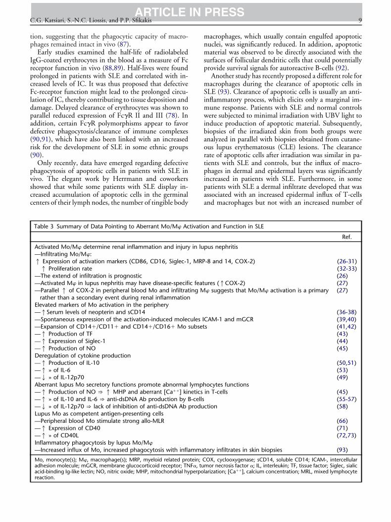
tp
IrpcFldpad(r(
pvscc
mnmsp
mSimwibaortpipaa
C.G. Katsiari, S.-N.C. Liossis, and P.P. Sfikakis 9ARTICLE IN PRESS
ion, suggesting that the phagocytic capacity of macro-hages remained intact in vivo (87).Early studies examined the half-life of radiolabeled
gG-coated erythrocytes in the blood as a measure of Fceceptor function in vivo (88,89). Half-lives were foundrolonged in patients with SLE and correlated with in-reased levels of IC. It was thus proposed that defectivec-receptor function might lead to the prolonged circu-
ation of IC, thereby contributing to tissue deposition andamage. Delayed clearance of erythrocytes was shown toarallel reduced expression of Fc�R II and III (78). Inddition, certain Fc�R polymorphisms appear to favorefective phagocytosis/clearance of immune complexes90,91), which have also been linked with an increasedisk for the development of SLE in some ethnic groups90).
Only recently, data have emerged regarding defectivehagocytosis of apoptotic cells in patients with SLE inivo. The elegant work by Herrmann and coworkershowed that while some patients with SLE display in-reased accumulation of apoptotic cells in the germinalenters of their lymph nodes, the number of tingible body
Table 3 Summary of Data Pointing to Aberrant Mo/M� Ac
Activated Mo/M� determine renal inflammation and injury—Infiltrating Mo/M�:1 Expression of activation markers (CD86, CD16, Siglec-11 Proliferation rate
—The extend of infiltration is prognostic—Activated M� in lupus nephritis may have disease-specifi—Parallel 1 of COX-2 in peripheral blood Mo and infiltrat
rather than a secondary event during renal inflammationElevated markers of Mo activation in the periphery—1Serum levels of neopterin and sCD14—Spontaneous expression of the activation-induced molec—Expansion of CD14�/CD11� and CD14�/CD16� Mo s—1 Production of TF—1 Expression of Siglec-1—1 Production of NODeregulation of cytokine production—1 Production of IL-10—1 » of IL-6—2 » of IL-12p70Aberrant lupus Mo secretory functions promote abnormal—1 Production of NO ) 1 MHP and aberrant [Ca��] kin—1 » of IL-10 and IL-6 ) anti-dsDNA Ab production by B—2 » of IL-12p70 ) lack of inhibition of anti-dsDNA Ab pLupus Mo as competent antigen-presenting cells—Peripheral blood Mo stimulate strong allo-MLR—1 Expression of CD40—1 » of CD40LInflammatory phagocytosis by lupus Mo/M�—Increased influx of Mo, increased phagocytosis with infla
Mo, monocyte(s); M�, macrophage(s); MRP, myeloid related proadhesion molecule; mGCR, membrane glucocorticoid receptor; TNacid-binding Ig-like lectin; NO, nitric oxide; MHP, mitochondrial hy
reaction.acrophages, which usually contain engulfed apoptoticuclei, was significantly reduced. In addition, apoptoticaterial was observed to be directly associated with the
urfaces of follicular dendritic cells that could potentiallyrovide survival signals for autoreactive B-cells (92).Another study has recently proposed a different role foracrophages during the clearance of apoptotic cells in
LE (93). Clearance of apoptotic cells is usually an anti-nflammatory process, which elicits only a marginal im-
une response. Patients with SLE and normal controlsere subjected to minimal irradiation with UBV light to
nduce production of apoptotic material. Subsequently,iopsies of the irradiated skin from both groups werenalyzed in parallel with biopsies obtained from cutane-us lupus erythematosus (CLE) lesions. The clearanceate of apoptotic cells after irradiation was similar in pa-ients with SLE and controls, but the influx of macro-hages in dermal and epidermal layers was significantlyncreased in patients with SLE. Furthermore, in someatients with SLE a dermal infiltrate developed that wasssociated with an increased epidermal influx of T-cellsnd macrophages but not with an increased number of
n and Function in SLE
Ref.
us nephritis
-8 and 14, COX-2) (26-31)(32-33)(26)
ures (1COX-2) (27)� suggests that Mo/M� activation is a primary (27)
(36-38)AM-1 and mGCR (39,40)
s (41,42)(43)(44)(45)
(50,51)(53)(49)
ocytes functionsin T-cells (45)
(55-57)ction (58)
(66)(71)(72,73)
tory infiltrates in skin biopsies (93)
OX, cyclooxygenase; sCD14, soluble CD14; ICAM-, intercellularmor necrosis factor �; IL, interleukin; TF, tissue factor; Siglec, sialicarization; [Ca��], calcium concentration; MRL, mixed lymphocyte
tivatio
in lup
, MRP
c feating M
ules ICubset
lymphetics-cellsrodu
mma
tein; CF�, tuperpol
altdlaScsaapaat
D
IfivltWsms
MAnnocdcmSme(cvtp
R
FpiatdAlo le on
10 Pathophysiologic role of monocytes and macrophages in SLEARTICLE IN PRESS
poptotic cells or epidermal deposition of immunoglobu-ins. Some lesional lupus macrophages displayed inges-ion of multiple apoptotic bodies, a process not previouslyescribed. Inflammatory lesions in these patients were
ocalized near accumulations of apoptotic keratinocytes,s was also evident in nonirradiated CLE skin lesions.uch inflammatory lesions in the vicinity of apoptoticells suggest not only that anti-inflammatory phagocyto-is may be inadequate but also that an inflammatory clear-nce of apoptotic cells takes place both in UVB-inducednd in CLE skin lesions in SLE patients. This study sup-orts the hypothesis that “inflammatory” phagocytosis ofpoptotic cells, ie, in the relative absence of protectiventi-inflammatory signals by Mo/M�, may play a role inhe development of SLE.
ISCUSSION
t has traditionally been held that lupus Mo/M� are de-ective. The model of “defective” function supports thedea that lupus Mo/M�, either overwhelmed by their en-ironment or because of intrinsic defects, become power-ess cells unable to perform their physiologic tasks con-ributing in this way to disease expression (Table 1).
hile defective function of Mo/M� is encountered inome patients with SLE, emerging experimental data inurine models (Table 2) and humans with SLE (Table 3)
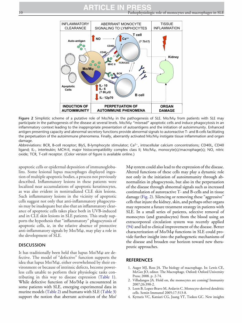
INFLAMMATORYCLEARANCE
INDUCTION OF INDUCTION OF AUTOIMMUNITYAUTOIMMUNITY
PERPETPERPETAUTOIMMUAUTOIMMUN
ABERRANSIGNALING T
CD40
CD40L
Apoptotic Cells
Auto-antigenNO
IL- 10IL- 6
IL- 12p70
(? BLyS)
MHC II
Mo/Mφ
igure 2 Simplistic scheme of a putative role of Mo/M� iarticipate in the pathogenesis of the disease at several levels
nflammatory context leading to the inappropriate presentatntigen presenting capacity and abnormal secretory functionshe perpetuation of the autoimmune phenomena. Finally, abamage.bbreviations: BCR, B-cell receptor; BlyS, B-lymphocyte stim
igand; IL-, interleukin; MCH-II, major histocompatibility coxide; TCR, T-cell receptor. (Color version of figure is availab
upport the notion that aberrant activation of the Mo/
� system could also lead to the expression of the disease.ltered functions of these cells may play a dynamic roleot only in the initiation of autoimmunity through ab-ormalities in phagocytosis, but also in the perpetuationf the disease through abnormal signals such as increasedostimulation of autoreactive T- and B-cells and in tissueamage (Fig. 2). Silencing or removing these “aggressive”ells that injure the kidney, skin, and perhaps other organsay represent a future treatment strategy in patients with
LE. In a small series of patients, selective removal ofonocytes (and granulocytes) from the blood using an
xtracorporeal circulation system was recently applied94) and led to clinical improvement of the disease. Betterharacterization of Mo/M� functions in SLE could pro-ide further insight into the pathogenetic mechanisms ofhe disease and broaden our horizon toward new thera-eutic approaches.
EFERENCES
1. Auger MJ, Ross JA. The biology of macrophage. In: Lewis CE,McGee JO, editor. The Macrophage. Oxford: Oxford UniversityPress; 2008. p. 2-74.
2. Villadangos JA. Hold on, the monocytes are coming! Immunity2007;26:390-2.
3. Leon B, Lopez-Bravo M, Ardavin C. Monocyte-derived dendriticcells. Semin Immunol 2005;17:313-8.
ON OF ON OF ENOMENAENOMENA
ORGAN ORGAN DAMAGEDAMAGE
NOCYTE PHOCYTES
TISSUEINFLAMMATION
3
+T cell
B cell
Kidney
(? Th17)
pathogenesis of SLE. Mo/M� from patients with SLE may� “misread” apoptotic cells and induce phagocytosis in an
autoantigens and the initiation of autoimmunity. Enhancedde abnormal signals to autoreactive T- and B-cells facilitatingly activated Mo/M� instigate tissue inflammation and organ
r; Ca2�, intracellular calcium concentrations; CD40L, CD40class II; Mo/M�, monocyte(s)/macrophage(s); NO, nitric
line.)
UATIUATINE PHE PH
T MOO LYM
TCR/CD
Ca+
BCR
n the. Mo/Mion ofprovi
errant
ulatomplex
4. Kyttaris VC, Katsiari CG, Juang YT, Tsokos GC. New insights
1
1
1
1
1
1
1
1
1
1
2
2
2
2
2
2
2
2
2
2
3
3
3
3
3
3
3
3
3
3
4
4
4
C.G. Katsiari, S.-N.C. Liossis, and P.P. Sfikakis 11ARTICLE IN PRESS
into the pathogenesis of systemic lupus erythematosus. CurrRheumatol Rep 2005;7:469-75.
5. Kuroiwa T, Lee EG. Cellular interactions in the pathogenesis oflupus nephritis: the role of T cells and macrophages in the ampli-fication of the inflammatory process in the kidney. Lupus 1998;7:597-603.
6. Gaipl US, Munoz LE, Grossmayer G, Lauber K, Franz S, SarterK, et al. Clearance deficiency and systemic lupus erythematosus(SLE). J Autoimmun 2007;28:114-21.
7. Riccardi PJ, Hausman PB, Raff HV, Stobo JD. The autologousmixed lymphocyte reaction in systemic lupus erythematosus. Ar-thritis Rheum 1982;25:820-3.
8. Shirakawa F, Yamashita U, Suzuki H. Reduced function of HLA-DR-positive monocytes in patients with systemic lupus erythem-atosus (SLE). J Clin Immunol 1985;5:396-403.
9. Tsokos GC, Kovacs B, Sfikakis PP, Theocharis S, Vogelgesang S,Via CS. Defective antigen-presenting cell function in patientswith systemic lupus erythematosus: role of the B7-1 (CD80) co-stimulatory molecule. Arthritis Rheum 1996;39:600-9.
0. Alcocer-Varela J, Laffon A, Alarcon-Segovia D. Defective mono-cyte production of, and T lymphocyte response to, interleukin-1in the peripheral blood of patients with systemic lupus erythem-atosus. Clin Exp Immunol 1984;55:125-32.
1. Scott RS, McMahon EJ, Pop SM, Reap EA, Caricchio R, CohenPL, et al. Phagocytosis and clearance of apoptotic cells is mediatedby MER. Nature 2001;411:207-11.
2. Lu Q, Lemke G. Homeostatic regulation of the immune systemby receptor tyrosine kinases of the Tyro 3 family. Science 2001;293:306-11.
3. Fadok VA, Bratton DL, Henson PM. Phagocyte receptors forapoptotic cells: recognition, uptake, and consequences. J ClinInvest 2001;108:957-62.
4. Devitt A, Parker KG, Ogden CA, Oldreive C, Clay MF, MelvilleLA, et al. Persistence of apoptotic cells without autoimmune dis-ease or inflammation in CD14�/� mice. J Cell Biol 2004;167:1161-70.
5. Patel VA, Longacre A, Hsiao K, Fan H, Meng F, Mitchell JE, et al.Apoptotic cells, at all stages of the death process, trigger charac-teristic signaling events that are divergent from and dominant tothose triggered by necrotic cells: implications for the delayed clear-ance model of autoimmunity. J Biol Chem 2006;281:4663-70.
6. Koh JS, Wang Z, Levine JS. Cytokine dysregulation induced byapoptotic cells is a shared characteristic of murine lupus. J Immu-nol 2000;165:4190-201.
7. Fan H, Patel VA, Longacre A, Levine JS. Abnormal regulation ofthe cytoskeletal regulator Rho typifies macrophages of the majormurine models of spontaneous autoimmunity. J Leukoc Biol2006;79:155-65.
8. Licht R, Jacobs CW, Tax WJ, Berden JH. No constitutive defectin phagocytosis of apoptotic cells by resident peritoneal macro-phages from pre-morbid lupus mice. Lupus 2001;10:102-7.
9. Jiang N, Reich CF, III, Pisetsky DS. Role of macrophages in thegeneration of circulating blood nucleosomes from dead and dyingcells. Blood 2003;102:2243-50.
0. Clynes R, Dumitru C, Ravetch JV. Uncoupling of immune com-plex formation and kidney damage in autoimmune glomerulone-phritis. Science 1998;279:1052-4.
1. Bergtold A, Gavhane A, D’Agati V, Madaio M, Clynes R. FcR-bearing myeloid cells are responsible for triggering murine lupusnephritis. J Immunol 2006;177:7287-95.
2. Nacionales DC, Kelly KM, Lee PY, Zhuang H, Li Y, WeinsteinJS, et al. Type I interferon production by tertiary lymphoid tissuedeveloping in response to 2,6,10,14-tetramethyl-pentadecane(pristane). Am J Pathol 2006;168:1227-40.
3. Ronnblom L, Eloranta ML, Alm GV. The type I interferon sys-tem in systemic lupus erythematosus. Arthritis Rheum 2006;54:408-20.
4. Nacionales DC, Kelly-Scumpia KM, Lee PY, Weinstein JS, Lyons R,
Sobel E, et al. Deficiency of the type I interferon receptor protectsmice from experimental lupus. Arthritis Rheum 2007;56:3770-83.
5. Lee PY, Weinstein JS, Nacionales DC, Scumpia PO, Li Y, But-filoski E, et al. A novel type I IFN-producing cell subset in murinelupus. J Immunol 2008;180:5101-8.
6. Yang N, Isbel NM, Nikolic-Paterson DJ, Li Y, Ye R, Atkins RC,et al. Local macrophage proliferation in human glomerulonephri-tis. Kidney Int 1998;54:143-51.
7. Tomasoni S, Noris M, Zappella S, Gotti E, Casiraghi F, Bonaz-zola S, et al. Upregulation of renal and systemic cyclooxygenase-2in patients with active lupus nephritis. J Am Soc Nephrol 1998;9:1202-12.
8. Hotta O, Yusa N, Ooyama M, Unno K, Furuta T, Taguma Y.Detection of urinary macrophages expressing the CD16 (Fc biggamma RIII) molecule: a novel marker of acute inflammatoryglomerular injury. Kidney Int 1999;55:1927-34.
9. Ikezumi Y, Suzuki T, Hayafuji S, Okubo S, Nikolic-Paterson DJ,Kawachi H, et al. The sialoadhesin (CD169) expressing a macro-phage subset in human proliferative glomerulonephritis. NephrolDial Transplant 2005;20:2704-13.
0. Peterson KS, Huang JF, Zhu J, DÆAgati V, Liu X, Miller N, et al.Characterization of heterogeneity in the molecular pathogenesisof lupus nephritis from transcriptional profiles of laser-capturedglomeruli. J Clin Invest 2004;113:1722-33.
1. Frosch M, Vogl T, Waldherr R, Sorg C, Sunderkotter C, Roth J.Expression of MRP8 and MRP14 by macrophages is a marker forsevere forms of glomerulonephritis. J Leukoc Biol 2004;75:198-206.
2. Hill GS, Delahousse M, Nochy D, Remy P, Mignon F, Mery JP,et al. Predictive power of the second renal biopsy in lupus nephri-tis: significance of macrophages. Kidney Int 2001;59:304-16.
3. Hill GS, Delahousse M, Nochy D, Tomkiewicz E, Remy P, Mi-gnon F, et al. A new morphologic index for the evaluation of renalbiopsies in lupus nephritis. Kidney Int 2000;58:1160-73.
4. Yamamoto K, Okamura D, Kurahara D, Liao K, Kimura L, Si-mafranca R, et al. Do urinary mononuclear cells reflect diseaseactivity in lupus nephritis? Cell Mol Biol (Noisy-le-grand) 2003;49:1333-7.
5. Wagrowska-Danilewicz M, Stasikowska O, Danilewicz M. Correla-tive insights into immunoexpression of monocyte chemoattractantprotein-1, transforming growth factor beta-1 and CD68� cells inlupus nephritis. Pol J Pathol 2005;56:115-20.
6. Jin O, Sun L, Zhou K, Zhang X, Feng X, Mok M, et al. Lympho-cyte apoptosis and macrophage function: correlation with diseaseactivity in systemic lupus erythematosus. Clin Rheumatol 2005;24:107-10.
7. Egerer K, Feist E, Rohr U, Pruss A, Burmester GR, Dorner T.Increased serum soluble CD14, ICAM-1 and E-selectin correlatewith disease activity and prognosis in systemic lupus erythemato-sus. Lupus 2000;9:614-21.
8. Nockher WA, Wigand R, Schoeppe W, Scherberich JE. Elevatedlevels of soluble CD14 in serum of patients with systemic lupuserythematosus. Clin Exp Immunol 1994;96:15-9.
9. Funauchi M, Ohno M, Minoda M, Horiuchi A. Abnormal ex-pression of intercellular adhesion molecule-1 on peripheral bloodmononuclear cells from patients with systemic lupus erythemato-sus. J Clin Lab Immunol 1993;40:115-24.
0. Spies CM, Schaumann DHS, Berki T, Mayer K, Jakstadt M,Huscher D, et al. Membrane glucocorticoid receptors are downregulated by glucocorticoids in patients with systemic lupus ery-thematosus and use a caveolin-1-independent expression path-way. Ann Rheum Dis 2006;65:1139-46.
1. Sullivan KE, Suriano A, Dietzmann K, Lin J, Goldman D, PetriMA. The TNFa locus is altered in monocytes from patients withsystemic lupus erythematosus. Clin Immunol 2007;123:74-81.
2. Figueroa-Vega N, Galindo-Rodriguez G, Bajana S, Portales-Perez D, Abud-Mendoza C, Sanchez-Torres C, et al. Pheno-
typic analysis of IL-10-treated monocyte-derived dendritic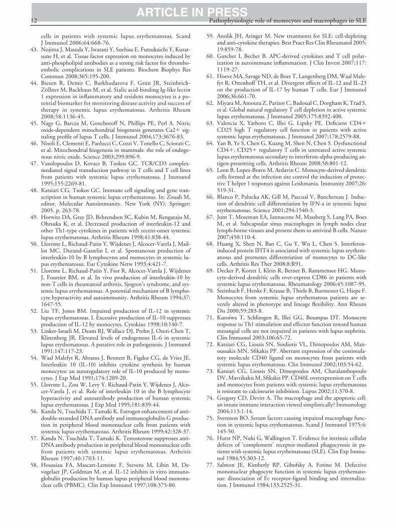
4
4
4
4
4
4
4
5
5
5
5
5
5
5
5
5
5
6
6
6
6
6
6
6
6
6
6
7
7
7
7
7
7
7
7
12 Pathophysiologic role of monocytes and macrophages in SLEARTICLE IN PRESS
cells in patients with systemic lupus erythematosus. ScandJ Immunol 2006;64:668-76.
3. Nojima J, Masuda Y, Iwatani Y, Suehisa E, Futsukaichi Y, Kurat-sune H, et al. Tissue factor expression on monocytes induced byanti-phospholipid antibodies as a strong risk factor for thrombo-embolic complications in SLE patients. Biochem Biophys ResCommun 2008;365:195-200.
4. Biesen R, Demir C, Barkhudarova F, Grün JR, Steinbrich-Zöllner M, Backhaus M, et al. Sialic acid-binding Ig-like lectin1 expression in inflammatory and resident monocytes is a po-tential biomarker for monitoring disease activity and success oftherapy in systemic lupus erythematosus. Arthritis Rheum2008;58:1136-45.
5. Nagy G, Barcza M, Gonchoroff N, Phillips PE, Perl A. Nitricoxide-dependent mitochondrial biogenesis generates Ca2� sig-naling profile of lupus T cells. J Immunol 2004;173:3676-83.
6. Nisoli E, Clementi E, Paolucci C, Cozzi V, Tonello C, Sciorati C,et al. Mitochondrial biogenesis in mammals: the role of endoge-nous nitric oxide. Science 2003;299:896-9.
7. Vassilopoulos D, Kovacs B, Tsokos GC. TCR/CD3 complex-mediated signal transduction pathway in T cells and T cell linesfrom patients with systemic lupus erythematosus. J Immunol1995;155:2269-81.
8. Katsiari CG, Tsokos GC. Immune cell signaling and gene tran-scription in human systemic lupus erythematosus. In: Zouali M,editor. Molecular Autoimmunity. New York (NY): Springer;2005. p. 263-78.
9. Horwitz DA, Gray JD, Behrendsen SC, Kubin M, Rengaraju M,Ohtsuka K, et al. Decreased production of interleukin-12 andother Th1-type cytokines in patients with recent-onset systemiclupus erythematosus. Arthritis Rheum 1998;41:838-44.
0. Llorente L, Richaud-Patin Y, Wijdenes J, Alcocer-Varela J, Mail-lot MC, Durand-Gasselin I, et al. Spontaneous production ofinterleukin-10 by B lymphocytes and monocytes in systemic lu-pus erythematosus. Eur Cytokine Netw 1993;4:421-7.
1. Llorente L, Richaud-Patin Y, Fior R, Alcocer-Varela J, WijdenesJ, Fourrier BM, et al. In vivo production of interleukin-10 bynon-T cells in rheumatoid arthritis, Sjogren’s syndrome, and sys-temic lupus erythematosus. A potential mechanism of B lympho-cyte hyperactivity and autoimmunity. Arthritis Rheum 1994;37:1647-55.
2. Liu TF, Jones BM. Impaired production of IL-12 in systemiclupus erythematosus. I. Excessive production of IL-10 suppressesproduction of IL-12 by monocytes. Cytokine 1998;10:140-7.
3. Linker-Israeli M, Deans RJ, Wallace DJ, Prehn J, Ozeri-Chen T,Klinenberg JR. Elevated levels of endogenous IL-6 in systemiclupus erythematosus. A putative role in pathogenesis. J Immunol1991;147:117-23.
4. Waal Malefyt R, Abrams J, Bennett B, Figdor CG, de Vries JE.Interleukin 10 (IL-10) inhibits cytokine synthesis by humanmonocytes: an autoregulatory role of IL-10 produced by mono-cytes. J Exp Med 1991;174:1209-20.
5. Llorente L, Zou W, Levy Y, Richaud-Patin Y, Wijdenes J, Alco-cer-Varela J, et al. Role of interleukin 10 in the B lymphocytehyperactivity and autoantibody production of human systemiclupus erythematosus. J Exp Med 1995;181:839-44.
6. Kanda N, Tsuchida T, Tamaki K. Estrogen enhancement of anti-double-stranded DNA antibody and immunoglobulin G produc-tion in peripheral blood mononuclear cells from patients withsystemic lupus erythematosus. Arthritis Rheum 1999;42:328-37.
7. Kanda N, Tsuchida T, Tamaki K. Testosterone suppresses anti-DNA antibody production in peripheral blood mononuclear cellsfrom patients with systemic lupus erythematosus. ArthritisRheum 1997;40:1703-11.
8. Houssiau FA, Mascart-Lemone F, Stevens M, Libin M, De-vogelaer JP, Goldman M, et al. IL-12 inhibits in vitro immuno-globulin production by human lupus peripheral blood mononu-
clear cells (PBMC). Clin Exp Immunol 1997;108:375-80.9. Anolik JH, Aringer M. New treatments for SLE: cell-depletingand anti-cytokine therapies. Best Pract Res Clin Rheumatol 2005;19:859-78.
0. Gutcher I, Becher B. APC-derived cytokines and T cell polar-ization in autoimmune inflammation. J Clin Invest 2007;117:1119-27.
1. Hoeve MA, Savage ND, de Boer T, Langenberg DM, Waal Male-fyt R, Ottenhoff TH, et al. Divergent effects of IL-12 and IL-23on the production of IL-17 by human T cells. Eur J Immunol2006;36:661-70.
2. Miyara M, Amoura Z, Parizot C, Badoual C, Dorgham K, Trad S,et al. Global natural regulatory T cell depletion in active systemiclupus erythematosus. J Immunol 2005;175:8392-400.
3. Valencia X, Yarboro C, Illei G, Lipsky PE. Deficient CD4�CD25 high T regulatory cell function in patients with activesystemic lupus erythematosus. J Immunol 2007;178:2579-88.
4. Yan B, Ye S, Chen G, Kuang M, Shen N, Chen S. DysfunctionalCD4�, CD25� regulatory T cells in untreated active systemiclupus erythematosus secondary to interferon-alpha-producing an-tigen-presenting cells. Arthritis Rheum 2008;58:801-12.
5. Leon B, Lopez-Bravo M, Ardavin C. Monocyte-derived dendriticcells formed at the infection site control the induction of protec-tive T helper 1 responses against Leishmania. Immunity 2007;26:519-31.
6. Blanco P, Palucka AK, Gill M, Pascual V, Banchereau J. Induc-tion of dendritic cell differentiation by IFN-a in systemic lupuserythematosus. Science 2001;294:1540-3.
7. Junt T, Moseman EA, Iannacone M, Massberg S, Lang PA, BoesM, et al. Subcapsular sinus macrophages in lymph nodes clearlymph-borne viruses and present them to antiviral B cells. Nature2007;450:110-4.
8. Huang X, Shen N, Bao C, Gu Y, Wu L, Chen S. Interferon-induced protein IFIT4 is associated with systemic lupus erythem-atosus and promotes differentiation of monocytes to DC-likecells. Arthritis Res Ther 2008;8:R91.
9. Decker P, Kotter I, Klein R, Berner B, Rammensee HG. Mono-cyte-derived dendritic cells over-express CD86 in patients withsystemic lupus erythematosus. Rheumatology 2006;45:1087-95.
0. Steinbach F, Henke F, Krause B, Thiele B, Burmester G, Hiepe F.Monocytes from systemic lupus erythematous patients are se-verely altered in phenotype and lineage flexibility. Ann RheumDis 2000;59:283-8.
1. Kuroiwa T, Schlimgen R, Illei GG, Boumpas DT. Monocyteresponse to Th1 stimulation and effector function toward humanmesangial cells are not impaired in patients with lupus nephritis.Clin Immunol 2003;106:65-72.
2. Katsiari CG, Liossis SN, Souliotis VL, Dimopoulos AM, Man-oussakis MN, Sfikakis PP. Aberrant expression of the costimula-tory molecule CD40 ligand on monocytes from patients withsystemic lupus erythematosus. Clin Immunol 2002;103:54-62.
3. Katsiari CG, Liossis SN, Dimopoulos AM, CharalambopouloDV, Mavrikakis M, Sfikakis PP. CD40L overexpression on T cellsand monocytes from patients with systemic lupus erythematosusis resistant to calcineurin inhibition. Lupus 2002;11:370-8.
4. Gregory CD, Devitt A. The macrophage and the apoptotic cell:an innate immune interaction viewed simplistically? Immunology2004;113:1-14.
5. Svensson BO. Serum factors causing impaired macrophage func-tion in systemic lupus erythematosus. Scand J Immunol 1975;4:145-50.
6. Hurst NP, Nuki G, Wallington T. Evidence for intrinsic cellulardefects of ‘complement’ receptor-mediated phagocytosis in pa-tients with systemic lupus erythematosus (SLE). Clin Exp Immu-nol 1984;55:303-12.
7. Salmon JE, Kimberly RP, Gibofsky A, Fotino M. Defectivemononuclear phagocyte function in systemic lupus erythemato-sus: dissociation of Fc receptor-ligand binding and internaliza-
tion. J Immunol 1984;133:2525-31.
7
7
8
8
8
8
8
8
8
8
8
8
9
9
9
9
9
C.G. Katsiari, S.-N.C. Liossis, and P.P. Sfikakis 13ARTICLE IN PRESS
8. Seres T, Csipo I, Kiss E, Szegedi G, Kavai M. Correlation of Fcgamma receptor expression of monocytes with clearance functionby macrophages in systemic lupus erythematosus. Scand J Immu-nol 1998;48:307-11.
9. Vazquez-Doval J, Sanchez-Ibarrola A. Defective mononuclearphagocyte function in systemic lupus erythematosus: relationshipof FcRII (CD32) with intermediate cytoskeletal filaments. J In-vestig Allergol Clin Immunol 1993;3:86-91.
0. Ren Y, Tang J, Mok MY, Chan AW, Wu A, Lau CS. Increasedapoptotic neutrophils and macrophages and impaired macro-phage phagocytic clearance of apoptotic neutrophils in systemiclupus erythematosus. Arthritis Rheum 2003;48:2888-97.
1. Donnelly S, Roake W, Brown S, Young P, Naik H, WordsworthP, et al. Impaired recognition of apoptotic neutrophils by theC1q/calreticulin and CD91 pathway in systemic lupus erythem-atosus. Arthritis Rheum 2006;54:1543-56.
2. Cairns AP, Crockard AD, McConnell JR, Courtney PA, Bell AL.Reduced expression of CD44 on monocytes and neutrophils insystemic lupus erythematosus: relations with apoptotic neutro-phils and disease activity. Ann Rheum Dis 2001;60:950-5.
3. Herrmann M, Voll RE, Zoller OM, Hagenhofer M, Ponner BB,Kalden JR. Impaired phagocytosis of apoptotic cell material bymonocyte-derived macrophages from patients with systemic lu-pus erythematosus. Arthritis Rheum 1998;41:1241-50.
4. Tas SW, Quartier P, Botto M, Fossati-Jimack L. Macrophagesfrom patients with SLE and rheumatoid arthritis have defectiveadhesion in vitro, while only SLE macrophages have impaireduptake of apoptotic cells. Ann Rheum Dis 2006;65:216-21.
5. Bijl M, Reefman E, Horst G, Limburg PC, Kallenberg CG. Re-duced uptake of apoptotic cells by macrophages in systemic lupuserythematosus: correlates with decreased serum levels of comple-ment. Ann Rheum Dis 2006;65:57-63.
6. Shoshan Y, Shapira I, Toubi E, Frolkis I, Yaron M, Mevorach D.
Accelerated Fas-mediated apoptosis of monocytes and maturingmacrophages from patients with systemic lupus erythematosus:relevance to in vitro impairment of interaction with iC3b-opsonized apoptotic cells. J Immunol 2001;167:5963-9.
7. Platt N, Suzuki H, Kodama T, Gordon S. Apoptotic thymocyteclearance in scavenger receptor class A-deficient mice is apparentlynormal. J Immunol 2000;164:4867.
8. Frank MM, Hamburger MI, Lawley TJ, Kimberly RP, Plotz PH.Defective reticuloendothelial system Fc-receptor function in sys-temic lupus erythematosus. N Engl J Med 1979;300:518-23.
9. Van der Woude FJ, van der Giessen M, Kallenberg CGM, Ou-wehand W, Beekhuis H, Beelen JM, et al. Reticuloendothelial Fcreceptor function in SLE patients. I. Primary HLA linked defector acquired dysfunction secondary to disease activity? Clin ExpImmunol 1984;55:473-80.
0. Salmon JE, Millard S, Schachter LA, Arnett FC, Ginzler EM,Gourley MF, et al. Fc gamma RIIA alleles are heritable risk factorsfor lupus nephritis in African Americans. J Clin Invest 1996;97:1348-54.
1. Dijstelbloem HM, Bijl M, Fijnheer R, Scheepers RH, Oost WW,Jansen MD, et al. Fcgamma receptor polymorphisms in systemiclupus erythematosus: association with disease and in vivo clear-ance of immune complexes. Arthritis Rheum 2000;43:2793-800.
2. Baumann I, Kolowos W, Voll RE, Manger B, Gaipl U, NeuhuberWL, et al. Impaired uptake of apoptotic cells into tingible bodymacrophages in germinal centers of patients with systemic lupuserythematosus. Arthritis Rheum 2002;46:191-201.
3. Reefman E, de Jong MC, Kuiper H, Jonkman MF, Limburg PC,Kallenberg CG, et al. Is disturbed clearance of apoptotic keratin-ocytes responsible for UVB-induced inflammatory skin lesions inSystemic Lupus Erythematosus? Arthritis Res Ther 2006;8:R156.
4. Soerensen H, Schneidewind-Mueller JM, Lange D, Kashiwagi N,Franz M, Yokoyama T, et al. Pilot clinical study of Adacolumncytapheresis in patients with systemic lupus erythematosus. Rheu-
matol Int 2006;26:409-15.