the pachymeter guide: a new device to facilitate accurate corneal thickness measurement
TRANSCRIPT
ELSEVIER
The Pachymeter Guide: A New Device to Facilitate Accurate Cornea1 Thickness Measurement
Tetsuro Oshika,* Fumiaki Yoshitomit and Kohtaro Oki”
*Department of Ophthalmology, University of Tokyo School of Medicine, Tokyo, Japan; fYoshitomi Eye Center, Dazaifi, Fukuoka, Japan; *Oki Eye Surgery Center, Tokyo, Japan
Abstract: A pachymeter guide has been developed to facilitate precise positioning of the ul- trasonic pachymeter tip, making cornea1 thickness measurement much faster and more re- producible. This guide also makes pachymetry more accurate by preventing dehydration of the cornea, avoiding undue pressure on the cornea, and limiting tilting of the pachymeter tip. Jpn J Opbthalmoll!N7;41:426-427 0 1997 Japanese Ophthalmological Society
Key Words: Astigmatic keratotomy, cornea1 thickness, optical zone, pachymetry.
Accurate measurement of cornea1 thickness with ultrasonic pachymetry is essential to ensure both the safety and predictability of incisional astigmatic keratotomy.’ Hand-guided pachymetry, however, is vulnerable to several sources of error, including in- appropriate measurement location, tilting of the in- strument, excess pressure, and undesirable drying or moistening of the cornea.2 Moreover, if the pachy- meter tip is not placed perpendicularly, ultrasound echo is poorly detected, resulting in repeated mea- surement attempts and dehydration of the cornea. This may lead to overestimation of the cornea1 thick- ness which increases the risk of cornea1 perforation and endothelial damage. 3,4 The present authors have developed a guide to aid in precise placement/posi- tioning of the pachymeter tip for accurate and rapid measurement of cornea1 thickness.
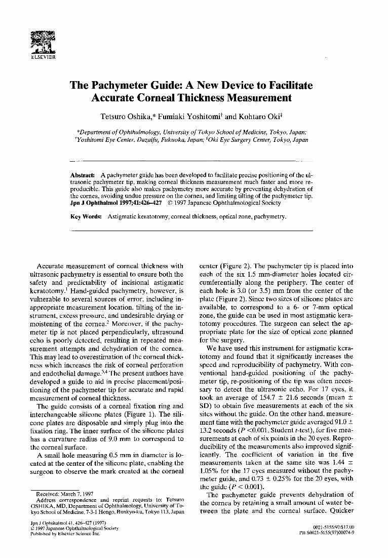
The guide consists of a cornea1 fixation ring and interchangeable silicone plates (Figure 1). The sili- cone plates are disposable and simply plug into the fixation ring. The inner surface of the silicone plates has a curvature radius of 9.0 mm to correspond to the cornea1 surface.
A small hole measuring 0.5 mm in diameter is lo- cated at the center of the silicone plate, enabling the surgeon to observe the mark created at the cornea1
Received: March 7,199l Address correspondence and reprint requests to: Tetsuro
OSHIKA, MD, Department of Ophthalmology, University of To- kyo School of Medicine, 7-3-l Hongo, Bunkyo-ku, Tokyo 113, Japan
Jpn J Ophthalmol41,42&427 (1997) 0 1997 Japanese Ophthalmological Society Published by Elsevier Science Inc.
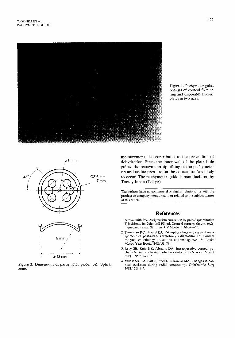
center (Figure 2). The pachymeter tip is placed into each of the six 1.5 mm-diameter holes located cir- cumferentially along the periphery. The center of each hole is 3.0 (or 3.5) mm from the center of the plate (Figure 2). Since two sizes of silicone plates are available, to correspond to a 6- or 7-mm optical zone, the guide can be used in most astigmatic kera- totomy procedures. The surgeon can select the ap- propriate plate for the size of optical zone planned for the surgery.
We have used this instrument for astigmatic kera- totomy and found that it significantly increases the speed and reproducibility of pachymetry. With con- ventional hand-guided positioning of the pachy- meter tip, re-positioning of the tip was often neces- sary to detect the ultrasonic echo. For 17 eyes, it took an average of 154.7 + 21.6 seconds (mean + SD) to obtain five measurements at each of the six sites without the guide. On the other hand, measure- ment time with the pachymeter guide averaged 91.0 + 13.2 seconds (P <O.OOl, Student t-test), for five mea- surements at each of six points in the 20 eyes. Repro- ducibility of the measurements also improved signif- icantly. The coefficient of variation in the five measurements taken at the same site was 1.44 2 1.05% for the 17 eyes measured without the pachy- meter guide, and 0.73 2 0.25% for the 20 eyes, with the guide (P < 0.001).
The pachymeter guide prevents dehydration of the cornea by retaining a small amount of water be- tween the plate and the cornea1 surface. Quicker
0021-5155/97/$17.00 PII SOO21-5155(97)00074-9
T. OSHIKA ET AL. PACHYMETER GUIDE
427
&I mm
OZ6mm 7mm
413mm j
Figure 2. Dimensions of pachymeter guide. OZ: Optical zone.
Figure 1. Pachymeter guide consists of cornea1 fixation ring and disposable silicone plates in two sizes.
measurement also contributes to the prevention of dehydration. Since the inner wall of the plate hole guides the pachymeter tip, tilting of the pachymeter tip and undue pressure on the cornea are less likely to occur. The pachymeter guide is manufactured by Tomey Japan (Tokyo).
The authors have no commercial or similar relationships with the product or company mentioned in or related to the subject matter of this article.
References 1. Arrowsmith PN. Astigmatism correction by paired quantitative
T-incisions. In: Brightbill FS, ed. Cornea1 surgery: theory, tech- nique, and tissue. St. Louis: CV Mosby, 1986:548-50.
2. Troutman RC, Buzard KA. Pathophysiology and surgical man- agement of post-radial keratotomy astigmatism. In: Cornea1 astigmatism: etiology, prevention, and management. St. Louis: Mosby Year Book, 1992:451-79.
3. Levy SB, Katz HR, Abrams DA. Intraoperative cornea1 pa- chymetry in eyes having radial keratotomy. J Cataract Refract Surg 1995;21:627-9.
4. Villasenor RA, Salz J, Steel D, Krasnow MA. Changes in cor- neal thickness during radial keratotomy. Ophthalmic Surg 1981;12:341-2.