the neuroscience of mindfulness meditation · the neuroscience of mindfulness meditation yi‑yuan...
TRANSCRIPT
Meditation can be defined as a form of mental training that aims to improve an individual’s core psychological capacities, such as attentional and emotional self-reg-ulation. Meditation encompasses a family of complex practices that include mindfulness meditation, mantra meditation, yoga, tai chi and chi gong1. Of these prac-tices, mindfulness meditation — often described as non-judgemental attention to present-moment experiences (BOX 1) — has received most attention in neuroscience research over the past two decades2–8.
Although meditation research is in its infancy, a number of studies have investigated changes in brain activation (at rest and during specific tasks) that are associated with the practice of, or that follow, training in mindfulness meditation. These studies have reported changes in multiple aspects of mental function in begin-ner and advanced meditators, healthy individuals and patient populations9–14.
In this Review, we consider the current state of research on mindfulness meditation. We discuss the methodological challenges that the field faces and point to several shortcomings in existing studies. Taking into account some important theoretical considerations, we then discuss behavioural and neuroscientific findings in light of what we think are the core components of medi-tation practice: attention control, emotion regulation and self-awareness (BOX 1). Within this framework, we describe research that has revealed changes in behaviour, brain activity and brain structure following mindfulness meditation training. We discuss what has been learned so far from this research and suggest new research strategies for the field. We focus here on mindfulness meditation practices and have excluded studies on other
types of meditation. However, it is important to note that other styles of meditation may operate via distinct neural mechanisms15,16.
Challenges in meditation researchFindings on the effects of meditation on the brain are often reported enthusiastically by the media and used by clinicians and educators to inform their work. However, most of the findings have not yet been replicated. Many researchers are enthusiastic meditators themselves. Although their insider perspective may be valuable for a deep understanding of meditation, these researchers must ensure that they take a critical view of study outcomes. In fact, for meditation studies there is a relatively strong bias towards the publication of positive or significant results, as was shown in a meta-analysis17.
The methodological quality of many meditation research studies is still relatively low. Few are actively controlled longitudinal studies, and sample sizes are small. As is typical for a young research field, many experiments are not yet based on elaborated theories, and conclusions are often drawn from post-hoc interpretations. These conclusions therefore remain tentative, and studies must be carefully replicated. Meditation research also faces several specific methodological challenges.
Cross-sectional versus longitudinal studies. Early medi-tation studies were mostly cross-sectional studies: that is, they compared data from a group of meditators with data from a control group at one point in time. These studies investigated practitioners with hundreds or thousands of hours of meditation experience (such as Buddhist monks) and compared them with control
1Department of Psychological Sciences, Texas Tech University, Lubbock, Texas 79409, USA.2Department of Psychology, University of Oregon, Eugene, Oregon 97403, USA.3Department of Neuroradiology, Technical University of Munich, 81675 Munich, Germany.4Massachusetts General Hospital, Charlestown, Massachusetts 02129, USA.*These authors contributed equally to this work.Correspondence to Y.‑Y.T. e‑mail: [email protected]:10.1038/nrn3916 Published online 18 March 2015
Longitudinal studiesStudy designs that compare data from one or more groups at several time points and that ideally include a (preferably active) control condition and random assignment to conditions.
Cross-sectional studiesStudy designs that compare data from an experimental group with those from a control group at one point in time.
The neuroscience of mindfulness meditationYi‑Yuan Tang1,2*, Britta K. Hölzel3,4* and Michael I. Posner2
Abstract | Research over the past two decades broadly supports the claim that mindfulness meditation — practiced widely for the reduction of stress and promotion of health — exerts beneficial effects on physical and mental health, and cognitive performance. Recent neuroimaging studies have begun to uncover the brain areas and networks that mediate these positive effects. However, the underlying neural mechanisms remain unclear, and it is apparent that more methodologically rigorous studies are required if we are to gain a full understanding of the neuronal and molecular bases of the changes in the brain that accompany mindfulness meditation.
R E V I E W S
NATURE REVIEWS | NEUROSCIENCE VOLUME 16 | APRIL 2015 | 213
© 2015 Macmillan Publishers Limited. All rights reserved
Correlational studiesStudies that assess the co-variation between two variables: for example, co-variation of functional or structural properties of the brain and a behavioural variable, such as reported stress.
groups of non-meditators matched on various dimen-sions9,18. The rationale was that any effects of meditation would be most easily detectable in highly experienced practitioners.
A number of cross-sectional studies revealed differ-ences in brain structure and function associated with meditation (see below). Although these differences may constitute training-induced effects, a cross-sectional study design precludes causal attribution: it is possible that there are pre-existing differences in the brains of meditators, which might be linked to their interest in meditation, personality or temperament2,19. Although correlational studies have attempted to discover whether more meditation experience is related to larger changes in brain structure or function, such correlations still cannot prove that meditation practice has caused the changes because it is possible that individuals with these particular brain characteristics may be drawn to longer meditation practice.
More recent research has used longitudinal designs, which compare data from one or more groups at sev-eral time points and ideally include a (preferably active) control condition and random assignment to condi-tions11–14,20–25. In meditation research, longitudinal stud-ies are still relatively rare. Among those studies, some have investigated the effects of mindfulness training over just a few days, whereas others have investigated programmes of 1 to 3 months. Some of these studies have revealed changes in behaviour, brain structure and function11–14,20–25. A lack of similar changes in the control
group suggests that meditation has caused the observed changes, especially when other potentially confounding variables are controlled for properly20–22.
Novice meditators versus expert meditators. Although most cross-sectional studies included long-term medi-tators9,17, longitudinal studies are often conducted in beginners or naive subjects. Thus, differences in the results of cross-sectional and longitudinal analyses might be attributed to the different brain regions used during learning of meditation versus those used during the continued practice of an acquired skill. It would be interesting to follow subjects over a long-term period of practice to determine whether changes induced by meditation training persist in the absence of continued practice. However, such long-term longitudinal studies would be compromised by feasibility constraints, and it is likely that future longitudinal studies will remain restricted to relatively short training periods2.
Control groups and interventions. It is important to con-trol for variables that may be confounded with meditation training, such as changes in lifestyle and diet that might accompany the meditation practice or the expectancy and intention that meditation beginners bring to their practice. Researchers must carefully determine which variables are integral aspects of the meditation training and which can be controlled for. Some earlier studies only controlled for the length of time that the individual has practised meditation and the effects of repeated testing,
Box 1 | Mindfulness meditation
Different styles and forms of meditation are found in almost all cultures and religions. Mindfulness meditation originally stems from Buddhist meditation traditions3. Since the 1990s, mindfulness meditation has been applied to multiple mental and physical health conditions, and has received much attention in psychological research2,4–7. In current clinical and research contexts, mindfulness meditation is typically described as non-judgemental attention to experiences in the present moment3–8. This definition encompasses the Buddhist concepts of mindfulness and equanimity158 and describes practices that require both the regulation of attention (in order to maintain the focus on immediate experiences, such as thoughts, emotions, body posture and sensations) and the ability to approach one’s experiences with openness and acceptance2–10,53,158–161. Mindfulness meditation can be subdivided into methods involving focused attention and those involving open monitoring of present-moment experience9.
The mindfulness practices that have been the subject of neuroscientific research comprise a broad range of methods and techniques, including Buddhist meditation traditions, such as Vipassana meditation, Dzogchen and Zen, as well as mindfulness-based approaches such as integrative body–mind training (IBMT), mindfulness-based stress reduction (MBSR) and clinical interventions based on MBSR4–6. Both MBSR and IBMT have adopted mindfulness practices from the Buddhist traditions and aim to develop moment-to-moment, non-judgemental awareness through various techniques8-11,53,73. IBMT has been categorized in the literature as open-monitoring mindfulness meditation9,10,161, whereas MBSR includes both focused attention and open-monitoring practices8.
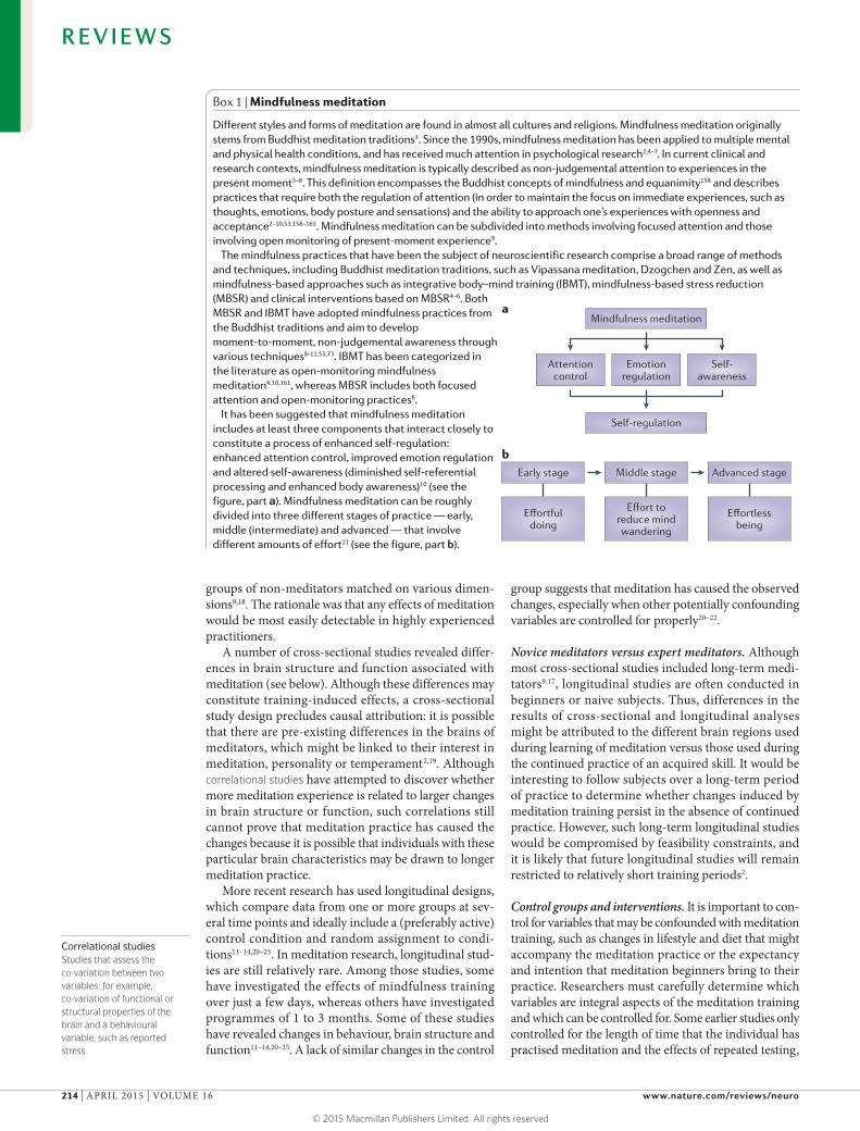
It has been suggested that mindfulness meditation includes at least three components that interact closely to constitute a process of enhanced self-regulation: enhanced attention control, improved emotion regulation and altered self-awareness (diminished self-referential processing and enhanced body awareness)10 (see the figure, part a). Mindfulness meditation can be roughly divided into three different stages of practice — early, middle (intermediate) and advanced — that involve different amounts of effort11 (see the figure, part b).
Nature Reviews | Neuroscience
a
b
Mindfulness meditation
Middle stage Advanced stageEarly stage
Effort toreduce mindwandering
Effortlessbeing
Effortfuldoing
Attentioncontrol
Emotionregulation
Self-awareness
Self-regulation
R E V I E W S
214 | APRIL 2015 | VOLUME 16 www.nature.com/reviews/neuro
© 2015 Macmillan Publishers Limited. All rights reserved
Blood-oxygen-level-dependent contrasts(BOLD contrasts). Signals that can be extracted with functional MRI and that reflect the change in the amount of deoxyhaemoglobin that is induced by changes in the activity of neurons and their synapses in a region of the brain. The signals thus reflect the activity in a local brain region.
Arterial spin labelling(ASL). An MRI technique that is capable of measuring cerebral blood flow in vivo. It provides cerebral perfusion maps without requiring the administration of a contrast agent or the use of ionizing radiation because it uses magnetically labelled endogenous blood water as a freely diffusible tracer.
Brain stateThe reliable patterns of brain activity that involve the activation and/or connectivity of multiple large-scale brain networks.
Fractional anisotropyA parameter in diffusion tensor imaging, which images brain structures by measuring the diffusion properties of water molecules. It provides information about the microstructural integrity of white matter.
but more recent studies have developed and included active interventions in control groups — such as stress management education26, relaxation training14,23,27 or health enhancement programmes20–22 — that can control for variables such as social interaction with the group and teachers, amount of home exercise, physical exercise and psychoeducation. These studies are therefore better able to extract and delineate the meditation-specific effects. For example, one study investigating short-term medita-tion training used a ‘sham meditation’ condition in which participants thought they were meditating, but did not receive proper meditation instructions, which allowed the researchers to control for factors such as expec-tancy, body posture and attention from the teacher28. Mechanistic studies ideally need to use interventions that are as effective as mindfulness meditation in producing the beneficial effects on target variables but that allow for assessment of the unique mechanism underlying the mindfulness practice23,29.
Control conditions in functional imaging. Although all functional neuroimaging studies must use appropriate comparison conditions, this challenge is particularly important when imaging meditative states (BOX 2). The comparison condition should be one in which a state of mindfulness meditation is not present. Many studies use resting comparison conditions, but a problem with this is that experienced practitioners are likely to enter into a state of meditation when at rest. However, other active tasks introduce additional brain activity that ren-ders the comparison difficult to interpret. Using imaging protocols that do not rely on blood-oxygen-level-dependent contrasts (BOLD contrasts), such as arterial spin labelling, might be a possible solution for this problem30.
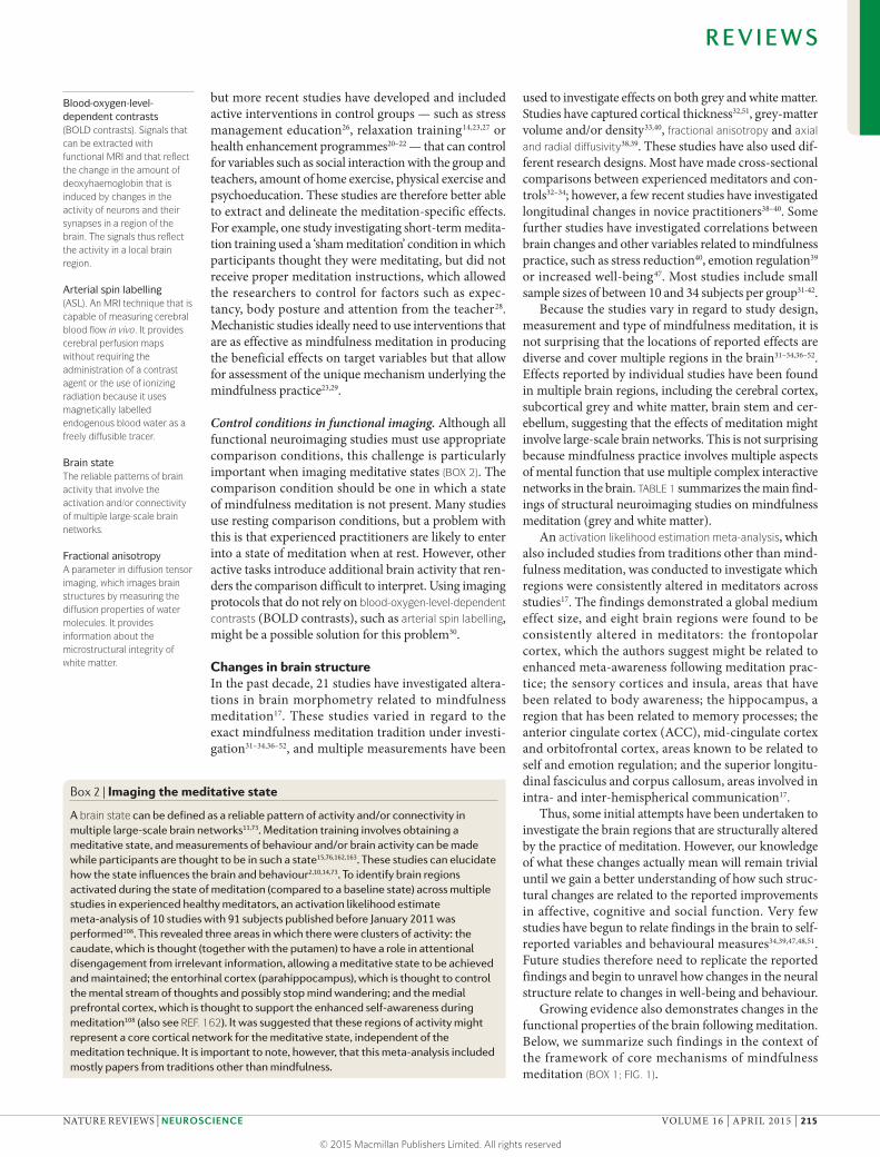
Changes in brain structureIn the past decade, 21 studies have investigated altera-tions in brain morphometry related to mindfulness meditation17. These studies varied in regard to the exact mindfulness meditation tradition under investi-gation31–34,36–52, and multiple measurements have been
used to investigate effects on both grey and white matter. Studies have captured cortical thickness32,51, grey-matter volume and/or density33,40, fractional anisotropy and axial and radial diffusivity38,39. These studies have also used dif-ferent research designs. Most have made cross-sectional comparisons between experienced meditators and con-trols32–34; however, a few recent studies have investigated longitudinal changes in novice practitioners38–40. Some further studies have investigated correlations between brain changes and other variables related to mindfulness practice, such as stress reduction40, emotion regulation39 or increased well-being47. Most studies include small sample sizes of between 10 and 34 subjects per group31-42.
Because the studies vary in regard to study design, measurement and type of mindfulness meditation, it is not surprising that the locations of reported effects are diverse and cover multiple regions in the brain31–34,36–52. Effects reported by individual studies have been found in multiple brain regions, including the cerebral cortex, subcortical grey and white matter, brain stem and cer-ebellum, suggesting that the effects of meditation might involve large-scale brain networks. This is not surprising because mindfulness practice involves multiple aspects of mental function that use multiple complex interactive networks in the brain. TABLE 1 summarizes the main find-ings of structural neuroimaging studies on mindfulness meditation (grey and white matter).
An activation likelihood estimation meta-analysis, which also included studies from traditions other than mind-fulness meditation, was conducted to investigate which regions were consistently altered in meditators across studies17. The findings demonstrated a global medium effect size, and eight brain regions were found to be consistently altered in meditators: the frontopolar cortex, which the authors suggest might be related to enhanced meta-awareness following meditation prac-tice; the sensory cortices and insula, areas that have been related to body awareness; the hippocampus, a region that has been related to memory processes; the anterior cingulate cortex (ACC), mid-cingulate cortex and orbitofrontal cortex, areas known to be related to self and emotion regulation; and the superior longitu-dinal fasciculus and corpus callosum, areas involved in intra- and inter-hemispherical communication17.
Thus, some initial attempts have been undertaken to investigate the brain regions that are structurally altered by the practice of meditation. However, our knowledge of what these changes actually mean will remain trivial until we gain a better understanding of how such struc-tural changes are related to the reported improvements in affective, cognitive and social function. Very few studies have begun to relate findings in the brain to self-reported variables and behavioural measures34,39,47,48,51. Future studies therefore need to replicate the reported findings and begin to unravel how changes in the neural structure relate to changes in well-being and behaviour.
Growing evidence also demonstrates changes in the functional properties of the brain following meditation. Below, we summarize such findings in the context of the framework of core mechanisms of mindfulness meditation (BOX 1; FIG. 1).
Box 2 | Imaging the meditative state
A brain state can be defined as a reliable pattern of activity and/or connectivity in multiple large-scale brain networks11,73. Meditation training involves obtaining a meditative state, and measurements of behaviour and/or brain activity can be made while participants are thought to be in such a state15,76,162,163. These studies can elucidate how the state influences the brain and behaviour2,10,14,73. To identify brain regions activated during the state of meditation (compared to a baseline state) across multiple studies in experienced healthy meditators, an activation likelihood estimate meta-analysis of 10 studies with 91 subjects published before January 2011 was performed108. This revealed three areas in which there were clusters of activity: the caudate, which is thought (together with the putamen) to have a role in attentional disengagement from irrelevant information, allowing a meditative state to be achieved and maintained; the entorhinal cortex (parahippocampus), which is thought to control the mental stream of thoughts and possibly stop mind wandering; and the medial prefrontal cortex, which is thought to support the enhanced self-awareness during meditation108 (also see REF. 162). It was suggested that these regions of activity might represent a core cortical network for the meditative state, independent of the meditation technique. It is important to note, however, that this meta-analysis included mostly papers from traditions other than mindfulness.
R E V I E W S
NATURE REVIEWS | NEUROSCIENCE VOLUME 16 | APRIL 2015 | 215
© 2015 Macmillan Publishers Limited. All rights reserved
Axial and radial diffusivityDerived from the eigenvalues of the diffusion tensor, their underlying biophysical properties are associated with axonal density and myelination, respectively.
Activation likelihood estimation meta-analysisA technique for coordinate-based meta-analysis of neuroimaging data. It determines the convergence of foci reported from different experiments, weighted by the number of participants in each study.
Mindfulness and attentionMany meditation traditions emphasize the necessity to cultivate attention regulation early in the practice9,53. A sufficient degree of attentional control is required to stay engaged in meditation, and meditators often report improved attention control as an effect of repeated prac-tice10,14. Multiple studies have experimentally investigated such effects54.
Components of attention. Attention is often subdivided into three different components: alerting (readiness in preparation for an impending stimulus, which includes tonic effects that result from spending time on a task (vigilance) and phasic effects that are due to brain changes induced by warning signals or targets); orienting (the selection of specific information from multiple sen-sory stimuli); and conflict monitoring (monitoring and resolution of conflict between computations in different
neural areas, also referred to as executive attention)55,56. Other distinctions between types of attention refer to combinations of these three components. For example, sustained attention refers to the sense of vigilance during long continued tasks and may involve both tonic alerting and orienting, whereas selective attention may involve either orienting (when a stimulus is present) or executive function (when stored information is involved).
Performance in these three basic domains can be measured with the attention network test (ANT)57. This test uses as a target an arrow pointing left or right. The target is surrounded by flankers, and subtracting reac-tion times to congruent stimuli (that is, those on the side of the screen indicated by the arrow) from reaction times to incongruent stimuli produces a measure of the time to resolve conflict. The inclusion of cues that indicate when or where the target will occur allows the measurement of alerting and orienting. These measures are used to
Table 1 | Structural changes in the brain associated with mindfulness meditation
Meditation tradition*
Control Sample size of meditation (M) and control (C) groups
Type of measurement Key areas affected‡ Refs
Cross-sectional studies (non-clinical samples)
Insight Non-meditators M: 20, C: 15 Cortical thickness Right anterior insula and right middle and superior frontal sulci
32
Zen Non-meditators M: 13, C: 13 Grey-matter volume Less age-related decline at left putamen 34
Insight Non-meditators M: 20, C: 20 Grey-matter density Right anterior insula, left inferior temporal gyrus and right hippocampus
31
Tibetan Dzogchen
Non-meditators M: 10, C: 10 Grey-matter density Medulla oblongata, left superior and inferior frontal gyrus, anterior lobe of the cerebellum (bilateral) and left fusiform gyrus
33
Zen Non-meditators M: 17, C: 18 Cortical thickness Right dorsal anterior cingulate cortex and secondary somatosensory cortices (bilateral)
51
MBSR Non-meditators M: 20, C: 16 Grey-matter volume Left caudate nucleus 52
Zen Non-meditators M: 10, C: 10 DTI: mean diffusivity and fractional anisotropy
Lower mean diffusivity in left posterior parietal white matter and lower fractional anisotropy in left primary sensorimotor cortex grey matter
37
Longitudinal studies (non-clinical samples)
IBMT (4 weeks)
Active control: relaxation training
M: 22, C: 23 DTI: FA and grey-matter volume
FA increased for left anterior corona radiata, superior corona radiata (bilateral), left superior longitudinal fasciculus, genu and body of corpus callosum. No effect on grey-matter volume
38
MBSR Individuals on a waiting list
M: 16, C: 17 Grey-matter density Left hippocampus, left posterior cingulate gyrus, cerebellum and left middle temporal gyrus
40
IBMT (2 weeks)
Active control: relaxation training
M: 34, C: 34 DTI: FA, radial diffusivity and axial diffusivity
Decrease of axial diffusivity in corpus callosum, corona radiata, superior longitudinal fasciculus, posterior thalamic radiation and sagittal striatum
39
Longitudinal studies (clinical samples)
MBI Usual care (patients with Parkinson disease)
M: 14, C: 13 Grey-matter density Caudate (bilateral), left inferior temporal lobe, hippocampus (bilateral), left occipital cuneus and other small clusters; anterior cerebellum increased in usual care group
42
MBSR Waiting list (patients with mild cognitive impairment)
M: 8, C: 5 Hippocampal volume (region of interest analysis)
Trend towards less hippocampal atrophy 41
DTI, diffusion tensor imaging; FA, fractional anisotropy; IBMT, integrative body–mind training; MBI, mindfulness-based intervention; MBSR, mindfulness-based stress reduction. *Studies that include meditators from traditions other than mindfulness or studies only investigating correlations with other variables are not listed. ‡Meditators show increased values, unless otherwise noted.
R E V I E W S
216 | APRIL 2015 | VOLUME 16 www.nature.com/reviews/neuro
© 2015 Macmillan Publishers Limited. All rights reserved
quantify efficiency in each of the three networks that support the individual components of attention. Alerting involves the brain’s noradrenaline system, which origi-nates in the locus coeruleus. Orienting involves frontal and parietal areas, including the frontal eye fields and inferior and superior parietal lobe. The executive net-work involved in conflict resolution involves the ACC, anterior insula and basal ganglia58,59.
Effects of mindfulness meditation on attention. The ANT and other experimental paradigms have been used to investigate the effects of meditation on atten-tional performance60. Improved conflict monitoring was reported in several studies14,61–64. For example, a longitu-dinal study showed that only 5 days (20 min per day) of integrative body–mind training (IBMT) led to improved conflict monitoring14. In addition, cross-sectional stud-ies of 3 months of mindfulness meditation showed a reduced attentional blink (a lapse in attention following a stimulus within a rapid stream of presented stimuli that has been related to executive function65,66) follow-ing training64 (also see REF. 67). Better performance in conflict monitoring has also been demonstrated in expe-rienced meditators in cross-sectional studies68. However, although altered attention is a common finding in these well-designed meditation studies, some studies investi-gating mindfulness-based stress reduction (MBSR) have not observed effects on conflict monitoring69,70.
Most of the studies on the effects of short-term (1 week) mindfulness meditation on alerting have not found significant effects, but studies investigating long-term meditators (ranging from months to years) did detect changes in alerting27,70–72. Enhanced orienting has been reported in some cross-sectional studies using longer periods of training. For example, 3 months of Shamatha mindfulness training improved tonic alert-ness (the ability to remain alert over time) and allowed for improved orienting towards a visual target in com-parison to controls71. However, 8 weeks of MBSR did not improve measures of sustained attention in a continuous
performance task that measured aspects of tonic alertness, but did show some improvement in orienting22. We do not know whether the differences in the findings of these studies are due to the type of training, type of control or other subtle factors.
A systematic review that compiled the findings of these studies (as well as the effects on other measures of cognition) concluded that early phases of mindfulness meditation might be associated with improvements in conflict monitoring and orienting, whereas later phases might be mainly associated with improved alerting60. It is currently still unclear how different meditation practices differentially affect the specific attentional components2,9,53. In addition, the length of practice needs to be defined more consistently in future research.
Neural mechanisms of enhanced attention control. Several functional and structural MRI studies on mind-fulness training have investigated neuroplasticity in brain regions supporting attention regulation. The brain region to which the effects of mindfulness training on atten-tion is most consistently linked is the ACC11,23,38,39,73–76. The ACC enables executive attention and control77–79 by detecting the presence of conflicts emerging from incom-patible streams of information processing. The ACC and the fronto-insular cortex form part of a network that facilitates cognitive processing through long-range con-nections to other brain areas11,80. Cross-sectional studies have reported enhanced activation of regions of the ACC in experienced meditators compared to controls during focused attention meditation76 or when mindfully antici-pating delivery of a painful stimulus81. Greater activation of the ventral and/or rostral ACC during the resting state following 5 days of IBMT was also found in an actively controlled, randomized, longitudinal study23. Although ACC activation may be enhanced in earlier stages of mindfulness meditation, it might decrease with higher levels of expertise, as demonstrated in a cross-sectional study18. Structural MRI data suggest that mindfulness meditation might be associated with greater cortical thickness51 and might lead to enhanced white-matter integrity in the ACC38,39.
Other attention-related brain regions in which func-tional changes have been observed following mindful-ness meditation include the dorsolateral prefrontal cortex (PFC), where responses were enhanced during executive processing82, as revealed by a randomized longitudi-nal study, and parietal attention regions, which showed greater activation following an MBSR course in people with social anxiety, as demonstrated by an uncontrolled longitudinal study83. Furthermore, a diminished age-related decline of grey-matter volume in the putamen as well as diminished age-related decline in sustained atten-tion performance were found in a cross-sectional study of Zen meditation practitioners34.
Although there is evidence that brain regions rele-vant for the regulation of attention show functional and structural changes following mindfulness meditation practice, it has not yet been determined whether these changes are actually related to the improved attentional performance. Longitudinal studies that use measures
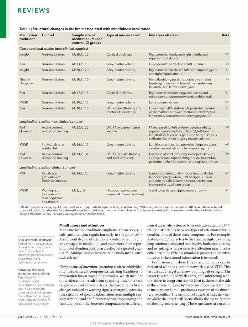
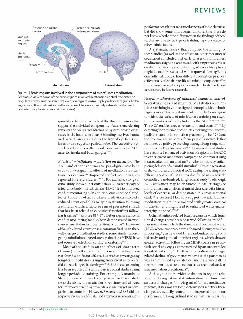
Figure 1 | Brain regions involved in the components of mindfulness meditation. Schematic view of some of the brain regions involved in attention control (the anterior cingulate cortex and the striatum), emotion regulation (multiple prefrontal regions, limbic regions and the striatum) and self-awareness (the insula, medial prefrontal cortex and posterior cingulate cortex and precuneus).
Nature Reviews | Neuroscience
Posterior cingulate cortex/precuneus
Multipleprefrontalregions
Medialprefrontalcortex
Insula
Striatum
Amygdala
Medial view Lateral view
Anterior cingulate cortex
R E V I E W S
NATURE REVIEWS | NEUROSCIENCE VOLUME 16 | APRIL 2015 | 217
© 2015 Macmillan Publishers Limited. All rights reserved
of attentional performance along with functional MRI (fMRI) are needed. If supported by more rigorous future research, the evidence of improved attention reg-ulation and strengthened brain activity in the regions underlying attentional control following mindfulness meditation might be promising for the treatment of psychiatric disorders in which there are deficiencies in these functions74,84,85.
Mindfulness and emotion regulationEnhanced emotion regulation has been suggested to underlie many of the beneficial effects of mindfulness meditation. Emotion regulation refers to strategies that can influence which emotions arise and when, how long they occur, and how these emotions are experienced and expressed. A range of implicit and explicit emotion regu-lation processes has been proposed86, and mindfulness-based emotion regulation may involve a mix of these processes, including attentional deployment (attending to mental processes, including emotions), cognitive change (altering typical patterns of appraisal regarding one’s emotions) and response modulation (decreasing tonic levels of suppression).
Effects of mindfulness meditation on emotion regulation. Improvements in emotion regulation associated with mindfulness meditation have been investigated through various approaches, including experimental studies, self-reporting studies, measurement of peripheral physiology and neuroimaging10. These studies have reported various positive effects of mindfulness meditation on emotional processing, such as a reduction in emotional interference by unpleasant stimuli87, decreased physiological reac-tivity and facilitated return to emotional baseline after response to a stressor film88, and decreased self-reported difficulties in emotion regulation89. Consequently, low-ered intensity and frequency of negative affect90,91 and improved positive mood states14,91,92 are reported to be associated with mindfulness meditation.
Neural mechanisms of improved emotion regulation. Neuroimaging studies that have probed the enhanced emotion regulation associated with mindfulness medita-tion in an attempt to identify the underlying brain acti-vation patterns typically present study participants with emotional pictures82,93–97, words and/or statements29,98 and instruct them to encounter these with a state of mindfulness or a simple baseline state.
The hypothesis that drives many of these studies is that mindful emotion regulation works by strengthen-ing prefrontal cognitive control mechanisms and thus downregulates activity in regions relevant to affect pro-cessing, such as the amygdala. Present-moment awareness and non-judgemental acceptance through mindfulness meditation8,10 are thought to be crucial in promoting cognitive control because they increase sensitivity to affective cues that help to signal the need for control99. Studies have therefore investigated whether mindfulness training exerts its effects through enhanced top-down control or facilitated bottom-up processing100. The find-ings (outlined below) suggest that the level of expertise
is important, with beginners showing a different pat-tern from expert meditators. However, although several studies have pointed to the involvement of fronto-limbic regions, very few studies have begun to relate changes in these regions to changes in measures of behaviour or well-being10.
A frequently reported finding is that mindfulness practice leads to (or is associated with) a diminished acti-vation of the amygdala in response to emotional stimuli during mindful states83,94,95 as well as in a resting state93, suggesting a decrease in emotional arousal. However, although such results have been reported for meditation beginners, they have less consistently been detected in experienced meditators95 (but see REF. 18).
Prefrontal activations are often enhanced as an effect of mindfulness meditation in novice meditators (but see REF. 29): for example, greater dorsolateral PFC responses were found during executive processing within an emo-tional Stroop task in healthy individuals after 6 weeks of mindfulness training82. Enhanced dorsomedial and dorsolateral PFC activation was also detected when par-ticipants expected to see negative images while engaging in a mindful state94. Moreover, after an MBSR course, an enhanced activation in the ventrolateral PFC in people suffering from anxiety was found when they labelled the affect of emotional images97. By contrast, experienced meditators have been found to show diminished acti-vation in medial PFC regions95. This finding could be interpreted as indicating reduced control (disengagement of elaboration and appraisal) and greater acceptance of affective states.
Neuroimaging studies of ameliorated pain process-ing through mindfulness meditation have also pointed to expertise-related differences in the extent of cogni-tive control over sensory experience. Meditation begin-ners showed increased activity in areas involved in the cognitive regulation of nociceptive processing (the ACC and anterior insula) and areas involved in reframing the evaluation of stimuli (the orbitofrontal cortex), along with reduced activation in the primary somatosensory cortex in a 4-day longitudinal study with no control group30, whereas meditation experts were characterized by decreased activation in dorsolateral and ventrolateral PFC regions and enhancements in primary pain processing regions (the insula, somatosensory cortex and thalamus) compared with controls in two cross-sectional studies35,81.
These findings are in line with the assumption that the process of mindfulness meditation is characterized as an active cognitive regulation in meditation begin-ners, who need to overcome habitual ways of internally reacting to one’s emotions and might therefore show greater prefrontal activation. Expert meditators might not use this prefrontal control. Rather, they might have automated an accepting stance towards their experience and thus no longer engage in top-down control efforts but instead show enhanced bottom-up processing100.
In the early stages of meditation training, achieving the meditation state seems to involve the use of atten-tional control and mental effort; thus, areas of the lat-eral prefrontal and parietal cortex are more active than before training11,16,100,101. This may reflect the higher level
R E V I E W S
218 | APRIL 2015 | VOLUME 16 www.nature.com/reviews/neuro
© 2015 Macmillan Publishers Limited. All rights reserved
of effort often found when participants struggle to obtain the meditation state in the early stages11,73,98,102. However, in the advanced stages, prefrontal–parietal activity is often reduced or eliminated, but ACC, striatum and insula activity remains9,10,53,73,76,101–103. Whether effort has a key role in PFC and ACC activation during or following meditation needs further investigation.
Analysis of functional connectivity between regions of the fronto-limbic network could help to further eluci-date the regulatory function of executive control regions. Only a few studies have included such analyses. One cross-sectional study on pain processing in meditators demonstrated decreased connectivity of executive and pain-related brain regions35, and one study of mindful-ness-naive smokers demonstrated reduced connectivity between craving-related brain regions during a mindful-ness condition compared to passive viewing of smoking-related images during cigarette craving96, suggesting a functional decoupling of involved regions. Another longitudinal, randomized study reported that people suffering from anxiety showed a change from a negative correlation between the activity of frontal regions and that of the amygdala before intervention (that is, negative connectivity) to a positive correlation between the activity of these regions (positive connectivity) after a mindful-ness intervention97. Because such a negative correlation will occur when prefrontal regions downregulate limbic activation104,105, it was speculated that the positive coupling between the activity of the two regions after mindfulness intervention might indicate that meditation involves monitoring of arousal rather than a downregulation or suppression of emotional responses, and that it might be a unique signature of mindful emotion regulation.
Importantly, this study also investigated the correlation between neural and self-reported findings and demon-strated that the changes in PFC–amygdala connectivity were correlated with anxiety symptom improvement. Further studies are needed to elucidate the complex interplay between regions of the fronto-limbic network in mindfulness meditation.
Although the proposed similarities between mind-fulness and the reappraisal strategy of emotion regula-tion have been much debated, there is some evidence that mindfulness also shares similarities with extinction processes (BOX 3).
Brain regions involved in motivation and reward pro-cessing also show functional alterations that are related to mindfulness training, such as stronger activity of the puta-men and caudate during a resting state following mindful-ness training23 and lower activation in the caudate nucleus during reward anticipation in experienced meditators106. These studies might indicate altered self-regulation in the motivational realm, with possibly reduced suscepti-bility to incentives and enhanced reward-related activity during rest.
Brain regions involved in the regulation of emotions have also shown structural changes following mindful-ness meditation31,32,38–41,48,51. Although these findings pro-vide some initial evidence that these brain regions are related to mindfulness practice, the question of whether they are involved in mediating the beneficial effects of mindfulness meditation remains largely unanswered.
Mindfulness and self-awarenessAccording to Buddhist philosophy, the identification with a static concept of ‘self ’ causes psychological distress. Dis-identification from such a static self-concept results in the freedom to experience a more genuine way of being. Through enhanced meta-awareness (making awareness itself an object of attention), mindfulness meditation is thought to facilitate a detachment from identification with the self as a static entity3,10,107 and a tendency to iden-tify with the phenomenon of ‘experiencing’ itself is said to emerge15,108–112. Currently, empirical research into this area is only just emerging111,113, and the few interpretations of connections between neuroimaging findings and self-reported data — which we will summarize briefly below — are suggestive at best.
Self-referential processing. Altered self-representation has been investigated with questionnaire studies. Early stud-ies reported mindfulness training to be associated with a more positive self-representation, higher self-esteem, higher acceptance of oneself114 and styles of self-concept that are typically associated with less-severe pathological symptoms115. Meditators have also been shown to score higher than non-meditators on a scale that measures non-attachment116: a construct that is based on insight into the constructed and impermanent nature of mental representations. Although such concepts are not easy to capture in experimental and neuroscientific studies, find-ings from a few recent studies seem to suggest that brain structures supporting self-referential processing might be affected by mindfulness meditation98,117,118.
Box 3 | Mindfulness meditation as exposure therapy
Exposure therapy aims for patients to extinguish a fear response and instead to acquire a sense of safety in the presence of a formerly feared stimulus by exposing them to that stimulus and preventing the usual response164. Mindfulness meditation resembles an exposure situation because practitioners ‘turn towards their emotional experience’, bring acceptance to bodily and affective responses, and refrain from engaging in internal reactivity towards it. Research on fear conditioning has helped to identify a network of brain regions that are crucial for the extinction of conditioned fear responses and the retention of extinction165. This network includes the ventromedial prefrontal cortex (vmPFC), which is important for a successful recall of the extinction; the hippocampus166, which is related to signalling the extinguished context (contextual safety); and the amygdala, which has a crucial role during the acquisition and expression of conditioned fear167 and is thought to be downregulated by the vmPFC and the hippocampus105,168. Activation in the vmPFC (subgenual anterior cingulate cortex) is primarily linked to the expression of fear learning during a delayed test of extinction and is critical for the retention of extinction169.
There is emerging evidence from MRI studies that the aforementioned brain regions show structural and functional changes following mindfulness meditation training (see main text). This overlap of involved brain regions, as well as the conceptual similarity between mindfulness and an exposure situation, suggest that mindfulness training might enhance the ability to extinguish conditioned fear by structurally and functionally affecting the brain network that supports safety signalling. The capacity for successful extinction memories reliably differentiates healthy from pathological conditions170,171, and is crucial in order to overcome maladaptive states. It helps individuals to learn to have no fear response to neutral stimuli when there is no adaptive function for a fear response. Instead, individuals can experience a sense of safety and can flexibly elicit other emotions and behaviours.
R E V I E W S
NATURE REVIEWS | NEUROSCIENCE VOLUME 16 | APRIL 2015 | 219
© 2015 Macmillan Publishers Limited. All rights reserved
Although there is much debate about its exact function, a widespread view holds that the default mode network (DMN)119,120 is involved in self-referential processing. This network includes midline structures of the brain, such as areas of the medial PFC, posterior cingulate cortex (PCC), anterior precuneus and inferior parietal lobule121,122. These structures show high activity during rest, mind wandering and conditions of stimulus-independent thought121 and have been suggested to support diverse mechanisms by which an individual can ‘project’ themselves into another perspective123. fMRI studies have investigated activity in the DMN in association with mindfulness practice. Regions of the DMN (the medial PFC and PCC) showed relatively little activity in meditators compared to con-trols across different types of meditation, which has been interpreted as indicating diminished self-referential processing117. Functional connectivity analysis revealed stronger coupling in experienced meditators between the PCC, dorsal ACC and dorsolateral PFC, both at baseline and during meditation, which was interpreted as indi-cating increased cognitive control over the function of the DMN117. Increased functional connectivity was also found between DMN regions and the ventromedial PFC in participants with more compared to less meditation experience118. It has been speculated that this increased connectivity with ventromedial PFC regions supports greater access of the default circuitry to information about internal states because this region is highly interconnected with limbic regions118.
Awareness of present-moment experiences. Evaluative self-referential processing is assumed to decrease as an effect of mindfulness meditation, whereas awareness of present-moment experiences is thought to be enhanced. Mindfulness practitioners often report that the practice of attending to present-moment body sensations results in an enhanced awareness of bodily states and greater per-ceptual clarity of subtle interoception. Empirical findings to support this claim are mixed. Although studies that assessed performance on a heartbeat detection task — a standard measure of interoceptive awareness — found no evidence that meditators had superior performance to non-meditators124,125, other studies found that meditators showed greater coherence between objective physiological data and their subjective experience in regard to an emo-tional experience126 and the sensitivity of body regions127.
Multiple studies have shown the insula to be impli-cated in mindfulness meditation: it shows stronger acti-vation during compassion meditation128 and following mindfulness training23,52,98, and has greater cortical thick-ness in experienced meditators32. Given its known role in awareness129, it is conceivable that enhanced insula activ-ity in meditators might represent the amplified awareness of present-moment experience.
Similarly, a study reported an uncoupling of the right insula and medial PFC and increased connectivity of the right insula with dorsolateral PFC regions in individuals after mindfulness training98. The authors interpret their findings as a shift in self-referential processing towards a more self-detached and objective analysis of interocep-tive and exteroceptive sensory events, rather than their
affective or subjective self-referential value. Furthermore, a preliminary analysis from a study of a state of ‘non-dual awareness’ (a state of awareness in which perceived duali-ties, such as the distinction between subject and object, are absent) showed a decreased functional connectivity of the central precuneus with the dorsolateral PFC. The author speculates that this finding might be indicative of a state in which awareness is itself the subject of awareness111.
Together, the findings from these studies have been taken to suggest that mindfulness meditation might alter the self-referential mode so that a previous narrative, evaluative form of self-referential processing is replaced by greater awareness98,111. We suggest that this shift in self-awareness is one of the major active mechanisms of the beneficial effects of mindfulness meditation. However, because these interpretations are built on a still-fragmentary understanding of the function of the involved brain regions, future research will need to test and elaborate these assumptions.
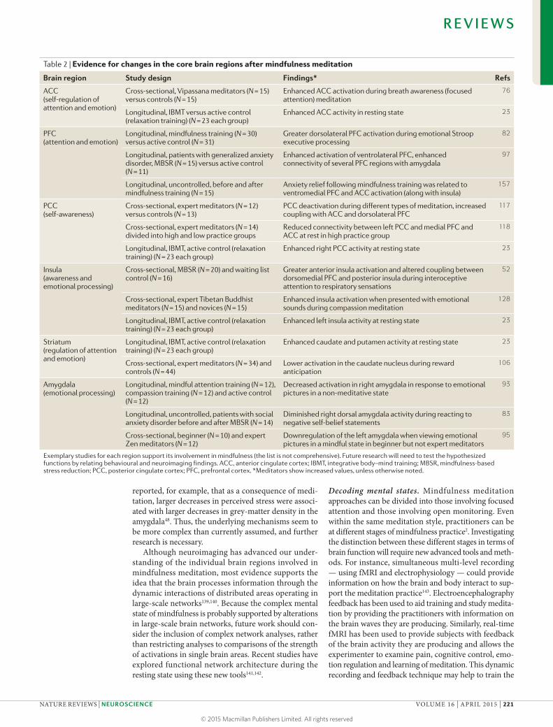
Across the functional and structural MRI studies that have been published to date, especially those based on the longitudinal, randomized, controlled studies with active control groups and meta-analyses, the ACC, PFC, PCC, insula, striatum (caudate and putamen) and amyg-dala seem to show consistent changes associated with mindfulness meditation9–11,13,17,23,34,73,108,130 (FIG. 1; TABLE 2). We consider these areas to be the core regions involved in self-regulation of attention, emotion and awareness following mindfulness training. However, we acknowl-edge that many other brain areas are also involved in mindfulness practice and warrant further investigation using rigorous randomized and controlled designs.
Future questionsMechanisms of mindfulness-induced changes. A num-ber of studies seem to suggest that mindfulness medita-tion induces changes in brain structure and function, raising the question of which underlying mechanisms support these processes. It is possible that engaging the brain in mindfulness affects brain structure by inducing dendritic branching, synaptogenesis, myelinogenesis or even adult neurogenesis. Alternatively, it is possible that mindfulness positively affects autonomic regulation and immune activity, which may result in neuronal preser-vation, restoration and/or inhibition of apoptosis14,23,131. It is well known that mindfulness-based techniques are highly effective in stress reduction, and it is possible that such stress reduction may mediate changes in brain function14,48,132–137 (BOX 4). A combination of all of these mechanisms may even occur.
It is also important to realize that the direction of the observed effects of mindfulness meditation has not been consistent across all studies. Although larger values in meditators compared to controls are predomi-nantly reported, a cross-sectional study also revealed smaller fractional anisotropy and cortical thickness values in meditators in some brain regions, including the medial PFC, postcentral and inferior parietal cor-tices, PCC and medial occipital cortex138. Along these lines, mindfulness-induced increases are predominantly observed in longitudinal studies. However, it was also
R E V I E W S
220 | APRIL 2015 | VOLUME 16 www.nature.com/reviews/neuro
© 2015 Macmillan Publishers Limited. All rights reserved
reported, for example, that as a consequence of medi-tation, larger decreases in perceived stress were associ-ated with larger decreases in grey-matter density in the amygdala48. Thus, the underlying mechanisms seem to be more complex than currently assumed, and further research is necessary.
Although neuroimaging has advanced our under-standing of the individual brain regions involved in mindfulness meditation, most evidence supports the idea that the brain processes information through the dynamic interactions of distributed areas operating in large-scale networks139,140. Because the complex mental state of mindfulness is probably supported by alterations in large-scale brain networks, future work should con-sider the inclusion of complex network analyses, rather than restricting analyses to comparisons of the strength of activations in single brain areas. Recent studies have explored functional network architecture during the resting state using these new tools141,142.
Decoding mental states. Mindfulness meditation approaches can be divided into those involving focused attention and those involving open monitoring. Even within the same meditation style, practitioners can be at different stages of mindfulness practice2. Investigating the distinction between these different stages in terms of brain function will require new advanced tools and meth-ods. For instance, simultaneous multi-level recording — using fMRI and electrophysiology — could provide information on how the brain and body interact to sup-port the meditation practice143. Electroencephalography feedback has been used to aid training and study medita-tion by providing the practitioners with information on the brain waves they are producing. Similarly, real-time fMRI has been used to provide subjects with feedback of the brain activity they are producing and allows the experimenter to examine pain, cognitive control, emo-tion regulation and learning of meditation. This dynamic recording and feedback technique may help to train the
Table 2 | Evidence for changes in the core brain regions after mindfulness meditation
Brain region Study design Findings* Refs
ACC (self-regulation of attention and emotion)
Cross-sectional, Vipassana meditators (N = 15) versus controls (N = 15)
Enhanced ACC activation during breath awareness (focused attention) meditation
76
Longitudinal, IBMT versus active control (relaxation training) (N = 23 each group)
Enhanced ACC activity in resting state 23
PFC (attention and emotion)
Longitudinal, mindfulness training (N = 30) versus active control (N = 31)
Greater dorsolateral PFC activation during emotional Stroop executive processing
82
Longitudinal, patients with generalized anxiety disorder, MBSR (N = 15) versus active control (N = 11)
Enhanced activation of ventrolateral PFC, enhanced connectivity of several PFC regions with amygdala
97
Longitudinal, uncontrolled, before and after mindfulness training (N = 15)
Anxiety relief following mindfulness training was related to ventromedial PFC and ACC activation (along with insula)
157
PCC (self-awareness)
Cross-sectional, expert meditators (N = 12) versus controls (N = 13)
PCC deactivation during different types of meditation, increased coupling with ACC and dorsolateral PFC
117
Cross-sectional, expert meditators (N = 14) divided into high and low practice groups
Reduced connectivity between left PCC and medial PFC and ACC at rest in high practice group
118
Longitudinal, IBMT, active control (relaxation training) (N = 23 each group)
Enhanced right PCC activity at resting state 23
Insula (awareness and emotional processing)
Cross-sectional, MBSR (N = 20) and waiting list control (N = 16)
Greater anterior insula activation and altered coupling between dorsomedial PFC and posterior insula during interoceptive attention to respiratory sensations
52
Cross-sectional, expert Tibetan Buddhist meditators (N = 15) and novices (N = 15)
Enhanced insula activation when presented with emotional sounds during compassion meditation
128
Longitudinal, IBMT, active control (relaxation training) (N = 23 each group)
Enhanced left insula activity at resting state 23
Striatum (regulation of attention and emotion)
Longitudinal, IBMT, active control (relaxation training) (N = 23 each group)
Enhanced caudate and putamen activity at resting state 23
Cross-sectional, expert meditators (N = 34) and controls (N = 44)
Lower activation in the caudate nucleus during reward anticipation
106
Amygdala (emotional processing)
Longitudinal, mindful attention training (N = 12), compassion training (N = 12) and active control (N = 12)
Decreased activation in right amygdala in response to emotional pictures in a non-meditative state
93
Longitudinal, uncontrolled, patients with social anxiety disorder before and after MBSR (N = 14)
Diminished right dorsal amygdala activity during reacting to negative self-belief statements
83
Cross-sectional, beginner (N = 10) and expert Zen meditators (N = 12)
Downregulation of the left amygdala when viewing emotional pictures in a mindful state in beginner but not expert meditators
95
Exemplary studies for each region support its involvement in mindfulness (the list is not comprehensive). Future research will need to test the hypothesized functions by relating behavioural and neuroimaging findings. ACC, anterior cingulate cortex; IBMT, integrative body–mind training; MBSR, mindfulness-based stress reduction; PCC, posterior cingulate cortex; PFC, prefrontal cortex. *Meditators show increased values, unless otherwise noted.
R E V I E W S
NATURE REVIEWS | NEUROSCIENCE VOLUME 16 | APRIL 2015 | 221
© 2015 Macmillan Publishers Limited. All rights reserved
Multivariate pattern analysisA method of analysing functional MRI data that is capable of detecting and characterizing information represented in patterns of activity distributed within and across multiple regions of the brain. Unlike univariate approaches, which only identify magnitudes of activity in localized parts of the brain, this approach can monitor multiple areas at once.
subjects effectively and allow their mental states at dif-ferent stages of mindfulness training to be decoded from their brain activity144–146, possibly by applying techniques such as multivariate pattern analysis147.
Interpretations of study outcomes remain tenta-tive until they are clearly linked to subjective reports or behavioural findings. Future studies should therefore increasingly draw connections between behavioural outcomes and neuroimaging data using the advanced multi-level analyses mentioned above.
Investigating individual differences. People respond to mindfulness meditation differently. These differences may derive from temperamental, personality or genetic differences. Studies in other fields have suggested that genetic polymorphisms may interact with experience to
influence the success of training148. Because mindfulness meditation affects the activation and connectivity of the ACC, PFC and other brain regions involved in cognitive control and emotion regulation, it might be helpful to examine these polymorphisms to determine their possi-ble influence on the success of meditation practice2,59,149. Moreover, individual differences in personality, lifestyle, life events and trainer–trainee dynamics are likely to have substantial influence on training effects, although little is known about these influences. Mood and personality have been used to predict individual variation in the improve-ment of creative performance following mindfulness meditation150. Capturing temperament and personality differences may serve to predict success in mindfulness training150,151 because different temperament and per-sonality traits are reported to be associated with different electroencephalography patterns and heart-rate variability in Zen meditators152.
Clinical application. Self-regulation deficits are associ-ated with diverse behavioural problems and mental dis-orders, such as increased risk of school failure, attention deficit disorder, anxiety, depression and drug abuse78,153. Convergent findings indicate that mindfulness medita-tion could ameliorate negative outcomes resulting from deficits in self-regulation and could consequently help patient populations suffering from diseases and behav-ioural abnormalities. Several clinical trials have explored the effects of mindfulness meditation on disorders such as depression154, generalized anxiety26, addictions155, attention deficit disorders156 and others42, and have begun to estab-lish the efficiency of mindfulness practice for these condi-tions. Only a few recent studies, however, have investigated the neuroplastic changes underlying these beneficial effects of mindfulness in clinical populations29,41,42,74,97,142,157. Although these studies are promising, future work needs to replicate and expand the emerging findings to optimally tailor interventions for clinical application.
ConclusionsInterest in the psychological and neuroscientific investiga-tion of mindfulness meditation has increased markedly over the past two decades. As is relatively common in a new field of research, studies suffer from low methodo-logical quality and present with speculative post-hoc inter-pretations. Knowledge of the mechanisms that underlie the effects of meditation is therefore still in its infancy. However, there is emerging evidence that mindfulness meditation might cause neuroplastic changes in the struc-ture and function of brain regions involved in regulation of attention, emotion and self-awareness. Further research needs to use longitudinal, randomized and actively con-trolled research designs and larger sample sizes to advance the understanding of the mechanisms of mindfulness meditation in regard to the interactions of complex brain networks, and needs to connect neuroscientific findings with behavioural data. If supported by rigorous research studies, the practice of mindfulness meditation might be promising for the treatment of clinical disorders and might facilitate the cultivation of a healthy mind and increased well-being.
Box 4 | Mindfulness meditation and stress
Stress reduction might be a potential mediator of the effects of mindfulness practice on neural function. Mindfulness meditation has been shown to reduce stress14,132–137; this is most consistently documented in self-reported data132,133. A review of mindfulness-based stress reduction (MBSR) studies showed a non-specific effect on stress reduction, which is similar to that of standard relaxation training134. However, findings in studies that have examined biomarkers of stress, such as cortisol levels, are less consistent: changes in cortisol levels have been found in association with mindfulness training in some studies14,136 but not in others132,135.
The brain is a target for stress and stress-related hormones. It undergoes functional and structural remodelling in response to stress in a manner that is adaptive under normal circumstances but can lead to damage when stress is excessive172. Evidence suggests that vulnerability to stress-induced brain plasticity is prominent in the prefrontal cortex (PFC), hippocampus, amygdala and other areas associated with fear-related memories and self-regulatory behaviours172,173. The interactions between these brain regions determine whether life experiences lead to successful adaptation or maladaptation and impaired mental and physical health173. A study has shown that chronic stress induces less flexibility in attention shifting in the rodent and human adult174. This was paralleled by a reduction in apical dendritic arborization in rodent medial PFC (specifically, in the anterior cingulate cortex) and fewer feedforward PFC connections in humans under stress, effects that recovered when the stressor was removed174. This suggests that the effects of chronic psychosocial stress on PFC function and connectivity are plastic and can change quickly as a function of mental state174. Studies have also shown that moderate to severe stress seems to increase the volume of the amygdala but reduce the volume of the PFC and hippocampus175. Mindfulness training, however, has been shown to enhance grey-matter density in the hippocampus40. Furthermore, after mindfulness training, reductions in perceived stress correlate with reductions in amygdala grey-matter density48. These findings suggest that mindfulness meditation might be a potential intervention and prevention strategy176. Thus, it is possible that mindfulness meditation reduces stress by improving self-regulation, which enhances neuroplasticity and leads to health benefits. It should be noted that mindfulness meditation might also directly modulate stress processing via a ‘bottom-up’ pathway, through which it alters the sympathetic–adrenal–medullary and hypothalamic–pituitary–adrenal axes by increasing activity in the parasympathetic nervous system; thus, mindfulness meditation could prevent sympathetic nervous system fight-or-flight stress responses177,178. Indeed, some research has suggested that mindfulness leads to increased activity of the parasympathetic nervous system23,179.
Brain-derived neurotrophic factor (BDNF) has been linked to numerous aspects of plasticity in the brain. Stress-induced remodelling of the PFC, hippocampus and amygdala coincides with changes in the levels of BDNF, supporting its role as a trophic factor modulating neuronal survival and regulating synaptic plasticity131. However, glucocorticoids and other molecules have been shown to act in conjunction with BDNF to facilitate both morphological and molecular changes. Because some forms of mindfulness meditation training have been found to reduce stress-induced cortisol secretion, this could potentially have neuroprotective effects by increasing levels of BDNF, and future research should explore this possible causal relationship136,149,180.
R E V I E W S
222 | APRIL 2015 | VOLUME 16 www.nature.com/reviews/neuro
© 2015 Macmillan Publishers Limited. All rights reserved
1. Ospina, M. B. et al. Meditation practices for health: state of the research. Evid. Rep. Technol. Assess. (Full Rep.) 155, 1–263 (2007).
2. Tang, Y.-Y. & Posner, M. I. Theory and method in mindfulness neuroscience. Soc. Cogn. Affect. Neurosci. 8, 118–120 (2013).
3. Hart, W. The Art of Living: Vipassana Meditation (Harper and Row, 1987).
4. Ivanovski, B. & Malhi, G. S. The psychological and neurophysiological concomitants of mindfulness forms of meditation. Acta Neuropsychiatr. 19, 76–91 (2007).
5. Chiesa, A. & Malinowski, P. Mindfulness-based approaches: are they all the same? J. Clin. Psychol. 67, 404–424 (2011).
6. Baer, R. A. Mindfulness training as a clinical intervention: a conceptual and empirical review. Clin. Psychol. Sci. Practice 10, 125–143 (2003).
7. Grossman, P. Defining mindfulness by how poorly I think I pay attention during everyday awareness and other intractable problems for psychology’s (re)invention of mindfulness: comment on Brown et al. Psychol. Assess. 23, 1034–1040 (2011).
8. Kabat-Zinn, J. Full Catastrophe Living: Using the Wisdom of Your Body and Mind to Face Stress, Pain, and Illness (Delta Trade Paperbacks, 1990).
9. Lutz, A., Slagter, H. A., Dunne, J. D. & Davidson, R. J. Attention regulation and monitoring in meditation. Trends Cogn. Sci. 12, 163–169 (2008).
10. Hölzel, B. K. et al. How does mindfulness meditation work? Proposing mechanisms of action from a conceptual and neural perspective. Perspect. Psychol. Sci. 6, 537–559 (2011).A review of the mechanisms of meditation.
11. Tang, Y.Y., Rothbart, M. K. & Posner, M. I. Neural correlates of establishing, maintaining and switching brain states. Trends Cogn. Sci. 16, 330–337 (2012).A review of the mechanisms of brain states associated with mental training.
12. Zeidan, F., Johnson, S. K., Diamond, B. J., David, Z. & Goolkasian, P. Mindfulness meditation improves cognition: evidence of brief mental training. Conscious. Cogn. 19, 597–605 (2010).
13. Ding, X. et al. Short-term meditation modulates brain activity of insight evoked with solution cue. Soc. Cogn. Affect. Neurosci. 10, 43–49 (2014).
14. Tang, Y. Y. et al. Short-term meditation training improves attention and self-regulation. Proc. Natl Acad. Sci. USA 104, 17152–17156 (2007).The first longitudinal, randomized study to document that brief training improves executive attention, mood and immune function, and reduces levels of stress hormones.
15. Manna, A. et al. Neural correlates of focused attention and cognitive monitoring in meditation. Brain Res. Bull. 82, 46–56 (2010).
16. Tomasino, B., Fregona, S., Skrap, M. & Fabbro, F. Meditation-related activations are modulated by the practices needed to obtain it and by the expertise: an ALE meta-analysis study. Front. Hum. Neurosci. 6, 346 (2012).
17. Fox, K. C. et al. Is meditation associated with altered brain structure? A systematic review and meta-analysis of morphometric neuroimaging in meditation practitioners. Neurosci. Biobehav. Rev. 43, 48–73 (2014).A review of structural alterations in the brain associated with meditation.
18. Brefczynski-Lewis, J. A., Lutz, A., Schaefer, H. S., Levinson, D. B. & Davidson, R. J. Neural correlates of attentional expertise in long-term meditation practitioners. Proc. Natl Acad. Sci. USA 104, 11483–11488 (2007).One of the first cross-sectional studies to document the neural correlates of focused meditation.
19. Davidson, R. J. Empirical explorations of mindfulness: conceptual and methodological conundrums. Emotion 10, 8–11 (2010).
20. MacCoon, D. G. et al. The validation of an active control intervention for Mindfulness Based Stress Reduction (MBSR). Behav. Res. Ther. 50, 3–12 (2012).One of the first studies to validate the active control conditions in mindfulness training.
21. Rosenkranz, M. A. et al. A comparison of mindfulness-based stress reduction and an active control in modulation of neurogenic inflammation. Brain Behav. Immun. 27, 174–184 (2013).
22. MacCoon, D. G., MacLean, K. A., Davidson, R. J., Saron, C. D. & Lutz, A. No sustained attention differences in a longitudinal randomized trial comparing mindfulness based stress reduction versus active control. PLoS ONE 9, e97551 (2014).
23. Tang, Y. Y. et al. Central and autonomic nervous system interaction is altered by short-term meditation. Proc. Natl Acad. Sci. USA 106, 8865–8870 (2009).
24. Erisman, S. M. & Roemer, L. The effects of experimentally induced mindfulness on emotional responding to film clips. Emotion 10, 72–82 (2010).
25. Leiberg, S., Klimecki, O. & Singer, T. Short-term compassion training increases prosocial behaviour in a newly developed prosocial game. PLoS ONE 6, e17798 (2011).
26. Hoge, E. A. et al. Randomized controlled trial of mindfulness meditation for generalized anxiety disorder: effects on anxiety and stress reactivity. J. Clin. Psychiatry 74, 786–792 (2013).
27. Tang, Y. Y., Yang, L., Leve, L. D. & Harold, G. T. Improving executive function and its neurobiological mechanisms through a mindfulness-based intervention: advances within the field of developmental neuroscience. Child Dev. Perspect. 6, 361–366 (2012).
28. Zeidan, F., Johnson, S. K., Gordon, N. S. & Goolkasian, P. Effects of brief and sham mindfulness meditation on mood and cardiovascular variables. J. Altern. Complement. Med. 16, 867–873 (2010).
29. Goldin, P., Ziv, M., Jazaieri, H., Hahn, K. & Gross, J. J. MBSR versus aerobic exercise in social anxiety: fMRI of emotion regulation of negative self-beliefs. Soc. Cogn. Affect. Neurosci. 8, 65–72 (2013).One of the first randomized mindfulness studies to document the neural mechanisms in social anxiety.
30. Zeidan, F. et al. Brain mechanisms supporting the modulation of pain by mindfulness meditation. J. Neurosci. 31, 5540–5548 (2011).
31. Hölzel, B. K. et al. Investigation of mindfulness meditation practitioners with voxel-based morphometry. Soc. Cogn. Affect. Neurosci. 3, 55–61 (2008).
32. Lazar, S. W. et al. Meditation experience is associated with increased cortical thickness. Neuroreport 16, 1893–1897 (2005).The first cross-sectional study to document that meditation is associated with structural changes in the brain.
33. Vestergaard-Poulsen, P. et al. Long-term meditation is associated with increased grey matter density in the brain stem. Neuroreport 20, 170–174 (2009).
34. Pagnoni, G. & Cekic, M. Age effects on grey matter volume and attentional performance in Zen meditation. Neurobiol. Aging 28, 1623–1627 (2007).
35. Grant, J. A., Courtemanche, J. & Rainville, P. A non-elaborative mental stance and decoupling of executive and pain-related cortices predicts low pain sensitivity in Zen meditators. Pain 152, 150–156 (2010).
36. Grant, J. A. et al. Cortical thickness, mental absorption and meditative practice: possible implications for disorders of attention. Biol. Psychol. 92, 275–281 (2013).
37. Fayed, N. et al. Brain changes in long-term zen meditators using proton magnetic resonance spectroscopy and diffusion tensor imaging: a controlled study. PLoS ONE 8, e58476 (2013).
38. Tang, Y. Y. et al. Short-term meditation induces white matter changes in the anterior cingulate. Proc. Natl Acad. Sci. USA 107, 15649–15652 (2010).The first longitudinal study to document that brief mindfulness training induces white-matter changes.
39. Tang, Y. Y., Lu, Q., Fan, M., Yang, Y. & Posner, M. I. Mechanisms of white matter changes induced by meditation. Proc. Natl Acad. Sci. USA 109, 10570–10574 (2012).
40. Hölzel, B. K. et al. Mindfulness practice leads to increases in regional brain grey matter density. Psychiatry Res. 191, 36–43 (2011).
41. Wells, R. E. et al. Meditation’s impact on default mode network and hippocampus in mild cognitive impairment: a pilot study. Neurosci. Lett. 556, 15–19 (2013).
42. Pickut, B. A. et al. Mindfulness based intervention in Parkinson’s disease leads to structural brain changes on MRI: a randomized controlled longitudinal trial. Clin. Neurol. Neurosurg. 115, 2419–2425 (2013).
43. Luders, E., Toga, A. W., Lepore, N. & Gaser, C. The underlying anatomical correlates of long-term meditation: larger hippocampal and frontal volumes of grey matter. Neuroimage 45, 672–678 (2009).
44. Luders, E., Clark, K., Narr, K. L. & Toga, A. W. Enhanced brain connectivity in long-term meditation practitioners. Neuroimage 57, 1308–1316 (2011).
45. Luders, E. et al. Bridging the hemispheres in meditation: thicker callosal regions and enhanced fractional anisotropy (FA) in long-term practitioners. Neuroimage 61, 181–187 (2012).
46. Luders, E. et al. Global and regional alterations of hippocampal anatomy in long-term meditation practitioners. Hum. Brain Mapp. 34, 3369–3375 (2013).
47. Singleton, O. et al. Change in brainstem grey matter concentration following a mindfulness-based intervention is correlated with improvement in psychological well-being. Front. Hum. Neurosci. 8, 33 (2014).
48. Hölzel, B. K. et al. Stress reduction correlates with structural changes in the amygdala. Soc. Cogn. Affect. Neurosci. 5, 11–17 (2010).
49. Luders, E., Kurth, F., Toga, A. W., Narr, K. L. & Gaser, C. Meditation effects within the hippocampal complex revealed by voxel-based morphometry and cytoarchitectonic probabilistic mapping. Front. Psychol. 4, 398 (2013).
50. Luders, E. et al. The unique brain anatomy of meditation practitioners: alterations in cortical gyrification. Front. Hum. Neurosci. 6, 34 (2012).
51. Grant, J. A., Courtemanche, J., Duerden, E. G., Duncan, G. H. & Rainville, P. Cortical thickness and pain sensitivity in zen meditators. Emotion 10, 43–53 (2010).
52. Farb, N. A., Segal, Z. V. & Anderson, A. K. Mindfulness meditation training alters cortical representations of interoceptive attention. Soc. Cogn. Affect. Neurosci. 8, 15–26 (2013).
53. Tang, Y.-Y. & Posner, M. I. Attention training and attention state training. Trends Cogn. Sci. 13, 222–227 (2009).
54. Tang, Y. Y. & Posner, M. I. in Handbook of Mindfulness: Theory, Research, and Practice Ch. 5 (eds Brown, K. W., Creswell, J. D. & Ryan, R. M.) 81–89 (Guildford Press, 2014).
55. Posner, M. I. & Petersen, S. E. The attention system of the human brain. Annu. Rev. Neurosci. 13, 25–42 (1990).
56. Petersen, S. E. & Posner, M. I. The attention system of the human brain: 20 years after. Annu. Rev. Neurosci. 35, 73–89 (2012).
57. Fan, J., McCandliss, B. D., Sommer, T., Raz, A. & Posner, M. I. Testing the efficiency and independence of attentional networks. J. Cogn. Neurosci. 14, 340–347 (2002).
58. Raz, A. & Buhle, J. Typologies of attentional networks. Nature Rev. Neurosci. 7, 367–379 (2006).
59. Posner, M. I. & Rothbart, M. K. Research on attention networks as a model for the integration of psychological science. Annu. Rev. Psychol. 58, 1–23 (2007).
60. Chiesa, A., Calati, R. & Serretti, A. Does mindfulness training improve cognitive abilities? A systematic review of neuropsychological findings. Clin. Psychol. Rev. 31, 449–464 (2011).
61. Chan, D. & Woollacott, M. Effects of level of meditation experience on attentional focus: is the efficiency of executive or orientation networks improved? J. Altern. Complement. Med. 13, 651–657 (2007).
62. Moore, A. & Malinowski, P. Meditation, mindfulness and cognitive flexibility. Conscious. Cogn. 18, 176–186 (2009).
63. Wenk-Sormaz, H. Meditation can reduce habitual responding. Altern. Ther. Health Med. 11, 42–58 (2005).
64. Slagter, H. A. et al. Mental training affects distribution of limited brain resources. PLoS Biol. 5, e138 (2007).
65. Pashler, H. Overlapping mental operations in serial performance with preview. Q. J. Exp. Psychol. A. 47, 161–191 (discussion 193–199, 201–205) (1994).
66. Posner, M. I. Measuring alertness. Ann. NY Acad. Sci. 1129, 193–199 (2008).
67. Van Leeuwen, S., Willer, N. G. & Melloni, L. Age effects on attentional blink performance in meditation. Conscious. Cogn. 18, 593–599 (2009).
68. Van den Hurk, P. A., Giommi, F., Gielen, S. C., Speckens, A. E. M. & Barendregt, H. P. Greater efficiency in attentional processing related to mindfulness meditation. Q. J. Exp. Psychol. (Hove) 63, 1168–1180 (2010).
69. Anderson, N. D., Lau, M. A., Segal, Z. V. & Bishop, S. R. Mindfulness-based stress reduction and attentional control. Clin. Psychol. Psychother. 14, 449–463 (2007).
70. Jha, A. P., Krompinger, J. & Baime, M. J. Mindfulness training modifies subsystems of attention. Cogn. Affect. Behav. Neurosci. 7, 109–119 (2007).
71. MacLean, K. A. et al. Intensive meditation training improves perceptual discrimination and sustained attention. Psychol Sci, 21, 829–839 (2010).
R E V I E W S
NATURE REVIEWS | NEUROSCIENCE VOLUME 16 | APRIL 2015 | 223
© 2015 Macmillan Publishers Limited. All rights reserved
72. Pagnoni, G. & Cekic, M. Age effects on grey matter volume and attentional performance in Zen meditation. Neurobiol. Aging 28, 1623–1627 (2007).
73. Tang, Y. Y. & Posner, M. I. Training brain networks and states. Trends Cogn. Sci. 18, 345–350 (2014).
74. Tang, Y. Y., Tang, R. & Posner, M. I. Brief meditation training induces smoking reduction. Proc. Natl Acad. Sci. USA 110, 13971–13975 (2013).
75. Cahn, B. R. & Polich, J. Meditation states and traits: EEG, ERP, and neuroimaging studies. Psychol. Bull. 132, 180–211 (2006).
76. Hölzel, B. K. et al. Differential engagement of anterior cingulate and adjacent medial frontal cortex in adept meditators and non-meditators. Neurosci. Lett. 421, 16–21 (2007).
77. Van Veen, V. & Carter, C. S. The anterior cingulate as a conflict monitor: fMRI and ERP studies. Physiol. Behav. 77, 477–482 (2002).
78. Posner, M. I., Sheese, B., Rothbart, M. & Tang, Y. Y. The anterior cingulate gyrus and the mechanism of self-regulation. Cogn. Affect. Behav. Neurosci. 7, 391–395 (2007).
79. Tang, Y. Y. & Tang, R. Ventral-subgenual anterior cingulate cortex and self-transcendence. Front. Psychol. 4, 1000 (2014).
80. Sridharan, D., Levitin, D. J. & Menon, V. A critical role for the right fronto-insular cortex in switching between central-executive and default-mode networks. Proc. Natl Acad. Sci. USA 105, 12569–12574 (2008).
81. Gard, T. et al. Pain attenuation through mindfulness is associated with decreased cognitive control and increased sensory processing in the brain. Cereb. Cortex 22, 2692–2702 (2012).
82. Allen, M. et al. Cognitive-affective neural plasticity following active-controlled mindfulness intervention. J. Neurosci. 32, 15601–15610 (2012).One of the first studies to document the effects of mindfulness using active controls.
83. Goldin, P. R. & Gross, J. J. Effects of mindfulness-based stress reduction (MBSR) on emotion regulation in social anxiety disorder. Emotion 10, 83–91 (2010).
84. Deckersbach, T., Hölzel, B. K., Eisner, L. R., Lazar, S. W. & Nierenberg, A. A. Mindfulness‑Based Cognitive Therapy for Bipolar Disorder (Guildford Press, 2014).
85. Passarotti, A. M., Sweeney, J. A. & Pavuluri, M. N. Emotion processing influences working memory circuits in pediatric bipolar disorder and attention-deficit/hyperactivity disorder. J. Am. Acad. Child Adolesc. Psychiatry 49, 1064–1080 (2010).
86. Gross, J. J. in Handbook of Emotion Regulation 2nd edn (ed. Gross, J. J.) 3–20 (Guildford Press, 2014).
87. Ortner, C. N. M., Kilner, S. J. & Zelazo, P. D. Mindfulness meditation and reduced emotional interference on a cognitive task. Motiv. Emot. 31, 271–283 (2007).
88. Goleman, D. J. & Schwartz, G. E. Meditation as an intervention in stress reactivity. J. Consult. Clin. Psychol. 44, 456–466 (1976).
89. Robins, C. J., Keng, S.-L., Ekblad, A. G. & Brantley, J. G. Effects of mindfulness-based stress reduction on emotional experience and expression: a randomized controlled trial. J. Clin. Psychol. 68, 117–131 (2012).
90. Chambers, R., Lo, B. C. Y. & Allen, N. B. The impact of intensive mindfulness training on attentional control, cognitive style, and affect. Cogn. Ther. Res. 32, 303–322 (2008).
91. Ding, X., Tang, Y. Y., Tang, R. & Posner, M. I. Improving creativity performance by short-term meditation. Behav. Brain Funct. 10, 9 (2014).
92. Jain, S. et al. A randomized controlled trial of mindfulness meditation versus relaxation training: effects on distress, positive states of mind, rumination, and distraction. Ann. Behav. Med. 33, 11–21 (2007).
93. Desbordes, G. et al. Effects of mindful-attention and compassion meditation training on amygdala response to emotional stimuli in an ordinary, non-meditative state. Front. Hum. Neurosci. 6, 292 (2012).
94. Lutz, J. et al. Mindfulness and emotion regulation — an fMRI study. Soc. Cogn. Affect. Neurosci. 9, 776–785 (2014).
95. Taylor, V. A. et al. Impact of mindfulness on the neural responses to emotional pictures in experienced and beginner meditators. Neuroimage 57, 1524–1533 (2011).
96. Westbrook, C. et al. Mindful attention reduces neural and self-reported cue-induced craving in smokers. Soc. Cogn. Affect. Neurosci. 8, 73–84 (2013).
97. Hölzel, B. K. et al. Neural mechanisms of symptom improvements in generalized anxiety disorder following mindfulness training. Neuroimage Clin. 2, 448–458 (2013).One of the first longitudinal, randomized mindfulness studies to document the neural mechanisms in generalized anxiety disorder.
98. Farb, N. A. S. et al. Attending to the present: mindfulness meditation reveals distinct neural modes of self-reference. Soc. Cogn. Affect. Neurosci. 2, 313–322 (2007).
99. Teper, R., Segal, Z. V. & Inzlicht, M. Inside the mindful mind: how mindfulness enhances emotion regulation through improvements in executive control. Curr. Dir. Psychol. 22, 449–454 (2013).
100. Chiesa, A., Serretti, A. & Jakobsen, J. C. Mindfulness: top-down or bottom-up emotion regulation strategy? Clin. Psychol. Rev. 33, 82–96 (2013).
101. Malinowski, P. Neural mechanisms of attentional control in mindfulness meditation. Front. Hum. Neurosci. 7, 8 (2013).
102. Jensen, C. G. et al. Mindfulness training affects attention — or is it attentional effort? J. Exp. Psychol. Gen. 141, 106–123 (2012).
103. Posner, M. I., Rothbart, M. K., Reuda, M. R. & Tang, Y. Y. in Effortless Attention: A New Perspective in the Cognitive Science of Attention and Action (ed. Bruya, B.) 410–424 (MIT Press, 2010).
104. Banks, S. J., Eddy, K. T., Angstadt, M., Nathan, P. J. & Phan, K. L. Amygdala-frontal connectivity during emotion-regulation. Soc. Cogn. Affect Neurosci. 2, 303–312 (2007).
105. Etkin, A., Egner, T., Peraza, D. M., Kandel, E. R. & Hirsch, J. Resolving emotional conflict: a role for the rostral anterior cingulate cortex in modulating activity in the amygdala. Neuron 51, 871–882 (2006).
106. Kirk, U., Brown, K. W. & Downar, J. Adaptive neural reward processing during anticipation and receipt of monetary rewards in mindfulness meditators. Soc. Cogn. Affect. Neurosci. http://dx.doi.org/ 10.1093/scan/nsu112 (2014).
107. Olendzki, A. Unlimiting Mind: The Radically Experiential Psychology of Buddhism (Wisdom Publications, 2010).
108. Sperduti, M., Martinelli, P. & Piolino, P. A neurocognitive model of meditation based on activation likelihood estimation (ALE) meta-analysis. Conscious. Cogn. 21, 269–276 (2012).
109. Fresco, D. M. et al. Initial psychometric properties of the experiences questionnaire: validation of a self-report measure of decentering. Behav. Ther. 38, 234–246 (2007).
110. Shapiro, S. L., Carlson, L. E., Astin, J. A. & Freedman, B. Mechanisms of mindfulness. J. Clin. Psychol. 62, 373–386 (2006).
111. Josipovic, Z. Neural correlates of nondual awareness in meditation. Ann. NY Acad. Sci. 1307, 9–18 (2014).
112. Kerr, C. E., Josyula, K. & Littenberg, R. Developing an observing attitude: an analysis of meditation diaries in an MBSR clinical trial. Clin. Psychol. Psychother. 18, 80–93 (2011).
113. Dor-Ziderman, Y., Berkovich-Ohana, A., Glicksohn, J. & Goldstein, A. Mindfulness-induced selflessness: a MEG neurophenomenological study. Front. Hum. Neurosci. 7, 582 (2013).
114. Emavardhana, T. & Tori, C. D. Changes in self-concept, ego defense mechanisms, and religiosity following seven-day Vipassana meditation retreats. J. Sci. Stud. Relig. 36, 194–206 (1997).
115. Haimerl, C. J. & Valentine, E. R. The effect of contemplative practice on intrapersonal, interpersonal, and transpersonal dimensions of the self-concept. J. Transpers. Psychol. 33, 37–52 (2001).
116. Sahdra, B. K., Shaver, P. R. & Brown, K. W. A scale to measure nonattachment: a Buddhist complement to Western research on attachment and adaptive functioning. J. Pers. Assess. 92, 116–127 (2010).
117. Brewer, J. A. et al. Meditation experience is associated with differences in default mode network activity and connectivity. Proc. Natl Acad. Sci. USA 108, 20254–20259 (2011).One of the first studies to document the alteration of the DMN by meditation.
118. Hasenkamp, W. & Barsalou, L. W. Effects of meditation experience on functional connectivity of distributed brain networks. Front. Hum. Neurosci. 6, 38 (2012).
119. Buckner, R. L., Andrews-Hanna, J. R. & Schacter, D. L. The brain’s default network: anatomy, function, and relevance to disease. Ann. NY Acad. Sci. 1124, 1–38 (2008).
120. Raichle, M. E. et al. A default mode of brain function. Proc. Natl Acad. Sci. USA 98, 676–682 (2001).
121. Northoff, G. et al. Self-referential processing in our brain: a meta-analysis of imaging studies on the self. Neuroimage 31, 440–457 (2006).
122. Sajonz, B. et al. Delineating self-referential processing from episodic memory retrieval: common and dissociable networks. Neuroimage 50, 1606–1617 (2010).
123. Buckner, R. L. & Carroll, D. C. Self-projection and the brain. Trends Cogn. Sci. 11, 49–57 (2007).
124. Khalsa, S. S. et al. Interoceptive awareness in experienced meditators. Psychophysiology 45, 671–677 (2008).
125. Nielsen, L. & Kaszniak, A. W. Awareness of subtle emotional feelings: a comparison of long-term meditators and nonmeditators. Emotion 6, 392–405 (2006).
126. Sze, J. A., Gyurak, A., Yuan, J. W. & Levenson, R. W. Coherence between emotional experience and physiology: does body awareness training have an impact? Emotion 10, 803–814 (2010).
127. Fox, K. C. R. et al. Meditation experience predicts introspective accuracy. PLoS ONE 7, e45370 (2012).
128. Lutz, A., Brefczynski-Lewis, J., Johnstone, T. & Davidson, R. J. Regulation of the neural circuitry of emotion by compassion meditation: effects of meditative expertise. PLoS ONE 3, e1897 (2008).
129. Craig, A. D. How do you feel — now? The anterior insula and human awareness. Nature Rev. Neurosci. 10, 59–70 (2009).
130. Monti, D. A. et al. Changes in cerebral blood flow and anxiety associated with an 8-week mindfulness programme in women with breast cancer. Stress Health 28, 397–407 (2012).
131. Grey, J. D., Milner, T. A. & McEwen, B. S. Dynamic plasticity: the role of glucocorticoids, brain-derived neurotrophic factor and other trophic factors. Neuroscience 239, 214–227 (2013).
132. Creswell, J. D., Pacilio, L. E., Lindsay, E. K. & Brown, K. W. Brief mindfulness meditation training alters psychological and neuroendocrine responses to social evaluative stress. Psychoneuroendocrinology 44, 1–12 (2014).
133. Tang, Y. Y., Tang, R., Jiang, C. & Posner, M. I. Short-term meditation intervention improves self-regulation and academic performance. J. Child Adolesc. Behav. 2, 4 (2014).
134. Chiesa, A., Serretti, A. Mindfulness-based stress reduction for stress management in healthy people: a review and meta-analysis. J. Altern. Complement. Med.15, 593–600 (2009).
135. Jacobs, T. L. et al. Self-reported mindfulness and cortisol during a Shamatha meditation retreat. Health Psychol. 32, 1104–1109 (2013).
136. Fan, Y., Tang, Y. Y. & Posner, M. I. Cortisol level modulated by integrative meditation in a dose-dependent fashion. Stress Health 30, 65–70 (2013).
137. Fan, Y., Tang, Y. Y., Ma, Y. & Posner, M. I. Mucosal immunity modulated by integrative meditation in a dose-dependent fashion. J. Altern. Complement. Med. 16, 151–155 (2010).
138. Kang, D. H. et al. The effect of meditation on brain structure: cortical thickness mapping and diffusion tensor imaging. Soc. Cogn. Affect. Neurosci. 8, 27–33 (2013).
139. Bressler, S. L. & Menon, V. Large-scale brain networks in cognition: emerging methods and principles. Trends Cogn. Sci. 14, 277–290 (2010).
140. Menon, V. Large-scale brain networks and psychopathology: a unifying triple network model. Trends Cogn. Sci. 15, 483–506 (2011).
141. Xue, S., Tang, Y. Y. & Posner, M. I. Short-term meditation increases network efficiency of the anterior cingulate cortex. Neuroreport 22, 570–574 (2011).
142. Gard, T. et al. Fluid intelligence and brain functional organization in aging yoga and meditation practitioners. Front. Aging Neurosci. 6, 76 (2014).
143. Lane, R. D. & Wager, T. D. The new field of brain-body medicine: what have we learned and where are we headed? Neuroimage 47, 1135–1140 (2009).
144. Garrison, K. M. et al. Real-time fMRI links subjective experience with brain activity during focused attention. Neuroimage 81, 110–118 (2013).
145. LaConte, S. M. Decoding fMRI brain states in real-time. Neuroimage 56, 440–454 (2011).
146. Zotev, V. et al. Self-regulation of amygdala activation using real-time fMRI neurofeedback. PLoS ONE 6, e24522 (2011).
147. Haynes, J. D. & Rees, G. Decoding mental states from brain activity in humans. Nature Rev. Neurosci. 7, 523–534 (2006).
R E V I E W S
224 | APRIL 2015 | VOLUME 16 www.nature.com/reviews/neuro
© 2015 Macmillan Publishers Limited. All rights reserved
148. van I.Jendoorn, M. H. et al. Gene-by-environment experiments: a new approach to finding the missing heritability. Nature Rev. Genet. 12, 881 (2011).
149. Jung, Y. H. et al. Influence of brain-derived neurotrophic factor and catechol O-methyl transferase polymorphisms on effects of meditation on plasma catecholamines and stress. Stress 15, 97–104 (2012).
150. Ding, X., Tang, Y. Y., Deng, Y., Tang, R. & Posner, M. I. Mood and personality predict improvement in creativity due to meditation training. Learn. Individ. Differ. 37, 217–221 (2014).
151. Rothbart, M. K. Becoming Who We Are (Guilford Press, 2011).
152. Takahashi, T. et al. Changes in EEG and autonomic nervous activity during meditation and their association with personality traits. Int. J. Psychophysiol. 55, 199–207 (2005).
153. Moffitt, T. E. et al. A gradient of childhood self-control predicts health, wealth, and public safety. Proc. Natl Acad. Sci. USA 108, 2693–2698 (2011).
154. Hofmann, S. G., Sawyer, A. T., Witt, A. A. & Oh, D. The effect of mindfulness-based therapy on anxiety and depression: a meta-analytic review. J. Consult. Clin. Psychol. 78, 169–183 (2010).A review of the effect of mindfulness-based therapy on anxiety and mood symptoms.
155. Bowen, S. et al. Relative efficacy of mindfulness-based relapse prevention, standard relapse prevention, and treatment as usual for substance use disorders: a randomized clinical trial. JAMA Psychiatry 71, 547–556 (2014).One of the first longitudinal studies to document the effects of mindfulness on drug use and heavy drinking.
156. Schoenberg, P. L. A. et al. Effects of mindfulness-based cognitive therapy on neurophysiological correlates of performance monitoring in adult attention-deficit/hyperactivity disorder. Clin. Neurophysiol. 125, 1407–1416 (2014).
157. Zeidan, F., Martucci, K. T., Kraft, R. A., McHaffie, J. G. & Coghill, R. C. Neural correlates of mindfulness meditation-related anxiety relief. Soc. Cogn. Affect. Neurosci. 9, 751–759 (2014).
158. Desbordes, G. et al. Moving beyond mindfulness: defining equanimity as an outcome measure in meditation and contemplative research. Mindfulness http://dx.doi.org/10.1007/s12671-013-0269-8 (2014).
159. Smith, J. C. Alterations in brain and immune function produced by mindfulness meditation: three caveats. Psychosom. Med. 66, 148–152 (2004).
160. Davidson, R. J., Kabat-Zinn, J. Response to Smith, J. C. Psychosom. Med. 66, 148–152 (2004).
161. Lippelt, D. P., Hommel, B. & Colzato, L. S. Focused attention, open monitoring and loving kindness meditation: effects on attention, conflict monitoring, and creativity — a review. Front. Psychol. 5, 1083 (2014).
162. Hasenkamp, W., Wilson-Mendenhall, C. D., Duncan, E. & Barsalou, L. W. Mind wandering and attention during focused meditation: a fine-grained temporal analysis of fluctuating cognitive states. Neuroimage 59, 750–760 (2012).One of the first studies to document brain activity during different phases of focused-attention meditation.
163. Pagnoni, G. Dynamical properties of BOLD activity from the ventral posteromedial cortex associated with meditation and attentional skills. J. Neurosci. 32, 5242–5249 (2012).
164. Öst, L. G. in Phobias: A Handbook of Theory, Research, and Treatment (ed. Davey, G. C. L.) 227–247 (John Wiley, 1997).
165. Milad, M. R. & Quirk, G. J. Fear extinction as a model for translational neuroscience: ten years of progress. Annu. Rev. Psychol. 63, 129–151 (2012).
166. Milad, M. R. et al. Recall of fear extinction in humans activates the ventromedial prefrontal cortex and hippocampus in concert. Biol. Psychiatry. 62, 446–454 (2007).
167. LeDoux, J. E. Emotion circuits in the brain. Annu. Rev. Neurosci. 23, 155–184 (2000).
168. Davidson, R. J., Jackson, D. C. & Kalin, N. H. Emotion, plasticity, context, and regulation: perspectives from affective neuroscience. Psychol. Bull. 126, 890–909 (2000).
169. Phelps, E. A., Delgado, M. R., Nearing, K. I. & LeDoux, J. E. Extinction learning in humans: role of the amygdala and vmPFC. Neuron 43, 897–905 (2004).
170. Holt, D. J. et al. Extinction memory is impaired in schizophrenia. Biol. Psychiatry 65, 455–463 (2009).
171. Milad, M. R. et al. Presence and acquired origin of reduced recall for fear extinction in PTSD: results of a twin study. J. Psychiatr. Res. 42, 515–520 (2008).
172. McEwen, B. S. & Morrison, J. H. The brain on stress: vulnerability and plasticity of the prefrontal cortex over the life course. Neuron 79, 16–29 (2013).
173. McEwen, B. S. & Gianaros, P. J. Stress- and allostasis-induced brain plasticity. Annu. Rev. Med. 62, 431–445 (2011).
174. Liston, C., McEwen, B. S. & Casey, B. J. Psychossocial stress sreversibly disrupts prefrontal processing and attentional control. Proc. Natl Acad. Sci. USA 106, 912–917 (2009).
175. Davidson, R. J. & McEwen, B. S. Social influences on neuroplasticity: stress and interventions to promote well-being. Nature Neurosci.15, 689–695 (2012).
176. McEwen, B. S. The brain on stress: toward an integrative approach to brain, body and behaviour. Perspect. Psychol. Sci. 8, 673–675 (2013).
177. Thayer, J. F. & Lane, R. D. A model of neurovisceral integration in emotion regulation and dysregulation. J. Affective Disord. 61, 201–216 (2000).
178. Creswell, J. D. in Handbook of Mindfulness: Theory, Research, and Practice Ch. 23 (eds Brown, K. W., Creswell, J. D. & Ryan, R. M.) (Guildford Press, 2014).
179. Ditto, B., Eclache, M. & Goldman, N. Short-term autonomic and cardiovascular effects of mindfulness body scan meditation. Ann. Behav. Med. 32, 227–234 (2006).
180. Xiong, G. L. & Doraiswamy, P. M. Does meditation enhance cognition and brain plasticity? Ann. NY Acad. Sci. 1172, 63–69 (2009).
AcknowledgementsThis work was supported by the US Office of Naval Research. We thank E. Luders for her contributions to an earlier version of this manuscript. We benefited from discussions with R. Davidson and A. Chiesa. We thank four anonymous review-ers for their constructive comments and R. Tang for manu-script preparation.
Competing interests statementThe authors declare no competing financial interests.
R E V I E W S
NATURE REVIEWS | NEUROSCIENCE VOLUME 16 | APRIL 2015 | 225
© 2015 Macmillan Publishers Limited. All rights reserved