the microbiome and inborn errors of metabolism: why we ... · the microbiome and inborn errors of...
TRANSCRIPT
The microbiome and inborn errors of metabolism: Why we should lookcarefully at their interplay?
Karina Colonetti1,2, Luiz Fernando Roesch3 and Ida Vanessa Doederlein Schwartz1,2,4
1Programa de Pós-Graduação em Genética e Biologia Molecular, Universidade Federal do Rio Grande do
Sul, Porto Alegre, RS, Brazil.2Laboratory of Basic Research and Advanced Investigations in Neurosciences (BRAIN), Hospital de Clínicas
de Porto Alegre, Porto Alegre, RS, Brazil.3Interdisciplinary Research Center on Biotechnology-CIP-Biotec, Universidade Federal do Pampa, Bagé,
RS, Brazil.4Medical Genetics Service, Hospital de Clínicas de Porto Alegre, Porto Alegre, RS, Brazil.
Abstract
Research into the influence of the microbiome on the human body has been shedding new light on diseases longknown to be multifactorial, such as obesity, mood disorders, autism, and inflammatory bowel disease. Although in-born errors of metabolism (IEMs) are monogenic diseases, genotype alone is not enough to explain the widephenotypic variability observed in patients with these conditions. Genetics and diet exert a strong influence on themicrobiome, and diet is used (alone or as an adjuvant) in the treatment of many IEMs. This review will describe howthe effects of the microbiome on the host can interfere with IEM phenotypes through interactions with organs such asthe liver and brain, two of the structures most commonly affected by IEMs. The relationships between treatment strat-egies for some IEMs and the microbiome will also be addressed. Studies on the microbiome and its influence in indi-viduals with IEMs are still incipient, but are of the utmost importance to elucidating the phenotypic variety observed inthese conditions.
Keywords: Inborn errors of metabolism, microbiome, microbiota, diet, treatment.
Received: July 28, 2017; Accepted: January 19, 2018.
Introduction
The human body host a large amount of non-human
genetic material, the microbiome, defined as the set of mi-
croorganisms, their genes, and the surrounding environ-
mental conditions (Marchesi and Ravel, 2015). The human
gut microbiome is believed to play an important role in the
development of basic physiological systems, such as the di-
gestive, immune, and nervous systems, and constitutes a
virtual metabolic organ of unquestionable importance
(Lopez-Legarrea et al., 2014; Suez et al., 2014; Maukonen
and Saarela, 2015). The gastrointestinal (GI) tract is a meta-
bolically rich environment that harbors approximately
three-quarters of the body’s immune cells, contains vagal
afferent endings which respond to immune cells and im-
mune and bacterial products (cytokines, proteases, 5-
Hydroxytryptimaine and CRH for corticotropin-releasing
hotmone, CRH, histamine), and has receptors for com-
pounds produced by neuroendocrine cells (Omran and
Aziz, 2014). Diet and genes related to the immune system
and metabolism are among the key factors with potential to
alter the bacterial community present in the gut. Thus, the
associations of diet, metabolism, the central nervous sys-
tem, and the immune system with the development and
composition of the gut microbiome has become the object
of intense interest among the scientific community (Mayer
et al., 2014).
Inborn errors of metabolism (IEM) are rare mono-
genic genetic diseases characterized by absent or deficient
activity of a given enzyme and which can sometimes be
managed with dietary strategies. The phenotypic heteroge-
neity found in IEMs is manifested mainly by the age at on-
set of symptoms, presence (or absence) of neurological
compromise, and response to the treatment. In untreated
phenylketonuria (PKU) and in propionic and methyl-
malonic acidemia patients, for instance, the neurological
and behavior impairment are highly variable. The develop-
ment of liver disease is common to several IEMs, such as
tyrosinemia type 1 and urea cycle disorders. Also, the re-
sponse to the treatment is not the same among patients with
the same genotype.
Genetics and Molecular Biology, 41, 3, 515-532 (2018)
Copyright © 2018, Sociedade Brasileira de Genética. Printed in Brazil
DOI: http://dx.doi.org/10.1590/1678-4685-GMB-2017-0235
Send correspondence to Karina Colonetti. Universidade Federal doRio Grande do Sul, Rua Ramiro Barcelos, 2350, 90035-903 PortoAlegre, RS, Brazil. E-mail: [email protected].
Review Article
Convergent efforts of professionals in different fields
have enabled the discovery of new mechanisms and pro-
cesses whereby the microbiome can exert local and sys-
temic effects. In this non-systematic review of the
literature, we will focus on how the gut microbiome could
influence the context of treatable IEMs.
The human gut microbiome
Among the various microbial habitats found in the
human body, the GI tract harbors the vast majority of mi-
crobial cells (Sender et al., 2016). The composition of the
microbiota varies along the GI tract, both quantitatively and
qualitatively, depending on the environmental conditions
(pH, oxygen, etc.) (Donaldson et al., 2016). In the small
bowel (particularly the duodenum), the composition is sim-
ilar to that of the stomach, while the large bowel (especially
the colon) contains the majority of the gut’s microbial pop-
ulation, as it is the site of fermentation, due to the availabil-
ity of nutrients obtained from digestion (Madigan and
Martinko, 2006).
Prior to the development of next-generation sequenc-
ing (NGS) techniques, the gene profile of these microor-
ganisms had never been determined accurately (Grenham
et al., 2011). The ability to obtain a large number of gene
sequences in a short period of time and at relatively low
cost led to the acquisition of an immense volume of data to
which biological significance could then be ascribed (Cho
and Blaser, 2012). Advances in these techniques, coupled
with the development of bioinformatics tools, have allowed
analysis of the gut microbiome to an extent that would have
been impossible with bacterial cultures alone (Hiergeist et
al., 2015). Furthermore, the use of NGS and bioinformatics
techniques, with the aid of databases and computational
and statistical algorithms, has allowed complex studies for
the detection, quantification, and functional analysis of the
human microbiome and its physiological associations, thus
expanding knowledge of microbial ecology beyond simple
pathogen vs. host relationships.
Initiatives such as the Human Microbiome Project,
created in the United States in 2008, have sought to charac-
terize the microbial communities of various sites in the hu-
man body, with a focus on analyzing the role of these
microorganisms in sickness and in health (Human Micro-
biome Project Consortium, 2012). In Europe, a similar ef-
fort known as MetaHIT, which took place from 2008 to
2012, sought to study the association of the gut microbiome
with several states of health and illness, prioritizing obesity
and inflammatory bowel disease (Metagenomics of the Hu-
man Intestinal Tract, MetaHIT.
The results of the aforementioned initiatives have led
to a new appreciation for the human microbiome from taxo-
nomic and functional points of view. The microbiota is both
functionally relevant and uniquely personal, differing even
between monozygotic twins, what suggests that childhood
exposure to different environmental factors is a determi-
nant of development of the adult microbiota (Turnbaugh
and Gordon, 2009). Despite great interpersonal variation in
the microbiota, the metabolic roles of its microorganisms
are highly conserved: enriching the biosynthesis of co-
factors and vitamins, in addition to a key role in central car-
bohydrate metabolism, aromatic amino acids (AA), and
ATP synthesis in the lower GI tract (Segata et al., 2011;
Human Microbiome Project Consortium, 2012). This has
given rise to the notion of a “functional core” of microor-
ganisms rather than a core set of microbial taxa, as the same
essential roles can be played by different taxa (Lloyd-Price
et al., 2016).
The gut microbiota is influenced by the environment
and affected by diet, medications, age, geographic factors,
surgical interventions, and host genetics, particularly genes
related to the immune system and metabolism (Yatsunenko
et al., 2012; Dabrowska and Witkiewicz, 2016; Goodrich et
al., 2016). The gut microbiome suffers drastic changes dur-
ing the first three years of life (Yatsunenko et al., 2012).
After that, diet is one of the main factors that shape the gut
microbiota (De Filippo et al., 2010; David et al., 2014), and
the microbiome continues to evolve all lifelong (Ottman et
al., 2012; Odamaki et al., 2016). Once diet is strongly cor-
related with cultural habits and is affected by geographic
factors, such as availability of nutrients and source of car-
bohydrates, fibers and fat, one can also consider that culture
affects the patterns found in the microbiome (Yatsunenko
et al., 2012). To study the microbiome is also to study ecol-
ogy. From an ecological point of view, maintaining suffi-
cient bacterial diversity and richness is important for gut
microbiota functional redundancy, adaptability and to pro-
vide a certain tolerance against environmental challenges,
resilience (Gill et al., 2006). Western diets, rich in calories
and refined sugar, are associated with lower richness in mi-
crobial communities at individual level (alpha diversity)
and higher variation among individuals (beta diversity)
when compared with diets high in fiber and relatively low
in calories (Martínez et al., 2015). Individuals who con-
sume a Western type diet with high-energy and high-fat in-
take present changes in metabolic and immune biomarkers,
such as a higher body mass index and higher levels of in-
flammatory markers than those who follow a high-fiber,
low-calorie diet (Cani et al., 2009). Taken together, these
facts have led to associations between microbial richness
and health. Once microbial richness is strongly associated
with diet patterns (De Filippo et al., 2010; Cotillard et al.,
2013; Sonnenburg and Sonnenburg 2014), both the compo-
sition and energy content of one’s diet are important modu-
lators of the microbiota (Oriach et al., 2016). Diet is a
crucial driver of the composition of the microbial commu-
nity from childhood to old age (Kashtanova et al., 2016)
and has the potential to alter the bacterial metabolite pro-
file, thus influencing the host’s metabolism both directly
and indirectly.
516 Colonetti et al.
The major bacterial metabolites known to influence
the host include short-chain fatty acids (SCFAs) and vita-
mins. SCFAs are organic monocarboxylic acids with six or
fewer carbon atoms, generated by anaerobic fermentation
of indigestible dietary fibers (such as cellulose, xylans, and
inulin) in the gut. The main SCFAs produced as a result of
these fermentation processes are butyrate, acetate, and pro-
pionate. SCFAs are absorbed by the host and are important
energy sources, corresponding to 10% of the energy source
in a Western diet. Portal and hepatic veins contain large
amounts of SCFAs (Cummings et al., 1987). SCFAs also
stimulate growth of bacteria in the genera Lactobacillus
and Bifidobacterium, these playing a key role in colon
physiology and metabolism (Roy et al., 2006) and influenc-
ing the immune and inflammatory responses (Maslowski
and Mackay, 2011; Tremaroli and Bäckhed, 2012; Lopez-
Legarrea et al., 2014). In vitro, SCFAs increase the produc-
tion of anti-inflammatory cytokines, such as IL-10, and de-
crease production of proinflammatory cytokines, such as
IL-1�, IL-6, and TNF-� (Vinolo et al., 2011). Production
of SCFAs also promotes transcription of the PTH1 gene,
which encodes tryptophan hydroxylase, the rate-limiting
enzyme of serotonin synthesis in the gut (Reigstad et al.,
2015). SCFAs are also generally involved in G-protein sig-
naling, modulation of cell signaling, cell–cell interactions,
gene expression, immune function, and neurotransmitter
synthesis and release (Nakao et al., 1998; Le Poul et al.,
2003; Nguyen et al., 2007; Han et al., 2014; Nankova et al.,
2014). Several physiological effects, including regulation
of energy homeostasis, obesity, immune system functions,
cancer, and cerebral function, as well as histone deace-
tylase (HDAC) inhibition, have been associated with butyr-
ate (Koh et al., 2016). Specific host transporters and
receptors are available for butyrate, and it is also used by
colon cells as a source of energy through beta-oxidation
(Stilling et al., 2016). Furthermore, acetate and propionate
can be used by the liver for lipogenesis and gluconeo-
genesis, respectively (Janssen and Kersten, 2015). The po-
tential for modulation of host metabolism and genetics by
the gut microbiota suggests that the role of this factor war-
rants closer attention. This is especially true in IEMs in
which metabolic pathways are originally altered, as the
microbiome may act to reinforce metabolic pathways that
are advantageous or disadvantageous to the host, with a di-
rect impact on phenotype.
The evidence for a role of the composition of the hu-
man gut microbiota and its metabolites in health and illness
becomes increasingly stronger (Sharon et al., 2014; Cole-
man and Nunes, 2016; Rooks and Garrett, 2016). Changes
in the GI tract microbiota induce metabolic changes with
systemic effects (Tremaroli and Bäckhed, 2012; Ochoa-
Repáraz and Kasper, 2014; Sharon et al., 2014), and current
research seeks to characterize microbiota–host interactions
to elucidate the depth and breadth of this influence.
Some conditions, such as liver and bowel diseases
and Clostridium difficile infection, are already being
treated with microbiota-modifying therapies. These in-
clude probiotics, prebiotics, antibiotics, and fecal trans-
plant (Sheth et al., 2016; Young, 2017). Probiotics are
living microorganisms that, when administered at an appro-
priate concentration, can confer health benefits to the host,
while prebiotics are indigestible components of foods that
benefit the host by promoting growth or activity of a spe-
cific bacterial species or community in the colon. Fecal
transplant is the administration of fecal matter from a
healthy donor to a diseased individual, with the objective of
restoring the typical microbial community of the healthy
gut. These strategies can be used jointly or in isolation to re-
store the balance of the intestinal microbial community in
the event of dysbiosis, which is any change to the composi-
tion of resident commensal communities relative to the
community found in healthy individuals.
Inborn Errors of Metabolism (IEM)
IEMs are individually rare diseases, but as a group
they are fairly common. Currently, more than 600 known
human diseases are classified as IEMs (Alfadhel et al.,
2016). Classically, IEMs are defined as a set of monogenic
(single-gene) diseases that cause protein dysfunction, with
partial or total loss of enzyme activity; however, IEMs can
be pleiotropic, and may involve virtually any organ or sys-
tem. Clinical onset may occur from even before birth up to
adulthood (Sharer, 2011), and environmental triggers may
be crucial determinants of individual phenotype (Lanpher
et al., 2006). In an individual IEM, one primary metabolite
flux is affected. In complex disease, however, a whole net-
work of metabolite fluxes might be subtly altered to con-
tribute to the overall phenotype. This concept of metabolic
flux is essential in the translation of genetic and environ-
mental factors into the phenotype or threshold for disease
(Lanpher et al., 2006). Even a single metabolite defect can
affect several secondary metabolic pathways, with a greater
or lesser degree of environmental influence, to contribute to
each patient’s specific phenotype.
The treatment and management of IEMs are always
individualized, based on each patient’s diagnosis and phe-
notype, and there is broad heterogeneity even within each
category (Argmann et al., 2016). Despite this heterogeneity
in management approaches, the specific treatment usually
falls into one of three classes: (I) enzyme replacement ther-
apy, to replenish the deficient enzyme; (II) substrate reduc-
tion therapy; or (III) dietary treatment, although organ
transplantation is also used in some cases (Ezgu, 2016).
Additional non-specific treatment may be necessary, de-
pending on the presence of comorbidities, such as neuro-
psychiatric disorders in PKU patients (Bilder et al., 2017),
or renal and neurologic impairment in patients with tyro-
sinemia type I (Santra et al., 2008; Chinsky et al., 2017).
Given the importance of diet to the microbiome, we will
Microbiome and IEM: an interplay 517
primarily address dietary therapy in this review, with a sec-
ondary focus on the importance of the microbiome in allo-
geneic hematopoietic stem-cell transplantation (HSCT).
Dietary treatment for IEMs may be employed as
monotherapy or adjuvant therapy. Its purpose is to elimi-
nate or reduce whichever toxic compound that accumulates
in the body (Schwartz et al., 2008). However, this form of
therapy has several limitations, including overload and/or
deficiency of certain food groups and nutrients (Crenn and
Maillot, 2007; Boyer et al., 2015). Theoretically, diets re-
stricted or excessively rich in certain nutrients may prompt
a state of intestinal dysbiosis with systemic effects, leading
to malnutrition, obesity (Henao-Mejia et al., 2012), type 1
(Wen et al., 2008) or type 2 diabetes (Larsen et al., 2010),
inflammatory bowel disease (Ashton et al., 2017; Geirnaert
et al., 2017) and liver disease (Lee and Sokol, 2015), as
well as a variety of disorders featuring an inflammatory
component, symptoms of autism spectrum disorders (De
Angelis et al., 2015), and even cancer (Jacqueline et al.,
2017; Xu and Jiang, 2017). Studies seeking to identify the
effects of dietary treatment and nutrient supplementation
on the microbiome of patients with IEMs are still scarce. A
summary of this research will be presented below and in
Table 1.
Organ transplantation (mainly liver transplantation
and HSCT) is also a treatment option for several IEMs
(Sirrs et al., 2013; Boelens et al., 2014). Within this con-
text, the microbiome was recently noted as a key factor in
graft-vs. -host disease (GVHD). Acute GVHD is character-
ized by rupture of the intestinal barrier, caused by the con-
ditioning regimen administered before HSCT and by
leakage of microbe-associated molecular patterns
(MAMPs, also known as pathogen-associated molecular
patterns or PAMPs), particularly lipopolysaccharide (LPS).
The proinflammatory response mounted against these mol-
ecules leads to systemic inflammation. Antibiotic treatment
in the perioperative period of allogeneic HSCT has been as-
sociated with a higher likelihood of GVHD and lower odds
of survival, which suggests a potentially pathogenic role of
antibiotics through depletion of gut microbiome diversity.
The finding that fecal transplant successfully treats GVHD
by reconstituting the microbiota has reinforced this theory
(Balmer et al., 2014; Melis et al., 2014; Kakihana et al.,
2016; Rashidi et al., 2017; Routy et al., 2017; Spindelboeck
et al., 2017). Efforts to characterize the influence of the
microbiome in complications resulting from organ trans-
plantation are paving the way for new avenues of treatment.
Administration of Lactobacillus, for instance, appears to be
a promising strategy for treatment of GVHD in allogeneic
HSCT recipients, although the mechanism of action has yet
to be fully understood (Staffas et al., 2017).
Influence of the microbiome on the majororgans affected by IEMs
The features of IEMs are highly heterogeneous; how-
ever, the nervous system central (CNS) and liver, due to
their high metabolic rate, are particularly susceptible to the
effects of any metabolic defect (Sahoo et al., 2012). These
organs are also closely related to microbiome activity, and a
summary of on this matter can be found in Figure 1.
The microbiome has wide-ranging influence on the
CNS, with probable effects on metabolism (Fu et al., 2015;
Montagner et al., 2016), coordination (Sampson et al.,
2016), mood (Slykerman et al., 2017), behavior (Tillisch et
al., 2013), cognition (Steenbergen et al., 2015), tempera-
ture control (Chevalier et al., 2015), and sensation (Chiu et
al., 2013). This influence may begin before birth, via the
maternal microbiome (Rautava et al., 2012), and may be
perpetuated throughout life, playing essential roles in the
development of the blood–brain barrier (Braniste et al.,
2014), maturation of the immune system (Chung et al.,
2012), and also myelination of the prefrontal cortex (Hoban
et al., 2016). Communication between the microbiome and
the CNS is two-way, occurring both through metabolites
and toxins produced by the bacterial community on the one
hand, and via the immune, metabolic, nervous, and endo-
crine systems on the other (Powell et al., 2017). Over the
years, disruption of the microbiome-brain-gut axis has been
associated with various diseases. A breach in system ho-
meostasis may occur at any point along this axis. Stressful
situations affecting the brain, for instance, may affect the
gut microbiome via the hypothalamic-pituitary-adrenal
(HPA) axis, with repercussions for immune cell activity
and bowel function (Moloney et al., 2014). Bacterial com-
ponents, in turn, can stimulate secretion of proinflamma-
tory cytokines from epithelial cells, dendritic cells, and
macrophages. Knowingly, several neuropsychiatric disor-
ders, including depression, anxiety, schizophrenia, and au-
tism spectrum disorders, are associated with elevated
circulating levels of proinflammatory cytokines (Liu et al.,
2015a; Petra et al., 2015). In addition to these pathways, ce-
rebral function can also be modulated by microbial metabo-
lites capable of crossing the blood–brain barrier (Li and
Zhou, 2016). Pierre and Pellerin (2005) reported that
monocarboxylate transporters (MCTs), which transport
lactate, pyruvate, ketone bodies, and other SCFAs, are
widely expressed in cerebral tissue, and especially so in the
cortex, hippocampus, striatum, and cerebellum (Pierre and
Pellerin, 2005). In rats, G protein-coupled receptors
(GPCRs) activated by propionic acid (PPA) are also highly
expressed in brain tissue (Bonini et al., 1997). Antibiotic
therapy, which is commonly used in the treatment of some
IEMs, depletes the microbiome and can affect levels of
neuromodulatory substances (tryptophan, monoamines,
and neuropeptides), thus influencing anxiety and cognition
patterns (Desbonnet et al., 2015).
518 Colonetti et al.
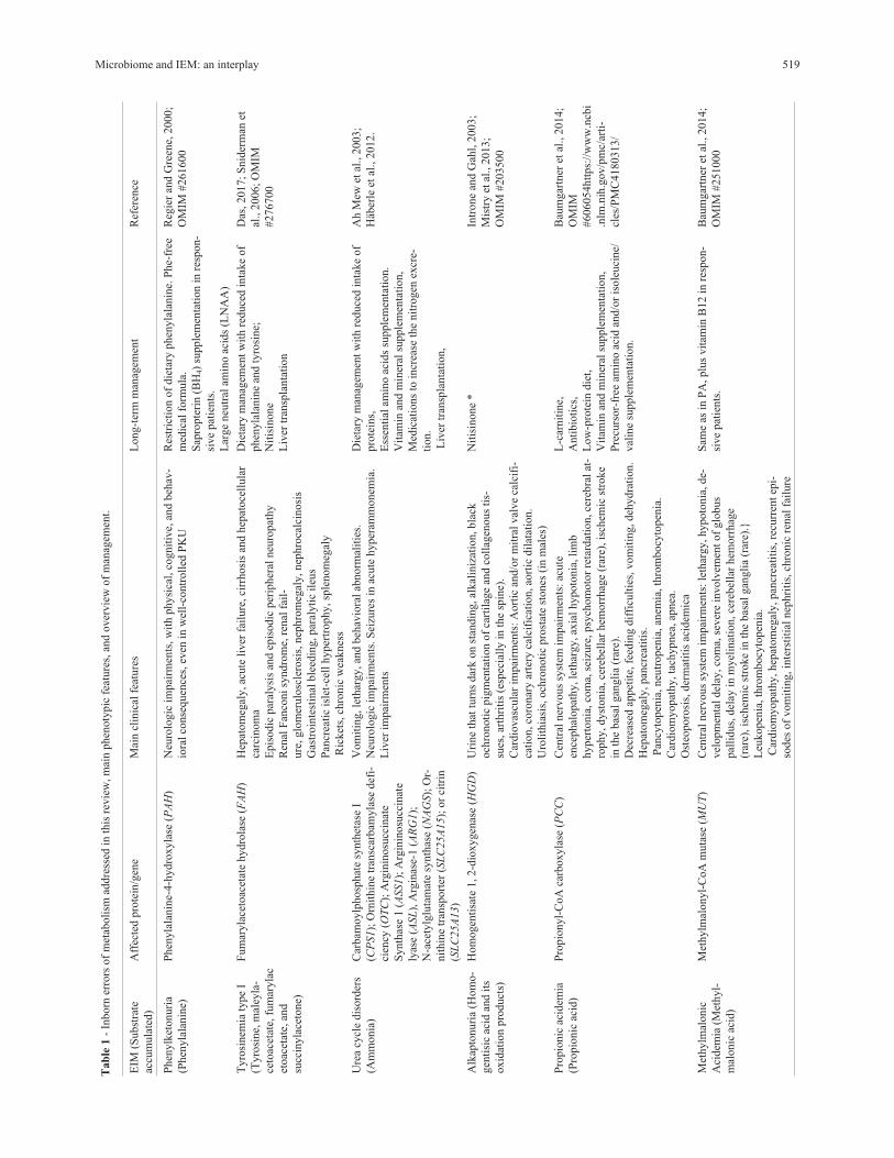
Microbiome and IEM: an interplay 519
Tab
le1
-In
born
erro
rsof
met
aboli
smad
dre
ssed
inth
isre
vie
w,m
ain
phen
oty
pic
feat
ure
s,an
dover
vie
wof
man
agem
ent.
EIM
(Subst
rate
accu
mula
ted)
Aff
ecte
dpro
tein
/gen
eM
ain
clin
ical
feat
ure
sL
ong-t
erm
man
agem
ent
Ref
eren
ce
Phen
ylk
etonuri
a
(Phen
yla
lanin
e)
Phen
yla
lanin
e-4-h
ydro
xyla
se(P
AH
)N
euro
logic
impai
rmen
ts,w
ith
physi
cal,
cognit
ive,
and
beh
av-
iora
lco
nse
quen
ces,
even
inw
ell-
contr
oll
edP
KU
Res
tric
tion
of
die
tary
phen
yla
lanin
e.P
he-
free
med
ical
form
ula
.
Sap
ropte
rin
(BH
4)
supple
men
tati
on
inre
spon-
sive
pat
ients
.
Lar
ge
neu
tral
amin
oac
ids
(LN
AA
)
Reg
ier
and
Gre
ene,
2000;
OM
IM#261600
Tyro
sinem
iaty
pe
I
(Tyro
sine,
mal
eyla
-
ceto
acet
ate,
fum
aryla
c
etoac
etat
e,an
d
succ
inyla
ceto
ne)
Fum
aryla
ceto
acet
ate
hydro
lase
(FA
H)
Hep
atom
egal
y,ac
ute
liver
fail
ure
,ci
rrhosi
san
dhep
atoce
llula
r
carc
inom
a
Epis
odic
par
alysi
san
dep
isodic
per
ipher
alneu
ropat
hy
Ren
alF
anco
ni
syndro
me,
renal
fail
-
ure
,glo
mer
ulo
scle
rosi
s,nep
hro
meg
aly,nep
hro
calc
inosi
s
Gas
troin
test
inal
ble
edin
g,par
alyti
cil
eus
Pan
crea
tic
isle
t-ce
llhyper
trophy,sp
lenom
egal
y
Ric
ket
s,ch
ronic
wea
knes
s
Die
tary
man
agem
ent
wit
hre
duce
din
take
of
phen
yla
lanin
ean
dty
rosi
ne;
Nit
isin
one
Liv
ertr
ansp
lanta
tion
Das
,2017;
Snid
erm
anet
al.,
2006;
OM
IM
#276700
Ure
acy
cle
dis
ord
ers
(Am
monia
)
Car
bam
oylp
hosp
hat
esy
nth
etas
eI
(CP
S1);
Orn
ithin
etr
ansc
arbam
yla
sedef
i-
cien
cy(O
TC
);A
rgin
inosu
ccin
ate
Synth
ase
1(A
SS1);
Arg
inin
osu
ccin
ate
lyas
e(A
SL
),A
rgin
ase-
1(A
RG
1);
N-a
cety
lglu
tam
ate
synth
ase
(NA
GS);
Or-
nit
hin
etr
ansp
ort
er(S
LC
25A
15);
or
citr
in
(SL
C25A
13)
Vom
itin
g,le
thar
gy,an
dbeh
avio
ral
abnorm
alit
ies.
Neu
rolo
gic
impai
rmen
ts.S
eizu
res
inac
ute
hyper
amm
onem
ia.
Liv
erim
pai
rmen
ts
Die
tary
man
agem
ent
wit
hre
duce
din
take
of
pro
tein
s,
Ess
enti
alam
ino
acid
ssu
pple
men
tati
on.
Vit
amin
and
min
eral
supple
men
tati
on,
Med
icat
ions
toin
crea
seth
enit
rogen
excr
e-
tion.
Liv
ertr
ansp
lanta
tion,
Ah
Mew
etal
.,2003;
Häb
erle
etal
.,2012.
Alk
apto
nuri
a(H
om
o-
gen
tisi
cac
idan
dit
s
oxid
atio
npro
duct
s)
Hom
ogen
tisa
te1,2-d
ioxygen
ase
(HG
D)
Uri
ne
that
turn
sdar
kon
stan
din
g,al
kal
iniz
atio
n,bla
ck
och
ronoti
cpig
men
tati
on
of
cart
ilag
ean
dco
llag
enous
tis-
sues
,ar
thri
tis
(esp
ecia
lly
inth
esp
ine)
.
Car
dio
vas
cula
rim
pai
rmen
ts:
Aort
ican
d/o
rm
itra
lval
ve
calc
ifi-
cati
on,co
ronar
yar
tery
calc
ific
atio
n,ao
rtic
dil
atat
ion.
Uro
lith
iasi
s,och
ronoti
cpro
stat
est
ones
(in
mal
es)
Nit
isin
one
*In
trone
and
Gah
l,2003;
Mis
try
etal
.,2013;
OM
IM#203500
Pro
pio
nic
acid
emia
(Pro
pio
nic
acid
)
Pro
pio
nyl-
CoA
carb
oxyla
se(P
CC
)C
entr
alner
vous
syst
emim
pai
rmen
ts:
acute
ence
phal
opat
hy,le
thar
gy,ax
ial
hypoto
nia
,li
mb
hyper
tonia
,co
ma,
seiz
ure
,psy
chom
oto
rre
tard
atio
n,ce
rebra
lat
-
rophy,dyst
onia
,ce
rebel
lar
hem
orr
hag
e(r
are)
,is
chem
icst
roke
inth
ebas
algan
gli
a(r
are)
.
Dec
reas
edap
pet
ite,
feed
ing
dif
ficu
ltie
s,vom
itin
g,deh
ydra
tion.
Hep
atom
egal
y,pan
crea
titi
s.
Pan
cyto
pen
ia,neu
tropen
ia,an
emia
,th
rom
bocy
topen
ia.
Car
dio
myopat
hy,ta
chypnea
,ap
nea
.
Ost
eoporo
sis,
der
mat
itis
acid
emic
a
L-c
arnit
ine,
Anti
bio
tics
,
Low
-pro
tein
die
t,
Vit
amin
and
min
eral
supple
men
tati
on,
Pre
curs
or-
free
amin
oac
idan
d/o
ris
ole
uci
ne/
val
ine
supple
men
tati
on.
Bau
mgar
tner
etal
.,2014;
OM
IM
#606054htt
ps:
//w
ww
.ncb
i
.nlm
.nih
.gov/p
mc/
arti
-
cles
/PM
C4180313/
Met
hylm
alonic
Aci
dem
ia(M
ethyl-
mal
onic
acid
)
Met
hylm
alonyl-
CoA
muta
se(M
UT
)C
entr
alner
vous
syst
emim
pai
rmen
ts:
leth
argy,hypoto
nia
,de-
vel
opm
enta
ldel
ay,co
ma,
sever
ein
volv
emen
tof
glo
bus
pal
lidus,
del
ayin
myel
inat
ion,ce
rebel
lar
hem
orr
hag
e
(rar
e),is
chem
icst
roke
inth
ebas
algan
gli
a(r
are)
.}
Leu
kopen
ia,th
rom
bocy
topen
ia.
Car
dio
myopat
hy,hep
atom
egal
y,pan
crea
titi
s,re
curr
ent
epi-
sodes
of
vom
itin
g,in
ters
titi
alnep
hri
tis,
chro
nic
renal
fail
ure
Sam
eas
inP
A,plu
svit
amin
B12
inre
spon-
sive
pat
ients
.
Bau
mgar
tner
etal
.,2014;
OM
IM#251000
As evidence mounts for a systemic effect of the gut
microbiome on the host, the liver has also been found to be
affected by changes in the microbiome. In addition to its
central role in intermediary metabolism (for instance, many
enzymes affected by IEM are only expressed in liver) and
bile secretion, the liver is the target organ of therapies for
metabolic disorders (Brunetti-Pierri and Lee, 2005) and can
also be considered a secondary lymphoid organ
(Macpherson et al., 2016). Changes in liver physiology are
probably caused primarily by DNA methylation processes,
covalent histone modifications, and regulation of gene ex-
pression by non-coding RNA (ncRNA) (Macpherson et al.,
2016). In addition to SCFAs, isothiocyanates and
polyphenols are also produced by the microbiome, and all
of these compounds have the potential to cause epigenetic
changes. As the liver receives blood from the gut through
the portal vein, it is susceptible to exposure to microbial by-
products that cross the intestinal barrier. In humans and
non-human animals alike, whenever liver or bowel disease
causes dysfunction of the barrier role played by these or-
gans, there is a breakdown in mutualism between the host
and the microbiome, which leads to systemic exposure to
gut bacteria and increased immune activation (Chassaing et
al., 2015). In these situations, the liver becomes a primary
immune barrier that mediates host–microbiome mutualism
(Balmer et al., 2014).
Hepatocytes are sensitive to microbial byproducts,
and may trigger an inflammatory immune response with
systemic effects: even exposure to low levels of LPS in-
duces IFN-� overexpression and IL-10 underexpression in
the liver in animal models of obesity, thus predisposing to
the development of steatohepatitis (Yang et al., 1997). On
the other hand, deletion of the flagellin receptor TLR5 in
mouse hepatocytes has been shown to predispose to hepatic
steatosis and fibrosis, as well as other features of the meta-
bolic syndrome. In this study, antibiotic treatment was able
to reverse steatosis and related aspects in TLR5 knockout
mice, suggesting that mechanisms for clearance of micro-
organisms capable of gut–liver translocation is essential for
maintenance of host systemic health, preventing the
chronic inflammation induced by microbial pathogens
(Etienne-Mesmin et al., 2016). Taking into account the im-
portant immune role of the liver, it makes sense that most
patients with cirrhosis and severe liver failure die of sepsis,
not of metabolic derangements (Leber et al., 2009), as
many of these infections are caused by oral commensals or
gut microbiota (Gustot et al., 2009). The dysbiosis state it-
self impulses inflammatory response and has potential for
causing disease. The role of the microbiome in liver disor-
ders is further supported by the efficiency of treating these
conditions with probiotics, prebiotics, and antibiotics.
Studying the microbiome, hence, may provide a better un-
derstanding of complex diseases and lay the groundwork
for new therapies (Tilg et al., 2016).
520 Colonetti et al.
EIM
(Subst
rate
accu
mula
ted)
Aff
ecte
dpro
tein
/gen
eM
ain
clin
ical
feat
ure
sL
ong-t
erm
man
agem
ent
Ref
eren
ce
Hem
och
rom
atosi
s
type
1(I
ron)
HF
Epro
tein
,H
emoch
rom
atosi
sgen
e
(HF
E1)
Hea
rtin
volv
emen
t:ca
rdio
myopat
hy,co
nges
tive
hea
rtfa
il-
ure
,ar
rhyth
mia
,ca
rdio
meg
aly.
Liv
erin
volv
emen
t:ci
rrhosi
s,hep
atom
egal
y,hep
atoce
llula
rca
r-
cinom
a.
Dia
bet
esm
elli
tus.
Art
hri
tis.
Hypogonad
otr
opic
hypogonad
ism
.
The
sever
eef
fect
sof
the
dis
ease
usu
ally
do
not
appea
runti
laf
-
ter
dec
ades
of
pro
gre
ssiv
eir
on
load
ing
Per
iodic
phle
boto
my
Sec
kin
gto
nan
d
Pow
ell,
2000;
OM
IM
#235200
Tri
met
hyla
min
uri
a
(Am
ino-t
rim
ethyla
-
min
e)
Fla
vin
-conta
inin
gm
onooxygen
ase
3
(FM
O3)
Beh
avio
ral/
psy
chia
tric
man
ifes
tati
ons:
dep
ress
ion,su
i-
cidal
,psy
choso
cial
pro
ble
ms
insc
hool.
Inso
me
pat
ients
:an
e-
mia
,neu
tropen
ia,pulm
onar
yin
fect
ions;
tach
yca
rdia
and
sever
e
hyper
tensi
on
afte
rea
ting
chee
se.
Die
tary
rest
rict
ion
of:
Tri
met
hyla
min
ean
dit
s
pre
curs
ors
incl
udin
gch
oli
ne
and
leci
thin
Tri
met
hyla
min
eN
-oxid
e;In
hib
itors
of
FM
O3
enzy
me
acti
vit
y,su
chas
indole
s.U
seof:
acid
soap
san
dbody
loti
ons,
acti
vat
edch
arco
alan
d
copper
chlo
rophyll
in,an
tibio
tics
,ri
bofl
avin
supple
men
ts.
Phil
lips
and
Shep
har
d,2007;
OM
IM
#602079
*U
nder
inves
tigat
ion
Tab
le1
-co
nt.
The microbiome and IEMs: the state of the art
The gut microbiome plays roles in amino acid and
carbohydrate metabolism, vitamin and cofactor bio-
synthesis, and production of SCFAs, in addition to influ-
encing the physiology of the liver, brain, and GI tract, all of
which are affected by IEMs. In light of the many important
activities of this virtual metabolic organ and its vast impact
on the host, some studies have considered the microbiome
as a factor that interferes with organic homeostasis in the
context of IEMs, and have sought to characterize possible
interactions, both endogenous (genetic defect) and exoge-
nous (treatment/diet), with host metabolic pathways, as
well as the probable consequences of the presence or ab-
sence of specific bacteria and their metabolites on the hu-
man body.
Studies of the association between microbiome and
IEMs have focused on aminoacidopathies (such as PKU,
tyrosinemia, and alkaptonuria), organic acidemias (methyl-
malonic acidemia and propionic acidemia), and hemochro-
matosis. The main characteristics of the IEMs addressed in
these studies, including their long-term management, are
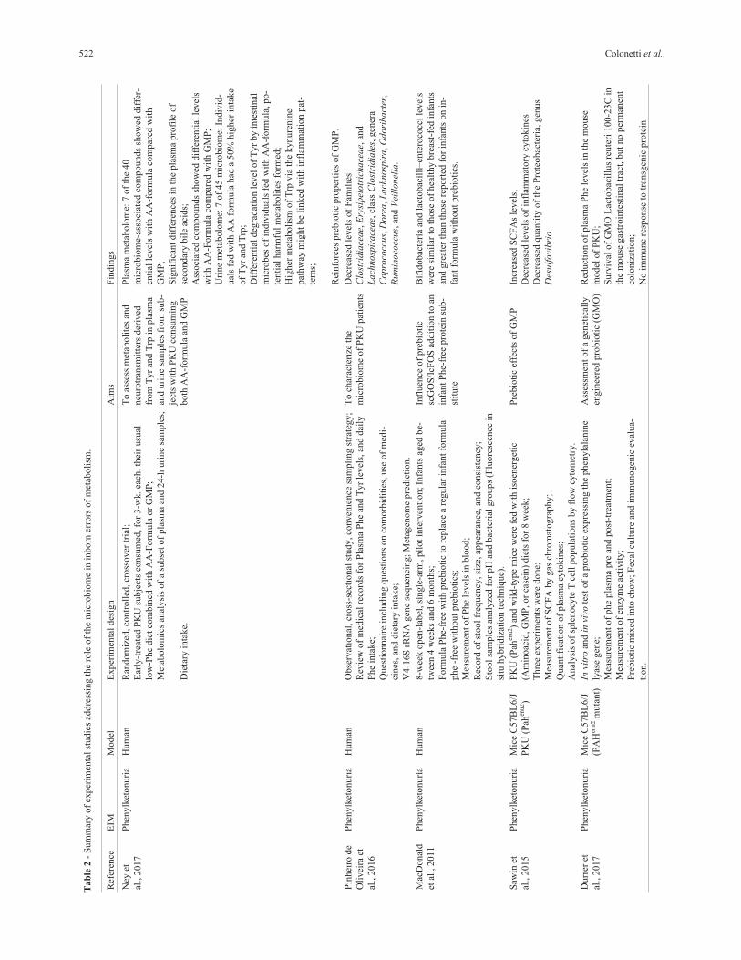
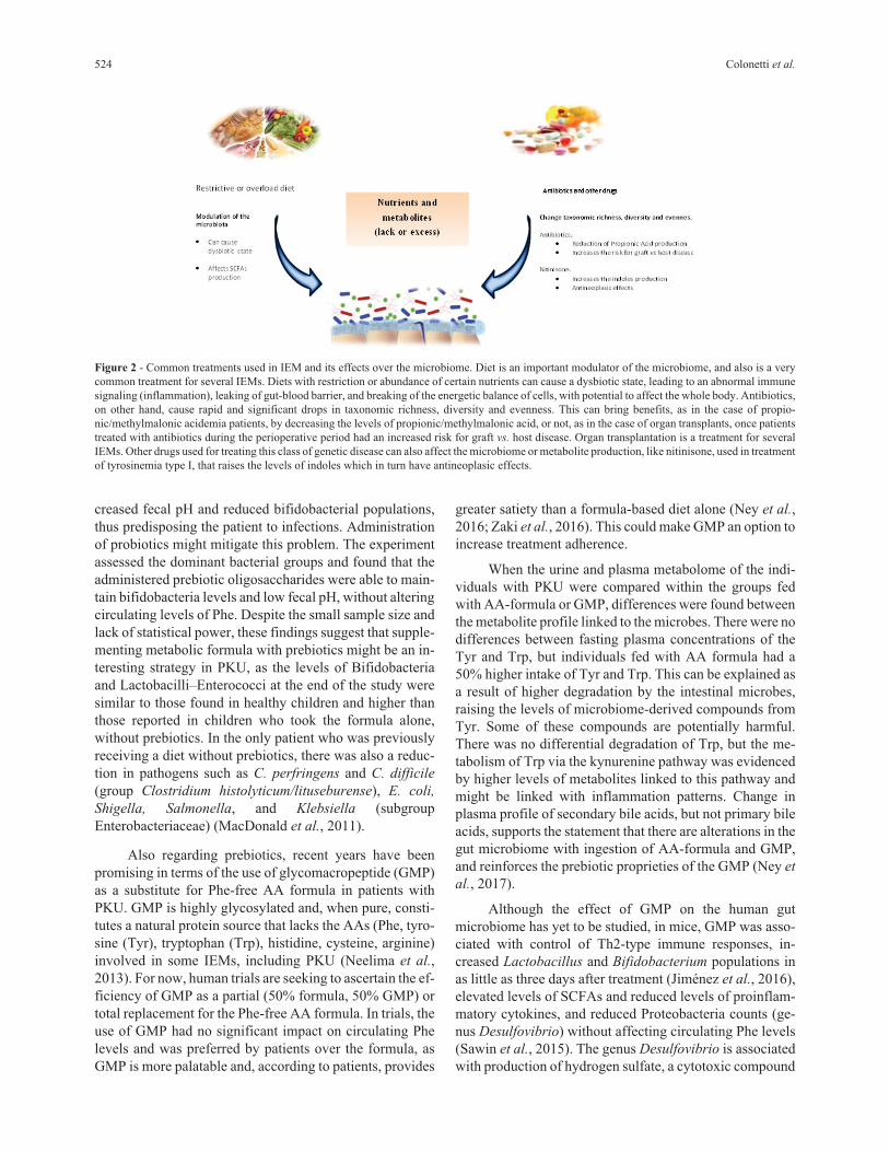
summarized in Table 2. Some possible effects of treatments
of IEM on microbiome are showed in Figure 2.
The majority of studies on microbiome–IEM interac-
tions has focused on PKU. One of the most thorough
among such studies compared the microbiome of eight pa-
tients with PKU to that of 10 healthy individuals by analy-
sis of the 16S rRNA gene. In this study, Pinheiro de Oli-
veira et al. (2016) demonstrated reduced abundance of bac-
teria in the families Clostridiaceae, Erysipelotrichaceae,
and Lachnospiraceae, class Clostridiales, and genera
Coprococcus, Dorea, Lachnospira, Odoribacter,
Ruminococcus, and Veillonella in patients with PKU, as
well as an increase in Prevotella, Akkermansia, and
Peptostreptococcaceae populations. Their metabolic pre-
diction was associated both with starch and glucose metab-
olism and with AA metabolism (Pinheiro de Oliveira et al.,
2016). The authors raised the hypothesis that bacterial en-
richment related to LPS biosynthesis, as observed in pa-
tients with PKU, might be associated with peripheral
inflammation, as indicated by the proinflammatory circu-
lating cytokine profile of these patients (Coakley et al.,
2014). In the same study, the authors found a correlation
between microbiotic profile and circulating levels of
phenylalanine (Phe), which might indicate a relationship
between these patients’ microbiome, their treatment re-
sponse, and their phenotype.
Focusing on the potential impacts of prebiotic treat-
ment in individuals with PKU, a study reported by Mac-
Donald et al. (2011) analyzed the effects of prebiotic
oligosaccharides (scGOS/lcFOS) as an adjunct to the meta-
bolic formula that forms the mainstay of PKU manage-
ment. As breastfeeding is highly restricted in children with
PKU, the authors theorized that a lack of the oligosaccha-
rides present in breast milk might be associated with in-
Microbiome and IEM: an interplay 521
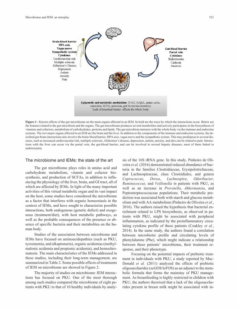
Figure 1 - Known effects of the gut microbiome on the main organs affected in an IEM. In bold are the ways by which the interactions occur. Below are
the features related to the gut microbiota and the organs. The gut microbiome produces several metabolites and actively participates in the biosynthesis of
vitamins and cofactors, metabolism of carbohydrates, proteins and lipids. The gut microbiota interacts with the whole body via the immune and endocrine
systems. The two major organs affected in an IEM are the brain and the liver. In addition to the components of the immune and endocrine systems, the de-
scribed gut-brain interactions also involve the brain-blood barrier, HPA axis, vagus nerve and the sympathetic system. This may predispose to several dis-
eases, such as increased cardiovascular risk, multiple sclerosis, Alzheimer’s disease, depression, autism, anxiety, and also can be related to pain. Interac-
tions with the liver can occur via the portal vein, the gut-blood barrier, and can be involved in several hepatic diseases, most of them linked to
endotoxemia.
522 Colonetti et al.
Tab
le2
-S
um
mar
yof
exper
imen
tal
studie
sad
dre
ssin
gth
ero
leof
the
mic
robio
me
inin
born
erro
rsof
met
aboli
sm.
Ref
eren
ceE
IMM
odel
Exper
imen
tal
des
ign
Aim
sF
indin
gs
Ney
et
al.,
2017
Phen
ylk
etonuri
aH
um
anR
andom
ized
,co
ntr
oll
ed,cr
oss
over
tria
l;
Ear
ly-t
reat
edP
KU
subje
cts
consu
med
,fo
r3-w
k.ea
ch,th
eir
usu
al
low
-Phe
die
tco
mbin
edw
ith
AA
-Form
ula
or
GM
P;
Met
abolo
mic
san
alysi
sof
asu
bse
tof
pla
sma
and
24-h
uri
ne
sam
ple
s;
Die
tary
inta
ke.
To
asse
ssm
etab
oli
tes
and
neu
rotr
ansm
itte
rsder
ived
from
Tyr
and
Trp
inpla
sma
and
uri
ne
sam
ple
sfr
om
sub-
ject
sw
ith
PK
Uco
nsu
min
g
both
AA
-form
ula
and
GM
P
Pla
sma
met
abolo
me:
7of
the
40
mic
robio
me-
asso
ciat
edco
mpounds
show
eddif
fer-
enti
alle
vel
sw
ith
AA
-form
ula
com
par
edw
ith
GM
P;
Sig
nif
ican
tdif
fere
nce
sin
the
pla
sma
pro
file
of
seco
ndar
ybil
eac
ids;
Ass
oci
ated
com
pounds
show
eddif
fere
nti
alle
vel
s
wit
hA
A-F
orm
ula
com
par
edw
ith
GM
P;
Uri
ne
met
abolo
me:
7of
45
mic
robio
me;
Indiv
id-
ual
sfe
dw
ith
AA
form
ula
had
a50%
hig
her
inta
ke
of
Tyr
and
Trp
;
Dif
fere
nti
aldeg
radat
ion
level
of
Tyr
by
inte
stin
al
mic
robes
of
indiv
idual
sfe
dw
ith
AA
-form
ula
,po-
tenti
alhar
mfu
lm
etab
oli
tes
form
ed;
Hig
her
met
aboli
smof
Trp
via
the
kynure
nin
e
pat
hw
aym
ight
be
linked
wit
hin
flam
mat
ion
pat
-
tern
s;
Rei
nfo
rces
pre
bio
tic
pro
per
ties
of
GM
P.
Pin
hei
rode
Oli
vei
raet
al.,
2016
Phen
ylk
etonuri
aH
um
anO
bse
rvat
ional
,cr
oss
-sec
tional
study,co
nven
ience
sam
pli
ng
stra
tegy;
Rev
iew
of
med
ical
reco
rds
for
Pla
sma
Phe
and
Tyr
level
s,an
ddai
ly
Phe
inta
ke;
Ques
tionnai
rein
cludin
gques
tions
on
com
orb
idit
ies,
use
of
med
i-
cines
,an
ddie
tary
inta
ke;
V4-1
6S
rRN
Agen
ese
quen
cing;
Met
agen
om
epre
dic
tion.
To
char
acte
rize
the
mic
robio
me
of
PK
Upat
ients
Dec
reas
edle
vel
sof
Fam
ilie
s
Clo
stri
dia
ceae,
Ery
sipel
otr
ichace
ae,
and
Lach
nosp
irace
ae,
clas
sC
lost
ridia
les,
gen
era
Copro
cocc
us,
Dore
a,L
ach
nosp
ira,O
dori
bact
er,
Rum
inoco
ccus,
and
Vei
llonel
la.
Mac
Donal
d
etal
.,2011
Phen
ylk
etonuri
aH
um
an8-w
eek
open
-lab
el,si
ngle
-arm
,pil
ot
inte
rven
tion;
Infa
nts
aged
be-
twee
n4
wee
ks
and
6m
onth
s;
Form
ula
Phe-
free
wit
hpre
bio
tic
tore
pla
cea
regula
rin
fant
form
ula
phe
-fre
ew
ithout
pre
bio
tics
;
Mea
sure
men
tof
Phe
level
sin
blo
od;
Rec
ord
of
stool
freq
uen
cy,si
ze,ap
pea
rance
,an
dco
nsi
sten
cy;
Sto
ol
sam
ple
san
alyze
dfo
rpH
and
bac
teri
algro
ups
(Flu
ore
scen
cein
situ
hybri
diz
atio
nte
chniq
ue)
.
Infl
uen
ceof
pre
bio
tic
scG
OS
/lcF
OS
addit
ion
toan
infa
nt
Phe-
free
pro
tein
sub-
stit
ute
Bif
idobac
teri
aan
dla
ctobac
illi
–en
tero
cocc
ile
vel
s
wer
esi
mil
arto
those
of
hea
lthy
bre
ast-
fed
infa
nts
and
gre
ater
than
those
report
edfo
rin
fants
on
in-
fant
form
ula
wit
hout
pre
bio
tics
.
Saw
inet
al.,
2015
Phen
ylk
etonuri
aM
ice
C57B
L6/J
PK
U(P
ahen
u2)
PK
U(P
ahen
u2)
and
wil
d-t
ype
mic
ew
ere
fed
wit
his
oen
erget
ic
(Am
inoac
id,G
MP
,or
case
in)
die
tsfo
r8
wee
k;
Thre
eex
per
imen
tsw
ere
done;
Mea
sure
men
tof
SC
FA
by
gas
chro
mat
ogra
phy;
Quan
tifi
cati
on
of
pla
sma
cyto
kin
es;
Anal
ysi
sof
sple
nocy
teT
cell
popula
tions
by
flow
cyto
met
ry.
Pre
bio
tic
effe
cts
of
GM
PIn
crea
sed
SC
FA
sle
vel
s;
Dec
reas
edle
vel
sof
infl
amm
atory
cyto
kin
es
Dec
reas
edquan
tity
of
the
Pro
teobac
teri
a,gen
us
Des
ulf
ovi
bri
o.
Durr
eret
al.,
2017
Phen
ylk
etonuri
aM
ice
C57B
L6/J
(PA
Hen
u2
muta
nt)
Invi
tro
and
invi
vote
stof
apro
bio
tic
expre
ssin
gth
ephen
yla
lanin
e
lyas
egen
e;
Mea
sure
men
tof
phe
pla
sma
pre
and
post
-tre
atm
ent;
Mea
sure
men
tof
enzy
me
acti
vit
y;
Pre
bio
tic
mix
edin
toch
ow
;F
ecal
cult
ure
and
imm
unogen
icev
alua-
tion.
Ass
essm
ent
of
agen
etic
ally
engin
eere
dpro
bio
tic
(GM
O)
Red
uct
ion
of
pla
sma
Phe
level
sin
the
mouse
model
of
PK
U;
Surv
ival
of
GM
OL
acto
bac
illu
sre
ute
ri100-2
3C
in
the
mouse
gas
troin
test
inal
trac
t,but
no
per
man
ent
colo
niz
atio
n;
No
imm
une
resp
onse
totr
ansg
enic
pro
tein
.
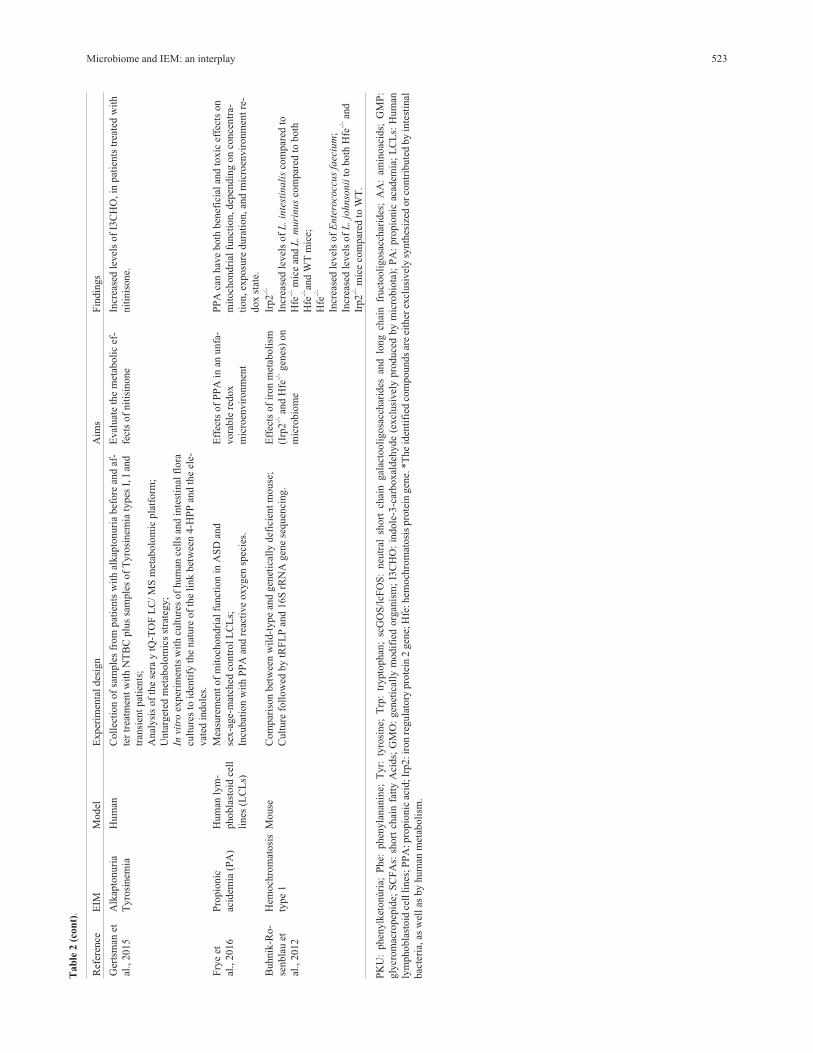
Microbiome and IEM: an interplay 523
Ref
eren
ceE
IMM
odel
Exper
imen
tal
des
ign
Aim
sF
indin
gs
Ger
tsm
anet
al.,
2015
Alk
apto
nuri
a
Tyro
sinem
ia
Hum
anC
oll
ecti
on
of
sam
ple
sfr
om
pat
ients
wit
hal
kap
tonuri
abef
ore
and
af-
ter
trea
tmen
tw
ith
NT
BC
plu
ssa
mple
sof
Tyro
sinem
iaty
pes
I,I
and
tran
sien
tpat
ients
;
Anal
ysi
sof
the
sera
ytQ
-TO
FL
C/
MS
met
abolo
mic
pla
tform
;
Unta
rget
edm
etab
olo
mic
sst
rate
gy;
Invi
tro
exper
imen
tsw
ith
cult
ure
sof
hum
ance
lls
and
inte
stin
alfl
ora
cult
ure
sto
iden
tify
the
nat
ure
of
the
link
bet
wee
n4-H
PP
and
the
ele-
vat
edin
dole
s.
Eval
uat
eth
em
etab
oli
cef
-
fect
sof
nit
isin
one
Incr
ease
dle
vel
sof
I3C
HO
,in
pat
ients
trea
ted
wit
h
nit
inis
one.
Fry
eet
al.,
2016
Pro
pio
nic
acid
emia
(PA
)
Hum
anly
m-
phobla
stoid
cell
lines
(LC
Ls)
Mea
sure
men
tof
mit
och
ondri
alfu
nct
ion
inA
SD
and
sex-a
ge-
mat
ched
contr
ol
LC
Ls;
Incu
bat
ion
wit
hP
PA
and
reac
tive
oxygen
spec
ies.
Eff
ects
of
PP
Ain
anunfa
-
vora
ble
redox
mic
roen
vir
onm
ent
PP
Aca
nhav
eboth
ben
efic
ial
and
toxic
effe
cts
on
mit
och
ondri
alfu
nct
ion,dep
endin
gon
conce
ntr
a-
tion,ex
posu
redura
tion,an
dm
icro
envir
onm
ent
re-
dox
stat
e.
Buhnik
-Ro-
senbla
uet
al.,
2012
Hem
och
rom
atosi
s
type
1
Mouse
Com
par
ison
bet
wee
nw
ild-t
ype
and
gen
etic
ally
def
icie
nt
mouse
;
Cult
ure
foll
ow
edby
tRF
LP
and
16S
rRN
Agen
ese
quen
cing.
Eff
ects
of
iron
met
aboli
sm
(Irp
2-/
-an
dH
fe-/
-gen
es)
on
mic
robio
me
Irp2
-/-
Incr
ease
dle
vel
sof
L.in
test
inali
sco
mpar
edto
Hfe
-/-m
ice
and
L.m
uri
nus
com
par
edto
both
Hfe
-/- an
dW
Tm
ice;
Hfe
-/-
Incr
ease
dle
vel
sof
Ente
roco
ccus
faec
ium
;
Incr
ease
dle
vel
sof
L.jo
hnso
nii
toboth
Hfe
-/-an
d
Irp2
-/-m
ice
com
par
edto
WT
.
PK
U:
phen
ylk
etonúri
a;P
he:
phen
yla
nan
ine;
Tyr:
tyro
sine;
Trp
:tr
ypto
phan
;sc
GO
S/l
cFO
S:
neu
tral
short
chai
ngal
acto
oli
gosa
cchar
ides
and
long
chai
nfr
uct
ooli
gosa
cchar
ides
;A
A:
amin
oac
ids;
GM
P:
gly
crom
acro
pep
ide;
SC
FA
s:sh
ort
chai
nfa
tty
Aci
ds;
GM
O:
gen
etic
ally
modif
ied
org
anis
m;
I3C
HO
:in
dole
-3-c
arboxal
deh
yde
(excl
usi
vel
ypro
duce
db
ym
icro
bio
ta);
PA
:pro
pio
nic
acad
emia
;L
CL
s:H
um
an
lym
phobla
stoid
cell
lines
;P
PA
:pro
pio
nic
acid
;Ir
p2:ir
on
regula
tory
pro
tein
2gen
e;H
fe:hem
och
rom
atosi
spro
tein
gen
e.*T
he
iden
tifi
edco
mpound
sar
eei
ther
excl
usi
vel
ysy
nth
esiz
edor
contr
ibute
dby
inte
stin
al
bac
teri
a,as
wel
las
by
hum
anm
etab
oli
sm.
Tab
le2
(con
t).
creased fecal pH and reduced bifidobacterial populations,
thus predisposing the patient to infections. Administration
of probiotics might mitigate this problem. The experiment
assessed the dominant bacterial groups and found that the
administered prebiotic oligosaccharides were able to main-
tain bifidobacteria levels and low fecal pH, without altering
circulating levels of Phe. Despite the small sample size and
lack of statistical power, these findings suggest that supple-
menting metabolic formula with prebiotics might be an in-
teresting strategy in PKU, as the levels of Bifidobacteria
and Lactobacilli–Enterococci at the end of the study were
similar to those found in healthy children and higher than
those reported in children who took the formula alone,
without prebiotics. In the only patient who was previously
receiving a diet without prebiotics, there was also a reduc-
tion in pathogens such as C. perfringens and C. difficile
(group Clostridium histolyticum/lituseburense), E. coli,
Shigella, Salmonella, and Klebsiella (subgroup
Enterobacteriaceae) (MacDonald et al., 2011).
Also regarding prebiotics, recent years have been
promising in terms of the use of glycomacropeptide (GMP)
as a substitute for Phe-free AA formula in patients with
PKU. GMP is highly glycosylated and, when pure, consti-
tutes a natural protein source that lacks the AAs (Phe, tyro-
sine (Tyr), tryptophan (Trp), histidine, cysteine, arginine)
involved in some IEMs, including PKU (Neelima et al.,
2013). For now, human trials are seeking to ascertain the ef-
ficiency of GMP as a partial (50% formula, 50% GMP) or
total replacement for the Phe-free AA formula. In trials, the
use of GMP had no significant impact on circulating Phe
levels and was preferred by patients over the formula, as
GMP is more palatable and, according to patients, provides
greater satiety than a formula-based diet alone (Ney et al.,
2016; Zaki et al., 2016). This could make GMP an option to
increase treatment adherence.
When the urine and plasma metabolome of the indi-
viduals with PKU were compared within the groups fed
with AA-formula or GMP, differences were found between
the metabolite profile linked to the microbes. There were no
differences between fasting plasma concentrations of the
Tyr and Trp, but individuals fed with AA formula had a
50% higher intake of Tyr and Trp. This can be explained as
a result of higher degradation by the intestinal microbes,
raising the levels of microbiome-derived compounds from
Tyr. Some of these compounds are potentially harmful.
There was no differential degradation of Trp, but the me-
tabolism of Trp via the kynurenine pathway was evidenced
by higher levels of metabolites linked to this pathway and
might be linked with inflammation patterns. Change in
plasma profile of secondary bile acids, but not primary bile
acids, supports the statement that there are alterations in the
gut microbiome with ingestion of AA-formula and GMP,
and reinforces the prebiotic proprieties of the GMP (Ney et
al., 2017).
Although the effect of GMP on the human gut
microbiome has yet to be studied, in mice, GMP was asso-
ciated with control of Th2-type immune responses, in-
creased Lactobacillus and Bifidobacterium populations in
as little as three days after treatment (Jiménez et al., 2016),
elevated levels of SCFAs and reduced levels of proinflam-
matory cytokines, and reduced Proteobacteria counts (ge-
nus Desulfovibrio) without affecting circulating Phe levels
(Sawin et al., 2015). The genus Desulfovibrio is associated
with production of hydrogen sulfate, a cytotoxic compound
524 Colonetti et al.
Figure 2 - Common treatments used in IEM and its effects over the microbiome. Diet is an important modulator of the microbiome, and also is a very
common treatment for several IEMs. Diets with restriction or abundance of certain nutrients can cause a dysbiotic state, leading to an abnormal immune
signaling (inflammation), leaking of gut-blood barrier, and breaking of the energetic balance of cells, with potential to affect the whole body. Antibiotics,
on other hand, cause rapid and significant drops in taxonomic richness, diversity and evenness. This can bring benefits, as in the case of propio-
nic/methylmalonic acidemia patients, by decreasing the levels of propionic/methylmalonic acid, or not, as in the case of organ transplants, once patients
treated with antibiotics during the perioperative period had an increased risk for graft vs. host disease. Organ transplantation is a treatment for several
IEMs. Other drugs used for treating this class of genetic disease can also affect the microbiome or metabolite production, like nitinisone, used in treatment
of tyrosinemia type I, that raises the levels of indoles which in turn have antineoplasic effects.
found at higher levels in patients with ulcerative colitis
(Rowan et al., 2010).
Regarding disorders of tyrosine metabolism, Gerts-
man et al. (2015) described the metabolic effect of niti-
sinone (NTBC or 2-(2-nitro-4-fluoromethylbenzoyl)-1,3-
cyclohexanedione) in patients with alkaptonuria. Analysis
of their metabolic profile showed that indole levels were in-
creased in treated patients as compared with controls.
Indoles play a key role in signaling pathways (as building
blocks for melanin and serotonin) and intercellular commu-
nication, facilitate quorum sensing, and have been uniquely
associated with dietary intake and microbial metabolism of
tryptophan. Among the indoles found to be increased,
indole-3-carboxaldehyde (I3CHO) is produced exclusively
by the microbiota, while the other two are produced by hu-
man cells (Gertsman et al., 2015). The authors stressed that
the reduced form of I3CHO, indole-3-carbinol, a com-
pound also found in cruciferous vegetables, is associated
with the prevention of several neoplasms.
Animal experiments also suggest that genetic defects
in the host may alter the composition of the gut microbiota,
leading to dysbiosis due to a buildup of substances in the
cells or lumen of the bowel (Buhnik-Rosenblau et al.,
2012). This effect has been observed in hemochromatosis.
Hemochromatosis is a disease caused by excess iron ab-
sorption by gut cells, which leads to iron overload. This
usually becomes clinically detectable in adulthood and is
damaging to many organs, including the liver, pancreas
(causing diabetes), heart, and skin (Babitt and Lin, 2011).
Mutations in the HFE gene account for the majority of
cases of hereditary hemochromatosis, especially in individ-
uals of Northern European descent (Barton, 2013). In a
study of mice with mutations in two genes that encode pro-
teins involved in regulation of iron homeostasis (HFE-/-and
Irp2 -/-), Buhnik-Rosenblau et al. (2012) found abnormali-
ties particularly in resident populations of lactic-acid bacte-
ria, both in Irp2-mutant and in HFE-mutant mice as
compared to controls.
The gut microbiome produces several metabolites,
including PPA, a SCFA implicated in several diseases. In
autistic populations, the level of the phylum Firmicutes is
increased and was largely attributable to Clostridia class
with Ruminococcaceae and Lachnospiraceae families. The
differences in Clostridia species in children with autism
spectrum disorder include greater abundance of
Clostridium clusters I, II, XI and C. bolteae (Finegold et al.,
2002; Song et al., 2004; Parracho et al., 2005; Williams et
al., 2011; Strati et al., 2017). Several Ruminococcaceae
and Lachnospiraceae are known butyrate producers and
may thus influence SCFA levels (Louis et al., 2010). So,
the treatment with antibiotics can affect producers of
SCFA. Some patients’ symptoms improve transiently when
antibiotics are administered (Sandler et al., 2000; Shaw
2010). Curiously, a similar effect is seen in patients with
propionic acidemia, who can experience the same neuro-
developmental complications seen in autism (Witters et al.,
2016). Among the various roles played by PPA, it was re-
cently reported to act as a modulator of mitochondrial func-
tion. In a study of autism and control cell lines, the effects
of PPA depended not only on the concentration of the acid,
but also on the level of reactive oxygen species (ROS) pres-
ent, as ROS influence mitochondrial ability to use PPA as
an energy source. Thus, PPA could have beneficial effects
in individuals without mitochondrial dysfunction, and
harmful effects in individuals with an unfavorable meta-
bolic status and elevated levels of ROS (Frye et al., 2016).
In methylmalonic acidemia, which shares several symp-
toms and management strategies with propionic acidemia,
vitamin B12 (cobalamin) is also used as treatment in respon-
sive patients, in addition to antibiotics. This vitamin is syn-
thesized by some gut bacteria, and is also a regulator of
microbiome composition and function (Baumgartner et al.,
2014; Degnan et al., 2014).
The microbiome can also be considered an exogenous
source of tetrahydrobiopterin (BH4), another important me-
tabolite of gut bacteria. BH4 is a key cofactor for several
regulatory enzymes, as Phenylalanine-4-hydroxylase,
which catalyzes the conversion of L-phenylalanine to L-ty-
rosine. The BH4 has also been shown to improve working
memory and cerebral activation (Christ et al., 2013). In ro-
dents, BH4 production is age-dependent and is related to the
presence of Actinobacteria in the bowel, especially
Adlercreutzia equolifaciens and Microbacterium
schleiferi. These same species have been identified in the
human gut microbiome (Belik et al., 2017). Very little is
known about the determinants of responsiveness to BH4
therapy and its effects on cerebral activity and cognition,
but these effects are known to be multifactorial, as they
vary across individuals with the same genotype (Pérez et
al., 2005). The discovery that BH4 is naturally produced by
gut microbiota has implications for translational medicine,
as this cofactor is used in the treatment of some patients
with PKU.
The long-term perspective is that elucidation of the
metabolic role of the microbiota and identification of which
species play these roles will pave the way for manipulating
the microbiome, so that pathways beneficial to the host are
stimulated, while those harmful to the host are inhibited. In
this line, some authors have raised the hypothesis of using
methanogenic bacteria normally present in the human
bowel to control metabolites such as trimethylamine
(TMA), bypassing the normal route of trimethylamine N-
oxide (TMAO) production as an intermediate for CH4 to an
alternative pathway (Brugère et al., 2014). In the liver, defi-
ciency in the pathway of TMA conversion into TMAO
leads to trimethylaminuria, an IEM that causes strong body
odor, impairing the patients’ quality of life and interper-
sonal relations (Mackay et al., 2011). Diets rich in com-
pounds such as phosphatidylcholine, choline, betaine, and
L-carnitine generate TMA via the gut microbiota, which is
Microbiome and IEM: an interplay 525
then converted in TMAO by the liver. High levels of
TMAO are associated with increased risk of cardiovascular
disease in the general population (Wang et al., 2011; Koeth
et al., 2013; Gregory et al., 2015; Liu et al., 2015b).
Making the transition from theory into practice, administra-
tion of the probiotic Lactobacillus reuteri, engineered to
express a phenylalanine lyase gene from the cyanobacteria
Anabaena variabilis, successfully treated mice with PKU.
Blood levels of Phe declined after the fourth day of treat-
ment and remained low throughout the experiment, with no
permanent colonization of the gut (Durrer et al., 2017), sug-
gesting potential for modified probiotics in the treatment of
IEMs.
The creation of genetically modified probiotics de-
sign especially to normalize defective metabolic pathways
in the host is only one of the many potential advantages of
microbiome research. IEMs are characterized by substan-
tial variability in presentation, and genotype alone cannot
explain patients’ clinical pictures. The microbiome may
contribute significantly to factors such as tolerance to cer-
tain nutrients and responsiveness to cofactors (and to treat-
ment itself). Studying the microbiomes of patients with
IEMs may provide valuable tools for clinical practice, both
advancing our understanding of phenotypes and facilitating
the development of new biomarkers and therapies.
Main questions about microbioma and IEM andhow to address them
There are some important issues involved in the study
of the human microbiome in IEM. First of all, most of the
diseases that compound the IEM class are rare, and usually
there are subclasses within the same IEM. This is the reason
why the studies normally have a small number of partici-
pants. Second, the microbiome is mainly influenced by
diet, and diet overload or restriction is one of most common
treatments for IEM. This is one of reasons that make obtain-
ing an adequate control group very difficult. Third, this
class of diseases is derived of a metabolic genetic defect,
and defects in a metabolic gene also affect the microbiome.
So, if a dysbiotic state is observed in this group of patients
will it reflect the genetic or the diet effect? Taken together,
all the facts above make it very hard to obtain a homoge-
neous and statistically valid group of untreated patients and
make difficult the comparison pre and post-treatment to
verify if the altered microbiome is mainly affected by ge-
netic or diet effects. Additional difficulty is added by the
fact that several metabolic diseases, if untreated, can lead to
severe impacts through life, so IEM patients should start to
be treated as soon as possible.
Despite the difficulties, studying the patterns of the
microbiome in groups of treated patients offers the possi-
bility to evaluate the real impact of the genetic defect and
diet on the microbiome. Patients need lifelong treatment,
and the intragroup study of phenotype, microbiome and
diet can be elucidative for some ancient questions that re-
main unknown. PKU patients, for instance, were studied in
light of the microbiome by Pinheiro de Oliveira et al.
(2016) (see Table 2). Even though not capable of answering
the question if alteration comes from diet or genetics, a
microbiome alteration correlated with Phe blood levels was
observed. This is exciting data, due to the fact that it can
help explain why some patients are more tolerant to Phe
than others, despite having the same genotypes.
In an IEM, the genetic defect and the diet factors co-
exist, so the measure of macro- and micronutrients ingested
is required. Diet has a strong impact on the microbiome,
and in spite of patients having similar lines of treatment all
over the world, the source of fibers, carbohydrates and pro-
teins can vary geographically and/or culturally. For this
reason, microbiome studies should not combine patients of
geographically distinct regions or culture to raise the num-
ber of participants. Rather, these studies must be done lo-
cally and then, if methodologically possible, make compar-
isons that take into account the dietetic/cultural/geographic
factors.
As detailed above, there are several other factors that
can influence and be influenced by the microbiome. Impor-
tant data as sex, age, body mass index, type of birth deliv-
ery, breast feeding (duration and transition to solid food),
antibiotic and other drug usage, vitamin supplementation,
as well as physical exercise, and other diseases (physical
and/or mental) must be collected and also analyzed. All
subjects included in studies that aim to characterize the
microbiome of certain IEMs should be three years or older
to avoid the period of drastic changes in microbiome com-
position due to the typical change in diet during this period.
Given that the microbiome varies according to the stage of
life and sex, and certain cultures can also exert some influ-
ence, the best way to avoid interference of age and sex is the
sex-age-matched strategy.
Another useful strategy is based on experimental
studies using animal models. This strategy is very impor-
tant since animal models have less genetic variation and are
maintained in a highly controlled environment (that in-
cludes diet and/or a germ-free environment). Also, a high
the number of subjects can be easily obtained in such re-
search. This is the better model for initial tests of geneti-
cally engineered probiotics and correlations with diseases
caused by the genetic defect in the absence or presence of
the treatment. This kind of study, besides not being capable
of fully reproducing the human reality, can work to gener-
ate hypotheses and help to provide better strategies and
comprehension of studies done in humans.
With the development of NGS tools, procedures are
no longer the main limitation for human microbiome stud-
ies. Microbiome data is currently obtained by three differ-
ent approaches: 1) by 16S rRNA gene partial sequencing,
2) by whole DNA shotgun metagenomic sequencing, or 3)
by metatranscriptomics (mRNA-seq), to access the active
526 Colonetti et al.
gene expression pattern For instance, the 16S rRNA gene
sequencing method is largely used and has been the first
choice method among researchers. Reasons for choosing
this approach include the availability of a comprehensive
database and scalability. Moreover, studies based in meta-
transcriptomics require a better control for sample collec-
tion to RNA/metabolites processing. Metagenomics, meta-
transcriptomics and all other “omics”, and the associated
bioinformatics techniques are allowing comparative analy-
ses in an unprecedented way. All of these tools allow for
testing a recent hypothesis related to the presence of a com-
mon set of microbial taxa universally present in healthy in-
dividuals (Turnbaugh et al., 2007), also known as microbial
core. However large variations in the taxonomic composi-
tion observed in the human microbiome rapidly refute such
a hypothesis (Bäckhed et al., 2012). Due to the well-known
microbial functional redundancy in nature, an alternative
hypothesis is the presence of a functional core represented
by a set of metabolic functions that are performed by the
microbiome within a particular habitat, but are not neces-
sarily provided by the same organisms in different people
(Shafquat et al., 2014). Still, studies devoted to better un-
derstand how deeply the microbiome can affect an organ-
ism with critical metabolic pathways that are naturally al-
tered, are just in the early stages. Multidisciplinary efforts
need to be done to aggregate modern techniques of se-
quencing and identification of metabolites that can lead to
the phenotype or drug effect in question. Microbial se-
quencing alone will not be capable of explaining the pheno-
type, but is a fundamental tool in the understanding of the
process. Additional techniques based on metabolomics
analysis and RNA-seq, as well as gathering information
about the immune system and SCFA levels can offer funda-
mental pieces of information in the process.
Conclusions
Studies on the microbiome in IEMs are scarce. The
effects of the genetic defect itself and of treatment in IEMs,
especially in the long term, have yet to be fully understood.
As IEMs are commonly managed through dietary interven-
tion (nutrient overload and/or restriction), dysbiosis is a
possibility. This dysbiotic status would alter the patients’
already compromised metabolic state even further, induc-
ing or worsening abnormalities in secondary metabolic
pathways, and thus contributing to phenotypic manifesta-
tions, especially liver and brain involvement. Dysbiosis can
be treated with antibiotic therapy, dietary prebiotics, or fe-
cal transplant, alone or in combination. The administration
of probiotics engineered to at least partly meet the meta-
bolic needs of the IEM-affected host has practically unex-
plored therapeutic potential and may constitute an
intervention that is simple to administer, yet has a major
impact on the patients’ lives. Collectively, microbiome re-
search in patients with IEMs can not only contribute signif-
icantly to our understanding of the pathophysiology of
these diseases and to the development of new biomarkers
and therapies, but also help to improve the long-term qual-
ity of life in affected patients.
Acknowledgments
The authors acknowledge the financial support of
CAPES, FAPERGS and FIPE-HCPA.
References
Alfadhel M, Benmeakel M, Hossain MA, Al Mutairi F, Al Othaim
A, Alfares AA, Al Balwi M, Alzaben A and Eyaid W (2016)
Thirteen year retrospective review of the spectrum of inborn
errors of metabolism presenting in a tertiary center in Saudi
Arabia. Orphanet J Rare Dis 11:126.
Argmann CA, Houten SM, Zhu J and Schadt EE (2016) A next
generation multiscale view of inborn errors of metabolism.
Cell Metab 23:13–26.
Ashton JJ, Colquhoun CM, Cleary DW, Coelho T, Haggarty R,
Mulder I, Batra A, Afzal NA, Beattie RM, Scott KP, et al.
(2017) 16S sequencing and functional analysis of the fecal
microbiome during treatment of newly diagnosed pediatric
inflammatory bowel disease. Medicine (Baltimore)
96:e7347.
Babitt JL and Lin HY (2011) The molecular pathogenesis of he-
reditary hemochromatosis. Semin Liver Dis 31:280–292.
Bäckhed F, Fraser CM, Ringel Y, Sanders ME, Sartor RB, Sher-
man PM, Versalovic J, Young V and Finlay BB (2012) De-
fining a healthy human gut microbiome: Current concepts,
future directions, and clinical applications. Cell Host Mi-
crobe 12:611–622.
Balmer ML, Slack E, de Gottardi A, Lawson MAE, Hapfelmeier
S, Miele L, Grieco A, Van Vlierberghe H, Fahrner R, Patuto
N, et al. (2014) The liver may act as a firewall mediating
mutualism between the host and its gut commensal micro-
biota. Sci Transl Med 6:237ra66.
Barton JC (2013) Hemochromatosis and iron overload: From
bench to clinic. Am J Med Sci 346:403–412.
Baumgartner MR, Hörster F, Dionisi-Vici C, Haliloglu G, Karall
D, Chapman KA, Huemer M, Hochuli M, Assoun M,
Ballhausen D, et al. (2014) Proposed guidelines for the diag-
nosis and management of methylmalonic and propionic
acidemia. Orphanet J Rare Dis 9:130.
Belik J, Shifrin Y, Arning E, Bottiglieri T, Pan J, Daigneault MC
and Allen-Vercoe E (2017) Intestinal microbiota as a
tetrahydrobiopterin exogenous source in hph-1 mice. Sci
Rep 7:39854.
Bilder DA, Kobori JA, Cohen-Pfeffer JL, Johnson EM, Jurecki
ER and Grant ML (2017) Neuropsychiatric comorbidities in
adults with phenylketonuria: A retrospective cohort study.
Mol Genet Metab 121:1–8.
Boelens JJ, Orchard PJ and Wynn RF (2014) Transplantation in
inborn errors of metabolism: current considerations and fu-
ture perspectives. Br J Haematol 167:293–303.
Bonini JA, Anderson SM and Steiner DF (1997) Molecular clon-
ing and tissue expression of a novel orphan G protein-
coupled receptor from rat lung. Biochem Biophys Res
Commun 234:190–193.
Boyer SW, Barclay LJ and Burrage LC (2015) Inherited meta-
bolic disorders: Aspects of chronic nutrition management.
Microbiome and IEM: an interplay 527
Nutr Clin Pract Off Publ Am Soc Parenter Enter Nutr
30:502–510.
Braniste V, Al-Asmakh M, Kowal C, Anuar F, Abbaspour A,
Tóth M, Korecka A, Bakocevic N, Ng LG, Guan NL, et al.
(2014) The gut microbiota influences blood-brain barrier
permeability in mice. Sci Transl Med 6:263ra158.
Brugère JF, Borrel G, Gaci N, Tottey W, O’Toole PW and
Malpuech-Brugère C (2014) Archaebiotics: Proposed thera-
peutic use of archaea to prevent trimethylaminuria and car-
diovascular disease. Gut Microbes 5:5–10.
Brunetti-Pierri N and Lee B (2005) Gene therapy for inborn errors
of liver metabolism. Mol Genet Metab 86:13–24.
Buhnik-Rosenblau K, Moshe-Belizowski S, Danin-Poleg Y and
Meyron-Holtz EG (2012) Genetic modification of iron me-
tabolism in mice affects the gut microbiota. Biometals Int J
Role Met Ions Biol Biochem Med 25:883–892.
Cani PD, Possemiers S, Van de Wiele T, Guiot Y, Everard A,
Rottier O, Geurts L, Naslain D, Neyrinck A, Lambert DM, et
al. (2009) Changes in gut microbiota control inflammation
in obese mice through a mechanism involving GLP-2-driven
improvement of gut permeability. Gut 58:1091–1103.
Chassaing B, Koren O, Goodrich JK, Poole AC, Srinivasan S, Ley
RE and Gewirtz AT (2015) Dietary emulsifiers impact the
mouse gut microbiota promoting colitis and metabolic syn-
drome. Nature 519:92–96.
Chevalier C, Stojanovic O, Colin DJ, Suarez-Zamorano N, Taral-
lo V, Veyrat-Durebex C, Rigo D, Fabbiano S, Stevanovic A,
Hagemann S, et al. (2015) Gut Microbiota Orchestrates En-
ergy Homeostasis during Cold. Cell 163:1360–1374.
Chinsky JM, Singh R, Ficicioglu C, van Karnebeek CDM,
Grompe M, Mitchell G, Waisbren SE, Gucsavas-Calikoglu
M, Wasserstein MP, Coakley K, et al. (2017) Diagnosis and
treatment of tyrosinemia type I: a US and Canadian consen-
sus group review and recommendations. Genet Med
19:1380
Chiu IM, Heesters BA, Ghasemlou N, Von Hehn CA, Zhao F,
Tran J, Wainger B, Strominger A, Muralidharan S, Horswill
AR, et al. (2013) Bacteria activate sensory neurons that
modulate pain and inflammation. Nature 501:52–57.
Cho I and Blaser MJ (2012) The Human Microbiome: at the inter-
face of health and disease. Nat Rev Genet 13:260–270.
Christ SE, Moffitt AJ, Peck D and White DA (2013) The effects of
tetrahydrobiopterin (BH4) treatment on brain function in in-
dividuals with phenylketonuria. NeuroImage Clin
3:539–547.
Chung H, Pamp SJ, Hill JA, Surana NK, Edelman SM, Troy EB,
Reading NC, Villablanca EJ, Wang S, Mora JR, et al. (2012)
Gut immune maturation depends on colonization with a
host-specific microbiota. Cell 149:1578–1593.
Coakley K, Douglas T and Singh R (2014) Phenylketonuria is a
condition of inflammation associated with high BMI and
low bone turnover (1034.2) FASEB J 28:1034.2.
Coleman OI and Nunes T (2016) Role of the microbiota in
colorectal cancer: Updates on microbial associations and
therapeutic implications. BioResearch Open Access
5:279–288.
Cotillard A, Kennedy SP, Kong LC, Prifti E, Pons N, Le Chatelier
E, Almeida M, Quinquis B, Levenez F, Galleron N, et al.
(2013) Dietary intervention impact on gut microbial gene
richness. Nature 500:585–588.
Crenn P and Maillot F (2007) Principes et contraintes des régimes
au cours des maladies neurométaboliques chez l’adulte. Rev
Neurol (Paris) 163:936–941.
Cummings JH, Pomare EW, Branch WJ, Naylor CP and Macfar-
lane GT (1987) Short chain fatty acids in human large intes-
tine, portal, hepatic and venous blood. Gut 28:1221–1227.
Dabrowska K and Witkiewicz W (2016) Correlations of host ge-
netics and gut microbiome composition. Front Microbiol
2016:1357
Das AM (2017) Clinical utility of nitisinone for the treatment of
hereditary tyrosinemia type-1 (HT-1). Appl Clin Genet
10:43–48.
David LA, Maurice CF, Carmody RN, Gootenberg DB, Button
JE, Wolfe BE, Ling AV, Devlin AS, Varma Y, Fischbach
MA, et al. (2014) Diet rapidly and reproducibly alters the
human gut microbiome. Nature 505:559–563.
De Angelis M, Francavilla R, Piccolo M, De Giacomo A and
Gobbetti M (2015) Autism spectrum disorders and intestinal
microbiota. Gut Microbes 6:207–213.
De Filippo C, Cavalieri D, Di Paola M, Ramazzotti M, Poullet JB,
Massart S, Collini S, Pieraccini G and Lionetti P (2010) Im-
pact of diet in shaping gut microbiota revealed by a compar-
ative study in children from Europe and rural Africa. Proc
Natl Acad Sci U S A 107:14691–14696.
Degnan PH, Taga ME and Goodman AL (2014) Vitamin B12 as a
modulator of gut microbial ecology. Cell Metab
20:769–778.
Desbonnet L, Clarke G, Traplin A, O’Sullivan O, Crispie F,
Moloney RD, Cotter PD, Dinan TG and Cryan JF (2015)
Gut microbiota depletion from early adolescence in mice:
Implications for brain and behaviour. Brain Behav Immun
48:165–173.
Donaldson GP, Lee SM and Mazmanian SK (2016) Gut biogeo-
graphy of the bacterial microbiota. Nat Rev Microbiol
14:20–32.
Durrer KE, Allen MS and Hunt von Herbing I (2017) Genetically
engineered probiotic for the treatment of phenylketonuria
(PKU); assessment of a novel treatment in vitro and in the
PAHenu2 mouse model of PKU. PLoS One 12:e0176286.
Etienne-Mesmin L, Vijay-Kumar M, Gewirtz AT and Chassaing
B (2016) Hepatocyte Toll-like Receptor 5 promotes bacte-
rial clearance and protects mice against high-fat diet-indu-
ced liver disease. Cell Mol Gastroenterol Hepatol
2:584–604.
Ezgu F (2016) Inborn Errors of Metabolism. Adv Clin Chem
73:195–250.
Finegold SM, Molitoris D, Song Y, Liu C, Vaisanen ML, Bolte E,
McTeague M, Sandler R, Wexler H, Marlowe EM, et al.
(2002) Gastrointestinal microflora studies in late-onset au-
tism. Clin Infect Dis 35:S6–S16.
Frye RE, Rose S, Chacko J, Wynne R, Bennuri SC, Slattery JC,
Tippett M, Delhey L, Melnyk S, Kahler SG, et al. (2016)
Modulation of mitochondrial function by the microbiome
metabolite propionic acid in autism and control cell lines.
Transl Psychiatry 6:e927.
Fu J, Bonder MJ, Cenit MC, Tigchelaar EF, Maatman A, Dekens
JAM, Brandsma E, Marczynska J, Imhann F, Weersma RK,
et al. (2015) The gut microbiome contributes to a substantial
proportion of the variation in blood lipids. Circ Res
117:817–824.
528 Colonetti et al.
Geirnaert A, Calatayud M, Grootaert C, Laukens D, Devriese S,
Smagghe G, Vos MD, Boon N and de Wiele TV (2017) Bu-
tyrate-producing bacteria supplemented in vitro to Crohn’s
disease patient microbiota increased butyrate production
and enhanced intestinal epithelial barrier integrity. Sci Rep
7:11450.
Gertsman I, Gangoiti JA, Nyhan WL and Barshop BA (2015) Per-
turbations of tyrosine metabolism promote the indolepy-
ruvate pathway via tryptophan in host and microbiome. Mol
Genet Metab 114:431–437.
Gill SR, Pop M, DeBoy RT, Eckburg PB, Turnbaugh PJ, Samuel
BS, Gordon JI, Relman DA, Fraser-Liggett CM and Nelson
KE (2006) Metagenomic analysis of the human distal gut
microbiome. Science 312:1355–1359.
Goodrich JK, Davenport ER, Waters JL, Clark AG and Ley RE
(2016) Cross-species comparisons of host genetic associa-
tions with the microbiome. Science 352:532–535.
Gregory JC, Buffa JA, Org E, Wang Z, Levison BS, Zhu W, Wag-
ner MA, Bennett BJ, Li L, DiDonato JA, et al. (2015) Trans-
mission of atherosclerosis susceptibility with gut microbial
transplantation. J Biol Chem 290:5647–5660.
Grenham S, Clarke G, Cryan JF and Dinan TG (2011)
Brain–gut–microbe communication in health and disease.
Front Physiol 2011:94.
Gustot T, Durand F, Lebrec D, Vincent JL and Moreau R (2009)
Severe sepsis in cirrhosis. Hepatol Baltim Md
50:2022–2033.
Häberle J, Boddaert N, Burlina A, Chakrapani A, Dixon M,
Huemer M, Karall D, Martinelli D, Crespo PS, Santer R, et
al. (2012) Suggested guidelines for the diagnosis and man-
agement of urea cycle disorders. Orphanet J Rare Dis 7:32.
Han A, Sung YB, Chung SY and Kwon MS (2014) Possible addi-
tional antidepressant-like mechanism of sodium butyrate:
targeting the hippocampus. Neuropharmacology
81:292–302.
Henao-Mejia J, Elinav E, Jin C, Hao L, Mehal WZ, Strowig T,
Thaiss CA, Kau AL, Eisenbarth SC, Jurczak MJ, et al.
(2012) Inflammasome-mediated dysbiosis regulates pro-
gression of NAFLD and obesity. Nature 482:179–185.
Hiergeist A, Gläsner J, Reischl U and Gessner A (2015) Analyses
of intestinal microbiota: Culture vs. sequencing. ILAR J
56:228–240.
Hoban AE, Stilling RM, Ryan FJ, Shanahan F, Dinan TG, Claes-
son MJ, Clarke G and Cryan JF (2016) Regulation of
prefrontal cortex myelination by the microbiota. Transl Psy-
chiatry 6:e774.
Human Microbiome Project Consortium (2012) Structure, func-
tion and diversity of the healthy human microbiome. Nature
486:207–214.
Jacqueline C, Brazier L, Faugère D, Renaud F, Thomas F and
Roche B (2017) Can intestinal microbiota be associated with
non-intestinal cancers? Sci Rep 7:12722.
Janssen AWF and Kersten S (2015) The role of the gut microbiota
in metabolic health. FASEB J 29:3111–3123.
Jiménez M, Cervantes-García D, Muñoz YH, García A, Haro LM
and Salinas E (2016) Novel mechanisms underlying the
therapeutic effect of glycomacropeptide on allergy: Change
in gut microbiota, upregulation of TGF-�, and inhibition of
mast cells. Int Arch Allergy Immunol 171:217–226.
Kakihana K, Fujioka Y, Suda W, Najima Y, Kuwata G, Sasajima
S, Mimura I, Morita H, Sugiyama D, Nishikawa H, et al.
(2016) Fecal microbiota transplantation for patients with
steroid-resistant acute graft-vs. -host disease of the gut.
Blood 128:2083–2088.
Kashtanova DA, Popenko AS, Tkacheva ON, Tyakht AB,
Alexeev DG and Boytsov SA (2016) Association between
the gut microbiota and diet: Fetal life, early childhood, and
further life. Nutrition 32:620–627.
Koeth RA, Wang Z, Levison BS, Buffa JA, Org E, Sheehy BT,
Britt EB, Fu X, Wu Y, Li L, et al. (2013) Intestinal micro-
biota metabolism of L-carnitine, a nutrient in red meat, pro-
motes atherosclerosis. Nat Med 19:576–585.
Koh A, De Vadder F, Kovatcheva-Datchary P and Bäckhed F
(2016) From dietary fiber to host physiology: Short-chain
fatty acids as key bacterial metabolites. Cell
165:1332–1345.
Lanpher B, Brunetti-Pierri N and Lee B (2006) Inborn errors of
metabolism: The flux from Mendelian to complex diseases.
Nat Rev Genet 7:449–460.
Larsen N, Vogensen FK, Berg FWJ van den, Nielsen DS, An-
dreasen AS, Pedersen BK, Al-Soud WA, Sørensen SJ, Han-
sen LH and Jakobsen M (2010) Gut microbiota in human
adults with type 2 diabetes differs from non-diabetic adults.
PLoS One 5:e9085.
Le Poul E, Loison C, Struyf S, Springael JY, Lannoy V, Decobecq
ME, Brezillon S, Dupriez V, Vassart G, Van Damme J, et al.
(2003) Functional characterization of human receptors for
short chain fatty acids and their role in polymorphonuclear
cell activation. J Biol Chem 278:25481–25489.
Leber B, Mayrhauser U, Rybczynski M and Stadlbauer V (2009)
Innate immune dysfunction in acute and chronic liver dis-
ease. Wien Klin Wochenschr 121:732–744.
Lee WS and Sokol RJ (2015) Intestinal microbiota, lipids and the
pathogenesis of intestinal failure-associated liver disease. J
Pediatr 167:519–526.
Li Q and Zhou JM (2016) The microbiota-gut-brain axis and its
potential therapeutic role in autism spectrum disorder. Neu-
roscience 324:131–139.
Liu J, Sun J, Wang F, Yu X, Ling Z, Li H, Zhang H, Jin J, Chen W,
Pang M, et al. (2015a) Neuroprotective effects of
Clostridium butyricum against vascular dementia in mice
via metabolic butyrate. BioMed Res Int 2015:412946
Liu TX, Niu HT and Zhang SY (2015b) Intestinal microbiota me-
tabolism and atherosclerosis. Chin Med J 128:2805–2811.
Lloyd-Price J, Abu-Ali G and Huttenhower C (2016) The healthy
human microbiome. Genome Med 8:51.
Lopez-Legarrea P, Fuller NR, Zulet MA, Martinez JA and Cater-
son ID (2014) The influence of Mediterranean, carbohydrate
and high protein diets on gut microbiota composition in the
treatment of obesity and associated inflammatory state. Asia
Pac J Clin Nutr 23:360–368.
Louis P, Young P, Holtrop G and Flint HJ (2010) Diversity of hu-
man colonic butyrate-producing bacteria revealed by analy-
sis of the butyryl-CoA:acetate CoA-transferase gene. Envi-
ron Microbiol 12:304–314.
MacDonald A, Cochrane B, Wopereis H and Loveridge N (2011)
Specific prebiotics in a formula for infants with Phenylke-
tonuria. Mol Genet Metab 104(Suppl):S55-S59.
Mackay RJ, McEntyre CJ, Henderson C, Lever M and George PM
(2011) Trimethylaminuria: Causes and diagnosis of a so-
cially distressing condition. Clin Biochem Rev 32:33–43.
Microbiome and IEM: an interplay 529
Macpherson AJ, Heikenwalder M and Ganal-Vonarburg SC
(2016) The liver at the nexus of host-microbial interactions.
Cell Host Microbe 20:561–571.
Madigan MT, Martinko JM and Brock TD (2006) Brock Biology
of Microorganisms. Pearson Prentice Hall, Nova Jersey, 992
p.
Marchesi JR and Ravel J (2015) The vocabulary of microbiome
research: A proposal. Microbiome 3:31.
Martínez I, Stegen JC, Maldonado-Gómez MX, Eren AM, Siba
PM, Greenhill AR and Walter J (2015) The gut microbiota
of rural Papua New Guineans: Composition, diversity pat-
terns, and ecological processes. Cell Rep 11:527–538. d
Maslowski KM and Mackay CR (2011) Diet, gut microbiota and
immune responses. Nat Immunol 12:5–9.
Maukonen J and Saarela M (2015) Human gut microbiota: does
diet matter? Proc Nutr Soc 74:23–36.
Mayer EA, Knight R, Mazmanian SK, Cryan JF and Tillisch K
(2014) Gut microbes and the Bbrain: Paradigm shift in neu-
roscience. J Neurosci 34:15490–15496.
Melis D, Della Casa R, Balivo F, Minopoli G, Rossi A, Salerno M,
Andria G and Parenti G (2014) Involvement of endocrine
system in a patient affected by glycogen storage disease 1b:
speculation on the role of autoimmunity. Ital J Pediatr 40:30.
Mistry JB, Bukhari M and Taylor AM (2013) Alkaptonuria. Rare
Dis 1:e27475.
Moloney RD, Desbonnet L, Clarke G, Dinan TG and Cryan JF
(2014) The microbiome: Stress, health and disease. Mamm
Genome 5:49–74.
Montagner A, Korecka A, Polizzi A, Lippi Y, Blum Y, Canlet C,
Tremblay-Franco M, Gautier-Stein A, Burcelin R, Yen YC,
et al. (2016) Hepatic circadian clock oscillators and nuclear
receptors integrate microbiome-derived signals. Sci Rep
6:20127.
Nakao S, Moriya Y, Furuyama S, Niederman R and Sugiya H
(1998) Propionic acid stimulates superoxide generation in
human neutrophils. Cell Biol Int 22:331–337.
Nankova BB, Agarwal R, MacFabe DF and La Gamma EF (2014)
Enteric bacterial metabolites propionic and butyric acid
modulate gene expression, including CREB-dependent
catecholaminergic neurotransmission, in PC12 cells—pos-
sible relevance to autism spectrum disorders. PLoS One
9:e103740.
Neelima, Sharma R, Rajput YS and Mann B (2013) Chemical and
functional properties of glycomacropeptide (GMP) and its
role in the detection of cheese whey adulteration in milk: a
review. Dairy Sci Technol 93:21–43.
Ney DM, Stroup BM, Clayton MK, Murali SG, Rice GM, Rohr F
and Levy HL (2016) Glycomacropeptide for nutritional
management of phenylketonuria: a randomized, controlled,
crossover trial. Am J Clin Nutr 104:334–345.
Ney DM, Murali SG, Stroup BM, Nair N, Sawin EA, Rohr F and
Levy HL (2017) Metabolomic changes demonstrate reduced
bioavailability of tyrosine and altered metabolism of trypto-
phan via the kynurenine pathway with ingestion of medical
foods in phenylketonuria. Mol Genet Metab 121:96–103.
Nguyen NHT, Morland C, Gonzalez SV, Rise F, Storm-Mathisen
J, Gundersen V and Hassel B (2007) Propionate increases
neuronal histone acetylation, but is metabolized oxidatively
by glia. Relevance for propionic acidemia. J Neurochem
101:806–814.
Ochoa-Repáraz J and Kasper LH (2014) Gut microbiome and the
risk factors in central nervous system autoimmunity. FEBS
Lett 588:4214–4222.
Odamaki T, Kato K, Sugahara H, Hashikura N, Takahashi S, Xiao
J, Abe F and Osawa R (2016) Age-related changes in gut
microbiota composition from newborn to centenarian: a
cross-sectional study. BMC Microbiol 16:90.
Omran YA and Aziz Q (2014) The Brain-Gut Axis in Health and
Disease. In: Lyte M and Cryan JF (eds) Microbial Endocri-
nology: The Microbiota-Gut-Brain Axis in Health and Dis-
ease. Springer, New York, pp 135–153.
Oriach CS, Robertson RC, Stanton C, Cryan JF and Dinan TG
(2016) Food for thought: The role of nutrition in the micro-
biota-gut–brain axis. Clin Nutr Exp 6:25–38.
Ottman N, Smidt H, de Vos WM and Belzer C (2012) The func-
tion of our microbiota: Who is out there and what do they
do? Front Cell Infect Microbiol 2:104.
Parracho HMRT, Bingham MO, Gibson GR and McCartney AL
(2005) Differences between the gut microflora of children
with autistic spectrum disorders and that of healthy children.
J Med Microbiol 54:987–991.
Pérez B, Desviat LR, Gómez-Puertas P, Martínez A, Stevens RC
and Ugarte M (2005) Kinetic and stability analysis of PKU
mutations identified in BH4-responsive patients. Mol Genet
Metab 86(Suppl 1):S11-S16.
Petra AI, Panagiotidou S, Hatziagelaki E, Stewart JM, Conti P and
Theoharides TC (2015) Gut-microbiota-brain axis and its ef-
fect on neuropsychiatric disorders with suspected immune
dysregulation. Clin Ther 37:984–995.
Pierre K and Pellerin L (2005) Monocarboxylate transporters in
the central nervous system: distribution, regulation and
function. J Neurochem 94:1–14.
Pinheiro de Oliveira F, Mendes RH, Dobbler PT, Mai V, Pylro
VS, Waugh SG, Vairo F, Refosco LF, Roesch LFW and
Schwartz IVD (2016) Phenylketonuria and gut microbiota:
A controlled study based on next-generation sequencing.
PloS One 11:e0157513.
Powell N, Walker MM and Talley NJ (2017) The mucosal im-
mune system: Master regulator of bidirectional gut-brain
communications. Nat Rev Gastroenterol Hepatol
14:143–159.
Rashidi A, Khoruts A and Weisdorf DJ (2017) Infection followed
by graft-vs. -host disease: Pathogenic role of antibiotics.
Biol Blood Marrow Transplant 23:1038–1039.
Rautava S, Kainonen E, Salminen S and Isolauri E (2012) Mater-
nal probiotic supplementation during pregnancy and breast-
feeding reduces the risk of eczema in the infant. J Allergy
Clin Immunol 130:1355–1360.
Reigstad CS, Salmonson CE, Rainey JF, Szurszewski JH, Linden
DR, Sonnenburg JL, Farrugia G and Kashyap PC (2015) Gut
microbes promote colonic serotonin production through an
effect of short-chain fatty acids on enterochromaffin cells.
FASEB J 29:1395–1403.
Rooks MG and Garrett WS (2016) Gut microbiota, metabolites
and host immunity. Nat Rev Immunol 16:341–352.
Routy B, Letendre C, Enot D, Chénard-Poirier M, Mehraj V,
Séguin NC, Guenda K, Gagnon K, Woerther PL, Ghez D, et
al. (2017) The influence of gut-decontamination prophylac-
tic antibiotics on acute graft-vs. -host disease and survival
following allogeneic hematopoietic stem cell transplanta-
tion. Oncoimmunology 6:e1258506.
530 Colonetti et al.
Rowan F, Docherty NG, Murphy M, Murphy B, Calvin Coffey J
and O’Connell PR (2010) Desulfovibrio bacterial species
are increased in ulcerative colitis. Dis Colon Rectum
53:1530–1536.
Roy CC, Kien CL, Bouthillier L and Levy E (2006) Short-chain
fatty acids: Ready for prime time? Nutr Clin Pract
21:351–366.
Sahoo S, Franzson L, Jonsson JJ and Thiele I (2012) A compen-
dium of inborn errors of metabolism mapped onto the hu-
man metabolic network. Mol Biosyst 8:2545–2558.
Sampson TR, Debelius JW, Thron T, Janssen S, Shastri GG, Ilhan
ZE, Challis C, Schretter CE, Rocha S, Gradinaru V, et al.
(2016) Gut microbiota regulate motor deficits and neuro-
inflammation in a model of Parkinson’s disease. Cell
167:1469-1480.e12.
Sandler RH, Finegold SM, Bolte ER, Buchanan CP, Maxwell AP,
Väisänen ML, Nelson MN and Wexler HM (2000) Short-
term benefit from oral vancomycin treatment of regres-
sive-onset autism. J Child Neurol 15:429–435.
Santra S, Preece MA, Hulton SA and McKiernan PJ (2008) Renal
tubular function in children with tyrosinaemia type I treated
with nitisinone. J Inherit Metab Dis 31:399–402.
Sawin EA, De Wolfe TJ, Aktas B, Stroup BM, Murali SG, Steele
JL and Ney DM (2015) Glycomacropeptide is a prebiotic
that reduces Desulfovibrio bacteria, increases cecal short-
chain fatty acids, and is anti-inflammatory in mice. Am J
Physiol Gastrointest Liver Physiol 309:G590-601.
Schwartz IV, Souza CFM and Giugliani R (2008) Treatment of in-
born errors of metabolism. J Pediatr 84:S8-S19.
Segata N, Izard J, Waldron L, Gevers D, Miropolsky L, Garrett
WS and Huttenhower C (2011) Metagenomic biomarker
discovery and explanation. Genome Biol 12:R60.
Sender R, Fuchs S and Milo R (2016) Revised estimates for the
number of human and bacteria cells in the body. PLoS Biol
14:e1002533.
Shafquat A, Joice R, Simmons SL and Huttenhower C (2014)
Functional and phylogenetic assembly of microbial commu-
nities in the human microbiome. Trends Microbiol 22:261-6
Sharer JD (2011) An overview of biochemical genetics. Curr
Protoc Hum Genet Chapter 17:17.1.
Sharon G, Garg N, Debelius J, Knight R, Dorrestein PC and
Mazmanian SK (2014) Specialized metabolites from the
microbiome in health and disease. Cell Metab 20:719–730.
Shaw W (2010) Increased urinary excretion of a 3-(3-hydro-
xyphenyl)-3-hydroxypropionic acid (HPHPA), an abnormal
phenylalanine metabolite of Clostridia spp. in the gastroin-
testinal tract, in urine samples from patients with autism and
schizophrenia. Nutr Neurosci 13:135–143.
Sheth RU, Cabral V, Chen SP and Wang HH (2016) Manipulating
bacterial communities by in situ microbiome engineering.
Trends Genet 32:189–200.
Sirrs SM, Faghfoury H, Yoshida EM and Geberhiwot T (2013)
Barriers to transplantation in adults with inborn errors of
metabolism. JIMD Rep 8:139–144.
Slykerman RF, Hood F, Wickens K, Thompson JMD, Barthow C,
Murphy R, Kang J, Rowden J, Stone P, Crane J, et al. (2017)
Effect of Lactobacillus rhamnosus HN001 in pregnancy on
postpartum symptoms of depression and anxiety: A random-
ised double-blind placebo-controlled trial. EBioMedicine
24:159–165.
Song Y, Liu C and Finegold SM (2004) Real-Time PCR auan-
titation of Clostridia in feces of autistic children. Appl Envi-
ron Microbiol 70:6459–6465.
Sonnenburg ED and Sonnenburg JL (2014) Starving our micro-
bial self: The deleterious consequences of a diet deficient in
microbiota-accessible carbohydrates. Cell Metab
20:779–786.
Spindelboeck W, Schulz E, Uhl B, Kashofer K, Aigelsreiter A,
Zinke-Cerwenka W, Mulabecirovic A, Kump PK, Halwachs
B, Gorkiewicz G, et al. (2017) Repeated fecal microbiota
transplantations attenuate diarrhea and lead to sustained
changes in the fecal microbiota in acute, refractory gastroin-
testinal graft-vs. -host-disease. Haematologica
102:e210–e213.
Staffas A, Burgos da Silva M and van den Brink MRM (2017) The
intestinal microbiota in allogeneic hematopoietic cell trans-
plant and graft-vs. -host disease. Blood 129:927–933.
Steenbergen L, Sellaro R, van Hemert S, Bosch JA and Colzato
LS (2015) A randomized controlled trial to test the effect of
multispecies probiotics on cognitive reactivity to sad mood.
Brain Behav Immun 48:258–264.
Stilling RM, van de Wouw M, Clarke G, Stanton C, Dinan TG and
Cryan JF (2016) The neuropharmacology of butyrate: The
bread and butter of the microbiota-gut-brain axis?
Neurochem Int 99:110–132.
Strati F, Cavalieri D, Albanese D, De Felice C, Donati C, Hayek J,
Jousson O, Leoncini S, Renzi D, Calabrò A, et al. (2017)
New evidences on the altered gut microbiota in autism spec-
trum disorders. Microbiome 5:24.
Suez J, Korem T, Zeevi D, Zilberman-Schapira G, Thaiss CA,
Maza O, Israeli D, Zmora N, Gilad S, Weinberger A, et al.
(2014) Artificial sweeteners induce glucose intolerance by
altering the gut microbiota. Nature 514:181–186.
Tilg H, Grander C and Moschen AR (2016) How does the micro-
biome affect liver disease? Clin Liver Dis 8:123–126.
Tillisch K, Labus J, Kilpatrick L, Jiang Z, Stains J, Ebrat B,
Guyonnet D, Legrain-Raspaud S, Trotin B, Naliboff B, et al.
(2013) Consumption of fermented milk product with pro-
biotic modulates brain activity. Gastroenterology
144:1394–1401.
Tremaroli V and Bäckhed F (2012) Functional interactions be-
tween the gut microbiota and host metabolism. Nature
489:242–249.
Turnbaugh PJ and Gordon JI (2009) The core gut microbiome, en-
ergy balance and obesity. J Physiol 587:4153-8.
Turnbaugh PJ, Ley RE, Hamady M, Fraser-Liggett C, Knight R
and Gordon JI (2007) The human microbiome project: ex-
ploring the microbial part of ourselves in a changing world.
Nature 449:804–810.
Vinolo MAR, Rodrigues HG, Nachbar RT and Curi R (2011)
Regulation of inflammation by short chain fatty acids. Nutri-
ents 3:858–876.
Wang Z, Klipfell E, Bennett BJ, Koeth R, Levison BS, DuGar B,
Feldstein AE, Britt EB, Fu X, Chung YM, et al. (2011) Gut
flora metabolism of phosphatidylcholine promotes cardio-
vascular disease. Nature 472:57–63.
Wen L, Ley RE, Volchkov PY, Stranges PB, Avanesyan L,
Stonebraker AC, Hu C, Wong FS, Szot GL, Bluestone JA, et
al. (2008) Innate immunity and intestinal microbiota in the
development of Type 1 diabetes. Nature 455:1109–1113.
Microbiome and IEM: an interplay 531
Williams BL, Hornig M, Buie T, Bauman ML, Cho Paik M, Wick
I, Bennett A, Jabado O, Hirschberg DL and Lipkin WI
(2011) Impaired carbohydrate digestion and transport and
mucosal dysbiosis in the intestines of children with autism
and gastrointestinal disturbances. PloS One 6:e24585.
Witters P, Debbold E, Crivelly K, Vande Kerckhove K, Corthouts
K, Debbold B, Andersson H, Vannieuwenborg L, Geuens S,
Baumgartner M, et al. (2016) Autism in patients with pro-
pionic acidemia. Mol Genet Metab 119:317–321.
Xu K and Jiang B (2017) Analysis of mucosa-associated micro-
biota in colorectal cancer. Med Sci Monit Int Med J Exp Clin
Res 23:4422–4430.
Yang SQ, Lin HZ, Lane MD, Clemens M and Diehl AM (1997)
Obesity increases sensitivity to endotoxin liver injury: Im-
plications for the pathogenesis of?steatohepatitis. Proc Natl
Acad Sci U S A 94:2557–2562.
Yatsunenko T, Rey FE, Manary MJ, Trehan I, Dominguez-Bello
MG, Contreras M, Magris M, Hidalgo G, Baldassano RN,
Anokhin AP, et al. (2012) Human gut microbiome viewed
across age and geography. Nature 486:222–227.
Young VB (2017) The role of the microbiome in human health
and disease: An introduction for clinicians. BMJ 356:j831.
Zaki OK, El-Wakeel L, Ebeid Y, Ez Elarab HS, Moustafa A,
Abdulazim N, Karara H and Elghawaby A (2016) The use of
glycomacropeptide in dietary management of
phenylketonuria. J Nutr Metab 2016:2453027.
Internet Resources
Ah Mew N, Simpson KL, Gropman AL, Brendan ML, Lanpher C,
Chapman KA and Summar ML (2003) Urea Cycle Disor-
ders Overview. 2003 Apr 29 [Updated 2017 Jun 22]. In:
Adam MP, Ardinger HH, Pagon RA, et al., editors.
GeneReviews® [Internet]. Seattle (WA): University of
Washington, Seattle; 1993-2017. Available from:
https://www.ncbi.nlm.nih.gov/books/NBK1217/ (Novem-
ber 01, 2017).
Introne WJ and Gahl WA (2003) Alkaptonuria. 2003 May 9 [Up-
dated 2016 May 12]. In: Pagon RA, Adam MP, Ardinger
HH, et al., editors. GeneReviews® [Internet]. Seattle (WA):
University of Washington, Seattle; 1993-2017. Available
from: https://www.ncbi.nlm.nih.gov/books/NBK1454/
(November 01, 2017).
Metagenomics of the Human Intestinal Tract (MetaHIT),
http://www.metahit.eu/ (December 09, 2017)
Online Mendelian Inheritance in Man (OMIM),
http://www.ncbi.nlm.nih.gov/OMIM (July 20, 2017)
Phillips IR and Shephard EA (2007) Primary Trimethylaminuria.
2007 Oct 8 [Updated 2015 Oct 1]. In: Pagon RA, Adam MP,
Ardinger HH, et al., editors. GeneReviews® [Internet]. Se-
attle (WA): University of Washington, Seattle; 1993-2017.
Available from:
https://www.ncbi.nlm.nih.gov/books/NBK1103/ (Novem-
ber 01, 2017).
Regier DS and Greene CL (2000) Phenylalanine Hydroxylase De-
ficiency. 2000 Jan 10 [Updated 2017 Jan 5]. In: Pagon RA,
Adam MP, Ardinger HH, et al., editors. GeneReviews®
[Internet]. Seattle (WA): University of Washington, Seattle;
1993-2017. Available from:
https://www.ncbi.nlm.nih.gov/books/NBK1504/ (Novem-
ber 01, 2017).
Sniderman King L, Trahms C and Scott CR (2006) Tyrosinemia
Type I. 2006 Jul 24 [Updated 2017 May 25]. In: Pagon RA,
Adam MP, Ardinger HH, et al., editors. GeneReviews®
[Internet]. Seattle (WA): University of Washington, Seattle;
1993-2017. Available from:
https://www.ncbi.nlm.nih.gov/books/NBK1515/ (Novem-
ber 01, 2017).
Seckington R and Powell L (2000) HFE-Associated Hereditary
Hemochromatosis. 2000 Apr 3 [Updated 2015 Sep 17]. In:
Pagon RA, Adam MP, Ardinger HH, et al., editors.
GeneReviews® [Internet]. Seattle (WA): University of
Washington, Seattle; 1993-2017. Available from:
https://www.ncbi.nlm.nih.gov/books/NBK1440 (Novem-
ber 01, 2017)
Associate Editor: Maria Rita Passos-Bueno
License information: This is an open-access article distributed under the terms of theCreative Commons Attribution License (type CC-BY), which permits unrestricted use,distribution and reproduction in any medium, provided the original article is properly cited.
532 Colonetti et al.