the larval development of pinnixa gracilipes coelho ... · resumo. o o desenvolvimentoolvimento...
TRANSCRIPT
Revista Brasileira de Zoologia 23 (2): 480–489, junho 2006
The literature have reported a lot of works on ecology andsystematic of adults of the Pinnotheridae family (GROVE & WOODIN
1996, ALVES & PEZZUTO 1997, 1998, CAMPOS 2002). However fewstudies are concerned on the morphological descriptions of lar-val stages. Actually, most studies are concentrated in the generaPinnixa White, 1846, Tunicotheres Campos, 1996 and DissodactylusSmith, 1870 (FAXON 1879, SEKIGUCHI 1978, BOUSQUETTE 1980,MARQUES & POHLE 1996a, b). Only two works on larval descrip-tions have been reported: P. rathbuni Sakai (SEKIGUCHI 1978) andP. longipes (Lockington, 1877) (BOUSQUETTE 1980).
COELHO (1997), after meticulous revision of the genusPinnixa, listed for Brazil nine species among which three wereregisted as new species: P. latissima Coelho, 1997, P. leptodactylaCoelho, 1997 and P. gracilipes Coelho, 1997. Recently, three ofthese species were moved to for the genus Austinixa Heard &Manning, 1997: A. aidae (Righi, 1967), A. patagoniensis (Rathbun,1918) and A. leptodactyla Coelho, 1997, remaining only sevenspecies for the genus Pinnixa. No species occurring in Brazil hadthe complete larval development described.
In the present work, P. gracilipes , a small crabectosymbiont with the thalassinid Lepidophthalmus siriboia
Felder & Rodrigues, 1993 is described and illustrated in detailfrom larvae reared in the laboratory. These results are brieflycompared with other descriptions previously reported onpinnotherid larvae.
MATERIAL AND METHODS
Four ovigerous females were obtained from Canela Is-land, northeast of Para State. The females were conditionedindividually in two 5-liters aquariums with constant aerationand filled with marine water.
After hatching, larvae were transferred into glass con-tainers of 500 ml capacity (approx. 30 larvae/container) filledwith filtered seawater and exposed to room temperature in thelaboratory, approximately 27°C. Salinity was maintained at30‰ and pH 8,2. Water and food were changed every threedays. The larval and postlarval stages were fed with rotifersBrachionus sp. Cultured diatom Thalassiosira sp. was added tothe culture.
Exuviae, some zoea larvae and a single megalopa werepreserved in ethylic alcohol 70%+glycerin (1:1) solution. Thelarvae were dissected with fine needles, measured and illus-
TTTTThe larvhe larvhe larvhe larvhe larval deval deval deval deval development of elopment of elopment of elopment of elopment of PinnixaPinnixaPinnixaPinnixaPinnixa grgrgrgrgracilipesacilipesacilipesacilipesacilipes Coelho Coelho Coelho Coelho Coelho(Deca(Deca(Deca(Deca(Decapoda,poda,poda,poda,poda, Pinnother Pinnother Pinnother Pinnother Pinnotheridae) ridae) ridae) ridae) ridae) reareareareareared in the labored in the labored in the labored in the labored in the laboratoratoratoratoratoryyyyy
Jô de F. Lima 1; Fernando Abrunhosa 1 & Petrônio A. Coelho 2
Núcleo de Estudos Costeiros, Universidade Federal do Pará. Campus de Bragança, Alameda Leandro Ribeiro, Aldeia,68600-000 Bragança, Pará, Brasil. E-mail: [email protected]; [email protected] Departamento de Oceanografia, Universidade Federal de Pernambuco. Avenida Arquitetura, Cidade Universitária,50670-901 Recife, Pernambuco, Brasil. E-mail: [email protected]
ABSTRACT. Pinnixa gracilipes Coelho, 1997 is a small pinnotherid crab living in association with ghost shrimpLepidophthalmus siriboia Felder & Rodrigues, 1993 in the northeastern region of Pará State, Brazil. Larvae of P.gracilipes were reared in the laboratory from hatching to the megalopa stage. The complete zoeal period averaged24 days. Mean duration for each larval stage was 5, 4, 4, 5 and 6 days, respectively. In the present study, five zoealand megalopal stages are described and illustrated in detail. Morphological comparisons with previous reportedworks on Pinnotheridae larvae are briefly discussed.KEY WORDS. Larval description; pinnotherid crab.
RESUMO. OOOOO desendesendesendesendesenvvvvvolvimentoolvimentoolvimentoolvimentoolvimento larvlarvlarvlarvlarvalalalalal dedededede PinnixaPinnixaPinnixaPinnixaPinnixa grgrgrgrgracilipesacilipesacilipesacilipesacilipes CoelhoCoelhoCoelhoCoelhoCoelho (Deca(Deca(Deca(Deca(Decapodapodapodapodapoda, PinnotherPinnotherPinnotherPinnotherPinnotheridae)idae)idae)idae)idae) cultivcultivcultivcultivcultivadoadoadoadoado emememememlaborlaborlaborlaborlaboratóratóratóratóratório.io.io.io.io. Pinnixa gracilipes Coelho, 1997 é um pequeno caranguejo pinoterídeo que vive em associação comLepidophthalmus siriboia Felder & Rodrigues, 1993 no nordeste do Estado do Pará, Brasil. Larvas de P. gracilipes foramcultivadas em laboratório desde o nascimento ao estágio megalopa. O desenvolvimento completo durou cercade 24 dias. O período médio de cada estágio foi 5, 4, 4, 5 e 6 dias, respectivamente. No presente trabalho, oscinco estágios zoeae e megalopa são descritos e ilustrados em detalhes. Comparações morfológicas com estudosanteriores sobre larvas da família Pinnotheridae são brevemente discutidas.PALAVRAS-CHAVE. Caranguejo pinoterídeo; descrição larval.
481The larval development of Pinnixa gracilipes reared in the laboratory
Revista Brasileira de Zoologia 23 (2): 480–489, junho 2006
trated, under a binocular microscope. The carapace length wasthe ocular length (OL), which corresponds he distance fromthe anterior portion of ocular region to the posterodorsal mar-gin of the carapace. All measurements were made with an ocu-lar micrometer.
The terminology used in the description follows SEKIGUCHI
(1978), BOUSQUETTE (1980), PEREYRA LAGO (1987, 1989), MARQUES
& POHLE (1996 a, b) and MAGALHÃES & MEDEIROS (1998).
RESULTS
The larval development of P. gracilipes consisted of five zoealand one megalopal stages. Only one single megalopa was obtainedfrom the larval culture. The Megalopa failed in molting into firstcrab probably due an inadequate shelter used in the experiment.The intermolting period of each larval stage and survival rate arepresented in the table I. The morphological features of the ZoeaIV are compared with those previously reported species: P. rathbuni,P. longipes and P. gracilipes are shown in the table II.
The first stage of P. gracilipes is described in detail. Onlymain morphological changes were described for stages follow-ing the first zoea.
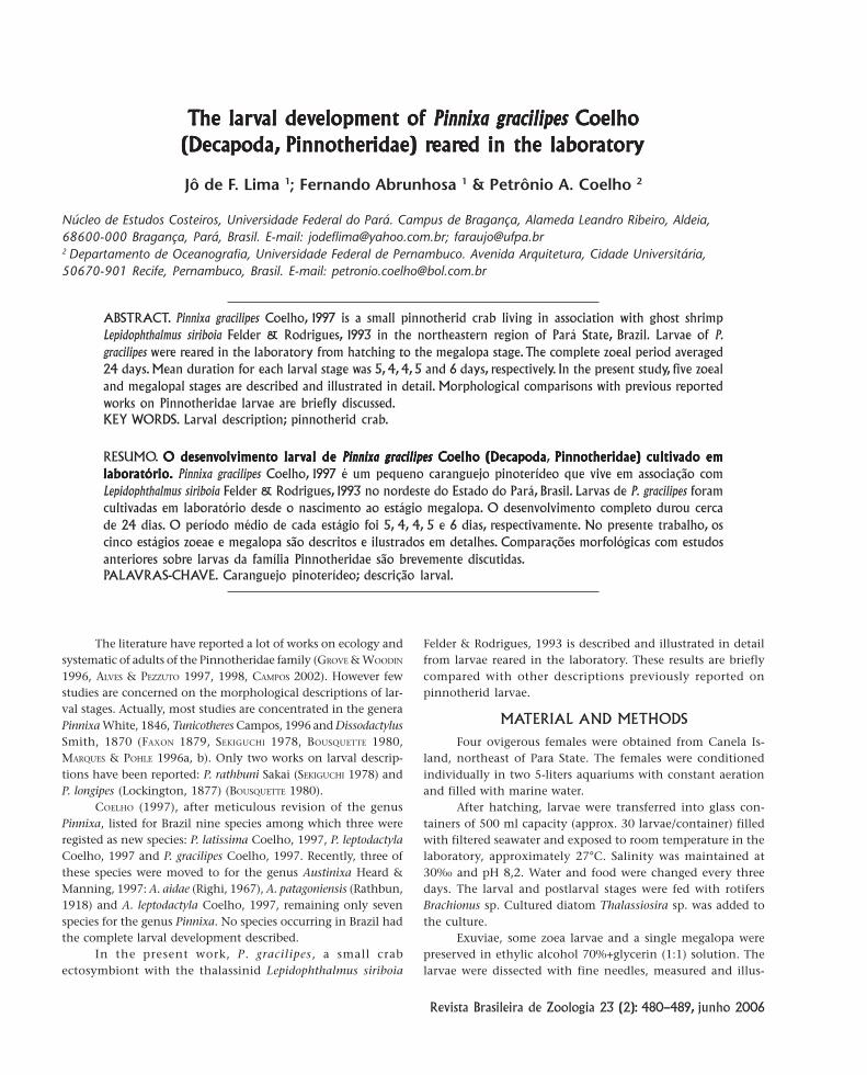
Zoea ICarapace length (OC): 0.33 mm (0.32-0.34 mm).Carapace (Fig.8): bearing one dorsal, one rostral and two
lateral spines. Eyes sessile.Antennule (Fig. 1): uniramous, unsegmented, smooth and
conical with two long and one short terminal aesthetascs.Antenna (Fig. 2): uniramous, elongate, with a tapered
protopodite and two rows of spinules distally and one simple
Table II. Morphological comparisons between features of the zoea IV of P. rathbuni, P. longipes and P. gracilipes. (S) Setation, (A) aesthetascs,(Seg) segment, (Exop) exopod, (BE) basal endite, (Bas) basipod, (CE) coxal endite, (End) endopod, (Scap) scaphognathite.
Appendages P. rathbuni (1) P. longipes (2) P. gracilipes (3)
Antennule:
End present as bud present as bud present as bud
Exop - A 8 aesthetascs 6 aesthetascs 7 aesthetascs
Antenna protopodite shorter thanendopodite
protopodite longer thanendopodite
protopodite absent and endopoditereaching medial portion of protopodite
Maxillule:
End Seg 2-segmented 2-segmented 2-segmented
Protopodite absent absent present
BE 10 setae 13 setae 9 setae
CE 6 setae 6 setae 4 setae
Maxilla:
Scap - S 32 setae 19 setae 18 setae
End - S 3 setae 3 setae 3 setae
BE - S 18 setae 14 setae 12 setae
CE - S 9 setae 8 setae 7 setae
Maxilliped I
Exop - S 12 setae 9 setae 10 setae
End - S 2+2+1+2+5 setae 2+2+1+2 +5 setae 2+2+1+2 +5 setae
Bas - S 9 setae 10 setae 11 setae
Maxilliped II
Exop - S 12 setae 9 setae 10 setae
End - S 0+5 setae 0+5 setae 0+4 setae
Bas - S 4 setae 4 setae 4 setae
1) SEKIGUCHI (1978), 2) BOUSQUETTE (1980), 3) This work.
Table I. Survival rate, intermolting period and accumulative daysof Pinnixa gracilipes reared in the laboratory.
Larval stagesIntermoltingperiod (days)
Cumulative(days)
Survival rate(%)
Zoea I 5 5 70
Zoea II 4 9 55
Zoea III 4 13 30
Zoea IV 5 18 10
Zoea V 6 24 3
Megalopa * * 1
*) Not recorded.
482 J. de F. Lima et al.
Revista Brasileira de Zoologia 23 (2): 480–489, junho 2006
median setae.Maxillule (Fig. 3): endopodite 2-segmented, distal seg-
ment showing four terminal long plumose setae. Basal enditewith four plumodenticulate and one simple setae. Coxal enditewith three distal plumodenticulate and two marginal smallsimple setae. Protopodite absent.
Maxilla (Fig. 4): scaphognathite with three to four plu-mose setae along the anterior margin, tapering to rounded ter-minal process bearing microtrichia. Endopodite unsegmentedwith 2+1 long plumose setae. Basal endite with proximal anddistal lobes fused showing five terminal plumodenticulate se-tae and 1 simple sub terminal seta. Coxal endite with proximaland distal lobes fused with four to five plumose setae.
First Maxilliped (Fig. 5): basipodite with internal marginbearing 2,1,2,2 setae. Endopodite 5-segmented with 2, 2, 1, 2,5setae, respectively. Exopod unsegmented, with four plumo-natatory setae.
Second Maxilliped (Fig. 6): basipodite with five to sixsetae. Endopodite 2-segmented with 0 and (4+1) setae, respec-tively. Exopod unsegmented with four plumo-natatory setae.
Abdomen and telson (Fig.7): Abdomen showing 5 ab-dominal somites; somites 2 and 3 with a pair of acute dorsolat-
eral spine projecting anteriorly; somite 5 with lateral exten-sions, which overlap the telson. Telson bifurcated with twolong lateral plumose spines. Internal margin showing 6 (3+3)plumose setae of diverse sizes.
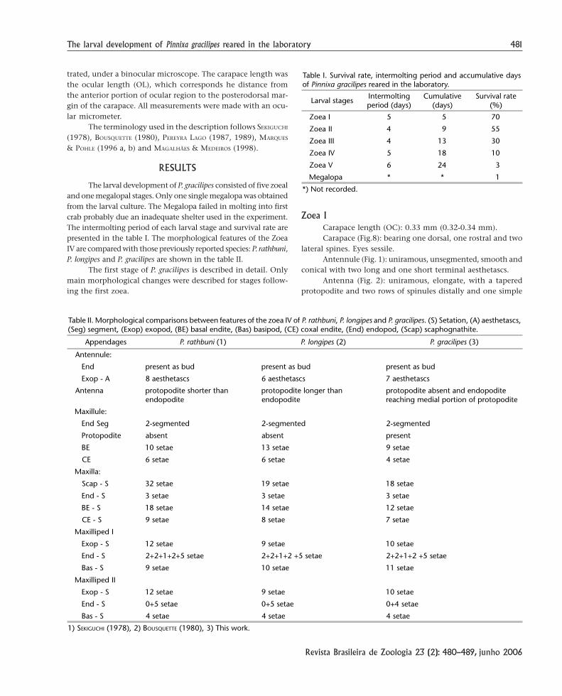
Zoea II
Carapace length (OL): 0.42 mm (0.43-0.44 mm).Carapace (Fig. 16): similar to previous stage. Eyes stalked.Antennule (Fig. 9): uniramous, unsegmented, smooth and
conical with 4 long and one short terminal aesthetascs.Antenna (Fig. 10): similar to previous stage.Maxillule (Fig. 11): basal endite shows two additional
plumodenticulate setae. Coxal endite with four distalplumodenticulate setae and two small simple basal setae.Protopodite with one long plumose seta.
Maxilla (Fig. 12): scaphognathite with six setae, taperingto delta in shape, terminal process bearing microtrichia. Basalendite with proximal and distal lobes fused showing nineplumodenticulate cuspidate setae. Coxal endite with proximaland distal lobes almost fused with four to five plumose setaeand marginal microtrichia.
First Maxilliped (Fig. 13): endopod 5-segmented with 2,
Figures 1-8. Zoea I of Pinnixa gracilipes: (1) antennule; (2) antenna; (3) maxillule; (4) maxilla; (5) first maxilliped; (6) second maxilliped;(7) abdomen and telson; (8) first zoea lateral view. Scale bar: 1-4 = 0.075 mm; 5-6 = 0.15 mm; 7 = 0.2 mm; 8 = 0.3 mm.
1 2
3 4
6 7 8
5
483The larval development of Pinnixa gracilipes reared in the laboratory
Revista Brasileira de Zoologia 23 (2): 480–489, junho 2006
2, 1, 2,(4+1) setae, respectively. Exopodite unsegmented, withsix plumo-natatory setae.
Second Maxilliped (Fig. 14): exopodite unsegmented withsix plumo-natatory setae.
Abdomen and telson (Fig. 15): abdomen without alteration.External margin of telson with two spines projecting laterally.
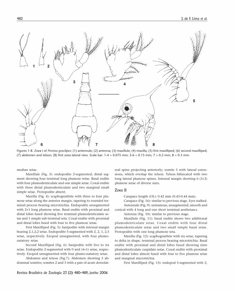
Zoea IIICarapace length (OC): 0.53 mm (0.52-0.54 mm).Carapace (Fig. 24): Similar to previous stage.Antennule (Fig. 17.): with five long aesthetascs and 1
simple seta.Antenna (Fig. 18): similar to previous stage.Maxillule (Fig. 19): basal endite with seven plumodenti-
culate setae. Protopodite present.Maxilla (Fig. 20): scaphognathite with 13 plumose setae.
Basal endite with 10 plumodenticulate setae. Coxal endite withproximal and distal lobes almost fused with four to five plu-mose setae and marginal microtrichia.
First Maxilliped (Fig. 21): basipod with nine simple se-tae. Exopod with eight plumo-natatory setae.
Second Maxilliped (Fig. 22): exopod with eight plumo-natatory setae.
Abdomen and telson (Fig. 23): similar to previous stage.
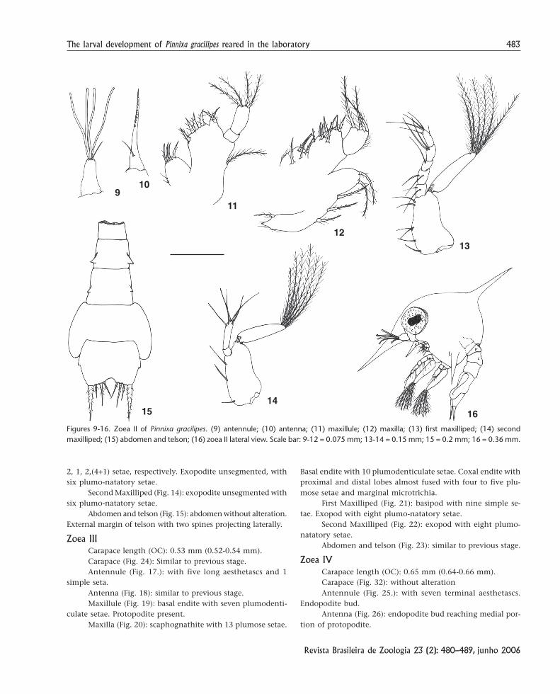
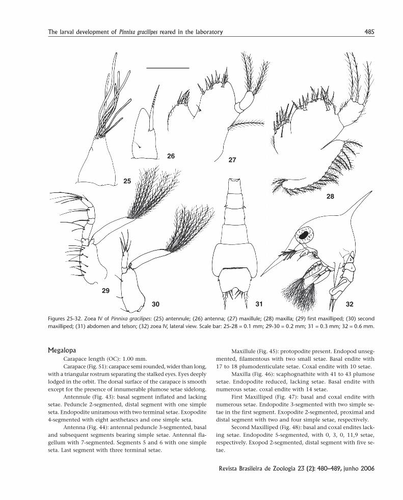
Zoea IVCarapace length (OC): 0.65 mm (0.64-0.66 mm).Carapace (Fig. 32): without alterationAntennule (Fig. 25.): with seven terminal aesthetascs.
Endopodite bud.Antenna (Fig. 26): endopodite bud reaching medial por-
tion of protopodite.
Figures 9-16. Zoea II of Pinnixa gracilipes. (9) antennule; (10) antenna; (11) maxillule; (12) maxilla; (13) first maxilliped; (14) secondmaxilliped; (15) abdomen and telson; (16) zoea II lateral view. Scale bar: 9-12 = 0.075 mm; 13-14 = 0.15 mm; 15 = 0.2 mm; 16 = 0.36 mm.
910
11
12
1514
13
16

484 J. de F. Lima et al.
Revista Brasileira de Zoologia 23 (2): 480–489, junho 2006
Maxillule (Fig. 27): basal endite with nine plumodenticu-late and 1 simple seta. Protopodite present.
Maxilla (Fig. 28): scaphognathite with 18 plumose setae.Basal endite with 12 to 13 plumodenticulate setae. Coxal enditewith seven plumose setae and marginal microtrichia.
First Maxilliped (Fig. 29): basipodite with 11 simple se-tae. Exopodite with 10 plumo-natatory setae.
Second Maxilliped (Fig. 30): exopodite with 10 plumo-natatory setae.
Abdomen and telson (Fig. 31): pleopods buds in the ab-dominal somites two to five. Telson similar to previous stage.
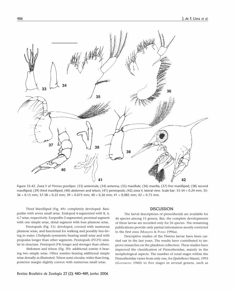
Zoea VCarapace length (OC): 0.76 mm (0.75-0.77 mm).Carapace (Fig. 42): similar to previous stage.Antennule (Fig. 33.): endopodite with seven terminal
aesthetascs. Endopodite bud.Antenna (Fig. 34): endopodite bud more developed than
previous stage with one small basal seta.Maxillule (Fig. 35): basal endite with 11 plumodenticulate
and five simple setae. Coxal endite eight plumose setae.Maxilla (Fig. 36): scaphognathite with 24 to 26 plumose
setae. Basal endite with 18 to 19 plumodenticulate setae. Coxalendite with 11 plumose setae.
First Maxilliped (Fig. 37): similar to previous stage.Second Maxilliped (Fig. 38): similar to previous stage.Third Maxilliped (Fig. 39): with endopod and exopod visible.Abdomen and telson (Fig. 40): with four pairs of pleo-
pods longer than previous stage, unsegmented and uniramous.Telson similar to previous stage.
Pereiopods (Fig. 41): with five pairs as illustrated.
Figures 17-24. Zoea III of Pinnixa gracilipes: (17) antennule; (18) antenna; (19) maxillule; (20) maxilla; (21) first maxilliped; (22) secondmaxilliped; (23) abdomen and telson; (24) zoea III, lateral view. Scale bar: 17-20 = 0.85 mm; 21-22 = 0.15 mm; 23 = 0.25 mm; 24 = 0.46 mm.
17
18
21
22
19
23
20
24
485The larval development of Pinnixa gracilipes reared in the laboratory
Revista Brasileira de Zoologia 23 (2): 480–489, junho 2006
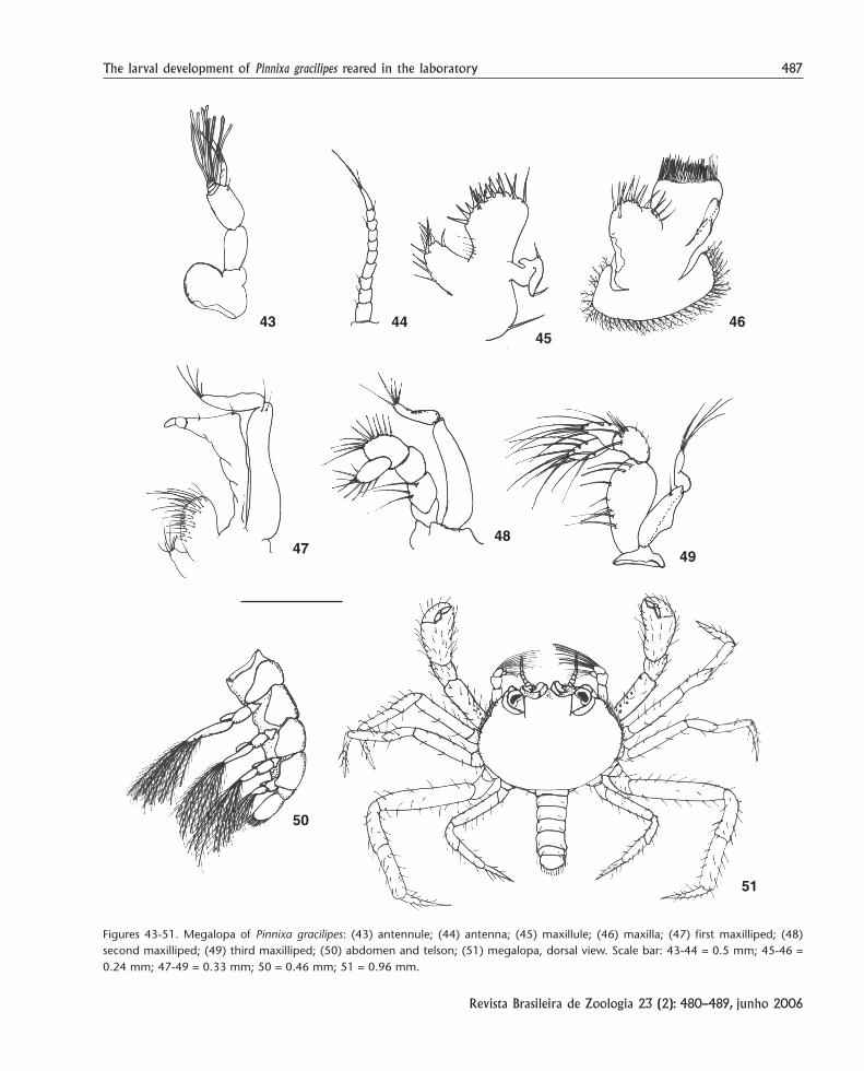
MegalopaCarapace length (OC): 1.00 mm.Carapace (Fig. 51): carapace semi rounded, wider than long,
with a triangular rostrum separating the stalked eyes. Eyes deeplylodged in the orbit. The dorsal surface of the carapace is smoothexcept for the presence of innumerable plumose setae sidelong.
Antennule (Fig. 43): basal segment inflated and lackingsetae. Peduncle 2-segmented, distal segment with one simpleseta. Endopodite uniramous with two terminal setae. Exopodite4-segmented with eight aesthetascs and one simple seta.
Antenna (Fig. 44): antennal peduncle 3-segmented, basaland subsequent segments bearing simple setae. Antennal fla-gellum with 7-segmented. Segments 5 and 6 with one simpleseta. Last segment with three terminal setae.
Maxillule (Fig. 45): protopodite present. Endopod unseg-mented, filamentous with two small setae. Basal endite with17 to 18 plumodenticulate setae. Coxal endite with 10 setae.
Maxilla (Fig. 46): scaphognathite with 41 to 43 plumosesetae. Endopodite reduced, lacking setae. Basal endite withnumerous setae. coxal endite with 14 setae.
First Maxilliped (Fig. 47): basal and coxal endite withnumerous setae. Endopodite 3-segmented with two simple se-tae in the first segment. Exopodite 2-segmented, proximal anddistal segment with two and four simple setae, respectively.
Second Maxilliped (Fig. 48): basal and coxal endites lack-ing setae. Endopodite 5-segmented, with 0, 3, 0, 11,9 setae,respectively. Exopod 2-segmented, distal segment with five se-tae.
Figures 25-32. Zoea IV of Pinnixa gracilipes: (25) antennule; (26) antenna; (27) maxillule; (28) maxilla; (29) first maxilliped; (30) secondmaxilliped; (31) abdomen and telson; (32) zoea IV, lateral view. Scale bar: 25-28 = 0.1 mm; 29-30 = 0.2 mm; 31 = 0.3 mm; 32 = 0.6 mm.
25
26 27
29
30 31
28
32
486 J. de F. Lima et al.
Revista Brasileira de Zoologia 23 (2): 480–489, junho 2006
Third Maxilliped (Fig. 49): completely developed. Basi-podite with seven small setae. Endopod 4-segmented with 8, 6,6,7 setae, respectively. Exopodite 2-segmented, proximal segmentwith one simple setae, distal segment with four plumose setae.
Pereiopods (Fig. 51): developed, covered with numerousplumose setae, and functional for walking and possibly free-liv-ing in water. Chelipeds symmetric bearing small setae and withpropodus longer than other segments. Pereiopods (P2-P3) simi-lar in structure. Pereiopod (P4) longer and stronger than others.
Abdomen and telson (Fig. 50): additional somite 6 bear-ing two simple setae. Other somites bearing additional simplesetae dorsally as illustrated. Telson semi-circular, wider than long,posterior margin slightly convex with numerous small setae.
DISCUSSIONThe larval descriptions of pinnotherids are available for
46 species among 15 genera. But, the complete developmentsof these larvae are recorded only for 26 species. The remainingpublications provide only partial information mostly restrictedto the first zoea (MARQUES & POHLE 1996a).
Descriptive studies of the Pinnixa larvae have been car-ried out in the last years. The results have contributed to im-prove researches on the plankton collection. These studies haveimproved the classification of Pinnotheridae, mainly in themorphological aspects. The number of zoeal stages within thePinnotheridae varies from only one, for Epulotheres Manni, 1993(GOODBODY 1960) to five stages in several genera, such as
Figure 33-42. Zoea V of Pinnixa gracilipes: (33) antennule; (34) antenna; (35) maxillule; (36) maxilla; (37) first maxilliped; (38) secondmaxilliped; (39) third maxilliped; (40) abdomen and telson; (41) pereiopods; (42) zoea V, lateral view. Scale bar: 33-34 = 0.24 mm; 35-36 = 0.15 mm; 37-38 = 0.25 mm; 39 = 0.075 mm; 40 = 0,36 mm; 41 = 0,085 mm; 42 = 0.73 mm.
34
35
37 39
40
41
38
42
3633
487The larval development of Pinnixa gracilipes reared in the laboratory
Revista Brasileira de Zoologia 23 (2): 480–489, junho 2006
Figures 43-51. Megalopa of Pinnixa gracilipes: (43) antennule; (44) antenna; (45) maxillule; (46) maxilla; (47) first maxilliped; (48)second maxilliped; (49) third maxilliped; (50) abdomen and telson; (51) megalopa, dorsal view. Scale bar: 43-44 = 0.5 mm; 45-46 =0.24 mm; 47-49 = 0.33 mm; 50 = 0.46 mm; 51 = 0.96 mm.
43 4445
46
4748
49
50
51
488 J. de F. Lima et al.
Revista Brasileira de Zoologia 23 (2): 480–489, junho 2006
Dissodactylus, Pinnixa and Pinnotheres Latreille, 1802 (MARQUES
& POHLE 1995).The morphology of the genera Pinnixa can be distin-
guished easily within of the family Pinnotheridae due to pres-ence of a structural enlargement of the fifth abdominal seg-ment. This appears to be a distinct characteristic of this genus,as observed in P. longipes (BOUSQUETTE 1980), P. rathbuni (SEKIGUCHI
1978) and in P. gracilipes in the present study.Abdomen and carapace of P. gracilipes showed few changes
during zoeal stages. Only the first zoea stage had telson withspines absent and second to fifth zoeal stages showed a pair ofspines projected laterally (Fig. 16). These spines appear to beunique for P. gracilipes and, they are not present in P. longipesnot in P. rathbuni. Other morphological feature showed onlyfor P. gracilipes is the shape of final portion of scaphognathiteof zoea I, which is tapering to sharp in P. longipes and P. rathbunibut visible rounded in P. gracilipes (Fig. 4).
Besides the morphologic differences, other characteris-tics can distinguish P. longipes and P. rathbuni from P. gracilipes.The larval rearing time averaged 24 days in P. gracilipes while,P. longipes 26 days. The intermolting period was shorter in P.gracilipes than P. longipes. Unfortunately, information on larvalperiod was not mentioned by SEKIGUCHI (1978) for P. rathbuni.Similarity is found in the number of larval stages; P. rathbuni, P.longipes and P. gracilipes have five zoeal stages.
The gross morphology of zoea stages is very similar in P.longipes, P. rathbuni and P. gracilipes but some differences are foundbetween these species. The zoea IV of P. gracilipes shows morpho-logical differences in relation to the other two species, mainly inthe appendages, the antenna, maxillule and maxilla (Tab. II).Other evident difference observed for P. gracilipes in the absenceof antennal protopodite in the zoea V, whereas in P. longipes thisprocess is larger than endopodite. The protopodite in P. rathbuniis smaller than endopodite. These characteristics intensify themorphological divergence of zoeal stages among Pinnixa species.
The megalopa stage of P. longipes, P. rathbuni and P.gracilipes showed similarity. The megalopa of P. gracilipes canbe easily distinguished from other species through the follow-ing morphologic characteristics: triangular rostrum separatingthe stalked eyes, 10-segmented antenna, fine and long pereio-pods, and semi-circular carapace. On the other hand, P. longipesshows rectangular rostrum separating the eyes, 7-segmentedantenna, strong pereiopods and semi-rectangular carapace. P.rathbuni has a set of large spines on each antero-lateral marginof carapace, a specific character for this species.
The descriptions in the present study are sufficient todistinguish P. gracilipes larvae from other described pinnothe-rids. However, further larval studies are needed to increase theunderstanding of this taxonomic group.
ACKNOWLEDGMENTS
To the Instituto do Milênio and to the project Manage-ment and Dynamics of Mangrove (MADAM) for the financial
support destined to this research; To the Universidade Federaldo Pará and Maria Iracilda Sampaio, Laboratory of MolecularBiology of University campus of Bragança, Pará.
REFERENCES
ALVES, E.S. & P.R. PEZZUTO. 1997. Population dynamics of Pinnixapatagoniensis Rathbun, 1918 (Brachyura: Pinnotheridae) asymbiotic crab of Sergio mirim (Thalassinidea: Callianassidae)in Cassino Beach, southern Brazil. Marine Ecology, Berlin,19 (1): 37-51.
ALVES, E.S. & P.R. PEZZUTO. 1998. Dispersão de Pinnixa patagoniensisRathbun, 1918 (Brachyura: Pinnotheridae) no médio litoraldo Cassino, RS, Brasil. Atlântica, Rio Grande, 20: 5-2.
BOUSQUETTE, G.D. 1980. The larval development of Pinnixa longipes(Lockington, 1877) (Brachyura: Pinnotheridae), reared in theLaboratory. Biological Bulletin, Miami, 159: 592-605.
CAMPOS, E. 2002. Two new genera of pinnotherid crabs fromthe tropical eastern Pacific (Decapoda: Brachyura: Pinnothe-ridae). Journal of Crustacean Biology, Lawrence, 22(2): 328-336.
COELHO, P. A. 1997. Revisão do gênero Pinnixa White, 1846, noBrasil (Crustacea: Decapoda: Pinnotheridae). Trabalhos Oce-anográficos da Universidade Federal de Pernambuco,Recife, 25: 163-193.
FAXON, W. 1879. On some young in the development of Hippa,Porcellana and Pinnixa. Bulletin of the Museum of Compa-rative Zoology, Harvard, 5: 253-268.
GOODBODY, I. 1960. Abbreviated development in a pinnotheridcrab. Nature, London, 185: 704-705.
GROVE, M.W. & S.A. WOODIN. 1996. Conspecific Recognition andhost choice in a pea crab, Pinnixa chaetopterana (Brachyura:Pinnotheridae). The Biological Bulletin, Massachusetts,190: 359-366.
MAGALHÃES, C. & N. MEDEIROS. 1998. The larval development ofpalaemonid shrimps from the Amazon Region reared in theLaboratory. VII. Abbreviated development of Pseudopalaemonamazonensis Ramos-Porto, 1979 (Crustacea: Decapoda: Cari-dea). Acta Amazônica, Manaus, 28 (4): 433-448.
MARQUES, F. & G. POOLE. 1995. Phylogenetic analysis of thePinnotheridae (Crustacea, Brachyura) based on larvalmorphology, with emphasis on the Dissodactylus speciescomplex. Zoologica Scripta, Oslo, 24 (4): 347-364.
MARQUES, F. & G. POHLE. 1996a. Laboratory-reared larval stagesof Dissodactylus mellitae (Decapoda: Brachyura:Pinnotheridae) and developmental patterns within theDissodactylus species complex. Canadian Journal ofZoology, Canada, 74: 47-62.
MARQUES, F. & G. POHLE. 1996b. Complete larval developmentof Clypeasterophilus stebbing (Decapoda: Brachyura: Pinno-theridae) and a comparison with other species within theDissodactylus complex. Bulletin of Marine Science, Miami,58 (1): 165-185.
PEREYRA LAGO, R. 1987. Larval development of Sesarma catenata
489The larval development of Pinnixa gracilipes reared in the laboratory
Revista Brasileira de Zoologia 23 (2): 480–489, junho 2006
Ortmann (Brachyura: Grapsidae: Sesarminae) reared in thelaboratory. South African Journal of Zoology, Grahams-town, 22 (3): 200-212.
PEREYRA LAGO, R. 1989. The larval development of the redmangrove crab Sesarma meinerti Ortmann (Brachyura:
Received in 14.VII.2005; accepted in 10.V.2006.
Grapsidae) reared in the laboratory. South African Journalof Zoology, Grahamstown, 24 (3): 199-211.
SEKIGUCHI, H. 1978. Larvae of a pinnotherid crab, Pinnixa rathbuniSakai. Proceeding of the Japanese Society of SystematicZoology, Tsu, 15: 36-46.