the immunoreactivity of cyclooxygenase(cox)-2 and relationship between expression of p53 and ki-67...
TRANSCRIPT
April 1998
G2768 RECTAL CARCINOMA: IS IT ABLE TO EXPECT STAGE AND PROGNOSIS BY COLONOSCOPE? T. Sakai. Y. Yamashita, T. Maekawa, T. Hideshima, T. Shirakusa. Second Department of Suegery, School of Medicine, Fukuoka University, Japan
Background/Aims: In the patients of rectal carcinoma, is it able to expect Dukes stage and prognosis before operation? The aim of this study was to investigate the correlation of the pathological findings and the tumor occupied rate by the colonoscope. Methods: Fifty-one patients with rectal carcinoma undergoing radical operation were enrolled in this study and were classified the following. Group A(26): Tumor occupied under 1 / 2 round of the colonic lumen. Group B(13): Tumor occupied over 1 / 2 under 2 / 3 round of the colonic lumen. Group C(12): Tumor occupied over 2 / 3 round of the colonic lumen. All subjects were examined pathologically. Results: In group A, Dukes A was 76.9%, Dukes B was 15.4% and Dukes C was 7.7%. In group B, Dukes A was 7.7%, Dukes B was 53.8% and Dukes C was 38.5%. In group C, Dukes A was 0%, Dukes B was 33.3% and Dukes C was 66.7%. Tumor occupied rate significantly correlated Dukes stage. In group A, there were no recurrence more than three years. On the other hand, 4 cases were recognized recurrence in group B and C. Conclusion: In the patients of rectal carcinoma, Dukes stage and prognosis are expected by tumor occupied rate.
• G2769 PRIMARY GASTRIC ADULT T-CELL LEUKEMIA/LYMPHOMA. H. Sakata. R. lwakiri, M. Mizuguchi*, A. Kuwahara, T. Yoshida, T. Ohyama, T. Koyama, K. Fujimoto. Departments of Internal Medicine, and * Radiology, Saga Medical School, Saga, Saga 849-8501, Japan.
We previously reviewed gastric lesions in 76 patients with adult T-cell leukemia/lymphoma (ATLL) evaluated by endoscopic examination (Cancer 78:396, 1996). ATLL is caused by human T-lymphotrophic virus type I. Most of primary gastric lymphomas are B-eell tumors, and a review of collected cases of primary gastric ATLL has not been reported. In this study, we reported primary gastric ATLL who visited our hospital. Materials and Methods. Between 1981 and 1995, 114 patients with ATLL came to our hospital. After informed consent was obtained, 78 patients underwent endoscopic examination of the upper gastrointestinal tract. Clinical subtypes of ATLL were divided into four groups: acute, lymphoma, chronic, and smoldering. Endoscopic features of gastric lesions of ATLL were classified into three types: diffuse type, tumor-forming type, and giant fold type. Results. Among 78 patients who underwent endoscopic examination, 48 patients were divided into lymphoma type regarding clinical subtypes. Eighteen patients in 48 lymphoma type patients had gastric lesions. As demonstrated in table, the most frequent gastric lesion configuration in lymphoma type of ATLL was the diffuse type with ulceration.
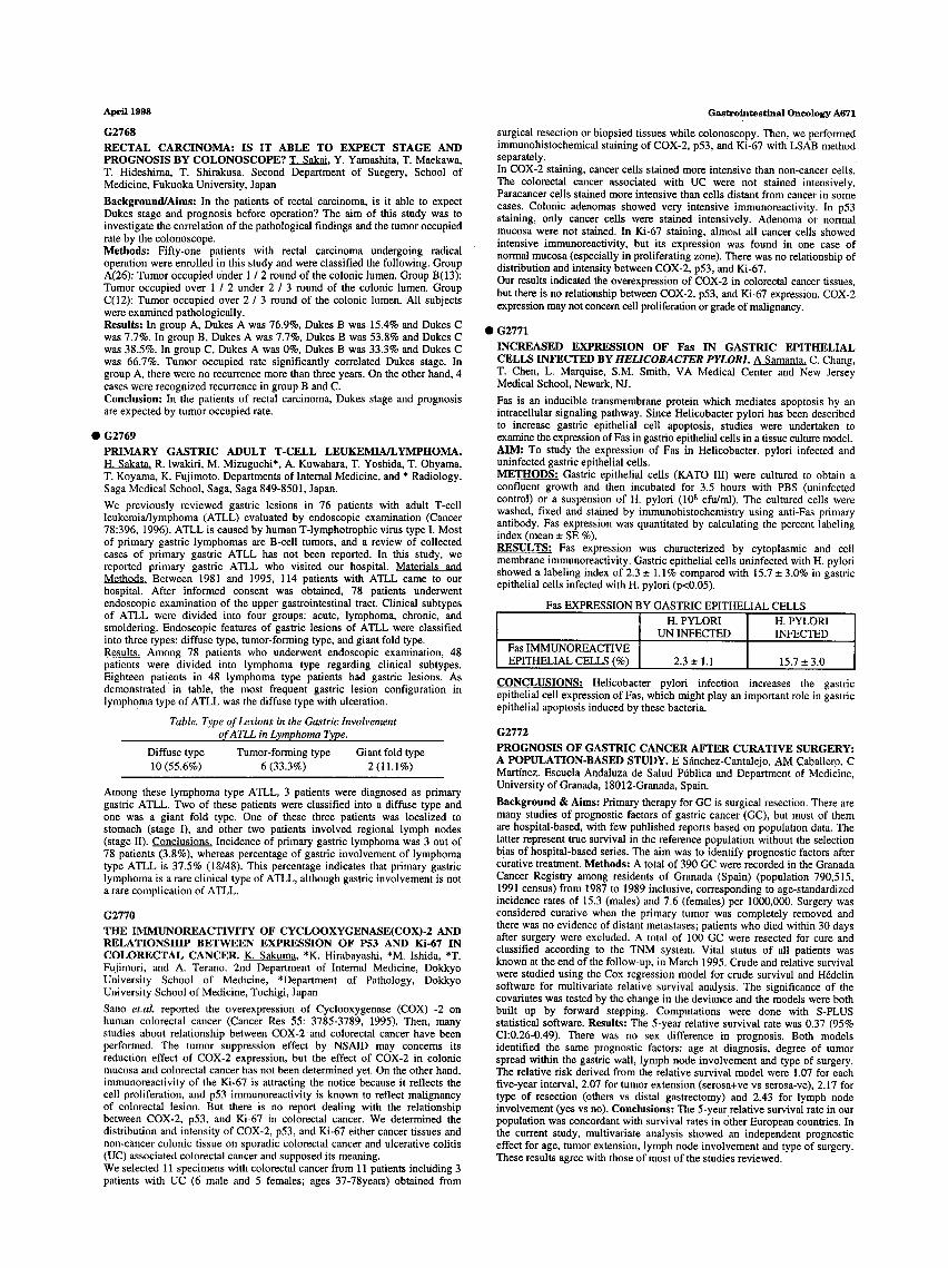
Table. Type of Lesions in the Gastric Involvement o, f ATLL in L~mphoma Type.
Diffuse type Tumor-forming type Giant fold type 10 (55.6%) 6 (33.3%) 2 (11.1%)
Among these lymphoma type ATLL, 3 patients were diagnosed as primary gastric ATLL. Two of these patients were classified into a diffuse type and one was a giant fold type. One of these three patients was localized to stomach (stage I), and other two patients involved regional lymph nodes (stage II). Conclusions. Incidence of primary gastric lymphoma was 3 out of 78 patients (3.8%), whereas percentage of gastric involvement of lymphoma type ATLL is 37.5% (18/48). This percentage indicates that primary gastric lymphoma is a rare clinical type of ATLL, although gastric involvement is not a rare complication of ATLL.
G2770
THE IMMUNOREACTIVITY OF CYCLOOXYGENASE(COX)-2 AND RELATIONSHIP BETWEEN EXPRESSION OF P53 AND Ki-67 IN COLORECTAL CANCER. K. Sakuma. *K. Hirabayashi, *M. Ishida, *T. Fujimori, and A. Terano. 2nd Department of Internal Medicine, Dokkyo University School of Medicine, *Department of Pathology, Dokkyo University School of Medicine, Tochigi, Japan
Sano et.al, reported the overexpression of Cyclooxygenase (COX) -2 on human colorectal cancer (Cancer Res 55: 3785-3789, 1995). Then, many studies about relationship between COX-2 and colorectal cancer have been performed. The tumor suppression effect by NSAID may concerns its reduction effect of COX-2 expression, but the effect of COX-2 in colonic mucosa and colorectal cancer has not been determined yet. On the other hand, immunoreactivity of the Ki-67 is attracting the notice because it reflects the cell proliferation, and p53 immunoreactivity is known to reflect malignancy of colorectal lesion. But there is no report dealing with the relationship between COX-2, p53, and Ki-67 in colorectai cancer. We determined the distribution and intensity of COX-2, p53, and Ki-67 either cancer tissues and non-cancer colonic tissue on sporadic Colorectal cancer and ulcerative colitis (UC) associated colorectal cancer and supposed its meaning. We selected 11 specimens with colorectal cancer from 11 patients including 3 patients with UC (6 male and 5 females; ages 37-78years) obtained from
Gastrointestinal Oncology A671
surgical resection or biopsied tissues while colonoscopy. Then, we performed immunohistochemical staining of COX-2, p53, and Ki-67 with LSAB method separately. In COX-2 staining, cancer cells stained more intensive than non-cancer cells. The colorectal cancer associated with UC were not stained intensively. Paracancer cells stained more intensive than cells distant from cancer in some cases. Colonic adenomas showed very intensive immunoreactivity. In p53 staining, only cancer cells were stained intensively. Adenoma or normal mucosa were not stained. In Ki-67 staining, almost all cancer cells showed intensive immunoreactivity, but its expression was found in one case of normal mucosa (especially in proliferating zone). There was no relationship of distribution and intensity between COX-2, p53, and Ki-67. Our results indicated the overexpression of COX-2 in colorectal cancer tissues, but there is no relationship between COX-2, p53, and Ki-67 expression. COX-2 expression may not concem cell proliferation or grade of malignancy.
• G2771
INCREASED EXPRESSION OF Fas IN GASTRIC EPITHELIAL CELLS INFECTED BY HELICOBACTER PYLORI. A Samanta. C. Chang, T. Chen, L. Marquise, S.M. Smith, VA Medical Center and New Jersey Medical School, Newark, NJ.
Fas is an inducible transmembrane protein which mediates apoptosis by an intracellular signaling pathway. Since Helicobacter pylori has been described to increase gastric epithelial cell apoptosis, studies were undertaken to examine the expression of Fas in gastrio epithelial cells in a tissue culture model. AIM; To study the expression of Fas in Helicobacter. pylori infected and uninfected gastric epithelial cells. METHODS: Gastric epithelial cells (KATO III) were cultured to obtain a confluent growth and then incubated for 3.5 hours with PBS (uninfected control) or a suspension of H. pylori (108 cfu/ml). The cultured cells were washed, fixed and stained by immunohistochemistry using anti-Fas primary antibody. Fas expression was quantitated by calculating the percent labeling index (mean _+ SE %). RESULTS: Fas expression was characterized by cytoplasmic and cell membrane immunoreactivity. Gastric epithelial cells uninfected with H. pylori showed a labeling index of 2.3 +_ 1.1% compared with 15.7 -+ 3.0% in gastric epithelial cells infected with H. pylori (p<0.05).
Fas EXPRESSION BY GASTRIC EPITHELIAL CELLS H. PYLORI H. PYLORI
UN INFECTED INFECTED Fas IMMUNOREACTIVE EPITHELIAL CELLS (%) 2.3 _+ 1.1 15.7 - 3.0
CONCLUSIONS: Helicobacter pylori infection increases the gastric epithelial cell expression of Fas, which might play an important role in gastric epithelial apoptosis induced by these bacteria.
G2772
PROGNOSIS OF GASTRIC CANCER AFTER CURATIVE SURGERY: A POPULATION-BASED STUDY. E Sfinchez-Cantalejo, AM Caballero, C Martfnez. Escuela Andaluza de Salud Pt~blica and Department of Medicine, University of Granada, 18012-Granada, Spain.
Background & Aims: Primary therapy for GC is surgical resection. There are many studies of prognostic factors of gastric cancer (GC), but most of them are hospital-based, with few published reports based on population data. The latter represent true survival in the reference population without the selection bias of hospital-based series. The aim was to identify prognostic factors after curative treatment. Methods: A total of 390 GC were recorded in the Granada Cancer Registry among residents of Granada (Spain) (population 790,515, 1991 census) from 1987 to 1989 inclusive, corresponding to age-standardized incidence rates of 15.3 (males) and 7.6 (females) per 1000,000. Surgery was considered curative when the primary tumor was completely removed and there was no evidence of distant metastases; patients who died within 30 days after surgery were excluded. A total of 100 GC were resected for cure and classified according to the TNM system. Vital status of all patients was known at the end of the follow-up, in March 1995. Crude and relative survival were studied using the Cox regression model for crude survival and H6delin software for multivariate relative survival analysis. The significance of the covariates was tested by the change in the deviance and the models were both built up by forward stepping. Computations were done with S-PLUS statistical software. Results: The 5-year relative survival rate was 0.37 (95% CI:0.26-0.49). There was no sex difference in prognosis. Both models identified the same prognostic factors: age at diagnosis, degree of tumor spread within the gastric wall, lymph node involvement and type of surgery. The relative risk derived from the relative survival model were 1.07 for each five-year interval, 2.07 for tumor extension (serosa+ve vs serosa-ve), 2.17 for type of resection (others vs distal gastrectomy) and 2.43 for lymph node involvement (yes vs no). Conclusions: The 5-year relative survival rate in our population was concordant with survival rates in Other European countries. In the current study, multivariate analysis showed an independent prognostic effect for age, tumor extension, lymph node involvement and type of surgery. These results agree with those of most of the studies reviewed.