the hematopoietic and lymphoid systems. hematopathology blood lymphoid organs –central: bone...
TRANSCRIPT
the hematopoietic and the hematopoietic and lymphoid systemslymphoid systems
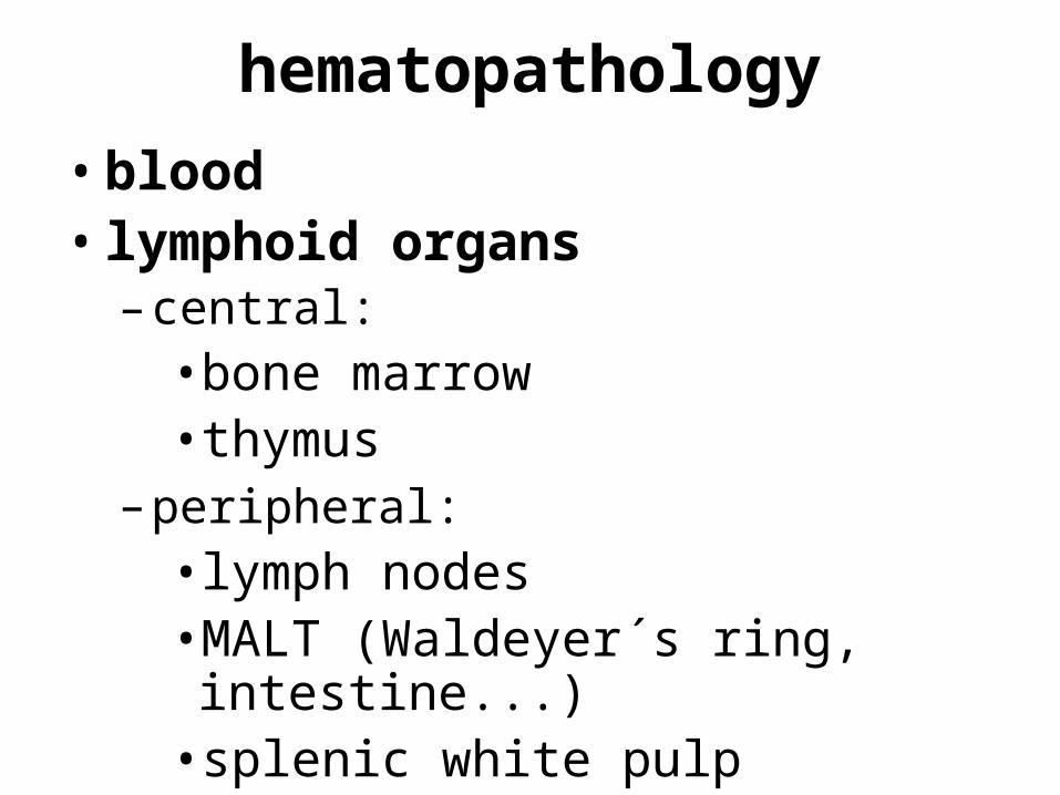
hematopathology
•blood• lymphoid organs
– central:•bone marrow•thymus
– peripheral:•lymph nodes•MALT (Waldeyer´s ring, intestine...)
•splenic white pulp
hematopathology
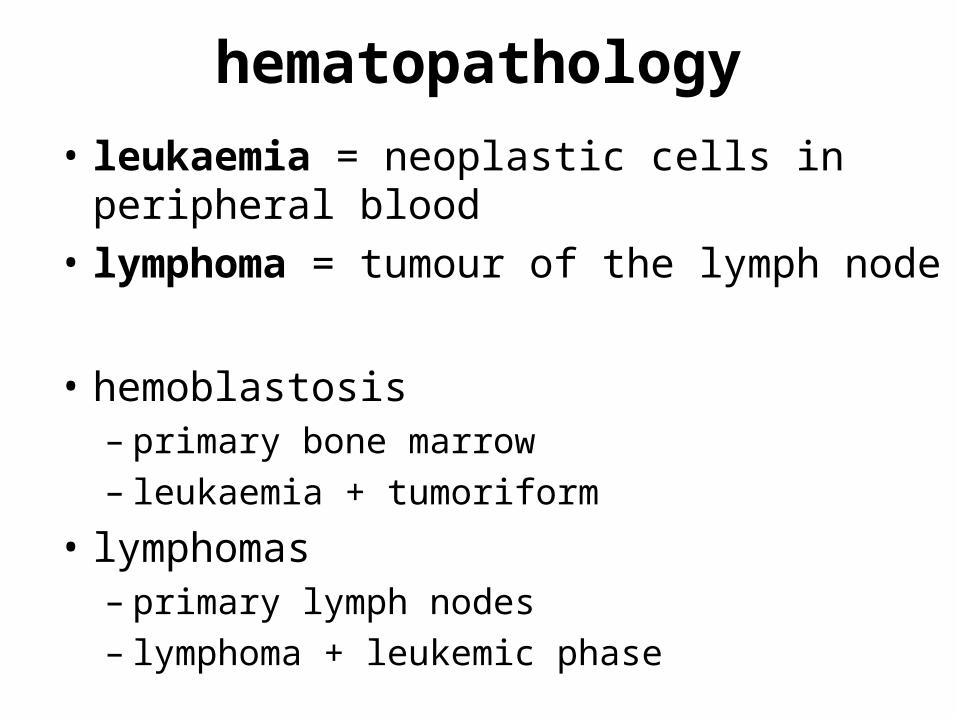
• leukaemia = neoplastic cells in peripheral blood
• lymphoma = tumour of the lymph node
• hemoblastosis – primary bone marrow– leukaemia + tumoriform
• lymphomas– primary lymph nodes– lymphoma + leukemic phase

bone marrow
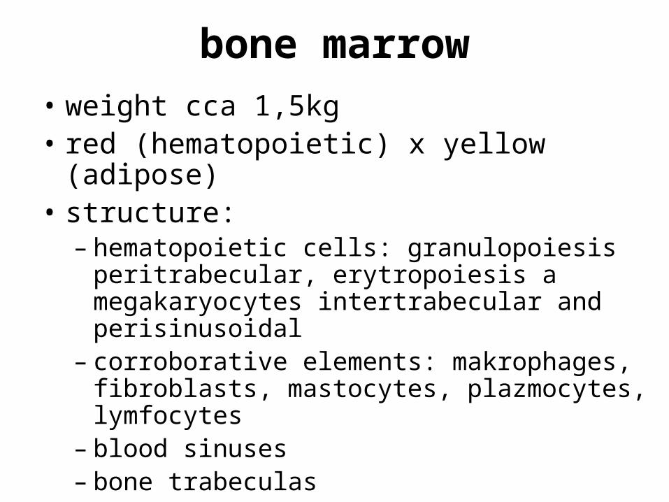
bone marrow• weight cca 1,5kg• red (hematopoietic) x yellow (adipose)• structure:
– hematopoietic cells: granulopoiesis peritrabecular, erytropoiesis a megakaryocytes intertrabecular and perisinusoidal
– corroborative elements: makrophages, fibroblasts, mastocytes, plazmocytes, lymfocytes
– blood sinuses– bone trabeculas
diminished hematopoiesis
A) total diminutionaplastic anemia (panmyelophtisis)• hereditary:
– Fanconi anemia• AR• death because of infectious and bleeding
complications• +/- turn into AML
• acquired:– infectious, irradiation, use of some drugs
diminished hematopoiesis
B) selective• one or more of hematopoietic lineages
critical is peripheral blood – marrow could be hypercelular = „ineffective hematopoiesis“
diminished hematopoiesis...anemia
1) anemia• ↓ total circulating RBC volume, +/-
↓Hb and ↓O2• hypoxia of tissues = clinical
symptoms
anemia...loss of RBC
a) hemorrhage: blood loss anemia• hypovolemia → normocytic
normochromic anemia → ↑ erytropoiesis (bone marrow) → reticulocytosis, hypochromic anemia
anemia...hemolytic
b) increased rate of RBC destruction: the hemolytic anemias
• anemia + reactive hyperplastic erytropoiesis
• bm: ↑erytropoiesis/myelopoiesis, gaucheroid cells
• +/- extramedullary hematopoiesis• Hb -emia, -uria
anemias..hemolytic..intrinsic
I) intrinsic (intracorpuscular) abnormalities of RBC
hereditary: 1) disorders of RBC membrane cytoskeleton
spherocytosis– erythrocytes spheroidal, less deformable
and vulnerable to splenic sequestration and destruction
– AD– anemia, splenomegalia a hemolytic icterus
anemias..hemolytic..intrinsic
2) RBC enzyme deficiencies3) disorders of Hb synthesis: hem+globin
deficient globin synthesis: thalassemia syndromes– lack of or decreased synthesis of globin chains:
α chains = α thalassemia β chains = β thalassemia
– ↓ synthesis of Hb → anemia (microcytic hypochromic) + excess of α chains in β thalassemia → insoluble aggregats → damage RBC membrane → reduction of plasticity → phagocytosis, inefective erytropoiesis
– heterozygous = thalassemia minor homozygous = thalassemia major
anemias..hemolytic..intrinsic
structurally abnormal globin synthesis (hemoglobinopathies): sickle cell anemia– structurally abnormal Hb S – on
deoxygenation polymerization = gelation or crystallization → microvascular obstruction → ischemic tissue damage + ↑ removing in the spleen = „autosplenectomy“
anemias..hemolytic..intrinsic
acquiredmembrane defect: paroxysmal
nocturnal hemoglobinuria)– ↓resistance against C3– granulocytes and plateles affected too →
hemolysis, +/- trombotic complications and ↑ susceptibility to infections
anemias..hemolytic..extrinsic
II) extrinsic (extracorpuscular) abnormalities
1) antibody mediated isohemagglutininserythroblastosis fetalis
– Rh (mother Rh-, father and child Rh+)– antibodies against fetal RBC– hydrops fetus universalis, mental
retardation, ↑extramedulary hematopoiesis
anemias..hemolytic..extrinsic
autoantibodies– idiopathic (primary), drug associated,
SLE– Coombs tests
anemias..hemolytic..extrinsic
2) mechanical trauma to RBCs mikroangiopathic hemolytic anemias
– DIC, TTP mechanic traumatization of
erythrocytes– dialysis, valves prosthesis
3) infections (malaria)
anemia...impaired RBC production
c) diminished erythropoiesis1) combination with the others in
aplastic anemia 2) pure „erytroblastophtisis“
Blackfan-Diamond syndrom• children• + thymomas and T-CLL
3) myelophtisic anemia• extensive replacement of the marrow by
tumours or other lesions → extramedullary hematopoiesis, leukoerythroblastosis
anemia...impaired RBC production
4) iron deficiency anemia• most common• mikrocytar hypochromic• ↓low intake (diets, malabsorptions) x
↑ demands (pregnancy, infancy, chronic blood loss)
• gross: hypoxic myocardial steatosis • marrow normal or hyperplastic
erythropoiesis, decline in serrum ferritin and depletion of stainable iron in the bone marrow
anemia...impaired RBC production
5) megaloblastic anemia• disturbance of proliferation and
differentiation of erythroblasts → megaloblasts, megakaryocytes
• nuclear-cytoplasmic asynchrony• giant metamyelocytes →
hypersegmented neutrophils• ineffektive erythropoiesis folate (folic acid) deficiency anemia• tetrahydrofolate• neurologic abnormalities do not occur
anemia...impaired RBC production
pernicious anemia• vitamin B12 (cobalamin) deficiency• diet, ↓intrinsic faktor (parietal
gastric cells), terminal ileum• gross: atrophic glossitis, gastritis,
demyelinization
anemia...impaired RBC production
6) lack of erythropoietin• kidney failure, parvovirosis (B19)
diminished hematopoiesis... leukopenia
2) leukopeniaa) lymfopenia• hereditary immunity disorders,
infections(viral), chronical diseases, steroid therapy
leukopeniab) neutropenia (granulocytopenia)• increased susceptibility to infections• marrow failure (aplastic anemia) →
agranulocytosis• inadequate or ineffective granulopoiesis:
certain drugs: benzen, purin and pyrimidin analogs, anthracyklin x idiosyncrastic reaction (chloramfenikol, chlorpromazin, fenylbutazon)
• accelerated removal or destruction of neutrophils: hypersplenism, certain drugs
• bm: depend on the underlying basis: ↑ or ↓ granulopoiesis and +/- reaction to infection
increased hematopoiesis
• transitory increasing of hematopoiesis1) ↑erythropoiesis = polycythemia• increased erythropoietin levels:
– appropriate: lung disease, high-altitude living, cyanotic heart disease
– inappropriate: erythropoietin-secreting tumours, „doping“
• bm hypercellular, inappropriate increasing of erythropoiesis
• no extramedullary hematopoiesis!
increased hematopoiesis
2) leukocytosisa) lymfocytosis: chronical infections (IM)b) granulocytosis: acute bacterial
infections (pyogenic organisms), sterile inflammation (tissue necrosis, burns) → leukemoid reaction (like in CML)
c) eosinophilia: allergic disorders, parasitic infestation, drug reaction, certain mlg
3) thrombocytosis: infections, chronical bleeding, tumours, iron deficiency
myelodysplastic syndromes
• heterogeneous group of disorders• some evidence of bone marrow failure
and dysplasia in one or more myeloid cell lineages
• may evolve to AML• chromosomal aberrations• primary x secondary (radiotherapy,
alkylating agent therapy) • bm hypercellular, ↑ erythropoiesis,
morphological changes, +/- fibrosis
myelodysplastic syndromes...histological
classification refractory anemia (RA) refractory anemia with ring
sideroblasts (RARS) refractory cytopenia with multilineage
dysplasia refractory anemia with excess blasts
(RAEB) MDS, unclassifiable
chronic myeloproliferative diseases
• CMPDs: clonal haematopoietic stem cell disorders characterised by proliferation in the bone marrow of one or more of the myeloid (i.e. granulocytic, erythroid and megakaryocytic) lineages
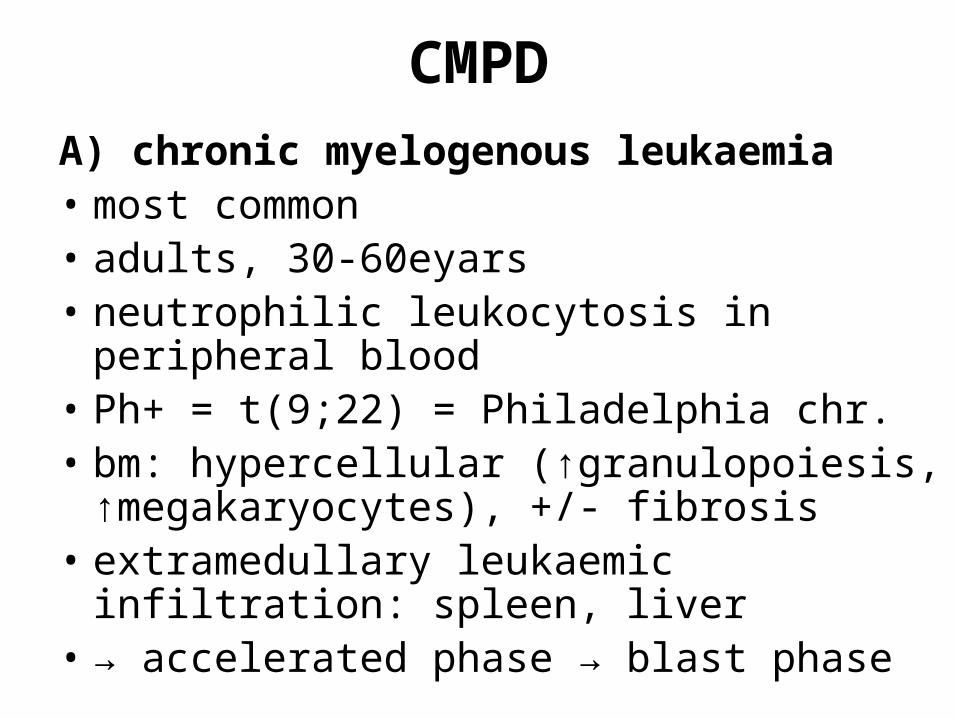
CMPDA) chronic myelogenous leukaemia• most common• adults, 30-60eyars• neutrophilic leukocytosis in peripheral
blood• Ph+ = t(9;22) = Philadelphia chr.• bm: hypercellular (↑granulopoiesis,
↑megakaryocytes), +/- fibrosis• extramedullary leukaemic infiltration:
spleen, liver• → accelerated phase → blast phase
CMPD
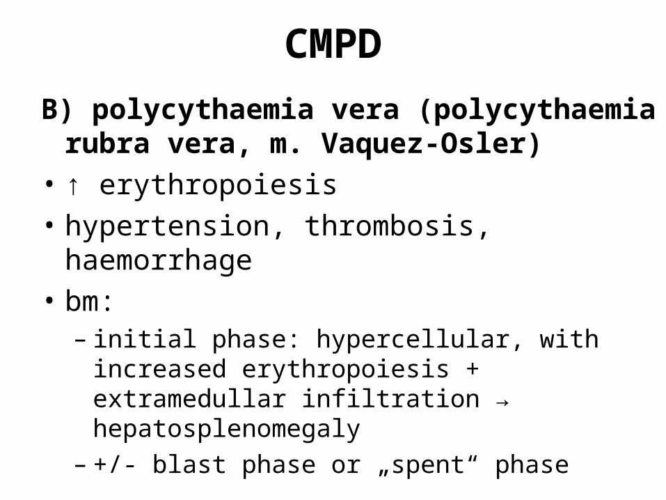
B) polycythaemia vera (polycythaemia rubra vera, m. Vaquez-Osler)
• ↑ erythropoiesis• hypertension, thrombosis,
haemorrhage• bm:
– initial phase: hypercellular, with increased erythropoiesis + extramedullar infiltration → hepatosplenomegaly
– +/- blast phase or „spent“ phase
CMPD
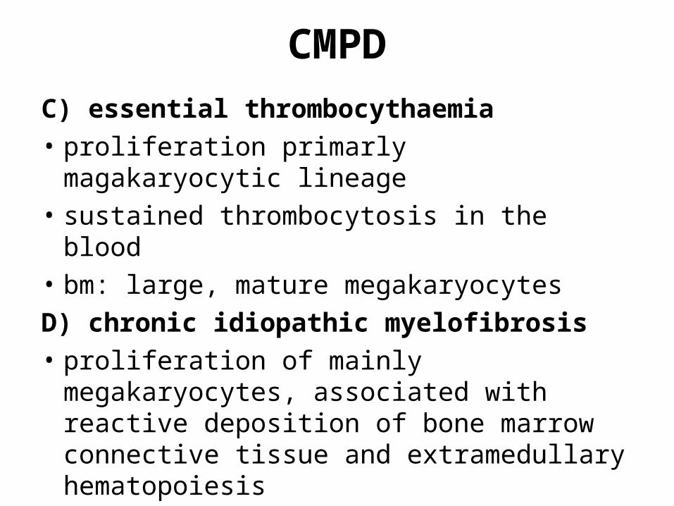
C) essential thrombocythaemia• proliferation primarly magakaryocytic
lineage• sustained thrombocytosis in the blood• bm: large, mature megakaryocytesD) chronic idiopathic myelofibrosis• proliferation of mainly
megakaryocytes, associated with reactive deposition of bone marrow connective tissue and extramedullary hematopoiesis
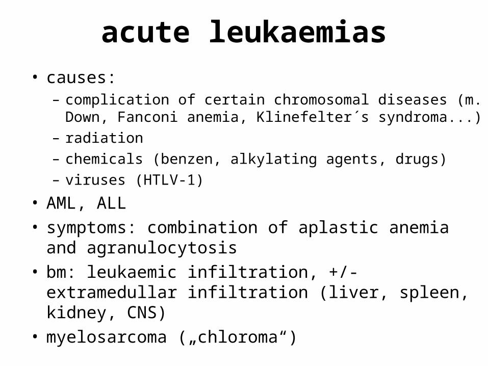
acute leukaemias• causes:
– complication of certain chromosomal diseases (m. Down, Fanconi anemia, Klinefelter´s syndroma...)
– radiation– chemicals (benzen, alkylating agents, drugs)– viruses (HTLV-1)
• AML, ALL• symptoms: combination of aplastic anemia
and agranulocytosis• bm: leukaemic infiltration, +/- extramedullar
infiltration (liver, spleen, kidney, CNS)• myelosarcoma („chloroma“)
acute myeloid leukaemias... histological
classification M0...acute myeloblastic l. minimally
differentiated M1 ...acute myeloblastic l. without
maturation M2...acute myeloblastic l. with
maturation M3...acute promyelocytic l. M4...acute myelomonocytic l. M5...acute monocytic l. M6...acute erythroid l. M7...acute megakaryoblastic l.
acute lymphoblastic leukaemias... histological
classification precursor B- and T- cell lymphoblastic
leukaemia/lymphoblastic lymphoma
proliferation of macrophages, histiocytosis
A) reactive proliferation of macrophages
• bone marrow, many causes (hemosiderosis, aiha, viral infections)
• lysosomal storage diseases (m. Gaucher, Niemann-Pick...)
proliferation of macrophages, histiocytosis
B) hemofagocytic syndroma• ↑ proliferation of macrophages or
histiocytic precursores → haemofagocytosis → cytopenia
• + hepatosplenomegaly, fever• proliferating macrophages: clonal (mlg
histiocytosis) x reaction (infection, Kawasaki, T lymphomas)
• fatal haemofagocytosis
proliferation of macrophages, histiocytosis
C) histiocytosis X (Langerhans cells histiocytosis)
1) solitary eosinophilic granuloma– bng– bones (unifocal lytic lesion), skin, lymph
nodes, lungs– Langerhans cells (Birbeck granules) +
eosinophils, +/- plasma cells and lymphocytes
2) m. Hand-Schüler-Christian– trias: multifocal lytic lesions of bone +
exophtalamus + diabetes insipidus
proliferation of macrophages, histiocytosis
3) m. Abt-Letterer-Siwe– mlg – children before 2 years of age– cutaneous lesions resembling seborrheic
skin eruptions + hepatosplenomegaly, lymphadenopathy, pulmonary lesions, osteolytic bone lesions → anemia and thrombocytopenia, reccurent infections
metastasis
• osteolytic x osteoplastic• prostate, breast, stomach, lung
cancer
bone marrow necrosis
• ischemia: – vascular collaps in hypercellular marrow– metastatic obstruction– sickle cell disease, DIC...
• symptoms: pain, fever, hematopoietic precursors in peripheral blood
transplantation
• transplantation: bone marrow, peripheral stem cells
• autologous x allogenneous (relatives, non-relatives)
• indications:– hematological: tumours,
immunodeficiency, anemias, b.m. aplasia– non-hematological: tumour metastasis
transplantation
• bone marrow suppression → graft • hypocellularity → proliferation• immunosuppression!• GvHD acute x chronic:
– skin, intestine, liver
Bleeding disorders
• cause:• defect in the vessel wall• platelet deficiency or dysfunction• coagulation factors disorder
bleeding disorders...vascular
A) defects in the vessel wall 1) hereditarya) m. Osler-Rendu-Weber (hereditary
hemorhagic teleangiektasias)– capillary aneurysms in the skin and
mucous membranes
b) connective tissue disorders m. Ehlers-Danlos Marfan´s syndrome
bleeding disorders...vascular
2) acquireda) avitaminosis C, ↑ corticosteroids
– cutaneous, intramuscular, mucosal bleeding
b) purpura Henoch-Schönlein– circulating IC → skin, kidney
bleeding disorders...plateles
B) plateles deficiency or dysfunction
1) thrombocytopeniaa) decresed production aplastic anemia hereditary disorders (sy Bernard-
Soulier, grey-plateles sy, m. Wiskott-Aldrich)
bleeding disorders...plateles
b) increased destruction splenomegaly, arteficial valves,...DIC (disseminated intravascular
coagulation)– activation of the coagulation sequence,
leading to formation of thrombi throughout the microcirculation → consumption of plateles and coagulation factors and secondarily activation of fibrinolysis
bleeding disorders...plateles
thrombotic thrombocytopenic purpura (TTP)– thrombocytopenia, fever, microvessel
obstruction symptoms– → microangiopathic hemolytic anemia– hyaline thrombi in the microcirculation
hemolytic-uremic syndrome (HUS)– E.coli– kidney cortex necrosis, intestinal bleeding
bleeding disorders...plateles
idiopathic thrombocytopenic purpura (ITP)– autoimmune origin– destruction in the spleen → splenectomy– bm +/- increased megakaryopoiesis
bleeding disorders...plateles
2) platelet dysfunction adhesion disorder (Bernard-Soulier, m.
von Willebrand) aggregation disorder (thrombasthenia
Glanzmann) secretion disorder: tromboxan A2
inhibition (aspirin)
bleeding disorders...coagulation factors
C) coagulation disorders1) hereditary deficienciesa) hemophilia A (classic hemophilia)
– f VIII (severe = activity < 1%!)– X chromosoma (new mutation x familiar)– easy bruising and massive hemorrhage
after trauma or operative procedures, „spontaneous“ hemorrhages – joints (hemarthroses) → progressive deformities
b) hemophilia B (Christmas disease)– f IX
bleeding disorders...coagulation factors
2) acquireda) DICb) liver diseases• synthesis of coagulation factors
(fibrinogen, prothrombin, fV, VII, IX-XI) + anticoagulation and fibrinolytic factors
c) vitamin K• food, synthesis in the large intestine
(bacterias) d) anticoagulation therapy
lymph vessels and nodes
lymphatic vessels
A) lymphoedema • lymph is protein-rich → lymphostasis
leads to fibroproduction, +/- infectious and ulcerative complications
1) hereditary = Milroy´s disease• valvular disorder2) acquired lymphoedema• lymphoedema praecox• secondary lymphoedema: obstruction
and lymphostasis (mlg, inflammatory changes)
lymphatic vessels
B) lymphangiectasia• focal extension of lymphatic vessels • skin, small intestine (chylangiectasia)• → lymforhea (chylothorax...)
lymphatic vessels
C) lymphangiitis• lymph vessels draining the primary
(infectious) focus• β hemolytic streptococci• + regional lymphadenitis• clinical: red subcutaneous line• histology:
– lymphangiitis simplex– lymphangiitis purulenta: pus + fibrin →
spreading → abscesses, trombophlebitis
lymfatic nodes...structure
• cells: lymphocytes, dendritic cells (FDRC, IDRC), macrophages with apoptotic bodies, NK cells
• follicles = B zone– lymphocytes from the bm → primary
follicle → immunity stimulation → germinal centres = secondary follicle, immunity answer → polarization of germinal centres
– germinal centres: B cells augmentation, selection Ag high affinity clones → plasma cells differentiation → migration into medulla, waiting to secondary immunity answer
lymph nodes...structure
• medulla– lymphatic tissue between medullar sinuses– small lymphocytes, plasma cells
• paracortex = T zone– mainly CD4 T cells, small venules – T lymphocytes 70% of lymphocytes in
lymph node and 80-90% in blood
• sinuses– incoming lymph vessels → subcapsular
(marginal) sinus → interfollicular → medulla → outgoing vessels
lymph nodes...regressive changes
A) regressive changes and circulatory disorders
1) infarction• vasculitis• tumorous infiltration vascular transformation of sinuses2) atrophy• lipomatous• hyalin3) pigmentation4) amyloidosis5) storage diseases
lymph nodes...inflammation
B) lymphadenitis1) acute nonspecific• inflammation of regional lymph node• clinicaly: enlarged, erythematous
lymph nodes• histology: ↑ follicles, mitoses, sinuses
filled with granulocytes, histiocytes• +/- healing with fibrous scarse
lymph nodes...inflammation
2) chronic nonspecific lymphadenitis• etiology:a) follicular hyperplasia• etio: tonsillitis, respiratory infections,
RA, syphilis, AIDS• histology: ↑ germinal centers,
fanciful shapes, many mitoses, blastic forms of cells – could be misinterpreted like mlg lymphoma!
lymph nodes...inflammation
progressive transformation of germinal centres– connection with HD (paragranuloma)
m. Castleman (angiofollicular hyperplasia)– „lolly pops“ follicles – unifocal bng x multifocal fatal
lymph nodes...inflammation
b) paracortical hyperplasia• etio: viruses (IM, HSV), inoculation,
some drugs• histology: enlarged paracortex, with
many IDRC, small follicles in the periphery of the lymph node, T imunoblasts
c) reactive sinusoidal histiocytosis • etio: reactive (different Ag)• histology: dilated sinuses filled with
histiocytes
lymph nodes...inflammation
m. Rosai-Dorfman (masive sinusoidal histiocytoses)– intrasinusoidal macrophages with
emperipolesis
d) mixed reactive hyperplasia• etio: toxoplasmosis (epitheloid
granulomas)
lymph nodes...inflammation
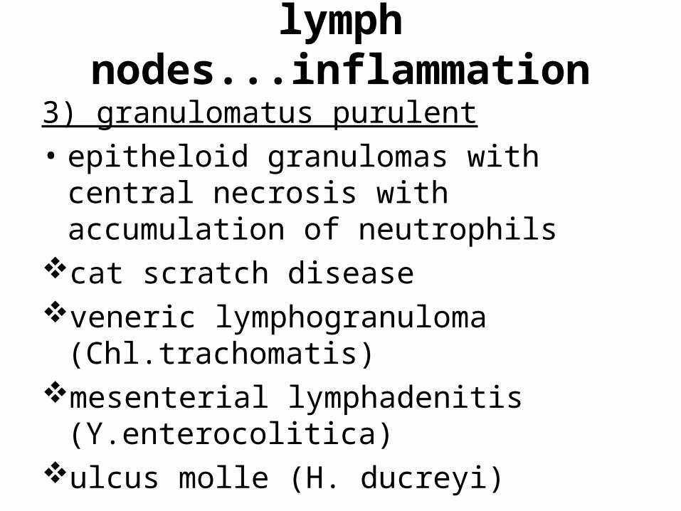
3) granulomatus purulent• epitheloid granulomas with central
necrosis with accumulation of neutrophils
cat scratch diseaseveneric lymphogranuloma
(Chl.trachomatis)mesenterial lymphadenitis
(Y.enterocolitica)ulcus molle (H. ducreyi)
lymph nodes...inflammation
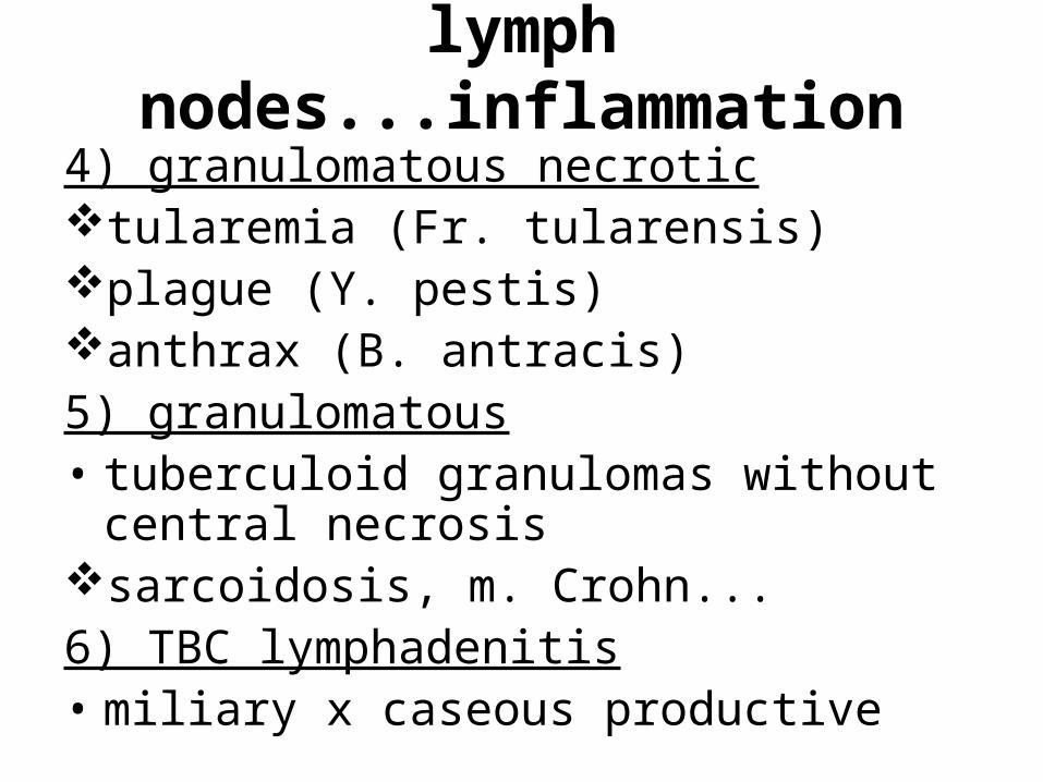
4) granulomatous necrotictularemia (Fr. tularensis)plague (Y. pestis)anthrax (B. antracis)5) granulomatous• tuberculoid granulomas without central
necrosis sarcoidosis, m. Crohn...6) TBC lymphadenitis• miliary x caseous productive
lymph nodes...inflammation
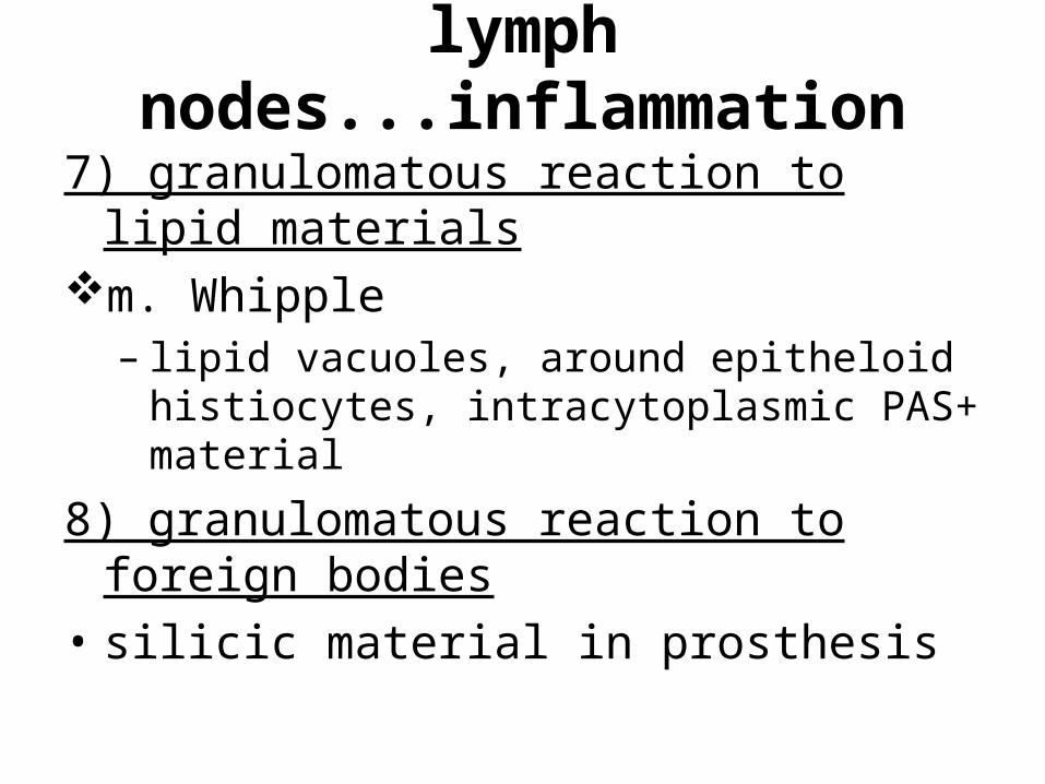
7) granulomatous reaction to lipid materials
m. Whipple – lipid vacuoles, around epitheloid
histiocytes, intracytoplasmic PAS+ material
8) granulomatous reaction to foreign bodies
• silicic material in prosthesis
lymph nodes...neoplasms
lymph nodes...neoplasms
C) neoplasms, malignant lymphomas1) m. Hodgkin (HD)• group of disesases• presence of distinctive neoplastic giant
cells: Reed-Sternberg cells, Hodgkin cells, admixed with a variable infiltrate of reactive, nonmalignant inflammatory cells
• young people
lymph nodes...neoplasms...HD
• classification: nodular lymphocytic predominance
Hodgkin lymphoma• „classic“: lymphocyte rich HL (LR-CHL) nodular sclerosis (NS-CHL) mixed cellularity (MC-CHL) lymfocyte depleted (LD-CHL)
lymp nodes...neoplasms...NHL2) non Hodgkin lymphomas• predominance of neoplastic cells• elder patients • B and T cells
lymph nodes...neoplasms...B-NHL
a) B-NHL chronic lymphocytic leukaemia/small
lymphocytic lymphoma (CLL/SLL) follicular lymphoma (FCL) mantle cell lymphoma (MCL) marginal zone lymphoma (MZL): SMZL,
ENMZL=MALT, NMZL hairy cell leukaemia (HCL) diffuse large B-cell lymphoma (DLBCL) Burkitt lymphoma
chronic lymphocytic leukaemia/small lymphocytic
lymphoma (CLL/SLL)
• elderly patients• naive lymphocytes• indolent type of lymphoma
follicular lymphoma (FCL)
• middle aged patients• centrocytes and centroblasts• indolent course but rellapsing!
mantle cell lymphoma (MCL)
• „diffuse centrocytoma“• agressive type of lymphoma• t(11;14) – cyklin D1
marginal zone lymphoma (MZL)
• mucosa associated lymphoid tissue (MALT) – GIT, bronchi…
• association with chronic inflammation
diffuse large B-cell lymphoma (DLBCL)
• „waste basket“• transformation of small cell
lymphomas• aggresive course x good reaction
to therapy
Burkitt lymphoma
• endemic x sporadic• younger patients• association with EBV infection
lymph nodes...neoplasms...B-NHL
• plasma cell neoplasms: monoclonal gammopathy of
undetermined significance (MGUS) lymphoplasmacytic lymphoma (LPL),
m. Waldenström plasmacytoma multiple myeloma
lymph nodes...neoplasms...T-NHL
b) T-NHL peripheral T cell lymphoma (PTL) anaplastic large T cell lymphoma
(ALCL) angioimmunoblastic T cell lymphoma
(AILT) adult T cell leukaemia mycosis fungoides/Sezary syndrom
anaplastic large T cell lymphoma (ALCL)
• young patients• typical translocation t(2;5)
mycosis fungoides/Sezary syndrom
• primary skin lymphoma → generalisation = Sezary syndrom

spleen
spleen
• structure:– white pulp: lymphoid tissue– red pulp: venous sinuses → hilus
• splenomegaly: venosthasis, inflammation, neoplasms
• hypersplenism = increased function → cytopenia
• hyposplenism → susceptibility to certain bacterial infections
spleen...regressive changes, circulatory disorders
A) inborn anomalies• accessory spleens = spleniculiB) regressive changes and
circulatory disorders1) amyloidosis• secondary (AA)• +/- hyposplenism
spleen...regressive changes, circulatory
disorders2) storage diseases3) hemolytic anemias• ↑ splenic function → splenomegaly• hereditary spherocytosis4) chronic perisplenitis5) splenic infarction• white (embolization, vasculitis)• red (thrombosis of lienal vein)
spleen...regressive changes, circulatory
disorders6) chronic venosthasis7) splenic rupture, bleeding• traumatic
spleen...inflammation
A) inflammation1) acute septic tumour• reaction to general infection x
tumour lysis• clinically: tense capsula, soft tissue• histology: red pulp cellular, small
abscesses (central pyemia)
spleen...inflammation
2) chronic inflammatory tumour• chronic infections (IE)• histology: red pulp hyperemia,
reactive hyperplasia of the white pulp
• malaria• TBC, histoplasmosis, leishmaniosis,
trypanosomiasis• AIDS
spleen...tumoursD) tumours and pseudotumours1) cystic formations• posttraumatic pseudocysts• parasitary cysts2) hamartoma (splenoma)• nodule• histology: chaotic sinuses a fibrous tissue
= incorrect arrangement of the red pulp• +/- hypersplenism
spleen...tumours
3) hemangioma• histology: cavernous blood spaces,
thrombosis4) littoral cell angioma• phagocytosis → pancytopenia5) inflammatory pseudotumour• histology: inflammatory cells +
fibroproduction
spleen...tumours
6) malignant lymphomas• primary: SMZL• secondary:• more often• secondary infiltration by NHL, HD, CML,
HCL7) epithelial metastasis• microscopically
thymus
thymus...structure
• structure:• lobulus• cortex and medulla mixture of T
lymphocytes and a epithelial cells = lymphoepithelial organ
• cortex: mainly T lymphocytes• lymphatic follicles without germinal
centres• medulla: thymocytes, Hassal bodies
thymus...function
• function:– production of small lymphocytes with
cellular immunity– TdT a CD1 → maturation → CD4 a CD8 →
postthymic lymfocytes in medulla: CD4 (helpers/inducers), CD8 (suppressor/cytotoxic), loss of TdT a CD1 → blood, peripheral lymphatic organs
• main role intrauterine and in childhood
thymus...dysgenesis
A) thymic dysgenesis• primary immunodeficiency syndromes
(diGeorge, Nezelof...)
thymus...regressive changes
B) regressive changes1) lipomatous atrophy (involution)• puberty – involution with increase of
adipous tissue = ↓ thymocytes, calcification of Hassal bodies...
2) acute (accidental) involution• etio: corticosteroids – stress• histology: fragmentation of cortical
thymocytes, cystic transformation of Hassal bodies, lymfocytes disappeared, in cortex only spindle epithelial cells
thymus...hyperplasia
C) thymic hyperplasiaprimary hyperplasiamyasthenia gravis• histology: lymphoid hyperplasia,
lymphatic follicles with germinal centres
• Ab anti acetylcholin-receptors
thymus...neoplasms
D) neoplasms1) thymomas• epithelial thymic cells + lymphocytes• local manifestation• association with myasthenia gravis
2) neuroendocrinne tumors3) germinal cells tumours (teratoma,
seminoma)• bng, cystic4) malignant lymphoma