the helper!. (1)
TRANSCRIPT

| Alfarabi college | كلية الفارابي لطب األسنان والتمريض
The helper in periodontics 2013
level 7 & 8
some basic info. Help you in the clinic and the general knowledge in the
periodontology
My Friends ,✌ I made these papers to Facilitates you ,
To access the information quickly
And remember main info, in our course I hope it benefit you
Osama almasry , level 8 28 / 2 / 2013
Pepared by : Osama a. almasry ☂
Course coordinator : Dr . khalid Azouni

Presenting the patient :
Mr| miss ( ) from Saudi arabia , and he ' she is 21 years old He | she is a smoker , non smoker
The C.C " Chief complaint " is …. His| her medical history :: Diabetes , hypertention , breathless , blood disorder , medications , allergy , any abnormalities , etc ..
His | her Dental history :: last visit , oral hygiene , Bleeding , plaque , bad taste , Resto , ortho , scaling , etc His | her habits are . . . .
The Diagnosis is :: Generalized marginal chronic gingivitis , Localized severe chronic periodontitis , etc ..
The G. prognosis is : Excellent , Fair , poor , Hopeless . etc ..
The treatment plan will be : non surgical treatment or sugrical Supragingival scaling , or Subgingival scaling , root planning , etc
OHI : oral hygiene instructions
weeks 6 -After 4 : Follow up re evaluation of the plaque , oral hygiene , codition of the gingiva , etc ..
-----------------------------------------------------------------------------------------------------------------------------------------------------------------------------------
Denti | Signs
You have to tell your patient about these signs " it's Facilitates your treatment "

The instruments :: ❞
Scaling instruments :
to remove supragingival calculus Sickle scaler :☛ - two cutting edges " sharp pointed tip " - straight shank to anterior and premolars teeth - contra – angled shanks to posterior teeth
: to remove subgingival calculus , root planning , removal of soft tissue teteCur☛ linning the pocket - two cutting edge , not sharp pointed tip - finer than scalers
Universal curette : Gracey " area specific " curette - two cutting edge , one plane - one cutting edge , two plane - blade not offset - blade offset - 90 degree angle of blade - the terminal shank 70 degree
★ te : Pull stroke ( UPWARD )teMethod of application of Cur ★
-------------------------------------------------------------------------------------------------
Gracey 1-2 , 3-4 | for anterior teeth Gracey 5-6 | for anterior teeth and premolars Gracey 7-8 , 9-10 | for posterior teeth ( Facial and lingual ) Gracey 11-12 | for posterior teeth ( mesial) Gracey 13- 14 | for posterior teeth (distal ) --------------------------------------------------------------------------------------------------------------
to remove tenacious subgingival calculus and altered el , Hoe , file scalers :Chis☛cementum .
for scaling and cleansing tooth surfaceUltrasonic : ☛ and soft tissue wall of perio pocket
tal tape rubber cup, brushes , den Cleaning and polishing instruments :☛ Air bowder abrasive system and prophy jet .
Rubber cup
Prophy jet
effective grasp for periodontal instruments :The most ★
Modified pen grasp
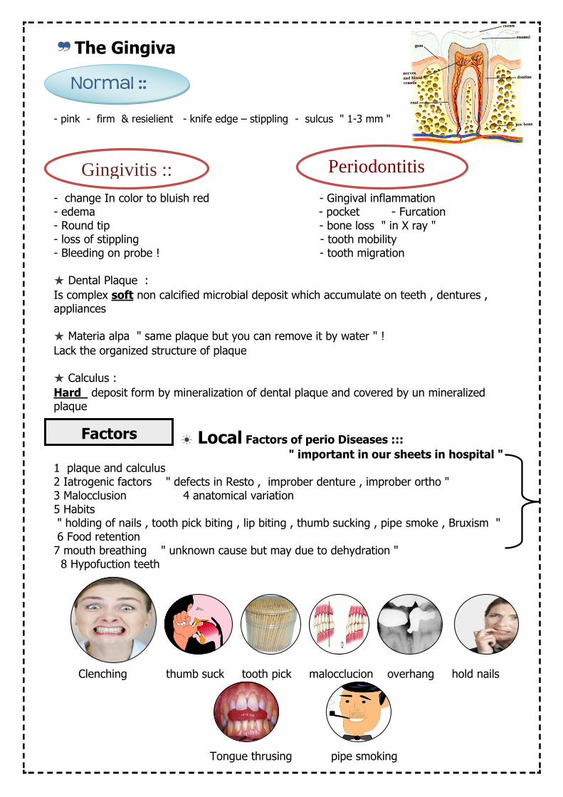
The Gingiva❞
- pink - firm & resielient - knife edge – stippling - sulcus " 1-3 mm "
- change In color to bluish red - Gingival inflammation - edema - pocket - Furcation - Round tip - bone loss " in X ray " - loss of stippling - tooth mobility - Bleeding on probe ! - tooth migration
Dental Plaque : ★
non calcified microbial deposit which accumulate on teeth , dentures , softIs complex appliances
Materia alpa " same plaque but you can remove it by water " ! ★
Lack the organized structure of plaque
Calculus : ★
form by mineralization of dental plaque and covered by un mineralized deposit Hard plaque
Factors of perio Diseases ::: Local ☀
" important in our sheets in hospital " 1 plaque and calculus 2 Iatrogenic factors " defects in Resto , improber denture , improber ortho " 3 Malocclusion 4 anatomical variation 5 Habits " holding of nails , tooth pick biting , lip biting , thumb sucking , pipe smoke , Bruxism " 6 Food retention 7 mouth breathing " unknown cause but may due to dehydration " 8 Hypofuction teeth
Clenching thumb suck tooth pick malocclucion overhang hold nails
Tongue thrusing pipe smoking
Gingivitis ::
Normal ::
Periodontitis
Factors

ystemic factors of perio disease ::S ☀ 1 Pregnancy 4 Abnormalities of endocrine system
eakened immune systemW 5 ubertyP 2
3 Diabetes 6 Smoking 7 Osteoporosis 8 Heart disease 9 Obesity
Medications can cause Gingival Disease ::
Pockets : ❞ Pathologically deep gingival sulcus
Classifications :
A - Gingival pocket " psuedo pocket " B - Periodontal pocket divided into :: " suprabony pocket : bone loss horizontal " intrabony pocket : bone loss vertical
Classification accord involved tooth surface :
* Simple pocket (A) * Compound Pocket (B) * Complex Pocket ( C )
: Gingival Recession❞ Is exposure of root surface by apical shifting
BY Nabers probe :ion involvement Furcat❞ Is invasion of bifurcation and trifurcation of multi rooted teeth .
Grade 0 0 mm no furcation Grade 1 1-3 mm Grade 2 > 3 mm surpassing half of buccolingual thic of tooth , not through and through + some interradicular bone attached Grade 3 > 3 mm Encompassing entire width of tooth , through and through + no bone attached Grade 4 > 3 mm Encompassing entire width of tooth , through and through + no bone attached + Gingival recession
^
Anticonvulsant drug
phenytoin
Hypertensive drug
Nifidipine
Immunosuppressive drug
Cyclosporine
Factors cause the Recession : age , inflammation , false brush , abnormal frenum , tooth malposition

Is loosening of tooth in socket :Tooth mobility ❞
Class 1 ☀ Slight mobility , up to 1 mm horizontal displacement " faciolingual direction "
Class 2 ☀ Moderate mobility , greater than 1 mm horizontal displacement " faciolingual direction "
Class 3 ☀ Severe mobility , greater than 1 mm horizontal displacement " faciolingual direction " With vertical displacement
~ Horizontal mobility " Exam it by tow dental instruments on either side " | Vertical mobility " Exam it by end of instrument handle "
Gingival index : ❞ 0 normal 1 sign of inflamation ( mild ) without bleeding 2 sign of inflamation ( moderate ) with bleeding on probing 3 sign of inflamation ( severe ) with spontaneous bleeding
Plaque index : ❞ 0 no plaque 1 film of plaque detectable on probe only 2 moderate plaque seen by eye and detectable by probe 3 abundance of plaque | more than 2/3 of the tooth covered by plaque
☉ Signs of inflammation :
Dolor (pain) Calor (heat Rubor (redness) Tumor (swelling)
Functio laesa (loss of function)
Ramfjord Teeth ❞
16 21 24 44 41 36
In the clininc we exam "Plaque or bleeding" on the ( Ramfjord teeth ) as indicator teeth If on of them missing we exam the adjacent one ..
Factors cause the mobility : Loss of support , Trauma , inflammation , perio surgery , sometimes pregnancy , use of contraceptive ,
Associated with menstrual cycle
Clinical attachment level ( C A L )❞ Is the distance from CEJ to location of the inserted probe tip . CAL = PD + GL pd = probing depth , GL = Gingival level
The land mark to measure the CAL Is : CEJ cemento enamel junction ☉
We measure the CAL by : a) PD (the depth to probe pentrates ) b ) level of gingival margin ( distance from CEJ to gingival margin )
▪﹁ if there is Recession " Positive number of GL "
CAL = PD + GL
▪﹁ If the gingival margin cover the CEJ " negative number of GL "
CAL = PD + ( - GL ) CAL = PD - GL
▪﹁ If the gingival margin at CEJ , GL will be = 0 mm
CAL = PD + GL
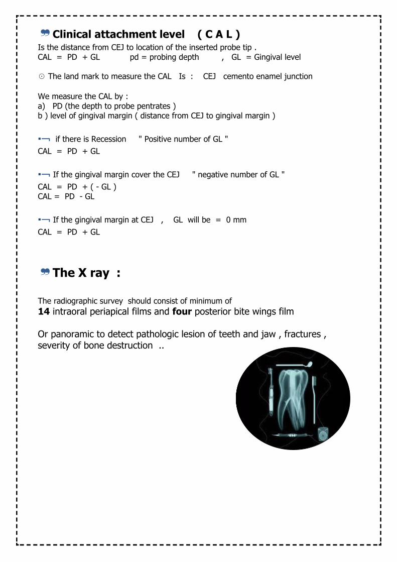
The X ray :❞ The radiographic survey should consist of minimum of
14 intraoral periapical films and four posterior bite wings film Or panoramic to detect pathologic lesion of teeth and jaw , fractures , severity of bone destruction ..

The Diagnosis ::❞
teeth% of the Localized <30♦ %of the teeth Generalized : > 30 ♦
Marginal : involve margin & papillae ♦ Papillary : papillae only ♦
gingiva Diffuse : involve margin & papillae & attached ♦
♦ The severity :: the classification of severity is as follows:
Mild: 1–2 mm
Moderate: 3–4 mm
Severe: ≥ 5 mm
إلحدى الحاالت severityلنحسب الـ مثال
6 ×27 =168
[ ال ننسى أن بعض الحاالت فقدت بعض األسنان ] نضرب عدد األسطح في عدد أسنان الحالة
,هو عدد األسطح لكل سن 6حيث [ يوجد خمس اسنان مفقودة لدى الحالة ] هو عدد األسنان الموجودة 27
لكل األسنان " PD " probind depthنحسب الـ
ومافوق mm 5أو الـ mm 4-3أو mm 2-1ونحسب كم عدد األسنان التي حصلت على
أسنان 9هو mm 2-1لنفرض أن عدد األسنان التي حصلت على سن 21هو mm 4-3وعدد األسنان التي حصلت على
أسنان 6هو ومافوق mm 5وعدد األسنان التي حصلت على :للمرض نعمل اآلتي severityلكي نحسب الـ
1-2 mm = ( 9 / 168 ) 100 = 5 %
3-4 mm = (12 / 168 ) 100 = 7 %
5 mm = ( 6 / 168 ) 100 = 3%
% 7 اذاٌ النسبة الكبرى هي
moderate = للمرض severityاذاً الـ
Generalized or localized
Severity : slight ,
moderate , severe Chronic (periodontitis or gingivitis etc .. )
Distribution : Marginal ,
papillary , Diffuse Diffuse
1 Location
2 Severity
Distribution 3 the disease
× ×
×

Prognosis The ❞
❞
Excellent No bone loss
Excellent ginigiva Good cooperation
No systemic factors
Good -Adequate remaining
bone support - adequate control of
etiological factors - Adequate cooperation - No systemic factors
Fair - less than adequate
remaining bone support - Some tooth mobility
- Grade I furcation - Acceptable cooperation – limited systemic factors
Poor - Modrate , advanced
bone loss - Tooth mobility
- Grade I & II furcation - doubtful cooperation - presence of systemic
factors
Questionable - Advanced bone loss
- Grade II or III furcation - Tooth mobility - in access area
- presence of systemic factors
Hopeless - Advanced bone loss
- non maintianable areas - Extraction indication
- presence of uncontrolled systemic
factors
Factors affect the prognosis : age , presence of plaque , calculus , local factors , severity , prosthetics

( OHI ) Oral Hygiene Instructions❞
A. Proper brushing Techniques and mouth rinse 1. Always use a soft bristled toothbrush
2. Use anti-cavity Fluoride toothpaste
3. Hold toothbrush at a 45-degree angle at the gum line, brushing in a
circular motion
minutes at least twice a day. three4. Brush teeth for a minimum of
5. Brush gums and tongue along with your teeth.
6. Don’t brush too hard because this can cause gingival (gum) recession.
7. Use mouthwash twice only 20 ml for thirty seconds then wash .
B. Proper flossing technique and the reason for flossing 1. Use an arms length (18 inches) of floss. Wrap around fingers mostly to one
side.
2. Floss each tooth forming a “C” shape with the floss each time.
3. A new area of floss should be introduced into each gingival pocket.
4. Don’t forget to floss behind your last molar.
5. Flossing removes plaque from behind your teeth that brushing misses.
6. Flossing helps prevent periodontal disease by removing plaque.
Types of oral disease and ways they can easily be prevented: 1. Dental Caries (cavities)
a) Brushing and flossing
b) Fluoride rinse from your dentist if he or she recommends it for you
c) Reduce your carbohydrate intake along with simple sugars
2. Periodontal Disease
a) Gingivitis is the first stage of periodontal disease
1. Bleeding gingival tissue while brushing teeth
2. Red puffy gingival tissue
3. May be sensitive to touch
4. Bad breath (Halitosis)
b) Periodontal disease
1. Recession of gingival tissue
2. May be sensitive to hot and cold liquids or foods
3. Root exposed due to gingival recession
4. Slight bone loss
5 Usually large amounts of plaque and calculus build up
6 Bone loss and periodontal ligament detachment
7 Teeth could be loose
8 Puss maybe within gingival pocket
9. Major bone loss along with damaged periodontal ligaments.
مراجعة التعليمات التي يتم إخبارها
حول العناية ] للمريض بعد الزيارة بالعربي[ باألسنان
األسنان يوميا مرتين على األقل لمدة تنظيف -دقائق وعدم التفريش بطريقة خاطئة 5 – 3
وشديدة حتى ال يتم إيذاء اللثة شهور 3تبديل الفرشاة كل - مرتين الفم غسولاستعمال -
وإزالة الرائحة الكريهة وإزالة الكميات الكبيرة 12] .. من البكتيريا والحماية من التسوس
لمدة ثالثين ثانية ثم بصق الكمية [ مل وغسل الفم بالماء وتجنب بلع أي كمية من
الغسول يا بين استعمال خيط األسنان إلزالة البقا -
األسناناستعمال معجون مضاد للحساسية إذا كان -
المريض يشكو من ذلك توصيته بالمراجعة بعد شهر من الزيارة -

ثالث مرات باليوم دقائق 5 – 3األسنان هو لتنظيفوإخباره بأن الوقت المناسب :: بإمكانك إطالع المريض على هذا الجزء لفرشاة كل ثالث أو أربع شهوروعليه أن يقوم بتغيير ا,
---------------------------------------------------------------------------------------------
Diseaseof Periodontal Treatments❞ 1. Root scaling to remove calculus and plaque below the gum line
2. Surgery to reattach gingival tissue to the surface of the tooth
3. Sometimes the gingival tissue may regenerate to fill the void
of the bone loss, but the bone itself will never regenerate.
DiseaseTypes of Periodontal ☛
Diagnosis Case Type Treatment by
Gingivitis Type I General Dentist
Mild Periodontitis Type II General Dentist
Moderate Periodontitis Type III General Dentist
Advanced Periodontitis Type IV Periodontist
---------------------------------------------------------------------------------------------
الخاص بالمريض كيفية الكتابة في الملف ☈
Procedureفي خانة
✍
D.hو الـ m.hوالـ C.cو الـ , نكتب اسم المريض .. للحالة Txوالـ Dxوالـ
Mr khalid , 21 years
c.c =pain in the lower gingival and bleeding m.h = NAD ( nothing abnormalities detectable) D.h = bad 0ral hygiene , bleeding on probing ,
Supragingival calculus X ray ? :: ..
The Dx : generalized marginal chronic gingivitis The Tx : Supragingival scaling , OHI
Follow up
The Resources : Carranza ,clininc Manual of periodontics , Wikipedia , Google picture !