the future role for colposcopy in europe · 2016-10-12 · the future role for colposcopy in europe...
TRANSCRIPT
The Future Role for Colposcopy
in Europe
Simon C. Leeson, FRCS, FRCOG,1 Tamar Alibegashvili, MD, PhD,2
Marc Arbyn, MD, MSc, DrTMH,3 Christine Bergeron, MD, PhD,4
Carmine Carriero, MD, PhD,5 Jean-Luc Mergui, MD,6
Pekka Nieminen, MD, PhD,7 Walter Prendiville, FRCOG,8
Charles W.E. Redman, MD,9 Gudrun C. Rieck, MD,1 Jens Quaas, MD,10
and K. Ulrich Petry, MD, PhD11
1Department of Obstetrics and Gynaecology, Betsi Cadwaladr University Health Board, Bangor,Gwynedd, UK; 2Department of Gynecology, National Screening Center, Tbilisi, Georgia; 3Unitof Cancer Epidemiology, Scientific Institute of Public Health, Brussels, Belgium; 4LaboratoireCerba, Paris, France; 5Department of Gynecology and Obstetrics, University of Bari, Bari, Italy;
6Hopital Tenon, Service de Gynecologie Obstetrique et Medecine de la Reproduction, Paris,France; 7Department of Obstetrics and Gynecology, Helsinki University Hospital, Finland;
8The Beacon Hospital, Sandyford, Dublin, Ireland; 9Department of Obstetrics andGynaecology, University Hospital of North Staffordshire, Stoke-on-Trent, UK;
10D-18437 Stralsund, Grunthal, Germany; and 11Department of Obstetrics andGynaecology, Klinikum Wolfsburg, Wolfsburg, Germany
h Abstract: Improvements in the performance of cervicalscreeningmay be limited by the diagnostic performance ofcolposcopy. Nonetheless, colposcopy remains the bestavailable tool to assess women considered at high risk forhaving or developing cervical cancer. The provision androle of colposcopy across Europe is variable. Introduction
of vaccination against human papillomavirus (HPV) types16 and 18 as well as the possible switch to HPV-basedscreening is likely to change the profiles of womenpresenting to colposcopy services and provide manage-ment difficulties for the colposcopist.
The standard of colposcopy in Europe can be maintainedor improved despite a variable availability of screening.The prevalence of cervical intraepithelial neoplasia grade3 may decrease for women having had HPV vaccination.The incidence of cervical intraepithelial neoplasia grade 3and cervical cancer in secondand subsequent roundsofHPV-based screeningare likely todecrease compared to cytology-based screening. In HPV-based screening, the numbers ofwomen with no detectable or minor abnormalities at col-poscopy and with screen-detected glandular disease arelikely to increase. We have considered how these issues willaffect states thathavevarying implementationoforganizedcervical screening programs and varying degrees of imple-mentation of HPV testing or vaccination.
The development of quality assurance across Europeaccompanying these program changes is discussed. h
Key Words: colposcopy, human papillomavirus 16, humanpapillomavirus 18, health care quality assurance, early detec-tion of cancer
Reprint requests to: Simon C. Leeson, FRCS, FRCOG, Department ofObstetrics and Gynaecology, Betsi Cadwaladr University Health Board(West), Gwynedd, Wales LL57 2PW, UK. E-mail: [email protected]
Dr Arbyn participated in the EUROGIN conference (Lisbon 2011) fundedby organizers of the conference. Dr Arbyn received financial support from(1) Directorate of SANCO of the European Commission, Luxembourg,Grand-Duchy of Luxembourg), through the ECCG project (European Co-operation on development and implementation of Cancer screening andprevention Guidelines, the IARC, Lyon, France); (2) the 7th FrameworkProgramme of DG Research of the European Commission through thePREHDICT project (grant no. 242061, coordinated by the Vrije UniversiteitAmsterdam, the Netherlands) and the HPV-AHEAD project (FP7-HEALTH-2011-282562, coordinated by IARC); and (3) the Belgian Foundation AgainstCancer (Brussels, Belgium). Dr Petry received unrestricted research grantfrom Sanofi Pasteur MSD for an HPV epidemiology study and a speaker’shonorarium from Roche, Qiagen, GSK, and Becton Dickinson Ad Board.
The other authors have declared they have no conflicts of interest.
� 2013, American Society for Colposcopy and Cervical Pathology
Journal of Lower Genital Tract Disease, Volume 18, Number 1, 2014, 70Y78
Copyright © 2013 American Society for Colposcopy and Cervical Pathology. Unauthorized reproduction of this article is prohibited.
Well-organized screening reduces the incidence and
death rate from cancer of the cervix. Incorpora-
tion of quality assurance for all aspects of the screening
program seems essential to ensure this outcome. Colpos-
copy has a subjectivity that may see its performance as
the gold standard for diagnosis lag behind advances in
screening and secondary testing. Cytology-based screen-
ing has a moderate sensitivity to detect high-grade cervical
intraepithelial neoplasia (CIN) and several developments
are at various stages of implementation, which will soon
impact on cervical screening programs.
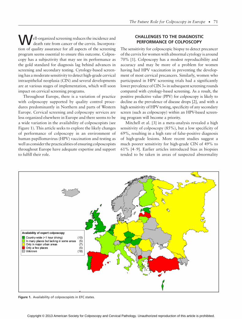
Throughout Europe, there is a variation of practice
with colposcopy supported by quality control proce-
dures predominantly in Northern and parts of Western
Europe. Cervical screening and colposcopy services are
less organized elsewhere in Europe and there seems to be
a wide variation in the availability of colposcopists (see
Figure 1). This article seeks to explore the likely changes
of performance of colposcopy in an environment of
human papillomavirus (HPV) vaccination and testing as
well as consider the practicalities of ensuring colposcopists
throughout Europe have adequate expertise and support
to fulfill their role.
CHALLENGES TO THE DIAGNOSTICPERFORMANCE OF COLPOSCOPY
The sensitivity for colposcopic biopsy to detect precancer
of the cervix for women with abnormal cytology is around
70% [1]. Colposcopy has a modest reproducibility and
accuracy and may be more of a problem for women
having had HPV vaccination in preventing the develop-
ment of most cervical precancers. Similarly, women who
participated in HPV screening trials had a significantly
lower prevalence of CIN 3+ in subsequent screening rounds
compared with cytology-based screening. As a result, the
positive predictive value (PPV) for colposcopy is likely to
decline as the prevalence of disease drops [2], and with a
high sensitivity of HPV testing, specificity of any secondary
screen (such as colposcopy) within an HPV-based screen-
ing program will become a priority.
Mitchell et al. [3] in a meta-analysis revealed a high
sensitivity of colposcopy (85%), but a low specificity of
69%, resulting in a high rate of false-positive diagnosis
of high-grade lesions. More recent studies suggest a
much poorer sensitivity for high-grade CIN of 49% to
61% [4Y9]. Earlier articles introduced bias as biopsies
tended to be taken in areas of suspected abnormality
Figure 1. Availability of colposcopists in EFC states.
The Future Role for Colposcopy in Europe & 71
Copyright © 2013 American Society for Colposcopy and Cervical Pathology. Unauthorized reproduction of this article is prohibited.
with an assumption that those cases without biopsies
did not have high-grade CIN. Colposcopic opinion also
seems to be influenced by prior knowledge of the cy-
tology [10]. Unlike PPV, sensitivity is difficult to calcu-
late for colposcopic opinion unless all colposcopies are
accompanied by a biopsy or biopsies. In general, through-
out Europe, biopsies are not usually taken from a normal-
appearing transformation zone if referral cytology is low
grade, which accounts for around 50% of all colposcopic
referrals [11, 12]. This effect will tend to inflate colpo-
scopic sensitivity, which will be inflated further in clinical
practice as the choice of punch biopsy site depends on the
skill of the colposcopist [10]. Random biopsies from the
squamocolumnar junction with or without accompanying
endocervical curettage do not improve the diagnostic yield
of high-grade CIN for low-grade referrals and should be
discouraged [4]. Furthermore, there is an enormous vari-
ation in colposcopic practice across Europe with its use
either in conjunction with screening (including where
colposcopy may form part of an annual gynecologic as-
sessment at the level of an individual gynecologist’s prac-
tice) or as an investigatory tool for abnormal cytology. In
both circumstances, the prevalence of high-grade CIN is
substantially different with a difference in anticipated
colposcopic performance.
As the incidence of cervical cancer is expected to de-
cline with HPV vaccination and HPV-based screening,
community-based clinicians and junior gynecologists will
not have seen many cases of overt cervical cancer limiting
expertise in diagnosing microinvasive or clinical disease.
DIFFICULTIES FOR FUTURE COLPOSCOPISTS
Colposcopic performance may be adversely affected by
3 changes to the screening program: the introduction of
vaccination, a change to HPV-based screening, and con-
sideration of the types of secondary triage tests as part of
that screening program.
I. HPV Vaccination
The consequences of vaccination on colposcopic prac-
tice are difficult to determine with any clarity. The in-
cidence of high-grade CIN and the need for treatment of
CIN will be reduced by at least 50% if there is uptake of
all the eligible population. A decline in high-grade CIN
and cervical glandular intraepithelial neoplasia (cGIN)
has been shown already in real life analysis [13].The
effect on low-grade disease is more difficult to predict,
but currently, there is no evidence of a different effect
between the 2 vaccines on the development of CIN 1.
HPV types 6 and 11 account for most condylomata, but
CIN 1 has a variety of causative HPV types. HPV-16
may be found in approximately 20% of CIN 1 [14]. If
screening becomes HPV-based then women with only
oncogenic HPV infection will be referred for colposcopy.
However, with continued cytology-based screening up
to 19% (range = 12%Y25%) of women with low-grade
cytology harbor high-grade CIN [15]. At least half of
these women would be prevented from developing high-
grade CIN by vaccination and would not be referred for
colposcopy. Overall, the future referral rates to colpos-
copy for women having had either of the 2 vaccines will
be lower than in the nonvaccinated population, but we
do not know the exact magnitude of this decline.
Nonetheless, the PPV for the colposcopic diagnosis of
CIN 2+ will decline in vaccinated women.
The specific major colposcopic changes (intense aceto-
white, distinct sharp margins, coarse mosaic or puncta-
tion, and lesion size) may not be a feature of high-grade
CIN but of HPV and, in particular, may be a feature of
HPV-16. Twenty expert colposcopists identified by the
American Society for Colposcopy and Cervical Pathol-
ogy reviewed images from the Atypical Squamous Cells
of Undetermined Significance (ASCUS) and Low-Grade
Squamous Intraepithelial Lesion (LSIL) Triage Study
(ALTS) after application of 5% acetic acid. There was
more agreement if the lesions were HPV-16 related rather
than by CIN grade, but otherwise, the associations be-
tween HPV type and lesion recognition were weak. Visual
appearances may be accountable to HPV-16 infection per
se rather than grade of CIN [16]. In a study examining
dynamic spectral imaging in 177 women referred for
colposcopy, HPV-16Ypositive lesions had biopsies taken
more often than nonYHPV-16 lesions despite colposco-
pists being unaware of HPV status (88.1% vs 72.5%).
However, this effect was not statistically significant
(p = .066) but was independent of lesion size. The au-
thors suggested that this may have been due to more
intense acetowhitening, which was detected by dynamic
spectral imaging [9]. An unpublished poster presentation
from Cruickshank et al. (2011) described 105 women
referred for colposcopy in Scotland who had their col-
poscopic impression compared to histologic outcome by
HPV type. The PPV for CIN 2+ was higher for lesions
containing HPV-16 (82%) or -16/18 (85%) than for those
which were neither HPV type (50%), but the numbers
were small. Perhaps some high-grade CIN not visualized
at colposcopy is nonYtype 16 CIN.
72 & L E E S O N E T A L .
Copyright © 2013 American Society for Colposcopy and Cervical Pathology. Unauthorized reproduction of this article is prohibited.
II. HPV-Based Screening
HPV-based screening will select a group of women who
differ from patients referred for cytology-based screen-
ing programs in clinical aspects that may increase the
failure rate of colposcopy:
1. The proportion of women without any cervical lesion or
with minor colposcopic changes is increased. In a pilot
project in Wolfsburg, the rate of HPV-positive women
without any lesion was 36% (normal colposcopy and/or
histology without any atypia). Cytology-negative/ HPV-
positive women were not immediately examined in this
pilot, and so theproportioncouldbe significantlyhigher.
A further problem is the performance of colposcopy in
subsequent HPV screening rounds because the preva-
lence of CIN 3+ as well as the PPV of colposcopy will
decline significantly. There have been no published data
as yet but one might speculate that seeing many healthy
women without lesions or with low-grade changes only
might distract the attention needed for colposcopy in a
high-risk population. Colposcopy would be likely to
perform with increasing difficulty in the vaccinated and
HPV-based screening population. Its use should be
confined to the diagnostic setting for women with ab-
normal screening such as HPV-positive women with
abnormal cytology.
2. Only women with persistent infections with high-risk
HPV types will develop new CIN 3 and cervical
cancer. Compared with the follow-up of women with
ASCUS or LSIL cytology, the risk of new lesions is
increased in these patients [17, 18]. If the evaluation
of colposcopy is based on the subsequent risk for
developing CIN 3+, this risk will be increased in HPV
screening compared to cytology-based screening.
3. More cGIN will be identified by HPV testing com-
pared with cytology-based screening [19]. These le-
sions are more difficult to be detected at colposcopy.
With more patients referred for colposcopy, more
women without CIN 2+ may have treatment with sub-
sequent morbidity such as pain, vaginal discharge, and
irregular bleeding. A single excisional treatment may
be associated with preterm delivery [20, 21]. Further-
more, there is the worry and inconvenience of having
abnormal screening tests with a need for more frequent
follow-up. Therefore, refined triage of positive HPV testing
is required. Colposcopic experience in detecting CIN is al-
most exclusively based on decades of Pap smear screening.
It is unclear if lesions with positive cervical cytology permit
easier colposcopic diagnosis than lesions associated with
normal cervical cytology. Within the ATHENA trial in-
vestigating the utility of HPV testing in primary screening
for cervical cancer in 47,208 women, as well as in the
FUTURE vaccine trials, colposcopic prediction showed
an underestimation of CIN 3+ in up to 57% of cases [22]
(Wright, Stoler, and Castle, personal communication).
Adherence to current colposcopic principles of identifing
the extent of the transformation zone, obtaining biopsies
from areas of abnormality, excision of an incompletely
visualized transformation zone in the presence of high-
grade referral cytology, and having quality-assured prac-
tice should avert a decline in sensitivity for the detection
of high-grade CIN seen with cytology-based screening.
This seems to be supported by a review of a primary HPV
screening pilot project over 5 years that rigorously followed
these principles. The failure rate of colposcopy defined as
CIN 3 lesions missed at the first colposcopy assessment
was significantly increased in HPV-positive women with
normal cytology compared with patients with abnormal
cytology. However, the overall failure rate of less than
5% was assumed to be acceptable. Remarkably, most
failures were related to type 3 transformation zones and
not explained by false-negative biopsies [23].
Castle et al. [24] considered that colposcopy is ap-
propriate if the cumulative risk of CIN 3+ over 2 years is
at least 10%, offering a risk-based referral to colposcopy
instead of one based on algorithm. The threshold for
referral for colposcopy set by Castle et al. should be
challenged. Should there be a prevalent risk for CIN 3+
at 10%; should this be for CIN 2+ or should the
threshold for colposcopy be changed? Ideally, colpos-
copy should be reserved exclusively for the management
of precancerous lesions (i.e., CIN 3 and cGIN). Until
ideal algorithms for referral to colposcopy are devised
within an HPV-based screening program, then the defi-
nition of Castle et al. seems wise but should be reappraised
when suitable performance data become available. Indeed,
a risk-based model would allow addition of further tests
such as p16 and other biomarkers to be added without
having to rewrite existing algorithms for all outcomes
of testing.
III. Use of Triage Tests for HPV-Based Screening
With a paradigm of carcinogenesis of HPV acquisition,
persistence, and progression from HPV infection to
precancer and then to invasion over several years, test-
ing for the presence or absence of HPV seems a crude
marker for cancer risk. More colposcopy referrals and
associated psychological morbidity [25] would be an-
ticipated with an HPV-based screening test positive rate
ranging from around 5% to 16% [19, 26Y28]. This
represents an approximate doubling of the referral rate
The Future Role for Colposcopy in Europe & 73
Copyright © 2013 American Society for Colposcopy and Cervical Pathology. Unauthorized reproduction of this article is prohibited.
to colposcopy for cytology-based screening with ASCUS
or worse [29].
Cervical cytology with its high specificity could pro-
vide triage and has been chosen in most of the popula-
tion pilot trials. An algorithm chosen by the Netherlands
and by Italy uses 2 cytology samples at 6 and 12 months.
The cumulative risk at 14 years for CIN 3 among cy-
tologically normal women with high-risk HPV at entry
was 28%, with 50% of these women detected at the next
screening round in a longitudinal study of 7,278 women
[30]. In this study, the prevalence of CIN 3 was highest
in those with HPV-16, with an odds ratio 3 to 4 times
higher than for other HPV types. HPV-16 was also more
than twice as likely to persist than any other HPV type.
HPV genotyping may therefore provide an alternative
triage tool with up to 27% of all HPV-positive cases
being due to HPV-16 [31Y33], accounting for up to 54%
of all HPV-positive CIN 3 [32, 34]. The ATHENA study
reported on 4,219 women who were high-risk HPV-
positive but cytology-negative and who were at least
30 years of age. The prevalent CIN 2+ rate on those
having HPV testing who were HPV-16/18-positive was
11.4%, whereas those who were high-risk HPV-positive
(but not HPV-16/18-positive) was 6.1%, and those who
were HPV-negative was 0.8%. The rate of CIN 2+ seems
age-dependent for 16/18 with the rate dropping with
increasing age [33]. Long-term follow-up shows the
same effect in the Kaiser Permanente population, with a
cumulative risk for CIN 3+ in a cytologically negative
population with HPV-16 at 10 years of 20.7%, 17.7%
for HPV-18, and 1.5% for high-risk HPV-negative women
[35]. Similar results for risk at 12 years were reported
by Kjaer et al. [18] from Denmark. In this latter study,
if a repeat test 2 years later remained positive for
HPV-16, then the 10-year risk increased to 47.4%, but
if the repeat test was negative, the 10-year risk dropped
to 3.0%. ATHENA data for women 30 years or older
shows that HPV-16/18 positivity as a triage to colpos-
copy was equivalent to ASCUS+ cytology and no HPV
testing with a sensitivity of 59.3% and a PPV of 15.5%
for CIN 3+ [34]. Other high-risk HPV types have an ab-
solute risk of CIN 3 of around 6% after 12 years of
follow-up [18]. The ATHENA study has an incident risk of
CIN 3+ for negative cytology and HPV-16/18+ of 9.8%
and colposcopy seems appropriate in this setting. Repeat
HPV testing and cytology in 12 months seems appropriate
for high-risk HPV-positive (but HPV-16/18-negative) cases.
A further alternative to cytology and genotyping for
triage of HPV-positive cases is the use of biomarkers.
A suitable candidate biomarker is p16, which had a
sensitivity of 88% for CIN 2+ in HPV-positive women in
the Italian NTCC study, with a relative sensitivity com-
pared to cytology of 1.53 for CIN 2+ in women 35 to
60 years without an increase in referral rate to colpos-
copy [36, 37]. However, there is an element of subjec-
tivity in assessing the degree of interpretation of p16
immunostaining, which is used with cytology or histol-
ogy. There seems to be wide variation in positivity when
applied to ASCUS and LSIL [38]. A combined immu-
nostain for p16 and Ki-67 has a high sensitivity and
specificity for CIN 3 and could be considered as a triage
for HPV testing if it proves to be reliable in clinical
practice [29]. Dual staining with p16/Ki-67 has been
shown to be reliable in diagnosing the remaining CIN 2+
in the group of HPV-positive, cytology-negative patients
within a German primary HPV screening pilot project
[39]. In this study, 25% of the HPV-positive, cytology-
negative population were p16/Ki-67-positive and dual
staining detected 90% of the remaining high-grade CIN.
Cytology may be suitable as a triage test if its referral
rate to colposcopy is no more than 2% in the general
population [40]; otherwise, genotyping or use of bio-
markers could be considered. Immediate testing is prefer-
able to reduce anxiety. In addition, any repeat testing
risks default.
POSSIBLE ASSISTANCE FORFUTURE COLPOSCOPISTS
There is a prospect of improving the objectivity and
performance of colposcopy with dynamic spectral imaging
(DySIS). Measurement of acetowhitening and modeling
of a color-coded map assists the identification of high-
grade CIN by localizing and grading the severity of any
cervical lesion. Several studies with DySIS-assisted colpos-
copy have shown promising results [7, 41, 42]. In a recent
Dutch study, the sensitivity of DySIS colposcopy to iden-
tify women with CIN 2+ was 79% versus 55% for con-
ventional colposcopy. When a DySIS color-coded map was
combined with conventional colposcopy, the sensitivity
was 88% [8]. Despite some technical issues that may limit
visualization of potential CIN (such as excessive blood or
mucus hindering a complete view of the transformation
zone), it seems that DySIS may have a future in cervical
screening. Indeed, the National Institute for Health and
Clinical Excellence has provided a draft recommendation
that DySIS is a cost-effective method of cervical assess-
ment, and colposcopy units should consider replacing col-
poscopes with DySIS despite the extended examination
time and cost in procuring the technology [43]. There are
74 & L E E S O N E T A L .
Copyright © 2013 American Society for Colposcopy and Cervical Pathology. Unauthorized reproduction of this article is prohibited.
several other optical and electrical impedance real-time
devices under development, but as yet, none are ready for
introduction into clinical practice.
THE CURRENT USE OF COLPOSCOPY INEUROPEVA WAY FORWARD
During a European Federation for Colposcopy (EFC)
satellite meeting in May 2011 in Berlin, authorized
representatives of 24 member societies answered ques-
tions on the role of colposcopy in their home states. The
rationale was to determine current practice and agree
basic standards for benchmarking performance across
Europe to encourage support for colposcopy services
where this was lacking. An objective was to support the
development of reports and guidelines to generate po-
litical interest in the development of national screening
programs. From their responses, colposcopy in Europe
is mostly used as assessment of abnormal screens, al-
though it is occasionally used as part of routine gyne-
cologic assessment (6 states). A large minority of states
permit colposcopy to be performed by all gynecologists
(7 states).
Six preliminary quality standards were also selected
as indicators for best practice (see Table 1), and a list of
final quality indicators will be identified by an EFC
Delphi consultation. Such standards must be applicable
to all colposcopists, whatever their circumstances and
however well organized their national screening may be.
The criteria for satisfying the standards must be achievable,
measureable, observable, understandable, and reasonable.
All women with abnormal screens must be investigated by
colposcopy if treatment is planned with liberal use of
colposcopically guided punch biopsies if high-grade CIN is
suspected. There should be a record of all colposcopic
findings with an adequate PPV for a colposcopic prediction
of CIN 2+. Overtreatment of CIN must be avoided, and
there should be suitable negative cytology or HPV status
after treatment. It is unclear how these standards will per-
form in an environment of HPV testing.
Multidisciplinary team meetings involving the cytol-
ogist, the pathologist, and colposcopist are required to
review cases where there is discrepancy between cytol-
ogy and histology, for glandular cases and for cancers.
Regular quality reports should be produced for each
colposcopist, local service, and state. Regular national
audits and perhaps a pan-European audit of cervical
cancer detected within the screening age range also
should be encouraged. This is in place in the United
Kingdom. An Internet-based voluntary quality assurance
system is being piloted in Germany. The EFC can coor-
dinate consensus building among member societies in
defining minimum standards on training and practicing
colposcopy as well as recommending tools to assess and
control the quality of all parts of colposcopy manage-
ment and education. Although the EFC is not in a position
to assess directly whether standards are being satisfied,
member societies can be advised how to achieve a trans-
parent and comparable quality assessment that will lead
to standardized high-quality colposcopy in all EFC mem-
ber states.
However, at present, data collection is limited and
practice is variable. Few states within Europe have col-
poscopists expected to see a defined number of patients.
Only Belgium, France, Germany, Italy, Israel, Netherlands,
Spain, and the United Kingdom reported seeing at least
30 cases per year per colposcopist. In Eastern Europe, no
qualifications are needed to practice colposcopy. Prac-
ticing colposcopists should have regular review of their
Table 1. The Berlin 2011 Consensus Quality Indicators
1. Quality of colposcopic examination / identification of SCJAim: Description/documentation of squamocolumnar junction and type ofTZ (IFCPC classification)
Indicator:& Proportion of documented colposcopies with description of SCJ andtype of TZ of all documented colposcopies (100%).
2. Quality of colposcopic predictionAim: A high PPV of colposcopic findings classified as major changes for ahistopathologic diagnosis of CIN 2+
Indicator:& Colposcopic findings classified as major changes should correlate with ahistologic diagnosis of CIN 2+ in most cases (975%).
PPV = (colposcopic opinion CIN 2+) / total CIN 2+ cases seen by that colposcopist)� 100
3. Quality of indication for invasive therapyAim: Good selection of CIN 2+, avoidance of overtreatmentIndicators:& Relation of CIN 2+ to eCIN 1 among all women who underwent invasivetreatment (CIN 2+ should outnumber eCIN 1).& High proportion of CIN 2+ of all treatment at first visit cases (985%).
4. Preference of minimal invasive therapyAim: Avoidance of cold knife conizations and hysterectomies in the treat-ment of CIN
Indicator:& Proportion of LLETZ or laser cone/ cold knife conization or hysterectomyin the treatment of CIN (998%).
5. Colposcopic guidance of minimal invasive CIN therapyAim:Minimal invasive therapies should bedoneunder colposcopic guidanceIndicator:& Proportion of treatment procedures for CIN performed by a trainedcolposcopist under colposcopic guidance/total treatments for CIN (995%).
6. Proof of cure following invasive treatment of CINAim: Assessment of the effectiveness of treatmentIndicator:& Proportion of treated CIN 2+ cases with negative tests (HPV or cytology)6Y12 months after treatment (985%).
SCJ, squamocolumnar junction; TZ, transformation zone; IFCPC, International Federa-tion for Cervical Pathology and Colposcopy; PPV, positive predictive value; CIN, cervicalintraepithelial neoplasia; LLETZ, large loop excision of the transformation zone.
The Future Role for Colposcopy in Europe & 75
Copyright © 2013 American Society for Colposcopy and Cervical Pathology. Unauthorized reproduction of this article is prohibited.
performance. All selected standards should be challenged
by a few good centers to see how the standards perform.
If screening programs are introduced in Eastern Europe
then more women will meet colposcopists who are
poorly trained and certification would be useful here for
young colposcopists where age is equated to experience.
Sufficient junior colposcopists must be trained and
registered to sustain screening programs and allow ad-
equate access for women to have colposcopy. Use of
paper-based or electronic training logbooks could fa-
cilitate these processes. Trainees from states with no
organized colposcopy training may train elsewhere to
return home with their skills and disseminate their ex-
pertise locally. Colposcopy needs to be of a high stan-
dard with practice of a small number of colposcopists
being nominated as experts in their area of practice and
training should provide the wherewithal to be an expert
colposcopist.
New innovations and technologies such as modified
screening algorithms, screening or triage tests, and the
use of DySIS can be added with measurable effect to
quality-assured screening and colposcopy services. Ap-
proaches to improve cervical screening must be to en-
hance and not undermine existing national programs.
This is indeed a daunting challenge but a task to which
pan-European health organizations have gained exper-
tise. The European Cervical Cancer Association and the
European Union are encouraging opportunities for
screening among member states. The EFC and its affil-
iated national colposcopy societies are collaborating to
provide workable standards for colposcopic practice as
well as providing training particularly for trainees from
Eastern Europe.
OUTLOOK AND CONCLUSIONS
Mass screening has to be available and it has to be
quality assured. Successful screening with a reduction in
the burden of cervical cancer is undoubtedly a desired
outcome but colposcopists must be prepared for the
changes imposed on practice. HPV vaccination and HPV
screening will reduce the burden of cervical cancer and
CIN 3 but will create new challenges with a lower PPV
for colposcopy and higher failure rates as well as an
increased risk of overtreatment, although the effects on
the appearance of CIN lesions currently remain specu-
lative. There will be fewer gynecologists who will have
seen or be familiar with the gross and colposcopic ap-
pearances of cervical cancer. Adherence to agreed qual-
ity standards for best practice in colposcopy will help
colposcopists manage cases at perceived increased risk
for cervical cancer and precancer in a standardized way,
although ongoing research is required to ensure that
following these colposcopic principles is sufficient to
address all challenges imposed by new cervical cancer
prevention programs. We should not be deterred from
providing high-quality training and standards of prac-
tice for colposcopists throughout Europe because what-
ever screening scenarios will be chosen, colposcopy will
remain the tool to diagnose precancerous lesions after a
positive screen. There is no better alternative to colpos-
copy, but there is no alternative for colposcopy than
better and more sophisticated standardized quality as-
surance. This may then realize the greatest benefit from
modified screening strategies translated into improved
cervical cancer diagnosis and mortality.
REFERENCES
1. Guido R, Schiffman M, Soloman D, Burke L. Post-
colposcopy management strategies for patients referred with
low-grade squamous intraepithelial lesions or human papil-
lomavirus DNA-positive atypical squamous lesions of un-
determined significance: a two-year prospective study. Am J
Obstet Gynecol 2003;188:1401Y5.
2. Franco EL, Cuzick J, Hildesheim A, de Sanjose S. Issues
in planning cervical cancer screening in the era of HPV vac-
cination. Ch 20. Vaccine 2006;24(suppl 3):S171Y7.
3. Mitchell MF, Schottenfeld D, Tortolero-Luna G,
Cantor SB, Richards-Kortum R. Colposcopy for the diagnosis
of squamous intraepithelial lesions: a meta-analysis. Obstet
Gynecol 1998;91:626Y31.
4. Pretorius RG, Zhang W-H, Belinson JL, Huang MN,
Wu LY, Zhang X, et al. Colposcopically directed biopsy, ran-
dom cervical biopsy, and endocervical curettage in the diag-
nosis of cervical intraepithelial neoplasia II or worse. Am J
Obstet Gynecol 2004;191:430Y4.
5. Jeronimo J, Schiffman M. Colposcopy at a crossroads.
Am J Obstet Gynecol 2006;195:349Y53.
6. Bekkers RL, van de Nieuwenhof HP, Neesham DE,
Hendriks JH, Tan J, Quinn MA. Does experience in colpos-
copy improve identification of high grade abnormalities? Eur J
Obstet Gynecol Reprod Biol 2008;141:75Y8.
7. Soutter WP, Diakomanolis E, Lyons D, Ghaem-
Maghami S, Ajala T, Haidopoulos D, et al. Dynamic spectral
imaging: improving colposcopy. Clin Cancer Res 2009;15:
1814Y20.
8. Louwers J, Zaal A, Kocken M, Ter Harmsel W, Graziosi
G, Spruijit J, et al. Dynamic spectral imaging colposcopy: higher
sensitivity for detection of premalignant cervical lesions. BJOG
2011;118:309Y18.
9. Zaal A, Louwers J, Berkhof J, Kocken M, Ter Harmsel
W, Graziosi G, et al. Agreement between colposcopic impres-
sion and histological diagnosis among human papillomavirus
76 & L E E S O N E T A L .
Copyright © 2013 American Society for Colposcopy and Cervical Pathology. Unauthorized reproduction of this article is prohibited.
type 16-positive women: a clinical trial using dynamic spectral
imaging colposcopy. BJOG 2012;119:537Y44.
10. Kierkegaard O, Byrjalsen C, Frandsen KH, Hansen
KC, Frydenberg M. Diagnostic accuracy of cytology and col-
poscopy in cervical squamous intraepithelial lesions. Acta
Obstet Gynecol Scand 1994;738:648Y51.
11. Jordan J, Martin-Hirsch P, Arbyn M, Schenk U, Baldauf
JJ, Da Silva D, et al. European guidelines for quality assurance
in cervical cancer screening: recommendations for clinical man-
agement of abnormal cervical cytology, part 1. Cytopathology
2008;19:342Y54.
12. Luesley D, Leeson S. Colposcopy and Programme
Management. 2nd Edition. Guidelines for the NHS Cervical
Screening Programme. NHSCSP Publication No. 20. Shefffeld,
UK: NHS Cancer Screening Programmes; 2010.
13. Brotherton JM, Fridman M, May CL, Chappell G,
Saville AM, Gertig DM. Early effect of the HPV vaccination
programme on cervical abnormalities in Victoria, Australia: an
ecological study. Lancet 2011;377:2085Y92.
14. Bergeron C, Barrasso R, Beaudenon S, Flamant P,
Croissant O, Orth G. Human papillomavirus associated with
cervical intraepithelial neoplasia. Am J Surg Pathol 1992;
16:641Y9.
15. Arbyn M, Sasieni P, Meijer CJ, Clavel C, Koliopoulos
G, Dillner J. Clinical applications of HPV testing: a summary
of meta-analyses. Ch 9. Vaccine 2006;24(suppl 3):S78Y89.
16. Jeronimo J, Massad LS, Schiffman M. Visual appear-
ance of the uterine cervix: correlation with human papillo-
mavirus detection and type. Am J Obstet Gynecol 2007;197:
47e1Y8.
17. Dillner J, Rebolj M, Birembaut P, Petry KU, Szarewski
A, Munk C, et al. Long term predictive values of cytology and
human papillomavirus testing in cervical cancer screening:
joint European cohort study. BMJ 2008;337:a1754.
18. Kjaer SK, Frederiksen K, Munk C, Iftner T. Long-term
absolute risk of cervical intraepithelial neoplasia grade 3 or
worse following human papillomavirus infection: role of per-
sistence. J Natl Cancer Inst 2010;102:1478Y88.
19. Katki HA, Kinney WK, Fetterman B, Lorey T, Poitras
NE, Cheung L, et al. Cervical cancer risk for 330,000 women
undergoing concurrent HPV testing and cervical cytology in
routine clinical practice at a large managed care organisation.
Lancet Oncol 2011;12:663Y72.
20. Kyrgiou M, Koliopoulos G, Martin-Hirsch P, Arbyn
M, Prendiville W, Paraskevaidis E. Obstetric outcomes after
conservative treatment for intraepithelial or early invasive cervical
lesions: systematic review and meta-analysis. Lancet 2006;367:
489Y98.
21. Castanon A, Brocklehurst P, Evans H, Peebles D, Singh
N, Walker P, et al. Risk of preterm delivery after treatment for
cervical intraepithelial neoplasia among women attending
colposcopy in England: retrospective-prospective cohort study.
BMJ 2012;345:e5174.
22. Stoler MH, Vichnin MD, Ferenczy A, Ferris DG, Perez
G, Paavonen J, et al. The accuracy of colposcopic biopsy:
analyses from the placebo arm of the Gardasil clinical trials.
Int J Cancer 2011;128:1354Y62.
23. Petry KU, Luyten A, Scherbring S. Accuracy of colpos-
copy management to detect CIN3 and invasive cancer in women
with abnormal screening tests: results from a primary HPV
screening project from 2006 to 2011 in Wolfsburg, Germany.
Gynecol Oncol 2013;128:282Y7.
24. Castle PE, Sideri M, Jeronimo J, Solomon D, Schiffman
M. Risk assessment to guide the prevention of cervical cancer.
Am J Obstet Gynecol 2007;197:356.e1Y6.
25. McCaffery K, Waller J, Forrest S, Cadman L, Szarewski
A, Wardle J. Testing positive for human papillomavirus in
routine cervical screening: examination of psychological im-
pact. BJOG 2004;111:1437Y43.
26. Munoz N, Bosch FX, de Sanjose S, Herrero R,
Castellsague X, Shah KV, et al. Epidemiologic classification of
human papillomavirus types associated with cervical cancer.
N Eng J Med 2003;348:518Y27.
27. Howell-Jones R, Bailey A, Beddows S, Sargent A, de
Silva N, Wilson G, et al. Multi-site study of HPV typeYspecific
prevalence inwomenwithcervical cancer, intraepithelial neoplasia
and normal cytology, in England. Br J Cancer 2010;103:209Y16.
28. Arbyn M, Ronco G, Meijer CJ, Naucler P. Trials com-
paring cytology with human papillomavirus screening.
Lancet Oncol 2009;10:935Y6.
29. Schiffman M, Wentzenson N, Wacholder S, Kinney W,
Gage JC, Castle PE. Human papillomavirus testing in the
prevention of cervical cancer. J Natl Cancer Inst 2011;103:
368Y83.
30. Peto J, Gilham C, Deacon J, Taylor C, Evans C, Binns
W, et al. Cervical infection and neoplasia in a large population-
based prospective study: the Manchester cohort. Br J Cancer
2004;91:942Y53.
31. Stoler MH, Wright TC, Sharma A, Apple R, Gutekunst
K, Wright TL. High-risk human papillomavirus testing in
women with ASC-US cytology. Results from the ATHENA
HPV study. Am J Clin Pathol 2011;135:468Y75.
32. Klug SJ, Hukelmann M, Hollwitz B, Duzenli N,
Schopp B, Petry KU, et al. Prevalence of human papillomavirus
types in women screened by cytology in Germany. J Med Virol
2007;79:616Y25.
33. Wright TC, Stoler MH, Sharma A, Zhang G, Behrens
Cn Wright TL. Evaluation of HPV-16 and HPV-18 genotyping
for the triage of women with high-risk HPV+ cytology-negative
results. Am J Clin Pathol 2011;136:578Y86.
34. Castle PE, Stoler MH, Wright TC, Sharma A, Wright
TL, Behrens CM. Intraepithelial Performance of carcinogenic
human papillomavirus (HPV) testing and HPV16 or HPV18
genotyping for cervical cancer screening of women aged 25 years
and older: a subanalysis of the ATHENA study. Lancet Oncol
2011;12:880Y90.
35. Khan MJ, Castle PE, Lorincz AT, Wacholder S, Sherman
M, Scott DR, et al. The elevated 10-year risk of cervical
The Future Role for Colposcopy in Europe & 77
Copyright © 2013 American Society for Colposcopy and Cervical Pathology. Unauthorized reproduction of this article is prohibited.
precancer and cancer in women with human papillomavirus
(HPV) type 16 of 18 and the possible utility of type-specific HPV
testing in clinical practice. J Natl Cancer Inst 2005;97:1072Y9.
36. Carozzi F, Confortini M, Dalla Palma P, Del Mistro
A, Gillio-Tos A, De Marco L, et al. Use of p16-INK4A over-
expression to increase the specificity of human papillomavirus
testing: a nested substudy of the NTCC randomised controlled
trial. Lancet Oncol 2008;9:937Y45.
37. Ronco G, Franceschi S, Segnan N. HPV16 and HPV18
genotyping in cervical cancer screening. Lancet Oncol 2011;
12:831Y2.
38. Tsoumpou I, Arbyn M, Kyrgiou M, Wentzensen N,
Koliopoulos G, Martin-Hirsch P, et al. p16INK4 immuno-
staining in cytological and histological specimens from the
uterine cervix: a systematic review and meta-analysis. Cancer
Treat Rev 2009;35:210Y20.
39. Petry KU, Schmidt D, Scherbring S, Luyten A, Reinecke-
Luthge A, Bergeron C, et al. Triaging pap cytology negative,
HPV positive cervical cancer screening results with p16/Ki-67
dual-stained cytology. Gynecol Oncol 2011;121:505Y9.
40. Rijkaart DC, Berkhof J, van Kemenade FJ, Coupe VM,
Hesselink AT, Rozendaal L, et al. Evaluation of 14 triage
strategies for HPV DNA-positive women in population-based
cervical screening. Int J Cancer 2012;130:602Y10.
41. Balas C. A novel optical imaging method for the early de-
tection, quantitative grading, and mapping of cancerous and pre-
cancerous lesions of cervix. Trans Biomed Eng 2001;48:96Y104.
42. Li W, Venkataraman S, Gustafsson U, Oyama JC,
Ferris DG, Lieberman RW. Using acetowhite opacity index for
detecting cervical intraepithelial neoplasia. J Biomed Opt 2009;
14:014020.
43. NICE. Adjunctive colposcopy technologies for examina-
tion of the uterine cervix Y DySIS and the Niris imaging system.
National Institute for Health and Clinical Excellence. Diagnostic
consultation document. Available at: http://guidance.nice.org.uk/
DT/5/Consultation/Latest. 2012. Accessed April 2, 2012.
78 & L E E S O N E T A L .
Copyright © 2013 American Society for Colposcopy and Cervical Pathology. Unauthorized reproduction of this article is prohibited.