the functions of estrogen receptor beta in the female...
TRANSCRIPT
Accepted Manuscript
Title: The Functions of Estrogen Receptor Beta in the FemaleBrain: A Systematic Review
Author: Kris G. Vargas Jelena Milic Asija Zaciragic Ke-xinWen Loes Jaspers Jana Nano Klodian Dhana Wichor M.Bramer Bledar Kraja Ed van Beeck M.Arfan Ikram TaulantMuka Oscar H. Franco
PII: S0378-5122(16)30122-0DOI: http://dx.doi.org/doi:10.1016/j.maturitas.2016.05.014Reference: MAT 6622
To appear in: Maturitas
Received date: 22-5-2016Accepted date: 31-5-2016
Please cite this article as: Vargas Kris G, Milic Jelena, Zaciragic Asija, Wen Ke-xin, Jaspers Loes, Nano Jana, Dhana Klodian, Bramer Wichor M, Kraja Bledar,Beeck Ed van, Ikram MArfan, Muka Taulant, Franco Oscar H.The Functionsof Estrogen Receptor Beta in the Female Brain: A Systematic Review.Maturitashttp://dx.doi.org/10.1016/j.maturitas.2016.05.014
This is a PDF file of an unedited manuscript that has been accepted for publication.As a service to our customers we are providing this early version of the manuscript.The manuscript will undergo copyediting, typesetting, and review of the resulting proofbefore it is published in its final form. Please note that during the production processerrors may be discovered which could affect the content, and all legal disclaimers thatapply to the journal pertain.
1
Highlights
Forty-nine studies that investigated the functions of estrogen receptor beta (ERβ) in the
female brain were included in this review. All included studies were performed in animal
models.
Estrogen receptor beta phosphorylated and activated intracellular second messenger
proteins, and regulated protein expression of genes involved in neurological functions. It
also promoted neurogenesis, modulated the neuroendocrine regulation of stress response,
conferred neuroprotection against ischemia and inflammation, and reduced anxiety- and
depression-like behaviors.
The estrogen receptor beta agonist diarylpropionitrile (DPN) may induce a significant
reduction of hippocampal ApoE mRNA and protein expression.
The results of the current systematic review show abundant functions of estrogen receptor
beta in the female brain and support the notion that future therapies targeting estrogen
receptor beta could constitute a novel preventive strategy and treatment for neurological
diseases in females.
2
The Functions of Estrogen Receptor Beta in the Female Brain: A Systematic Review
Kris G. Vargasa, Jelena Milic,a Asija Zaciragica, Ke-xin Wena, Loes Jaspersa, Jana Nanoa,
Klodian Dhanaa, Wichor M. Bramerb, Bledar Krajaa,c,d, Ed van Beeck,e M. Arfan Ikrama,f,g,
Taulant Mukaa,h, Oscar H. Francoa.
a Department of Epidemiology, Erasmus MC, Rotterdam, The Netherlands
b Medical Library, Erasmus MC, Rotterdam, The Netherlands
c Department of Biomedical Sciences, Faculty of Medicine, University of Medicine, Tirana,
Albania
d University Clinic of Gastrohepatology, University Hospital Center Mother Teresa, Tirana, Albania
e Department of Public Health, Erasmus University Medical Center, The Netherlands
f Department of Neurology, Erasmus University Medical Center, The Netherlands
g Department of Radiology, Erasmus University Medical Center, The Netherlands
Short title: Estrogen receptor β in the female brain.
hCorresponding author:
Taulant Muka, MD, MSc, DSc., Department of Epidemiology, Erasmus University Medical
Center, Dr. Molewaterplein 50, Office NA29-14, PO Box 2040, 3000 CA Rotterdam, The
Netherlands. Tel: +31 10 7043399. Email: [email protected]
3
ABSTRACT
Females have unique and additional risk factors for neurological disorders. Among classical
estrogen receptors, estrogen receptor beta (ERβ) has been suggested as a therapeutic target.
However, little is known about the role of ERβ in the female brain. Six electronic databases were
searched for articles evaluating the role of ERβ in the female brain and the influence of age and
menopause on ERβ function. After screening 3186 titles and abstracts, 49 articles were included
in the review, all of which were animal studies. Of these, 19 focused on cellular signaling, 7 on
neuroendocrine pathways, 8 on neurological disorders, 4 on neuroprotection and 19 on
psychological and psychiatric outcomes (6 studies evaluated two or more outcomes). Our
findings showed that ERβ phosphorylated and activated intracellular second messenger proteins
and regulated protein expression of genes involved in neurological functions. It also promoted
neurogenesis, modulated the neuroendocrine regulation of stress response, conferred
neuroprotection against ischemia and inflammation, and reduced anxiety- and depression-like
behaviors. Targeting ERβ may constitute a novel treatment for menopausal symptoms, including
anxiety, depression, and neurological diseases. However, to establish potential therapeutic and
preventive strategies targeting ERβ, future studies should be conducted in humans to further our
understanding of the importance of ERβ in women’s mental and cognitive health.
Keywords: estrogen receptor beta, neurological disorders, psychiatry, brain, menopause, systematic
review
4
1. INTRODUCTION
Non-communicable diseases including neurological disorders constitute a significant and increasing
public health problem [1]. The highest prevalence of many of the most common neurological disorders,
including stroke, dementia, Parkinson’s disease, and depression are found in the female population [2].
Women are more prone to neurological disorders due to additional and unique risk factors:
hypercoagulable states in relation to pregnancy and hormonal contraceptives, as well as longer lifespan
predisposing women to Alzheimer’s disease (AD) and stroke [3]. In addition, postmenopausal women
lose the protective anti-inflammatory effects previously conferred by estrogen, and replenishment of the
hypoestrogenic state through hormonal replacement therapy seems to be insufficient.[3] Estrogen,
including estradiol, has many physiological roles in the body and brain, all of which are mediated by its
receptors. Estrogen receptor beta (ERβ) has multiple functions in the human body and has been found to
be widely distributed throughout the brain [4, 5]. Consequently, high expression of ERβ in hippocampus,
amygdala, and dorsal raphe nucleus has raised the question of whether these estrogenic receptors could be
used as targets for novel therapeutic agents against common neurological and behavioral disorders [4].
However, the definitive role of ERβ and its mechanism of action in estradiol-regulated brain areas remain
to be further elucidated.
We conducted a systematic review of all the available evidence evaluating the function of ERβ in the
female brain.
2. METHODS
2.1 Literature Search
This review was conducted using a predefined protocol in accordance with the PRISMA and
MOOSE guidelines (Appendix A and B). Six electronic databases (Medline, Embase, Web of Science,
5
PubMed, Cochrane and Google Scholar) were searched until September 17th 2015 (date last searched)
without any language or study design restriction, with the help of an experienced medical information
specialist (WMB). The search strategy combined terms related to exposure (e.g., estrogen receptor beta)
and outcome (e.g., nervous system, mental function, depression, AD, Parkinson’s disease, cognition). In
databases where a thesaurus was available (Embase and Medline), articles were searched by
thesaurus terms and in title and / or abstract. In other databases, they were searched only by title and
/ or abstract. The full search strategies of all databases are provided in Appendix C. In total, we
identified 3186 potentially relevant citations.
2.2 Study Selection and Inclusion Criteria
We included studies that evaluated the function of ERβ in the female brain on at least one of the
following outcomes: 1) cellular signaling, including metabolic regulation, regulation of gene expression
in the brain, posttranslational modification, and neurogenesis; 2) neuroendocrine pathways, i.e.,
hypothalamic-pituitary-adrenal axis; 3) neurological outcomes, such as hippocampal-dependent learning
tasks or memory; 4) neuroprotection, including protection against neuroinflammation, and finally; 5)
psychological and psychiatric disorders including cognitive function, anxiety-related behavior,
aggression, pain, emotions and sexual dysfunction, depression, AD and Parkinson’s disease. We
considered studies that assessed the function of ERβ via its gene deletion, use of ERβ ligand, for example,
diarylpropionitrile (DPN), antibodies/vectors or its expression levels. Studies performed either in female
animals or adult humans were also included. We excluded conference abstracts, narrative reviews, studies
evaluating the localization of ERβ only, as well as studies that assessed ERβ function in tissues other than
the brain. Furthermore, we excluded studies that did not report the sex of participants or that did not show
sex-specific results when both males and females were included.

6
Two independent reviewers (KGV and AZ) screened the retrieved titles and abstracts and selected eligible
studies. Any disagreements were discussed and resolved by consensus with the participation of a third
investigator TM). Full texts were retrieved for studies that satisfied all selection criteria.
2.3 Data Extraction
Two reviewers (KGV and AZ) independently performed the extraction of the data. A predesigned
extraction form was used to collect relevant information from the selected full-text articles, including lead
author, year of publication, sample type, sex, age, and number of participants, menopausal status, brain
region involved, method used to assess ERβ function, outcome measures, and results of each study. In
case of disagreement, a decision was made through consensus or consultation with a third
independent reviewer (TM).
2.4 Outcome Assessment
For each animal study, we defined whether a positive (enhanced), negative (decreased), or null effect was
reported. For population-based studies, we reported the effect magnitude, direction and significance.
3. RESULTS
A total of 3186 potentially relevant citations were identified (Figure 1). After removing duplicates and
excluding studies by titles and abstracts, 141 articles were retrieved for detailed assessment. Of these, 92
did not meet our selection criteria. Thus, 49 were included in our analysis.
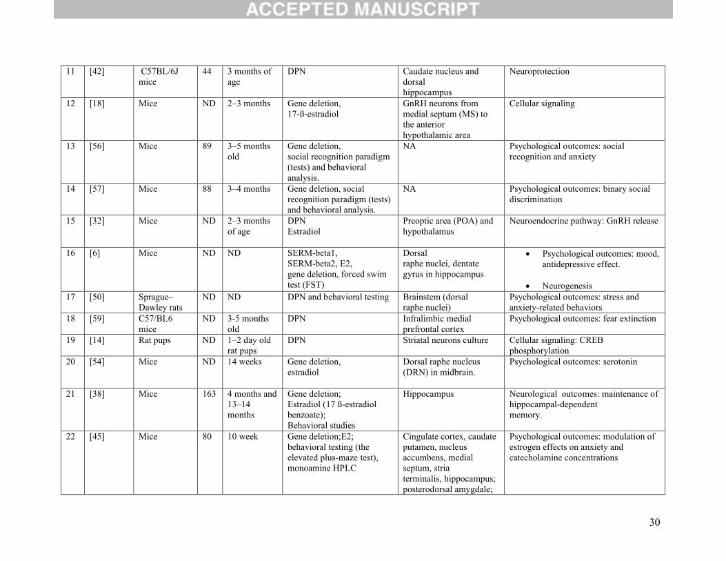
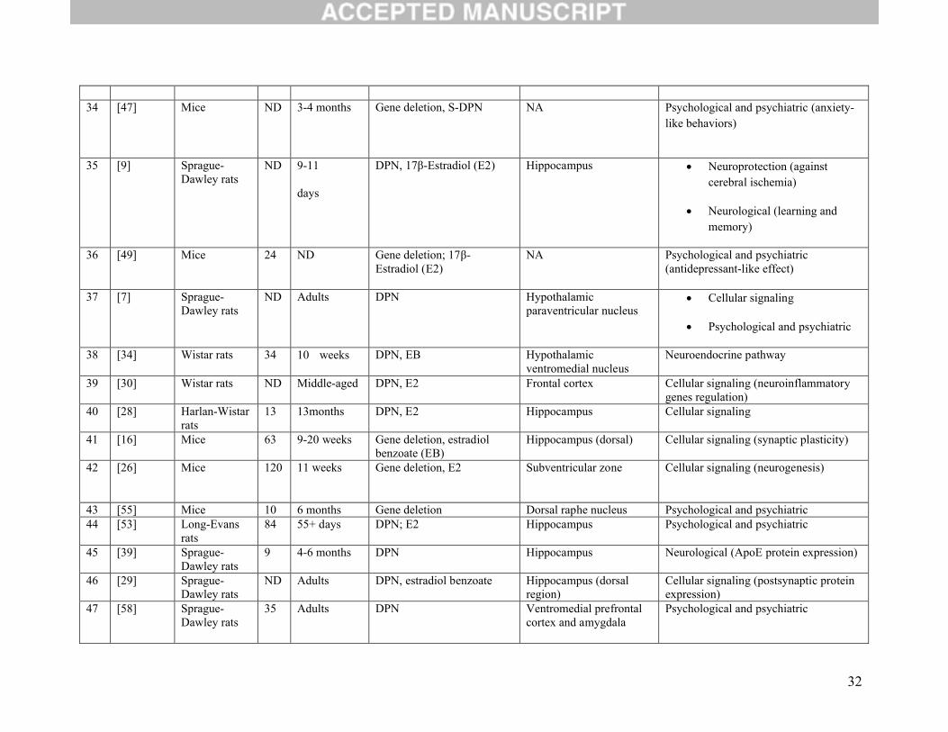
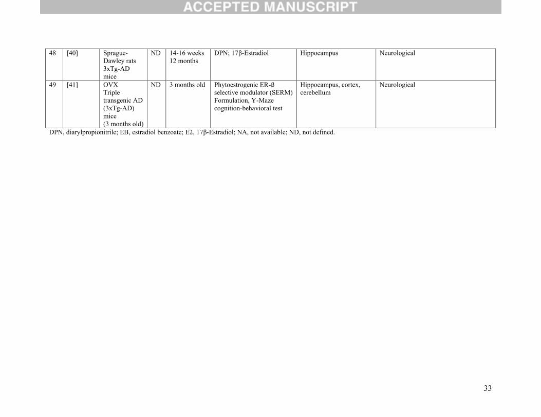
3.1 General Characteristics of the Included Studies
General characteristics of the included studies are reported in Table 1. All studies were performed in
animal models (24 used tissues from mice, 24 from rats and 1 used combined tissue from both mice and
rats). Of the included studies, 19 focused on the function of ERß on cellular signaling, 7 focused on the
neuroendocrine pathway, 4 studied whether neuroprotection was conferred, 8 dealt with neurological
7
outcomes and 19 with psychological and psychiatric outcomes. Six studies evaluated and reported results
on two or more major outcomes [6-11].
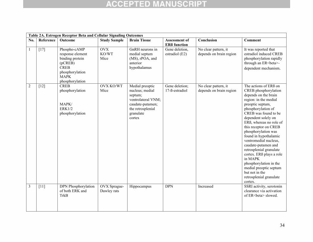
3.2 Cellular signaling:
Nineteen studies evaluated the function of ERβ on cellular signaling (Table 2A). Of these, nine studies
assessed the effect of phosphorylation in different subunits [9, 11-18]. Within these nine, four studies
[11, 12, 15, 16] reported that activation of ERβ was associated with increased immunoreactivity and
expression of phosphorylated mitogen-activated protein kinase (MAPK/ERK), protein kinase B (Akt),
and tropomyosin receptor kinase B (TrKB) receptor. Also, one study [18] found that ERβ was involved
in the phosphorylation of ERK1/2 in gonadotropin-releasing hormone (GnRH) neurons. Akt and TrkB
receptors participate in synaptic plasticity involved in spatial memory, cognition, and other hippocampal-
dependant behaviors [16]. MAPK/ERK signaling regulates a variety of cellular activities including
proliferation, differentiation, survival, and death and has been implicated in the development of AD,
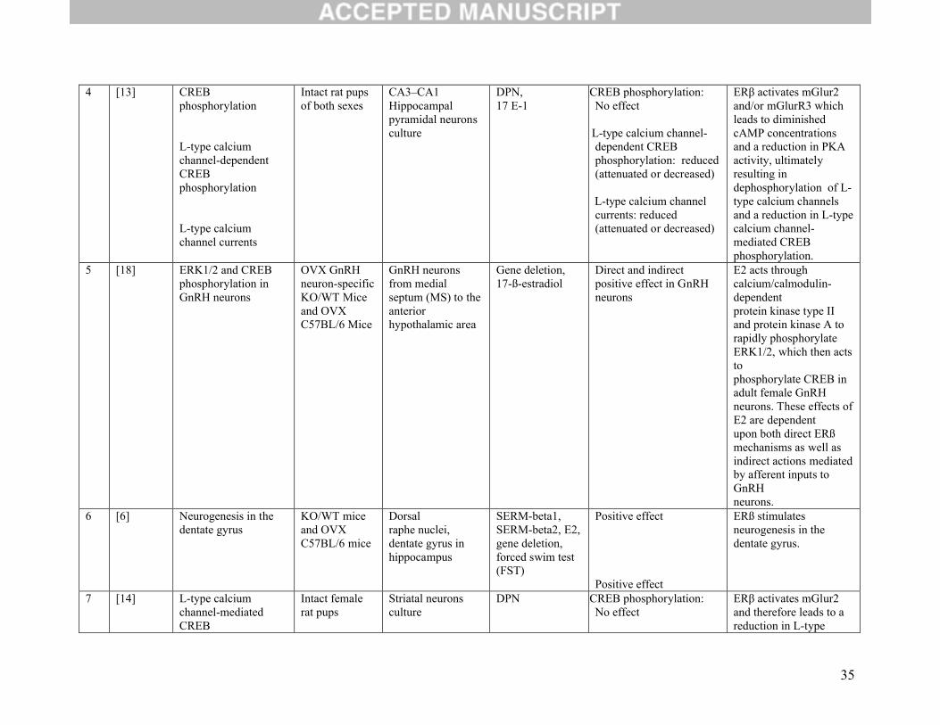
Parkinson’s Disease, and amyotrophic lateral sclerosis [19]. Two other studies [13, 14] showed that
ERβ, via activation of metabotropic glutamate receptor (mGlur) 2 but not mGlur3, dephosphorylates L-
type calcium channels and, therefore causes a reduction in L-type calcium channel-mediated cAMP
response element-binding protein (CREB). Furthermore, two studies [17, 18] reported an involvement of
ERβ in CREB phosphorylation in GnRH neurons whereas another study [12] found regional actions of
ERβ on CREB phosphorylation. In the medial preoptic septum, phosphorylation of CREB was found to
be dependent solely on ERß, whereas no role of this receptor on CREB phosphorylation was found in
hypothalamic ventromedial nucleus, caudate-putamen, or retrosplenial granulate cortex. Moreover, one
study tested periodic ERβ-activation on CREB phosphorylation in the hippocampus and showed
increased protein expression of phosphorylated CREB [9]. CREB has been found to be involved in the
formation of long-term memories, long-term potentiation, and in the development of drug addiction,
physiological dependence and Huntington’s Disease [20-24].
8
Three studies assessed the function of ERβ on neurogenesis [6, 25, 26]. The first two studies were
performed in the hippocampus under normal conditions, and the third in the subventricular zone after an
induced stroke injury. Their conclusions were consistent in that the number of newborn cells was
significantly increased with the activation of ERβ, and therefore support a role of ERβ in neurogenesis.
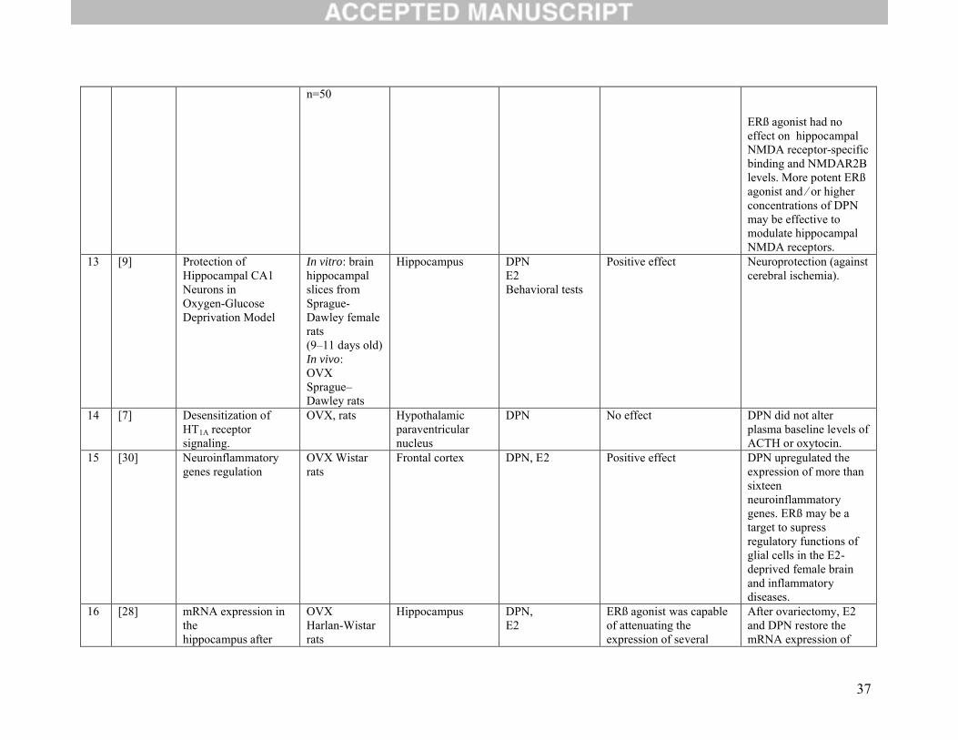
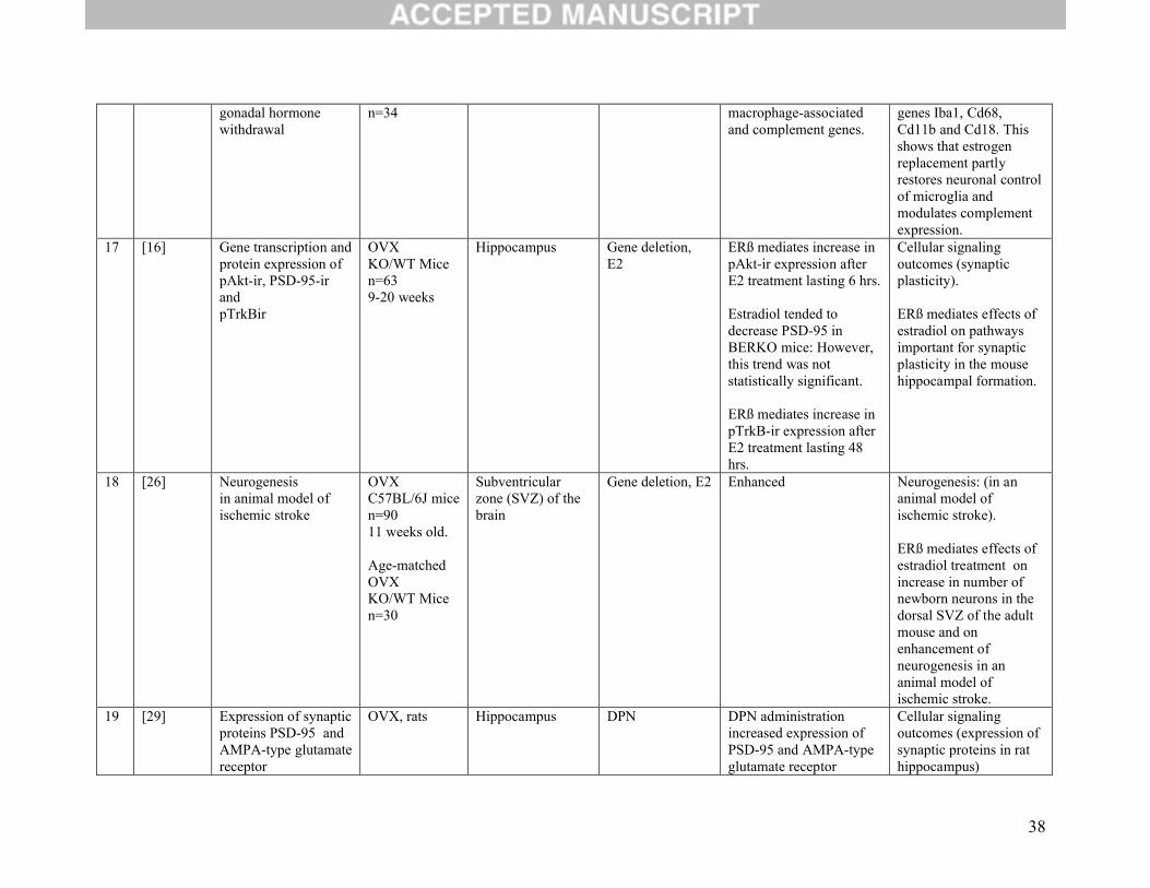
Five studies evaluated gene and protein expression [10, 27-30]. One study found that ERβ activation was
required in order to potentiate mitochondrial function in the brain [27]. The second study found that ERβ
agonist affected the expression of a specified set of genes (Iba1, Cd68, Cd11b and Cd18) that, in turn,
affects neuronal control of microglia and complement expression.[28]. Two studies concluded that ERβ
increased the regulation of post-synaptic PSD-95 and AMPA-type glutamate receptor subunit GluR1 [10,
29]. One study evaluated the effect of DPN on the modulation of N-methyl-D-aspartate (NMDA)
receptor specific binding and found no modulating effect [31]. One other study concluded that DPN
upregulated the expression of more than 16 neuroinflammatory genes, thus leading to suspicion that ERβ
may be a target to supress regulatory functions of glial cells in the E2-deprived female brain and
inflammatory diseases [30].
Finally, one study assessed whether DPN had an effect on the desensitization of serotonin 1A (5-HT1A)
receptor signaling and on the plasma levels of oxytocin and adrenocorticotropic hormone (ACTH) and
found no effect [7].
3.3 Neuroendocrine Pathway Outcomes:
Seven studies evaluated the functions of ERβ on neuroendocrine pathway outcomes (Table 2B). Of
these, one study found that ERß agonist activates GnRH firing and increases gamma-aminobutyric acid
(GABA) transmission and postsynaptic response in GnRH neurons [32].
Two studies evaluated the involvement of ERβ in modulation of hypothalamus-pituitary-adrenal (HPA)
axis reactivity [8, 33]. The first study found that administration of DPN reduced adrenal

9
corticosterone (CORT) and ACTH responses to restraint stress. Based on the observed findings,
the authors concluded that ERβ may modulate HPA/neuroendocrine stress reactivity [8]. The second
study found that ERβ isoforms had a positive effect on regulation of corticotropin-releasing hormone
(CRH) promoter activity; indicating possible involvement of ERβ in the mechanisms of the stress
response and HPA axis disorders pathogenesis [33]. Thus, it was inferred that ERβ might not play a role
in central regulation of serotonin via receptor 5-HT1A by mediating ACTH release.
The role of ERβ in the induction of progesterone receptors (PRs), which are critical for female sexual
behaviour, was assessed in two studies [6, 34]. One study showed that ERβ activation in hypothalamic
ventromedial nucleus averts the action of ERα in the induction of PRs [34], whereas one study reported
that the ERβ agonist selective estrogen receptor modulator beta 2 (SERM-beta 2) dose-dependently
increased PRs in the murine dorsal raphe nucleus but not in the hippocampus [6].
Finally, one study assessed the role of ERβ in the expression and function of T-type calcium
channels (subtype Cav3.2) in the hypothalamus. The authors found that the effect of estradiol on
Cav3.2 was dependent on ERβ as well as on ERα indicating involvement of ERβ in excitability of
hypothalamic neurons; namely, the study has shown that E2-treatment increases Cav3.1 in the
pituitary, but decreases Cav3.2 and Cav3.3 in an ERα dependant manner. [35]
3.4 Neurological outcomes:
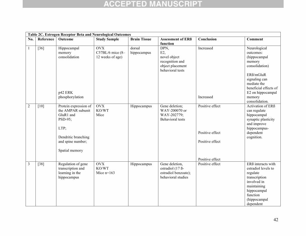
Eight studies evaluated the function of ERβ on neurological outcomes (Table 2C). Of these, four
investigated memory as their main outcome [9, 10, 36, 37]. One study demonstrated that ERβ in dorsal
hippocampus, through activation of mGluR1 signaling, enhanced novel object recognition and object
placement memory [36]. Another study [10] concluded that along with increased protein expression of
the AMPA receptor (AMPAR) subunit GluR1 and PSD-95, ERβ activation induced morphological
changes in hippocampal neurons, including increased dendritic branching and increased density of
10
mushroom-type spines. Furthermore, the same study showed that ERβ improved performance in
hippocampus-dependent memory tasks. This finding implies that activation of ERβ could regulate
hippocampal synaptic plasticity and improve hippocampus-dependent cognition [10]. Jacome at al. found
that ERβ activation increased recognition memory and altered the levels of monoamines (3-methoxy-4-
hydroxyphenylglycol (MHPG) or the MHPG/norepinephrine (NE) ratio), dopamine’s metabolite
(homovanillic acid (HVA)), and serotonin metabolite (5-hydroxyindole acetic acid (5-HIAA)) in several
areas of the brain, including the prefrontal cortex, ventral hippocampus and dentate gyrus, but not in the
striatum or medial septum. This finding implies that ERβ may enhance recognition memory through
alterations in monoaminergic containing systems primarily in the prefrontal cortex and hippocampus.[37].
One study assessed spatial learning, memory, and post-ischemic neuronal survival. It showed
improvement in all these outcomes when ERβ agonist (DPN) was given periodically [9].
The four other studies evaluated a neurological outcome based on gene regulation and protein expression
[38-41]. The first investigated the role of ERβ in regulating gene transcription and learning in the
hippocampus. It found a positive effect and concluded that ERβ might regulate transcription involved in
maintaining hippocampal function during aging.[38] The second study on gene and protein expression
demonstrated a decrease in apolipoprotein E (ApoE) mRNA protein expression (detrimental in AD) with
the use of DPN [39]. The third publication studied whether any changes were produced at the insulin-
degrading enzyme (IDE) level. IDE is an enzyme involved in the catabolism of amyloid beta (Aβ) protein
in the brain and has been associated with the etiology and development of AD. The third publication
found that DPN significantly increased IDE in hippocampal neurons via activation of
phosphatidylinositol 3-kinase (PI3-K) [40]. Finally, the fourth study sought to determine the efficacy of a
SERM formulation in the regulation of early stages of AD. A nine-month dietary supplementation of β-
SERM formulation was found to promote physical health, prolonged survival and improved spatial
11
recognition memory. It also revealed attenuation of both Aβ deposition and plaque formation in AD mice
[41].
3.5 Neuroprotection outcomes:
Four studies set out to determine the role of ERβ on neuroprotection (Table 2D). Three of these
considered whether protection against ischemia was conferred either by way of DPN [9, 42] or WAY
200070-3 [43]. Regardless of the ERβ agonist used, their results were all similar in that protection of
neurons in caudate nucleus and hippocampus against global ischemia-induced death was conferred. The
fourth study evaluated two outcomes: regulation of cerebral inflammatory cytokine and chemokine levels
as well as regulation of the blood brain barrier permeability. The authors found a positive effect for both
and concluded that ERβ may also confer protection against neuroinflammation [44].
3.6 Psychological and psychiatric outcomes:
Nineteen studies assessed the function of ERβ agonists or behavioural testing after gene deletion
or a combination of both (Table 2E). Of these, three studies considered anxiety-like behavior as their
main outcome. The first evaluated anxiety-like behavior and measurement of brain serotonin and
dopamine levels. It found enhanced anxiety and decreased concentrations of these neurotransmitters in the
absence of functional ERβ [45]. The second studied the contribution of estrogen receptors in modulation
of anxiety and analyzed the effect of deleting ERβ gene in mice. Absence of this receptor was associated
with expression of anxiety disorders and with a reduced threshold for synaptic plasticity in the amygdala,
suggesting a role of ERβ in the processing of emotional behaviour [46]. The third study showed that S-
enantiomer of diarylpropionitrile (S-DPN) reduced anxiety-like behavior and attenuated stress-induced
corticosterone (CORT) and ACTH in wild-type mice, but not in mice lacking the ERβ gene, suggesting
anxiolytic effects mediated by ERβ [47].
12
Seven studies focused on depression-like behaviour [6, 11, 48-52]. The first [48] described that
activation of ERβ in the CA3 region of hippocampus induced by DPN slowed serotonin clearance via
MAPK/ERK1/2 signaling and interactions with both TrKB and IGF-1 receptors whereas no role of
PI3K/Akt signaling in mediating this effect was shown. These results show an antidepressant-like effect
of ERβ agonist. Moreover, the other six studies used the widely used behavioural assays/tests, Porsolt
test or forced swim test and found an anti-depressant effect of ERβ activation [6, 11, 49-52].
Two other studies evaluated both anxiety- and depression-like behaviors simultaneously. They reached
the same conclusion in that both outcomes were decreased after the use of ERβ agonists [8, 53].
Four studies focused on the role of ERβ in tryptophan hydroxylase (TPH) 1 mRNA expression, which is
involved in the synthesis of serotonin. Three of these studies found that ERβ was associated with
increased expression of TPH1 [6, 50, 54]. A fourth study concluded that high TPH levels could be
maintained by using the selective ERβ agonist, LY3201) even after ovariectomy [55].
Two publications dealt with social recognition. [56, 57]. Both used gene deletion, social recognition
paradigm tests, and behavioral analysis. Their results were similar: ERβ is necessary for social
discrimination and modulation of social behaviour [56, 57].
Three publications looked to study the role of ERβ on extinction recall, which is the process of learning
not to fear. Two of these authors concluded that DPN administration facilitated extinction memory
consolidation [58, 59]. However, one study found that fear generalization was increased via an effect on
fear memory retrieval mechanisms through activation of ERβ [60].
4. DISCUSSION
This review summarizes 49 studies published worldwide that investigate the functions of ERβ in the
female brain. The studies suggest multiple functions of ERβ might contribute to a diversity of normal
neurophysiologic functions (Figure 2). Our results support non-genomic actions of ERβ on the
13
phosphorylation status and activity of multiple different signalling pathways in the brain. ERβ may act in
the female brain through MAPK/ERK1/2 and PI3K/Akt signalling pathways and interactions of ERβ with
TrkB and IGF-1R may facilitate activation of these kinases. Akt and TrkB are markers of hippocampal
synaptic plasticity [61]. Spenser-Segal et al have demonstrated that ERβ mediates the increase in pAkt-ir
expression after treatment with estradiol for 6 hours. The kinase pathways in the brain play a role in cell
differentiation, survival, apoptosis, and cell proliferation among other functions, all of which might
determine the initiation and progression of neurodegenerative disorders [16]. Also, ERβ interacts with
metabotropic glutamate receptors (mGluRs) signalling, which may depend upon caveolin proteins that are
essential for the trafficking and clustering of signalling molecules [13]. Among mGluRs, ERβ may
activate cell membrane-localized mGluRs type 2 and 3. Their activation would lead to decreased
concentrations of cAMP and a reduction in PKA activity, which would, in turn, result in
dephosphorylation of L-type calcium ion channels and a reduction of L-type calcium ion channel-
mediated CREB phosphorylation. Phosphorylation of CREB in the medial septum, for example, is
undoubtedly due to action of ERβ since it is the only estrogen receptor type expressed in that part of the
brain. Alternatively, other regions devoid of both ERα and ERβ, such as the caudate putamen or granulate
cortex, do not show signs of CREB phosphorylation. CREB-mediated transcription has been described as
promoting synapse formation and transmission in long-term memory and conferring protection against
striatal neuronal death. Both of these attributes may be used as rationale for potential therapeutic
interventions against Huntington’s disease [10, 62]. Accordingly, an alteration of CREB functioning has
also been associated with major depressive disorder, drug addiction and psychological dependence;
conditions which may be better treated with a more thorough understanding of ERβ’s biological function
[63, 64]. Similarly, mGluRs act as a second messenger in the signalling pathway that modulates synaptic
transmission and neuronal excitability, suggesting a potential therapeutic utility in AD, Parkinson disease,
schizophrenia, anxiety and depression [65, 66]. Consequently, further research into these components
may prove to be potential opportunities for treatment for Huntington’s disease, AD, epilepsy, and trauma
[67, 68].
14
The selected studies in this review consistently showed that ERβ agonists increased hippocampal proteins
PSD-95, synaptophysin and AMPA-receptor subunit GluR1, and dendritic branching and mushroom-type
spines. Altogether, these effects of ERβ may suggest a role of this receptor in enhancing synaptic
plasticity by persistent strengthening of synapses (long-term potentiation) and improving hippocampus-
dependent memory and cognition . Further evidence in this systematic review demonstrates that mGluR1
from the dorsal hippocampus interacts with other membrane components such as p42 ERK and Gq
proteins to enhance novel object recognition and object placement memory consolidation. These may
constitute potential targets for treatment of memory impairment conditions. The effect of ERβ on object
recognition and object placement memory tasks, however, may only be exerted in certain regions of the
brain and in response to a dose-dependent administration of estradiol. In this regard, ERβ and its agonist
DPN have been associated with alteration of the monoamines 3-methoxy-4-hydroxyphenylglycol
(MHPG) and metabolites of dopamine and serotonin: homovanillic acid (HVA) and 5-hydroxyindole
acetic acid, respectively. These findings were noted only in the prefrontal cortex and dentate gyrus but not
in the striatum or medial septum. [37]. Although ERβ is present in many areas of the neocortex, it is
highly expressed in the hippocampus and frontal cortex, areas that are important for memory [69].
ERβ also plays a key role in neurogenesis. Three of the studies included in this systematic review show a
consistent effect of increased cell formation in the hippocampal dentate gyrus and subventricular zone [6,
25, 26], a finding supported by results from previous studies in adult animal models. [70, 71]. These
particular areas of the brain are known to produce a high expression of ERβ, which may explain their
unique function of producing newborn cells. Neurogenesis in the hippocampus promotes the formation of
new episodic memories and may even contribute to the therapeutic actions of antidepressant treatment
[72]. Similarly, newly formed astrocytes from the subventricular zone could stimulate brain repair after
ischemic injury and may limit the extension of neurodegenerative changes and traumatic brain injuries
[26]. However, the functional outcome of cell proliferation may become more important if we consider
the exclusive presence and potential action of ERβ in other cell types of the brain, such as neurons of the
15
paraventricular, suprachiasmatic, and tuberal hypothalamic nuclei, as well as the cerebellum and pineal
gland [73].
Other mechanisms of ERβ action have been described in the neuroendocrine system. The activation of the
HPA axis normally responds to a stress stimulus and is controlled by neurons in the paraventricular
nucleus of the hypothalamus, an area of the brain rich in ERβ mRNA. Part of this response involves the
action of CRH whose promoter activity may be regulated by ERβ isoforms. Furthermore, ERβ may also
decrease stress-induced HPA activation through oxytocin neurons in the hypothalamus. ERβ and oxytocin
are highly co-expressed in that part of the brain and both their actions may be mediated by CRH. Control
of ERβ signalling may be better appreciated in the female population given that females are known to
secrete higher levels of glucocorticoid in response to stress than are their male counterparts. Thus,
targeting ERβ may be of benefit in the treatment of anorexia nervosa and depression, conditions
characterized by dysregulation of the HPA axis [8, 33]. ERβ was also found to increase postsynaptic
response of GnRH neurons and could therefore prove to be important in regulating ovulation, fertility,
and maternal behaviour [32, 74]. However, the display of lordosis, another female sexual behaviour in
mice, could not be attributed to ERβ. Its lack of relationship to ERβ was implied from the null effect on
the expression of progesterone receptors in the hypothalamic ventromedial nucleus [34]. The raphe
nucleus, on the other hand, may respond to the ERβ agonist SERM Beta2 in a dose-dependent manner in
order to increase progesterone receptor expression [6]. Another significant contribution of ERβ may be
observed in the hypothalamus by way of regulation of low voltage-activated (T-type) calcium channels
that participate in burst firing and neurotransmission. These phenomena, however, may also be dependent
on the co-expression of other estrogen receptors, including ERα [35].
ERβ has also gained particular interest in the prevention of AD. Several studies included in this
systematic review have shown that ERβ agonist DPN may induce a significant reduction of hippocampal
ApoE mRNA and protein expression, an established risk factor for late-onset AD. In an aging study
sample with low estradiol level, ERβ may compensate to maintain hippocampal function, provided that
estradiol level is increased [38, 39, 41]. Another mechanism may involve catabolism of Aβ by insulin-
16
degrading enzyme (IDE). ERβ, in conjunction with the activation of PI3-K, may regulate the expression
of this protease in normal and early-stage AD brains. IDE induction, which is reported also to respond
rapidly to estradiol administration, may prove to be a target for prevention of AD [40]. Functional
significance of ERβ is also supported by findings from clinical studies in which genetic variations of ERβ
were found to increase the risk of AD in women.[75]
Neuroprotection has also been consistently reported in this systematic review. ERβ may reduce global
ischemia in the caudate nucleus and CA1 pyramidal layer by enhancing expression of estrogen-regulated
genes such as ApoE or bcl-2. Other proposed vehicles for neuroprotection are activation of CREB or
preservation of mitochondrial function [9, 42]. As ischemic neuronal death is mainly caused by
mitochondrial dysfunction disrupting calcium homeostasis and increasing oxidative stress, interest has
increased in studying the pathway by which ERβ may potentiate mitochondrial function [9, 27]. Although
the exact mechanism of action for this protective effect remains to be further explained, its potential
benefits may help prevent loss of synaptic transmission after cerebral ischemia as well as age-related
decline in cognition. Reducing neuroinflammation may also confer brain protection. A number of
cytokines and chemokines (interleukins) including IL-1 β, IL-6 and IL-12p40 in the brain may be
regulated by ERβ through alteration of the blood brain barrier. These findings may contribute to decrease
proinflammatory cytokines commonly described in postmenopausal females and thought to induce
subclinical states of neurodegeneration and cognitive decline [76].
This systematic review also shows consistency in the role of ERβ in modulating estrogen signaling in the
process of emotional behaviour [8, 33, 45-47] and other types of behaviour such as social recognition and
extinction recall[56-58]. The use of ERβ knockout animal models and adapted behavioural tests were
particularly useful in determining its functional role. Studies in this review have demonstrated that
anxiety-like behaviour was enhanced, while serotonin levels decreased, with the absence of functional
ERβ. These effects may be region-specific to the stria terminalis, preoptic area, hippocampus, and
possibly the dorsal raphe nucleus. The reduction of other monoamines such dopamine and
dihydroxyphenylacetate may also be associated with increased anxiety in the absence of ERβ. Similarly,
17
the addition of ERβ specific agonist DPN was found to attenuate the levels of stress-induced CORT and
ACTH; which may contribute to decrease anxiety-like behaviours in different test settings. Furthermore,
ERβ may reduce not only anxiety but also depression-like behaviour, which may be mediated by genomic
effects of ERβ in 5-hydroxytryptamine (5-HT) neurons of the dorsal raphe nucleus. This effect was
described after testing (e.g., the open field, elevated plus maze, and the forced swim test) (Benmansour et
al., 2015; Donner and Handa 2009). ERβ activation may increase expression of SERT and thereby induce
slower serotonin clearance.[48]. Consequently, the intersynaptic serotonin gaps would be present in
higher concentrations and would have more time to exert its antidepressant effect; further suggesting that
ERβ may promote an antidepressant-like effect.
To our knowledge, this is the first systematic review assessing the role of ERβ receptor in female brain,
following an a priori designed protocol with clearly defined inclusion and exclusion criteria. So far
narrative reviews have mainly focused on estrogen signalling not targeting specific functions of ERβ [4,
73, 77, 78]. Contrary to our study, the above mentioned narrative reviews covered only limited areas of
ERβ brain functions. Some limitations of this review need to be considered. We included studies that
were highly heterogeneous with respect to input parameters, assumptions, and study design. Therefore,
performing a quantitative pooling of the data was unfeasible. Also, all the included studies used animal
tissue and so caution should be taken in consideration when extrapolating the results of this review to
human subjects. Finally, publication bias may be a concern, as with all systematic reviews. However, we
tried to minimize the impact of publication bias by employing a thorough search strategy in six databases
in which no restrictions were applied on language or time of publication. In addition, reference lists were
checked for further studies.
Overall, the results of the current systematic review included only animal studies showing abundant
functions of ERβ in the female brain and supporting the notion that future therapies targeting ERβ could
constitute a novel preventive and/or treatment strategy for neurological diseases in females. ERβ agonists
can mimic the actions of 17β-estradiol in the brain without causing other physiological responses
mediated by other estrogen receptors. In view of the high burden of depressive disorders and

18
neurodegenerative disease and despite advances in their prevention and treatment, transfer of these novel
therapeutic venues on the field of neurological diseases could constitute a suitable alternative in the
future. However, in order to establish potential therapeutic and preventive strategies targeting ERβ in the
female brain, future studies in humans would first be required.
Contributors
TM and OHF conceived the study.
KGV, JM, AZ and TM designed and performed the literature review, designed the search strategy,
screened titles and abstracts, obtained full text, determined eligibility of articles, participated in data
extraction, data synthesis/analysis, data interpretation, coordination and writing the manuscript.
WMB designed and performed the literature review.
K-xW, LJ, JN, KD and BK screened titles and abstracts.
EvB and MAI participated in data interpretation and writing the manuscript.
All authors contributed to the critical revision of the manuscript.
OHF participated in study design and drafting of the final manuscript.
TM and OHF are guarantors.
All authors approved the final version.
KGV and JM contributed equally.
Conflict of interest
K-xW, LJ and TM received research support from Metagenics Inc. JM, AZ, JN, KD and BK have been
financially supported by Erasmus Mundus Western Balkans (ERAWEB), a project funded by the
19
European Commission. OHF received grants or research support from Metagenics Inc. KGV, WMB, EvB
and MAI have nothing to disclose.
Funding
This study was sponsored and funded by Metagenics Inc. Metagenics Inc. with the steering committee
was involved in study design; collection, analysis and interpretation of data; writing of the report; and
decision to submit for publication. The funder/sponsor did not have the ability to veto publication of study
results.
Provenance and peer review This article has undergone peer review.
REFERENCES
[1] World Health Organization.).[2] A. Hofman, P.T. de Jong, C.M. van Duijn, M.M. Breteler, Epidemiology of neurological diseases in elderly people: what did we learn from the Rotterdam Study?, Lancet Neurol 5(6) (2006) 545-50.[3] M.A. O'Neal, Neurologic diseases in women Neurology Clinical Practice 3(3) (2013) 217-223.[4] N. Sugiyama, R.P. Barros, M. Warner, J.A. Gustafsson, ERbeta: recent understanding of estrogen signaling, Trends Endocrinol Metab 21(9) (2010) 545-52.[5] T. Muka, K.G. Vargas, L. Jaspers, K.X. Wen, K. Dhana, A. Vitezova, J. Nano, A. Brahimaj, V. Colpani, A. Bano, B. Kraja, A. Zaciragic, W.M. Bramer, G.M. Dijk, M. Kavousi, O.H. Franco, Estrogen receptor beta actions in the female cardiovascular system: A systematic review of animal and human studies, Maturitas 86 (2016) 28-43.[6] J.A. Clark, S. Alves, C. Gundlah, B. Rocha, E.T. Birzin, S.J. Cai, R. Flick, E. Hayes, K. Ho, S. Warrier, L. Pai, J. Yudkovitz, R. Fleischer, L. Colwell, S. Li, H. Wilkinson, J. Schaeffer, R. Wilkening, E. Mattingly, M. Hammond, S.P. Rohrer, Selective estrogen receptor-beta (SERM-beta) compounds modulate raphe nuclei tryptophan hydroxylase-1 (TPH-1) mRNA expression and cause antidepressant-like effects in the forced swim test, Neuropharmacology 63(6) (2012) 1051-63.[7] D.V. Rossi, Y. Dai, P. Thomas, G.A. Carrasco, L.L. DonCarlos, N.A. Muma, Q. Li, Estradiol-induced desensitization of 5-HT1A receptor signaling in the paraventricular nucleus of the hypothalamus is independent of estrogen receptor-beta, Psychoneuroendocrinology 35(7) (2010) 1023-33.
20
[8] A.E. Kudwa, R.F. McGivern, R.J. Handa, Estrogen receptor beta and oxytocin interact to modulate anxiety-like behavior and neuroendocrine stress reactivity in adult male and female rats, Physiol Behav 129 (2014) 287-96.[9] A.P. Raval, R. Borges-Garcia, W. Javier Moreno, M.A. Perez-Pinzon, H. Bramlett, Periodic 17beta-estradiol pretreatment protects rat brain from cerebral ischemic damage via estrogen receptor-beta, PLoS One 8(4) (2013) e60716.[10] F. Liu, M. Day, L.C. Muniz, D. Bitran, R. Arias, R. Revilla-Sanchez, S. Grauer, G. Zhang, C. Kelley, V. Pulito, A. Sung, R.F. Mervis, R. Navarra, W.D. Hirst, P.H. Reinhart, K.L. Marquis, S.J. Moss, M.N. Pangalos, N.J. Brandon, Activation of estrogen receptor-beta regulates hippocampal synaptic plasticity and improves memory, Nat Neurosci 11(3) (2008) 334-43.[11] S. Benmansour, O.S. Adeniji, A.A. Privratsky, A. Frazer, Effects of Long-Term Treatment with Estradiol and Estrogen Receptor Subtype Agonists on Serotonergic Function in Ovariectomized Rats, Neuroendocrinology (2015).[12] I.M. Abraham, M.G. Todman, K.S. Korach, A.E. Herbison, Critical in vivo roles for classical estrogen receptors in rapid estrogen actions on intracellular signaling in mouse brain, Endocrinology 145(7) (2004) 3055-61.[13] M.I. Boulware, J.P. Weick, B.R. Becklund, S.P. Kuo, R.D. Groth, P.G. Mermelstein, Estradiol activates group I and II metabotropic glutamate receptor signaling, leading to opposing influences on cAMP response element-binding protein, J Neurosci 25(20) (2005) 5066-78.[14] D. Grove-Strawser, M.I. Boulware, P.G. Mermelstein, Membrane estrogen receptors activate the metabotropic glutamate receptors mGluR5 and mGluR3 to bidirectionally regulate CREB phosphorylation in female rat striatal neurons, Neuroscience 170(4) (2010) 1045-55.[15] H.H. Le, S.M. Belcher, Rapid signaling actions of environmental estrogens in developing granule cell neurons are mediated by estrogen receptor ss, Endocrinology 151(12) (2010) 5689-99.[16] J.L. Spencer-Segal, M.C. Tsuda, L. Mattei, E.M. Waters, R.D. Romeo, T.A. Milner, B.S. McEwen, S. Ogawa, Estradiol acts via estrogen receptors alpha and beta on pathways important for synaptic plasticity in the mouse hippocampal formation, Neuroscience 202 (2012) 131-46.[17] I.M. Abraham, S.K. Han, M.G. Todman, K.S. Korach, A.E. Herbison, Estrogen receptor beta mediates rapid estrogen actions on gonadotropin-releasing hormone neurons in vivo, J Neurosci 23(13) (2003) 5771-7.[18] R.Y. Cheong, A. Kwakowsky, Z. Barad, R. Porteous, A.E. Herbison, I.M. Abraham, Estradiol acts directly and indirectly on multiple signaling pathways to phosphorylate cAMP-response element binding protein in GnRH neurons, Endocrinology 153(8) (2012) 3792-803.[19] E.K. Kim, E.J. Choi, Pathological roles of MAPK signaling pathways in human diseases, Biochim Biophys Acta 1802(4) (2010) 396-405.[20] R. Bourtchuladze, B. Frenguelli, J. Blendy, D. Cioffi, G. Schutz, A.J. Silva, Deficient long-term memory in mice with a targeted mutation of the cAMP-responsive element-binding protein, Cell 79(1) (1994) 59-68.[21] A. Nazarian, W.L. Sun, L. Zhou, L.M. Kemen, S. Jenab, V. Quinones-Jenab, Sex differences in basal and cocaine-induced alterations in PKA and CREB proteins in the nucleus accumbens, Psychopharmacology (Berl) 203(3) (2009) 641-50.[22] Y. Wang, A. Ghezzi, J.C. Yin, N.S. Atkinson, CREB regulation of BK channel gene expression underlies rapid drug tolerance, Genes Brain Behav 8(4) (2009) 369-76.[23] D.P. DiRocco, Z.S. Scheiner, C.B. Sindreu, G.C. Chan, D.R. Storm, A role for calmodulin-stimulated adenylyl cyclases in cocaine sensitization, J Neurosci 29(8) (2009) 2393-403.
21
[24] C. Rouaux, J.P. Loeffler, A.L. Boutillier, Targeting CREB-binding protein (CBP) loss of function as a therapeutic strategy in neurological disorders, Biochem Pharmacol 68(6) (2004) 1157-64.[25] C.A. Mazzucco, S.E. Lieblich, B.I. Bingham, M.A. Williamson, V. Viau, L.A.M. Galea, Both estrogen receptor alpha and estrogen receptor beta agonists enhance cell proliferation in the dentate gyrus of adult female rats, Neuroscience 141(4) (2006) 1793-1800.[26] S. Suzuki, L.M. Gerhold, M. Bottner, S.W. Rau, C. Dela Cruz, E. Yang, H. Zhu, J. Yu, A.B. Cashion, M.S. Kindy, I. Merchenthaler, F.H. Gage, P.M. Wise, Estradiol enhances neurogenesis following ischemic stroke through estrogen receptors alpha and beta, J Comp Neurol 500(6) (2007) 1064-75.[27] R.W. Irwin, J. Yao, J. To, R.T. Hamilton, E. Cadenas, R.D. Brinton, Selective oestrogen receptor modulators differentially potentiate brain mitochondrial function, J Neuroendocrinol 24(1) (2012) 236-48.[28] M. Sarvari, I. Kallo, E. Hrabovszky, N. Solymosi, Z. Liposits, Ovariectomy and subsequent treatment with estrogen receptor agonists tune the innate immune system of the hippocampus in middle-aged female rats, PLoS One 9(2) (2014) e88540.[29] E.M. Waters, K. Mitterling, J.L. Spencer, S. Mazid, B.S. McEwen, T.A. Milner, Estrogen receptor alpha and beta specific agonists regulate expression of synaptic proteins in rat hippocampus, Brain Res 1290 (2009) 1-11.[30] M. Sarvari, E. Hrabovszky, I. Kallo, N. Solymosi, K. Toth, I. Liko, J. Szeles, S. Maho, B. Molnar, Z. Liposits, Estrogens regulate neuroinflammatory genes via estrogen receptors alpha and beta in the frontal cortex of middle-aged female rats, J Neuroinflammation 8 (2011) 82.[31] M. Morissette, M. Le Saux, T. Di Paolo, Effect of oestrogen receptor alpha and beta agonists on brain N-methyl-D-aspartate receptors, J Neuroendocrinol 20(8) (2008) 1006-14.[32] Z. Chu, J. Andrade, M.A. Shupnik, S.M. Moenter, Differential regulation of gonadotropin-releasing hormone neuron activity and membrane properties by acutely applied estradiol: dependence on dose and estrogen receptor subtype, J Neurosci 29(17) (2009) 5616-27.[33] W.J. Miller, S. Suzuki, L.K. Miller, R. Handa, R.M. Uht, Estrogen receptor (ER)beta isoforms rather than ERalpha regulate corticotropin-releasing hormone promoter activity through an alternate pathway, J Neurosci 24(47) (2004) 10628-35.[34] S.I. Sa, P.A. Pereira, V. Malikov, M.D. Madeira, Role of estrogen receptor alpha and beta in the induction of progesterone receptors in hypothalamic ventromedial neurons, Neuroscience 238 (2013) 159-67.[35] M.A. Bosch, J. Hou, Y. Fang, M.J. Kelly, O.K. Ronnekleiv, 17Beta-estradiol regulation of the mRNA expression of T-type calcium channel subunits: role of estrogen receptor alpha and estrogen receptor beta, J Comp Neurol 512(3) (2009) 347-58.[36] M.I. Boulware, J.D. Heisler, K.M. Frick, The memory-enhancing effects of hippocampal estrogen receptor activation involve metabotropic glutamate receptor signaling, J Neurosci 33(38) (2013) 15184-94.[37] L.F. Jacome, C. Gautreaux, T. Inagaki, G. Mohan, S. Alves, L.S. Lubbers, V. Luine, Estradiol and ERbeta agonists enhance recognition memory, and DPN, an ERbeta agonist, alters brain monoamines, Neurobiol Learn Mem 94(4) (2010) 488-98.[38] X. Han, K.K. Aenlle, L.A. Bean, A. Rani, S.L. Semple-Rowland, A. Kumar, T.C. Foster, Role of estrogen receptor alpha and beta in preserving hippocampal function during aging, J Neurosci 33(6) (2013) 2671-83.

22
[39] J.M. Wang, R.W. Irwin, R.D. Brinton, Activation of estrogen receptor alpha increases and estrogen receptor beta decreases apolipoprotein E expression in hippocampus in vitro and in vivo, Proc Natl Acad Sci U S A 103(45) (2006) 16983-8.[40] L. Zhao, J. Yao, Z. Mao, S. Chen, Y. Wang, R.D. Brinton, 17beta-Estradiol regulates insulin-degrading enzyme expression via an ERbeta/PI3-K pathway in hippocampus: relevance to Alzheimer's prevention, Neurobiol Aging 32(11) (2011) 1949-63.[41] L. Zhao, Z. Mao, S. Chen, L.S. Schneider, R.D. Brinton, Early intervention with an estrogen receptor beta-selective phytoestrogenic formulation prolongs survival, improves spatial recognition memory, and slows progression of amyloid pathology in a female mouse model of Alzheimer's disease, J Alzheimers Dis 37(2) (2013) 403-19.[42] H.V. Carswell, I.M. Macrae, L. Gallagher, E. Harrop, K.J. Horsburgh, Neuroprotection by a selective estrogen receptor beta agonist in a mouse model of global ischemia, Am J Physiol Heart Circ Physiol 287(4) (2004) H1501-4.[43] N.R. Miller, T. Jover, H.W. Cohen, R.S. Zukin, A.M. Etgen, Estrogen can act via estrogen receptor alpha and beta to protect hippocampal neurons against global ischemia-induced cell death, Endocrinology 146(7) (2005) 3070-9.[44] C.M. Brown, T.A. Mulcahey, N.C. Filipek, P.M. Wise, Production of proinflammatory cytokines and chemokines during neuroinflammation: novel roles for estrogen receptors alpha and beta, Endocrinology 151(10) (2010) 4916-25.[45] D.B. Imwalle, J.A. Gustafsson, E.F. Rissman, Lack of functional estrogen receptor beta influences anxiety behavior and serotonin content in female mice, Physiol Behav 84(1) (2005) 157-63.[46] W. Krezel, S. Dupont, A. Krust, P. Chambon, P.F. Chapman, Increased anxiety and synaptic plasticity in estrogen receptor beta -deficient mice, Proc Natl Acad Sci U S A 98(21) (2001) 12278-82.[47] M.G. Oyola, W. Portillo, A. Reyna, C.D. Foradori, A. Kudwa, L. Hinds, R.J. Handa, S.K. Mani, Anxiolytic effects and neuroanatomical targets of estrogen receptor-beta (ERbeta) activation by a selective ERbeta agonist in female mice, Endocrinology 153(2) (2012) 837-46.[48] S. Benmansour, A.A. Privratsky, O.S. Adeniji, A. Frazer, Signaling mechanisms involved in the acute effects of estradiol on 5-HT clearance, Int J Neuropsychopharmacol 17(5) (2014) 765-77.[49] B.A. Rocha, R. Fleischer, J.M. Schaeffer, S.P. Rohrer, G.J. Hickey, 17 Beta-estradiol-induced antidepressant-like effect in the forced swim test is absent in estrogen receptor-beta knockout (BERKO) mice, Psychopharmacology (Berl) 179(3) (2005) 637-43.[50] N. Donner, R.J. Handa, Estrogen receptor beta regulates the expression of tryptophan-hydroxylase 2 mRNA within serotonergic neurons of the rat dorsal raphe nuclei, Neuroscience 163(2) (2009) 705-18.[51] S. Bansal, K. Chopra, Differential role of estrogen receptor modulators in depression-like behavior and memory impairment in rats with postmenopausal diabetes, Menopause 22(10) (2015) 1117-24.[52] C.P. Bastos, L.M. Pereira, T.H. Ferreira-Vieira, L.E. Drumond, A.R. Massensini, M.F. Moraes, G.S. Pereira, Object recognition memory deficit and depressive-like behavior caused by chronic ovariectomy can be transitorialy recovered by the acute activation of hippocampal estrogen receptors, Psychoneuroendocrinology 57 (2015) 14-25.
23
[53] A.A. Walf, C.A. Frye, Administration of estrogen receptor beta-specific selective estrogen receptor modulators to the hippocampus decrease anxiety and depressive behavior of ovariectomized rats, Pharmacol Biochem Behav 86(2) (2007) 407-14.[54] C. Gundlah, S.E. Alves, J.A. Clark, L.Y. Pai, J.M. Schaeffer, S.P. Rohrer, Estrogen receptor-beta regulates tryptophan hydroxylase-1 expression in the murine midbrain raphe, Biol Psychiatry 57(8) (2005) 938-42.[55] H. Suzuki, R.P. Barros, N. Sugiyama, V. Krishnan, B.C. Yaden, H.J. Kim, M. Warner, J.A. Gustafsson, Involvement of estrogen receptor beta in maintenance of serotonergic neurons of the dorsal raphe, Mol Psychiatry 18(6) (2013) 674-80.[56] E. Choleris, J.A. Gustafsson, K.S. Korach, L.J. Muglia, D.W. Pfaff, S. Ogawa, An estrogen-dependent four-gene micronet regulating social recognition: a study with oxytocin and estrogen receptor-alpha and -beta knockout mice, Proc Natl Acad Sci U S A 100(10) (2003) 6192-7.[57] E. Choleris, S. Ogawa, M. Kavaliers, J.A. Gustafsson, K.S. Korach, L.J. Muglia, D.W. Pfaff, Involvement of estrogen receptor alpha, beta and oxytocin in social discrimination: A detailed behavioral analysis with knockout female mice, Genes Brain Behav 5(7) (2006) 528-39.[58] M.A. Zeidan, S.A. Igoe, C. Linnman, A. Vitalo, J.B. Levine, A. Klibanski, J.M. Goldstein, M.R. Milad, Estradiol modulates medial prefrontal cortex and amygdala activity during fear extinction in women and female rats, Biol Psychiatry 70(10) (2011) 920-7.[59] C. Galvin, I. Ninan, Regulation of the mouse medial prefrontal cortical synapses by endogenous estradiol, Neuropsychopharmacology 39(9) (2014) 2086-94.[60] J.F. Lynch, 3rd, D. Dejanovic, P. Winiecki, J. Mulvany, S. Ortiz, D.C. Riccio, A.M. Jasnow, Activation of ERbeta modulates fear generalization through an effect on memory retrieval, Horm Behav 66(2) (2014) 421-9.[61] T. Nakai, T. Nagai, M. Tanaka, N. Itoh, N. Asai, A. Enomoto, M. Asai, S. Yamada, A.B. Saifullah, M. Sokabe, M. Takahashi, K. Yamada, Girdin phosphorylation is crucial for synaptic plasticity and memory: a potential role in the interaction of BDNF/TrkB/Akt signaling with NMDA receptor, J Neurosci 34(45) (2014) 14995-5008.[62] Y.S. Choi, B. Lee, H.Y. Cho, I.B. Reyes, X.A. Pu, T.C. Saido, K.R. Hoyt, K. Obrietan, CREB is a key regulator of striatal vulnerability in chemical and genetic models of Huntington's disease, Neurobiol Dis 36(2) (2009) 259-68.[63] R.H. Belmaker, G. Agam, Major depressive disorder, N Engl J Med 358(1) (2008) 55-68.[64] C.S. McPherson, A.J. Lawrence, The nuclear transcription factor CREB: involvement in addiction, deletion models and looking forward, Curr Neuropharmacol 5(3) (2007) 202-12.[65] C.M. Niswender, P.J. Conn, Metabotropic glutamate receptors: physiology, pharmacology, and disease, Annu Rev Pharmacol Toxicol 50 (2010) 295-322.[66] L. Pomierny-Chamiolo, K. Rup, B. Pomierny, E. Niedzielska, P.W. Kalivas, M. Filip, Metabotropic glutamatergic receptors and their ligands in drug addiction, Pharmacol Ther 142(3) (2014) 281-305.[67] Z.Z. Chong, Y.C. Shang, S. Wang, K. Maiese, A Critical Kinase Cascade in Neurological Disorders: PI 3-K, Akt, and mTOR, Future Neurol 7(6) (2012) 733-748.[68] F. Boulle, G. Kenis, M. Cazorla, M. Hamon, H.W. Steinbusch, L. Lanfumey, D.L. van den Hove, TrkB inhibition as a therapeutic target for CNS-related disorders, Prog Neurobiol 98(2) (2012) 197-206.[69] S.W. Mitra, E. Hoskin, J. Yudkovitz, L. Pear, H.A. Wilkinson, S. Hayashi, D.W. Pfaff, S. Ogawa, S.P. Rohrer, J.M. Schaeffer, B.S. McEwen, S.E. Alves, Immunolocalization of estrogen

24
receptor beta in the mouse brain: comparison with estrogen receptor alpha, Endocrinology 144(5) (2003) 2055-67.[70] P. Tanapat, N.B. Hastings, E. Gould, Ovarian steroids influence cell proliferation in the dentate gyrus of the adult female rat in a dose- and time-dependent manner, J Comp Neurol 481(3) (2005) 252-65.[71] B.K. Ormerod, T.T. Lee, L.A. Galea, Estradiol initially enhances but subsequently suppresses (via adrenal steroids) granule cell proliferation in the dentate gyrus of adult female rats, J Neurobiol 55(2) (2003) 247-60.[72] J.E. Malberg, A.J. Eisch, E.J. Nestler, R.S. Duman, Chronic antidepressant treatment increases neurogenesis in adult rat hippocampus, J Neurosci 20(24) (2000) 9104-10.[73] M.J. Weiser, C.D. Foradori, R.J. Handa, Estrogen receptor beta in the brain: from form to function, Brain Res Rev 57(2) (2008) 309-20.[74] L.R. Brooks, C.D. Le, W.C. Chung, P.S. Tsai, Maternal behavior in transgenic mice with reduced fibroblast growth factor receptor function in gonadotropin-releasing hormone neurons, Behav Brain Funct 8 (2012) 47.[75] M. Pirskanen, M. Hiltunen, A. Mannermaa, S. Helisalmi, M. Lehtovirta, T. Hanninen, H. Soininen, Estrogen receptor beta gene variants are associated with increased risk of Alzheimer's disease in women, Eur J Hum Genet 13(9) (2005) 1000-6.[76] M. Abu-Taha, C. Rius, C. Hermenegildo, I. Noguera, J.M. Cerda-Nicolas, A.C. Issekutz, P.J. Jose, J. Cortijo, E.J. Morcillo, M.J. Sanz, Menopause and ovariectomy cause a low grade of systemic inflammation that may be prevented by chronic treatment with low doses of estrogen or losartan, J Immunol 183(2) (2009) 1393-402.[77] H.J. Kim, G. Casadesus, Estrogen-mediated effects on cognition and synaptic plasticity: what do estrogen receptor knockout models tell us?, Biochim Biophys Acta 1800(10) (2010) 1090-3.[78] S. Lokuge, B.N. Frey, J.A. Foster, C.N. Soares, M. Steiner, The rapid effects of estrogen: a mini-review, Behav Pharmacol 21(5-6) (2010) 465-72.

25
Legend for Figure 1.
Flowchart of studies investigating the function of estrogen receptor beta in the female brain
26
Legend for Figure 2
Potential pathways for ERβ function in the female brain.
ApoE mRNA, Apolipoprotein E messenger RNA; HPA, hypothalamus-pituitary-adrenal; IDE,
insulin-degrading enzyme; AMPA, α-amino-3-hydroxy-5-methyl-4-isoxazolepropionic acid;
NMDA, N-Methyl-D-aspartic acid; Akt, Protein kinase B; MAPK, Mitogen-activated protein
kinase; ERK, extracellular signal-regulated kinases; CREB, c-AMP response-element binding
protein.

27
Figure 1
Records given full text detailed assessment
(n = 141)
Studies included (n =49)
Records identified through database searching (n = 3186)
Records after duplicates removed and to be screened by titles and abstracts (n=3051)
Full-text articles excluded (n = 92)
Reasons for exclusion:1.Not the appropriate exposure or
outcome (n=39)2.No female tissue or results not sex-
specific (n=25)3.Results are not ER-beta specific
(n=5)4.No full text available (n=4)5.Other: data extraction unfeasible,
conference abstracts, commentary, duplicates (n=19)
Records excluded (n = 2910)
Iden
tifi
cati
onE
ligi
bili
tyS
cree
ning
Incl
uded

28
Figure 2

29
Table 1. General Characteristics of the Animal Studies Included in this ReviewNo. Reference Animal Type N Age Assessment of ERβ
function Tissue Outcome
1 [17] Mice ND 40–54 days Gene deletion,estradiol (E2)
GnRH neurons in medial septum , rostral preoptic area, and anterior hypothalamus
Cellular signaling
2 [12] Mice ND ND Gene deletion; 17-ß-estradiol
Medial preoptic nucleus; medial septum;; caudate-putamen; the retrosplenialgranulatecortex
Cellular signaling: phosphorylation; rapid intracellular signaling
3 [51] Rats (Sprague-Dawley)
8-12
ND DPN NA Psychological and psychiatric(depression-like and memory impairment)
4 [52] C57BL/6mice
ND 8-12 weeks DPN, E2 Hippocampus Psychological and psychiatric (depression-like and non-spatial memory)
5 [48] Sprague–Dawley rats
ND ND DPN,E2
CA3 region of the hippocampus
Psychological outcomes (mood)
6 [11] Sprague–Dawley rats
12-28
4 months DPN Hippocampus Cellular signaling: Psychological and psychiatric outcomes (depression-like)
7 [35] Mice ND ND Gene deletion,E2
Hypothalamic nuclei and pituitary calcium channel subunit: Cav3.1
Neuroendocrine pathway: excitability of hypothalamic neurons and modulation of pituitarysecretion
8 Rat pups ND 1- to 2-days-old rat pups
DPN,17 E- 1
CA3–CA1 Hippocampal pyramidal neurons
Cellular signaling: CREB phosphorylation
9 [36] C57BL/6 mice
ND 8–12 weeks of age
DPN,E2,novel object recognition and object placement behavioral tests
Dorsal hippocampus Neurological outcomes: (hippocampal memory consolidation)
10 [44] Mice ND 10–15 weeksof age
Gene deletion, E2, cytokine/chemokine quantification.
Cortex andstriatum
Neuroprotection: anti-inflammatory effects (mediation of neuroinflammatory response)
30
11 [42] C57BL/6J mice
44 3 months of age
DPN Caudate nucleus and dorsalhippocampus
Neuroprotection
12 [18] Mice ND 2–3 months Gene deletion,17-ß-estradiol
GnRH neurons from medial septum (MS) to the anteriorhypothalamic area
Cellular signaling
13 [56] Mice 89 3–5 months old
Gene deletion,social recognition paradigm (tests) and behavioral analysis.
NA Psychological outcomes: social recognition and anxiety
14 [57] Mice 88 3–4 months Gene deletion, social recognition paradigm (tests) and behavioral analysis.
NA Psychological outcomes: binary social discrimination
15 [32] Mice ND 2–3 months of age
DPNEstradiol
Preoptic area (POA) and hypothalamus
Neuroendocrine pathway: GnRH release
16 [6] Mice ND ND SERM-beta1,SERM-beta2, E2,gene deletion, forced swim test (FST)
Dorsalraphe nuclei, dentate gyrus in hippocampus
Psychological outcomes: mood, antidepressive effect.
Neurogenesis17 [50] Sprague–
Dawley ratsND ND DPN and behavioral testing Brainstem (dorsal
raphe nuclei)Psychological outcomes: stress and anxiety-related behaviors
18 [59] C57/BL6 mice
ND 3-5 months old
DPN Infralimbic medial prefrontal cortex
Psychological outcomes: fear extinction
19 [14] Rat pups ND 1–2 day old rat pups
DPN Striatal neurons culture Cellular signaling: CREB phosphorylation
20 [54] Mice ND 14 weeks Gene deletion,estradiol
Dorsal raphe nucleus (DRN) in midbrain.
Psychological outcomes: serotonin
21 [38] Mice 163 4 months and 13–14 months
Gene deletion; Estradiol (17 ß-estradiol benzoate);Behavioral studies
Hippocampus Neurological outcomes: maintenance of hippocampal-dependentmemory.
22 [45] Mice 80 10 week Gene deletion;E2; behavioral testing (the elevated plus-maze test),monoamine HPLC
Cingulate cortex, caudate putamen, nucleus accumbens, medialseptum, striaterminalis, hippocampus; posterodorsal amygdale;
Psychological outcomes: modulation of estrogen effects on anxiety and catecholamine concentrations

31
substantia nigra; dorsal raphe, locuscoeruleus
23 [27] Sprague–Dawley rats
ND 4–6 months old
DPN,E2
Isolated brain mitochondria (of forebrain)
Cellular signaling: modulation of mitochondrial function in the brain
24 [37] Sprague–Dawley rats
32–34
2 months old DPN; estradiol benzoate;behavioral tests (object recognition / placement memory tasks)
NA Neurological outcomes: mediation of sub-chronic and acute effectsof estrogens on recognition memory
25 [46] Mice ND 8-month-old Gene deletion; behavioral testing;electrophysiological evaluation of amygdala functions
Amygdala Psychological outcomes: emotionalbehavior
26 [8] Sprague–Dawley rats
118 ND DPN, behavioral and restraint testing
NA Neuroendocrine pathway: HPA reactivity.
Psychological outcomes (anxiety-like behaviors).
27 [15] Neonatal Rats(Sprague Dawley)
ND ND Xenoestrogens, DPN Immature cerebellar granule cell cultures
Cellular signaling: rapid intracellular signaling
28 [10] Mice ND ND Gene deletion; WAY-200070 or WAY-202779;behavioral tests
Hippocampus Neurological outcomes: hippocampal-dependent memory + hippocampal synaptic plasticity
29 [60] Long-Evans rats
146 90 days old DPN; behavioral test NA Psychological outcomes:fear generalization
30 [25] Sprague-Dawley rats
69 Adults DPN Hippocampus(Dentate gyrus)
Cellular signaling (cellular proliferation)
31 [33] Sprague-Dawley rats
5 Adults Transfected ER beta 1 Hypothalamus Neuroendocrine pathway
32 [43] Sprague-Dawley rats
8 21 days WAY 200070-2 Hippocampus Neuroprotection
33 [31] Sprague-Dawley rats
10 Adults DPN Prefrontal cortex and hippocampal regions
Cellular signaling(NMDA receptor modulation)
32
34 [47] Mice ND 3-4 months Gene deletion, S-DPN NA Psychological and psychiatric (anxiety-like behaviors)
35 [9] Sprague-Dawley rats
ND 9-11
days
DPN, 17β-Estradiol (E2) Hippocampus Neuroprotection (against cerebral ischemia)
Neurological (learning and memory)
36 [49] Mice 24 ND Gene deletion; 17β-Estradiol (E2)
NA Psychological and psychiatric (antidepressant-like effect)
37 [7] Sprague-Dawley rats
ND Adults DPN Hypothalamic paraventricular nucleus
Cellular signaling
Psychological and psychiatric
38 [34] Wistar rats 34 10 weeks DPN, EB Hypothalamic ventromedial nucleus
Neuroendocrine pathway
39 [30] Wistar rats ND Middle-aged DPN, E2 Frontal cortex Cellular signaling (neuroinflammatory genes regulation)
40 [28] Harlan-Wistarrats
13 13months DPN, E2 Hippocampus Cellular signaling
41 [16] Mice 63 9-20 weeks Gene deletion, estradiol benzoate (EB)
Hippocampus (dorsal) Cellular signaling (synaptic plasticity)
42 [26] Mice 120 11 weeks Gene deletion, E2 Subventricular zone Cellular signaling (neurogenesis)
43 [55] Mice 10 6 months Gene deletion Dorsal raphe nucleus Psychological and psychiatric44 [53] Long-Evans
rats84 55+ days DPN; E2 Hippocampus Psychological and psychiatric
45 [39] Sprague-Dawley rats
9 4-6 months DPN Hippocampus Neurological (ApoE protein expression)
46 [29] Sprague-Dawley rats
ND Adults DPN, estradiol benzoate Hippocampus (dorsal region)
Cellular signaling (postsynaptic protein expression)
47 [58] Sprague-Dawley rats
35 Adults DPN Ventromedial prefrontal cortex and amygdala
Psychological and psychiatric
33
48 [40] Sprague-Dawley rats3xTg-AD mice
ND 14-16 weeks12 months
DPN; 17β-Estradiol Hippocampus Neurological
49 [41] OVXTriple transgenic AD (3xTg-AD) mice(3 months old)
ND 3 months old Phytoestrogenic ER-ß selective modulator (SERM)Formulation, Y-Maze cognition-behavioral test
Hippocampus, cortex, cerebellum
Neurological
DPN, diarylpropionitrile; EB, estradiol benzoate; E2, 17β-Estradiol; NA, not available; ND, not defined.
34
Table 2A. Estrogen Receptor Beta and Cellular Signaling OutcomesNo. Reference Outcome Study Sample Brain Tissue Assessment of
ERß function Conclusion Comment
1 [17] Phospho-cAMP response element binding protein (pCREB) CREB phosphorylation MAPK phosphorylation
OVX KO/WTMice
GnRH neurons in medial septum (MS), rPOA, and anterior hypothalamus
Gene deletion, estradiol (E2)
No clear pattern, it depends on brain region
It was reported that estradiol induced CREB phosphorylation rapidly through an ER<beta>-dependent mechanism.
2 [12] CREB phosphorylation
MAPK/ERK1/2 phosphorylation
OVX KO/WTMice
Medial preoptic nucleus; medial septum; ventrolateral VNM; caudate-putamen; the retrosplenial granulatecortex
Gene deletion; 17-ß-estradiol
No clear pattern, it depends on brain region
The actions of ERß on CREB phosphorylation depends on the brain region: in the medial preoptic septum, phosphorylation of CREB was found to be dependent solely on ERß, whereas no role of this receptor on CREB phosphorylation was found in hypothalamic ventromedial nucleus, caudate-putamen and retrosplenial granulate cortex. ERß plays a role in MAPK phosphorylation in the medial preoptic septum but not in the retrosplenial granulate cortex.
3 [11] DPN Phosphorylation of both ERK and TrkB
OVX Sprague-Dawley rats
Hippocampus DPN Increased SSRI activity, serotonin clearance via activation of ER<beta> slowed.
35
4 [13] CREB phosphorylation
L-type calcium channel-dependent CREB phosphorylation
L-type calcium channel currents
Intact rat pups of both sexes
CA3–CA1 Hippocampal pyramidal neurons culture
DPN,17 E-1
CRCREB phosphorylation: No effect
L-type calcium channel-dependent CREB phosphorylation: reduced (attenuated or decreased)
L-type calcium channel currents: reduced (attenuated or decreased)
ERβ activates mGlur2 and/or mGlurR3 which leads to diminished cAMP concentrations and a reduction in PKA activity, ultimately resulting in dephosphorylation of L-type calcium channels and a reduction in L-type calcium channel-mediated CREB phosphorylation.
5 [18] ERK1/2 and CREB phosphorylation in GnRH neurons
OVX GnRH neuron-specific KO/WT Miceand OVX C57BL/6 Mice
GnRH neurons from medial septum (MS) to the anteriorhypothalamic area
Gene deletion,17-ß-estradiol
Direct and indirect positive effect in GnRH neurons
E2 acts through calcium/calmodulin-dependentprotein kinase type II and protein kinase A to rapidly phosphorylate ERK1/2, which then acts tophosphorylate CREB in adult female GnRH neurons. These effects of E2 are dependentupon both direct ERß mechanisms as well as indirect actions mediated by afferent inputs to GnRHneurons.
6 [6] Neurogenesis in the dentate gyrus
KO/WT miceand OVX C57BL/6 mice
Dorsalraphe nuclei, dentate gyrus in hippocampus
SERM-beta1,SERM-beta2, E2,gene deletion, forced swim test (FST)
Positive effect
Positive effect
ERß stimulatesneurogenesis in the dentate gyrus.
7 [14] L-type calciumchannel-mediated CREB
Intact female rat pups
Striatal neurons culture
DPN CRCREB phosphorylation: No effect
ERβ activates mGlur2 and therefore leads to a reduction in L-type

36
phosphorylationMglur2 and Mglu3
L-type calcium channel-dependent CREB phosphorylation: reduced (attenuated or decreased)
ERβ activates mGlur3 but not mGlurR2
calcium channel-mediated CREB phosphorylation.
8 [27] Mitochondrial DNA-encoded COX I expression;
OVX Sprague–Dawley Rats
Isolated brain mitochondria from forebrain
DPNE2
Positive effect Activation of ERß isdifferentially required to potentiate mitochondrial function in brain.
9 [15] ERK phosphorylation Intact neonatal femaleSprague Dawley rats
Immature cerebellar granule cell cultures
Xenoestrogens;DPN
Positive effect Rapid intracellular signaling of estrogen is mediated by ER beta.
10 [10] Protein expression of the AMPAR subunit GluR1 andPSD-95;
LTP;
Dendritic branching and spine number;
Spatial memory
OVX KO/WTMice
Hippocampus Gene deletion; WAY-200070 or WAY-202779;Behavioral tests
Positive effect
Positive effect
Positive effect
Positive effect
Activation of ERß can regulate hippocampal synaptic plasticity and improve hippocampus-dependent cognition.
11 [25] hippocampal neurogenesis (hippocampal cell proliferation)
OVXSprague–Dawley ratsn=95
Hippocampus DPN,E2
Increased Neurogenesis:(hippocampal cell proliferation)
ERß is involved inestradiol-enhanced cell proliferation.
12 [31] Modulation of NMDA receptor specific binding
OVXSprague–Dawley rats
Prefrontal cortex and hippocampus
DPN,E2
No effect Cellular signalingoutcomes: (modulation of NMDA receptors)
37
n=50
ERß agonist had no effect on hippocampalNMDA receptor-specific binding and NMDAR2B levels. More potent ERß agonist and ⁄ or higher concentrations of DPN may be effective to modulate hippocampalNMDA receptors.
13 [9] Protection of Hippocampal CA1 Neurons inOxygen-Glucose Deprivation Model
In vitro: brain hippocampal slices from Sprague-Dawley female rats(9–11 days old)In vivo:OVXSprague–Dawley rats
Hippocampus DPNE2Behavioral tests
Positive effect Neuroprotection (against cerebral ischemia).
14 [7] Desensitization of HT1A receptor signaling.
OVX, rats Hypothalamic paraventricularnucleus
DPN No effect DPN did not alter plasma baseline levels of ACTH or oxytocin.
15 [30] Neuroinflammatory genes regulation
OVX Wistar rats
Frontal cortex DPN, E2 Positive effect DPN upregulated the expression of more than sixteen neuroinflammatory genes. ERß may be a target to supress regulatory functions of glial cells in the E2-deprived female brain and inflammatory diseases.
16 [28] mRNA expression in thehippocampus after
OVXHarlan-Wistar rats
Hippocampus DPN,E2
ERß agonist was capable of attenuating the expression of several
After ovariectomy, E2 and DPN restore the mRNA expression of
38
gonadal hormone withdrawal
n=34 macrophage-associated and complement genes.
genes Iba1, Cd68, Cd11b and Cd18. This shows that estrogen replacement partly restores neuronal control of microglia and modulates complement expression.
17 [16] Gene transcription andprotein expression of pAkt-ir, PSD-95-ir and pTrkBir
OVXKO/WT Micen=639-20 weeks
Hippocampus Gene deletion,E2
ERß mediates increase in pAkt-ir expression after E2 treatment lasting 6 hrs.
Estradiol tended to decrease PSD-95 in BERKO mice: However, this trend was notstatistically significant.
ERß mediates increase in pTrkB-ir expression after E2 treatment lasting 48 hrs.
Cellular signalingoutcomes (synaptic plasticity).
ERß mediates effects of estradiol on pathways important for synaptic plasticity in the mouse hippocampal formation.
18 [26] Neurogenesisin animal model of ischemic stroke
OVXC57BL/6J micen=9011 weeks old.
Age-matched OVXKO/WT Micen=30
Subventricular zone (SVZ) of the brain
Gene deletion, E2 Enhanced Neurogenesis: (in ananimal model of ischemic stroke).
ERß mediates effects of estradiol treatment on increase in number of newborn neurons in the dorsal SVZ of the adult mouse and on enhancement of neurogenesis in ananimal model of ischemic stroke.
19 [29] Expression of synaptic proteins PSD-95 and AMPA-type glutamate receptor
OVX, rats Hippocampus DPN DPN administration increased expression of PSD-95 and AMPA-type glutamate receptor
Cellular signalingoutcomes (expression of synaptic proteins in rat hippocampus)
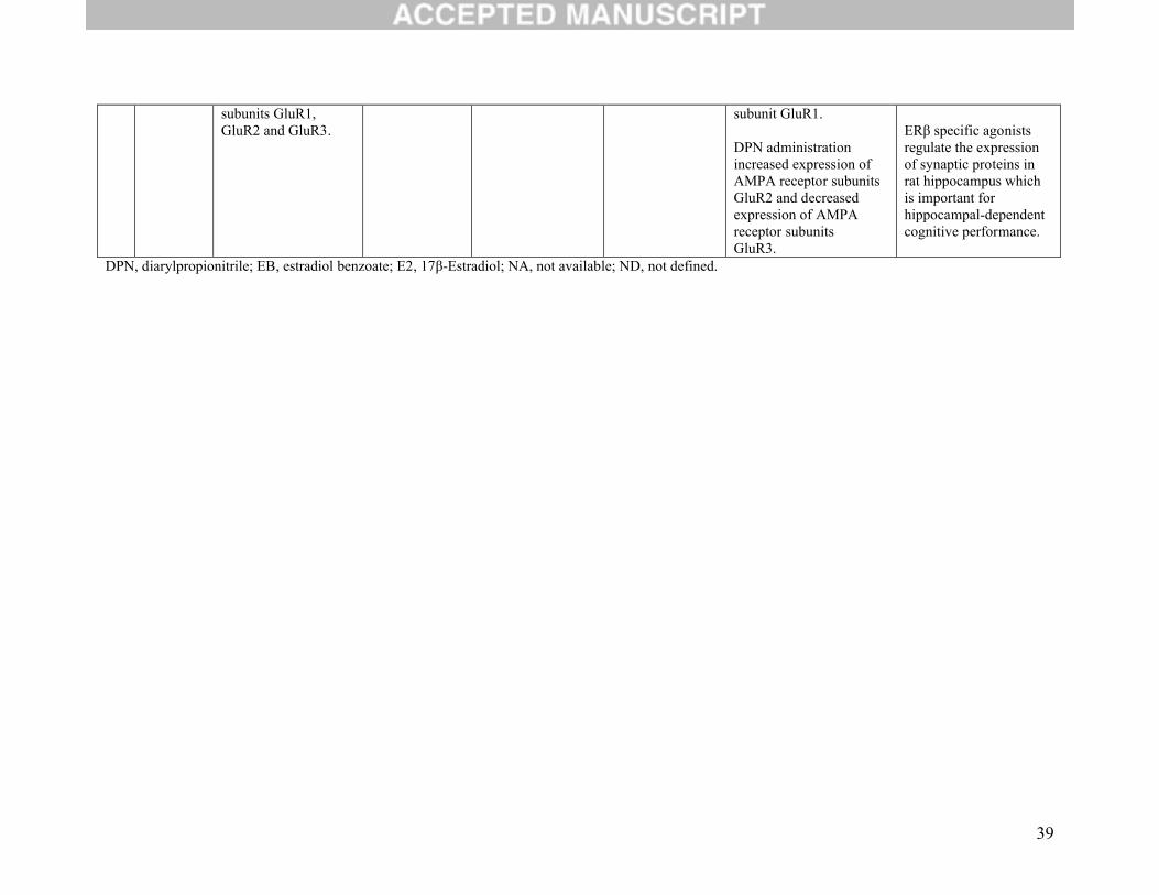
39
subunits GluR1, GluR2 and GluR3.
subunit GluR1.
DPN administration increased expression of AMPA receptor subunitsGluR2 and decreased expression of AMPA receptor subunitsGluR3.
ERβ specific agonists regulate the expression of synaptic proteins in rat hippocampus which is important for hippocampal-dependent cognitive performance.
DPN, diarylpropionitrile; EB, estradiol benzoate; E2, 17β-Estradiol; NA, not available; ND, not defined.

40
Table 2B. Estrogen Receptor Beta and Neuroendocrine Pathway OutcomesNo. Reference Outcome Study Sample Brain Tissue Assessment of
ERß function Conclusion Comment
1 [35] T-type calciumchannel (subtype Cav3.2) expression and function
KO/WT Miceand OVX C57BL/6 mice
Hypothalamic nuclei and pituitary calcium channel subunit: Cav3.1
Gene deletion, Estradiol (E2)
Positive effect Involvement in excitability of hypothalamic neurons.
2 [6] Tryptophan hydroxylase-1 (TPH-1) mRNA expression;progesterone receptorexpression
KO/WT Miceand OVX C57BL/6 Mice
Dorsalraphe nuclei; dentate gyrus in hippocampus
SERM-beta1,SERM-beta2, E2,gene deletion, forced swim test (FST)
Positive effect SERM-beta1 and2 exhibited antidepressant-like effects / ERß may play an important role in modulating mood
3 [32] Firing of GnRH neuronsGABA transmission
OVXMice expressing enhancedgreen fluorescent protein under the control of theGnRH promoter
Preoptic area (POA) / hypothalamus
Estradiol,DPN
Supporting effect ERß agonist activates GnRH firing, reduces after hyperpolarizing potential (AHP) and increased slow afterdepolarization amplitudes (ADP), and reduced IAHP and enhanced IADP.Also, ERß agonist increased GABA transmission and postsynaptic response in GnRH neuron
4 [8] CORT and ACTH response to restraint stress
Sprague–Dawley rats of both sexes(OVX females)n=118
NA DPN, behavioral and restraint testing
DPN reduced CORT and ACTH responses in bothmales and females.
ERβ may modulate HPA (neuroendocrine stress) reactivity.
5 [33] regulation of the corticotropin-releasing hormone (CRH) promoter
OVXSprague–Dawley ratsn=5
Hypothalamus Expressionvectors: ERß isoforms
Enhanced (Increased) (positive effect)
Neuroendocrine pathway: (regulation of the corticotropin-releasing hormone promoter).
ERß regulates CRH in
41
the PVH(paraventricular nucleus) of the hypothalamus.
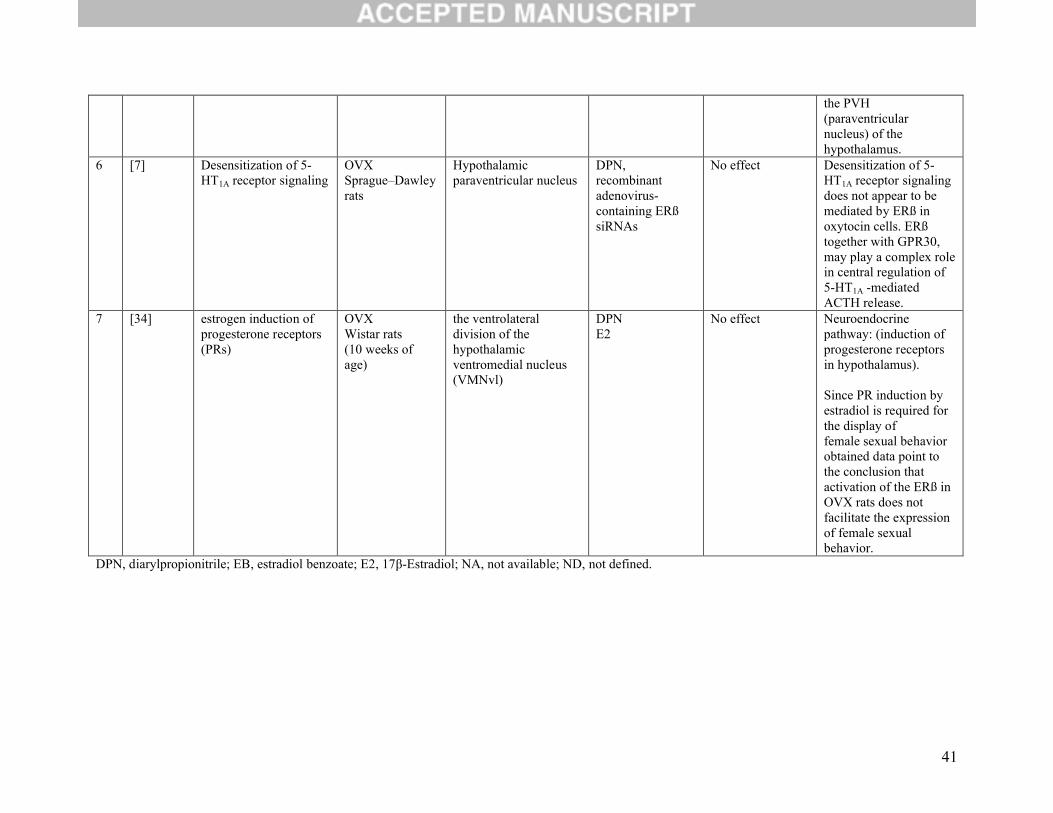
6 [7] Desensitization of 5-HT1A receptor signaling
OVXSprague–Dawley rats
Hypothalamic paraventricular nucleus
DPN,recombinant adenovirus-containing ERß siRNAs
No effect Desensitization of 5-HT1A receptor signalingdoes not appear to be mediated by ERß in oxytocin cells. ERßtogether with GPR30, may play a complex role in central regulation of 5-HT1A -mediated ACTH release.
7 [34] estrogen induction of progesterone receptors(PRs)
OVXWistar rats(10 weeks of age)
the ventrolateral division of the hypothalamicventromedial nucleus (VMNvl)
DPNE2
No effect Neuroendocrine pathway: (induction of progesterone receptors in hypothalamus).
Since PR induction by estradiol is required for the display offemale sexual behavior obtained data point to the conclusion that activation of the ERß in OVX rats does not facilitate the expression of female sexualbehavior.
DPN, diarylpropionitrile; EB, estradiol benzoate; E2, 17β-Estradiol; NA, not available; ND, not defined.
42
Table 2C. Estrogen Receptor Beta and Neurological OutcomesNo. Reference Outcome Study Sample Brain Tissue Assessment of ERß
function Conclusion Comment
1 [36] Hippocampal memory consolidation
p42 ERKphosphorylation
OVXC57BL/6 mice (8–12 weeks of age)
dorsal hippocampus
DPN,E2,novel object recognition and object placement behavioral tests
Increased
Increased
Neurological outcomes: (hippocampal memory consolidation)
ERß/mGluR signaling can mediate the beneficial effects of E2 on hippocampal memory consolidation.
2 [10] Protein expression of the AMPAR subunit GluR1 andPSD-95;
LTP;
Dendritic branching and spine number;
Spatial memory
OVX KO/WTMice
Hippocampus Gene deletion; WAY-200070 or WAY-202779;Behavioral tests
Positive effect
Positive effect
Positive effect
Positive effect
Activation of ERßcan regulate hippocampal synaptic plasticity and improve hippocampus-dependent cognition.
3 [38] Regulation of gene transcription and learning in the hippocampus
OVX KO/WTMice n=163
Hippocampus Gene deletion, estradiol (17 ß-estradiol benzoate);behavioral studies
Positive effect ERß interacts with estradiol levels to regulate transcription involved inmaintaining hippocampal function (hippocampal dependent
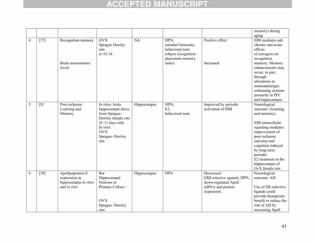
43
memory) during aging
4 [37] Recognition memory
Brain monoamines levels
OVXSprague Dawley ratsn=32-34
NA DPN, estradiol benzoate;behavioral tests (object recognition / placement memory tasks)
Positive effect
Increased
ERß mediates sub chronic and acute effectsof estrogens on recognition memory. Memory enhancements may occur, in part, throughalterations in monoaminergic-containing systems primarily in PFC and hippocampus.
5 [9] Post-ischemicLearning and Memory
In vitro: brain hippocampal slices from Sprague-Dawley female rats(9–11 days old)In vivo:OVXSprague–Dawley rats
Hippocampus DPN,E2,behavioral tests
Improved by periodic activation of ERß
Neurological outcome: (learning and memory).
ERß intracellular signaling mediates improvement of post-ischemic outcome and cognition induced by long-term periodicE2 treatment in the hippocampus of OvX female rats.
6 [39] Apolipoprotein Eexpression in hippocampus in vitro and in vivo
RatHippocampal Neurons in Primary Culture /
OVXSprague–Dawley rats
Hippocampus DPN Decreased /ERβ selective agonist, DPN, down-regulated ApoE mRNA and protein expression.
Neurological outcome: AD
Use of ER-selective ligands could provide therapeutic benefit to reduce the risk of AD byincreasing ApoE
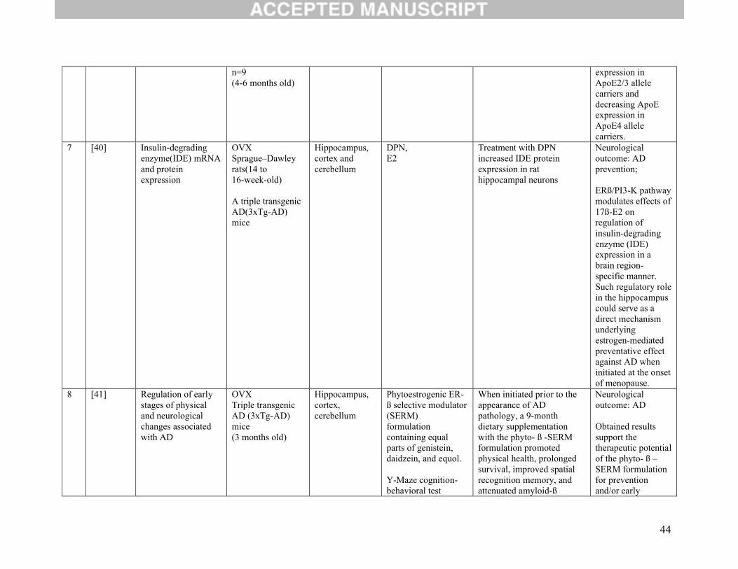
44
n=9(4-6 months old)
expression in ApoE2/3 allele carriers and decreasing ApoE expression in ApoE4 allele carriers.
7 [40] Insulin-degrading enzyme(IDE) mRNA and protein expression
OVXSprague–Dawley rats(14 to16-week-old)
A triple transgenic AD(3xTg-AD) mice
Hippocampus, cortex andcerebellum
DPN,E2
Treatment with DPN increased IDE protein expression in rat hippocampal neurons
Neurological outcome: AD prevention;
ERß/PI3-K pathway modulates effects of 17ß-E2 on regulation of insulin-degrading enzyme (IDE) expression in a brain region-specific manner. Such regulatory role in the hippocampus could serve as a direct mechanism underlying estrogen-mediated preventative effect against AD when initiated at the onset of menopause.
8 [41] Regulation of earlystages of physical and neurological changes associated with AD
OVXTriple transgenic AD (3xTg-AD) mice(3 months old)
Hippocampus, cortex, cerebellum
Phytoestrogenic ER-ß selective modulator (SERM)formulation containing equal parts of genistein, daidzein, and equol.
Y-Maze cognition-behavioral test
When initiated prior to the appearance of AD pathology, a 9-month dietary supplementationwith the phyto- ß -SERM formulation promoted physical health, prolonged survival, improved spatial recognition memory, andattenuated amyloid-ß
Neurological outcome: AD
Obtained results support the therapeutic potential of the phyto- ß –SERM formulation for prevention and/or early

45
deposition and plaque formation in the brains of treated AD mice.
intervention of AD, and warrant further investigations in human studies.
DPN, diarylpropionitrile; EB, estradiol benzoate; E2, 17β-Estradiol; NA, not available; ND, not defined.

46
Table 2D. Estrogen Receptor Beta and Neuroprotection Outcomes
No. Reference Outcome Study Sample Brain Tissue Assessment of ERß function
Conclusion Comment
1 [44] Regulation of brain cytokine and chemokinelevels;
Regulation of BBB permeability
OVX KO/WTMice
Cortex andstriatum
Gene deletion; E2; Cytokine/chemokine quantification.
Positive effect
Positive effect
ERß regulates proinflammatory cytokine and chemokine productionthrough E2-dependent and E2-independent mechanisms.
ERß is essential for E2-mediatedregulation of BBB permeability.
2 [42] Induction of transient global ischemia by bilateral carotid arteryocclusion
OVXC57BL/6J micen=44
Caudate nucleus and dorsalhippocampus
DPN DPN significantly reduced ischemicdamage by 70% in the caudate nucleus and 55% in the CA1 regioncompared with vehicle controls.
ERß activation is neuroprotective in global cerebral ischemia mouse model.
3 [43] Protection of hippocampal neuronsin a global ischemia model
OVXSprague–Dawley rats(21 days old)
Hippocampus WAY 200070-3 ERß-selective agonistWAY 200070–3 produced nearly complete protectionof CA1 neurons in approximately 50% of the animals.
Neuroprotection: (protection of hippocampal neuronsin global ischemia model)
ERß mediates estradiol protection of CA1 hippocampal neurons from globalischemia-induced

47
cell death. 4 [9] Protection of
Hippocampal CA1 Neurons inOxygen-Glucose Deprivation Model
In vitro: brain hippocampal slices from Sprague-Dawley female rats(9–11 days old)In vivo:OVXSprague–Dawley rats
Hippocampus DPNE2Behavioral tests
Positive effect Neuroprotection (against cerebral ischemia).
DPN, diarylpropionitrile; EB, estradiol benzoate; E2, 17β-Estradiol; NA, not available; ND, not defined.
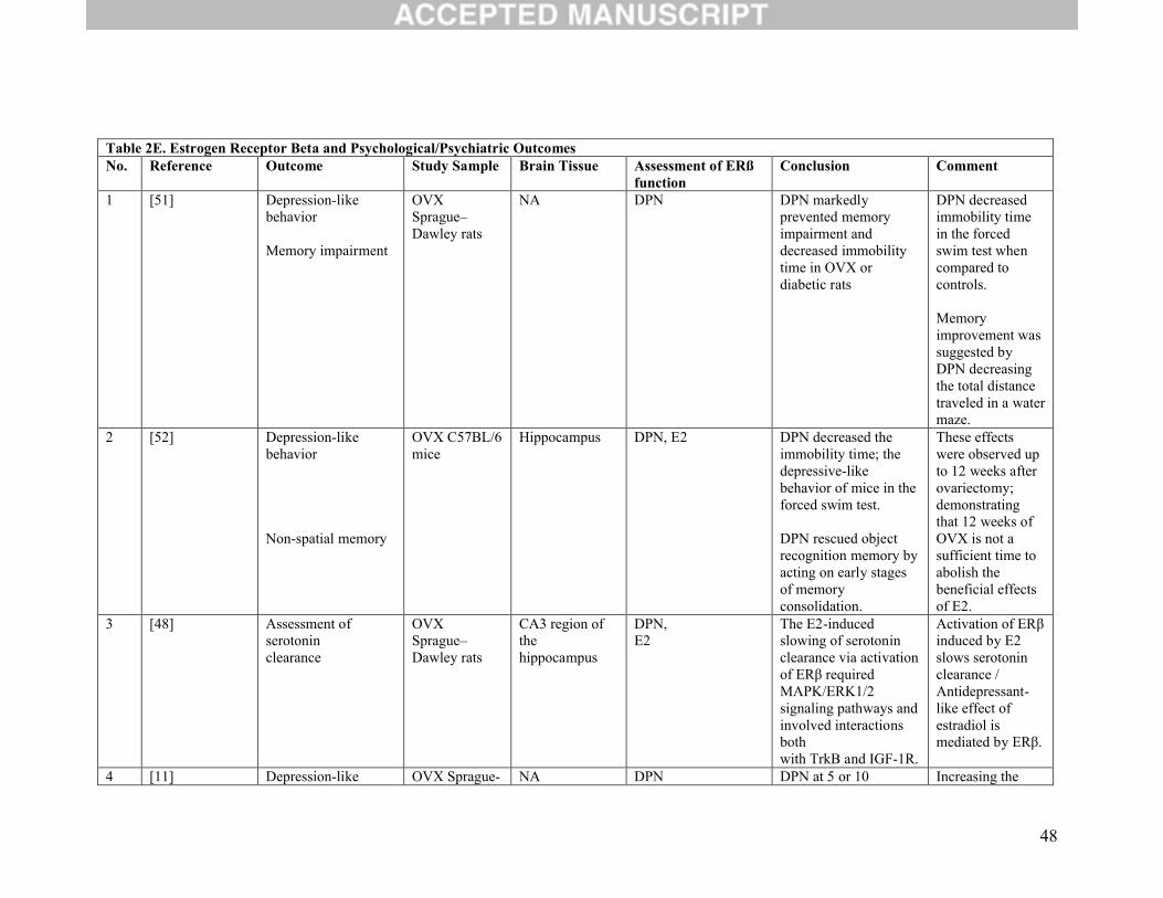
48
Table 2E. Estrogen Receptor Beta and Psychological/Psychiatric OutcomesNo. Reference Outcome Study Sample Brain Tissue Assessment of ERß
function Conclusion Comment
1 [51] Depression-like behavior
Memory impairment
OVXSprague–Dawley rats
NA DPN DPN markedly prevented memory impairment and decreased immobility time in OVX or diabetic rats
DPN decreased immobility time in the forced swim test when compared to controls.
Memory improvement was suggested by DPN decreasing the total distance traveled in a water maze.
2 [52] Depression-like behavior
Non-spatial memory
OVX C57BL/6 mice
Hippocampus DPN, E2 DPN decreased the immobility time; the depressive-like behavior of mice in the forced swim test.
DPN rescued object recognition memory by acting on early stages of memory consolidation.
These effects were observed up to 12 weeks after ovariectomy; demonstrating that 12 weeks of OVX is not a sufficient time to abolish the beneficial effects of E2.
3 [48] Assessment of serotoninclearance
OVXSprague–Dawley rats
CA3 region of the hippocampus
DPN,E2
The E2-induced slowing of serotoninclearance via activation of ERβ required MAPK/ERK1/2signaling pathways and involved interactions bothwith TrkB and IGF-1R.
Activation of ERβinduced by E2 slows serotonin clearance /Antidepressant-like effect of estradiol ismediated by ERβ.
4 [11] Depression-like OVX Sprague- NA DPN DPN at 5 or 10 Increasing the

49
behavior Dawley rats micrograms induced antidepressant-like effects during the forced swim test.
dosage from 5 micrograms to 10 of DPN was not found to produce antidepressant-like effects.
5 [56] Social recognition
Social anxiety
KO/WT Mice and OVX Swiss Webstermicen=89
NA Gene deletion,social recognition paradigm (tests) and behavioral analysis.
ß-ERKO mice wereselectively impaired in social recognition but not in overall activity.
The reduced social anxiety was found in the ß-ERKO mice.
Genes forERß play a crucial role in oxytocin-dependent socialrecognition.
Estrogen modulation of femalepsychosocial anxiety depends on ER-ß.
6 [57] Social recognition (binary social discriminationassay)
KO/WT Mice and OVX Swiss Webster micen=88
NA Gene deletion, social recognition paradigm (tests) and behavioral analysis.
ß-ERKO mice showed partially impaired social discrimination.
ERß genes arenecessary for social discriminationand, thus, for the modulation of social behavior (e.g.,aggression, affiliation).
7 [6] Tryptophan hydroxylase-1 (TPH-1) mRNA expression;progesterone receptorexpression
KO/WT Miceand OVX C57BL/6 Mice
Dorsalraphe nuclei; dentate gyrus in hippocampus
SERM-beta1,SERM-beta2, E2,gene deletion, forced swim test (FST)
Positive effect SERM-beta1 and2 exhibited antidepressant-like effects / ERß may play an important role in modulating mood

50
8 [50] Tryptophan-Hydroxylase 2 mRNA expression
Anxiety-like behaviors
Despair-like behavior
OVXSprague–Dawley rats
Brainstem (dorsalraphe nuclei)
Systemic and local application of DPN,
Behavioral testing
Positive effect
Systemic delivery of DPNdecreased anxiety-like behavior, while local administrationof DPN failed to have the same effect.
Local DPN administration in animals showed decreased despair-likebehavior. does not alter anxiety-likebehaviors, but enhances active stress-coping behavior.
ERß acts at the level of the ratDRN to modulate tph2 mRNAexpression and thereby influenceserotoninsynthesis in DRN subregions.
Local activation of ERß neurons in the DRN may be sufficient to decrease despair-like behavior, but notanxiolytic behaviors.
9 [59] Fear extinction C57/BL6 mice Infralimbic medial prefrontal cortex
DPN Increased ERß activation facilitates the infralimbic medial prefrontal cortex to undergo potentiation; necessary for the regulation of fear extinction.
10 [54] TPH1 mRNA expression
KO/WT miceand OVX C57BL/6mice
Dorsal raphe nucleus (DRN) in midbrain
Gene deletion,estradiol
Positive effect ERß might be responsible for mediating estrogen regulated TPH1 expression in the murine DRN. /ERß might be a key mediator of estrogen
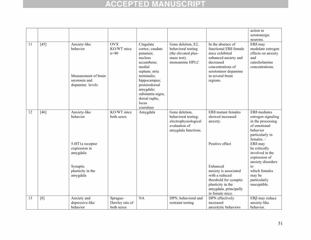
51
action in serotonergic neurons.
11 [45] Anxiety-like behavior
Measurement of brain serotonin and dopamine levels
OVX KO/WT micen=80
Cingulate cortex; caudate putamen; nucleus accumbens; medialseptum; striaterminalis; hippocampus; posterodorsal amygdale; substantia nigra; dorsal raphe, locuscoeruleus
Gene deletion, E2, behavioral testing (the elevated plus-maze test),monoamine HPLC
In the absence of functional ERß female mice exhibited enhanced anxiety anddecreased concentrations of serotoninor dopamine in several brain regions.
ERß may modulate estrogeneffects on anxiety and catecholamine concentrations.
12 [46] Anxiety-like behavior
5-HT1a receptor expression in amygdala
Synapticplasticity in the amygdala
KO/WT miceboth sexes
Amygdala Gene deletion, behavioral testing;electrophysiological evaluation of amygdala functions.
ERß mutant females showed increased anxiety.
Positive effect
Enhancedanxiety is associated with a reduced threshold for synapticplasticity in the amygdala, principally in female mice.
ERß mediates estrogen signaling in the processing of emotionalbehavior particularly in females. /ERß maybe critically involved in the expression of anxiety disorders towhich females may be particularly susceptible.
13 [8] Anxiety and depressive-like behavior
Sprague–Dawley rats of both sexes
NA DPN, behavioral and restraint testing
DPN effectively increasedanxiolytic behaviors
ERβ may reduce anxiety-likebehavior.
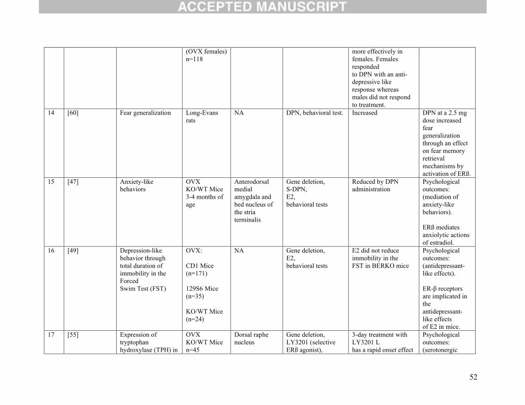
52
(OVX females)n=118
more effectively in females. Females respondedto DPN with an anti-depressive like response whereas males did not respond to treatment.
14 [60] Fear generalization Long-Evans rats
NA DPN, behavioral test. Increased DPN at a 2.5 mg dose increased fear generalization through an effect on fear memory retrieval mechanisms by activation of ERß.
15 [47] Anxiety-like behaviors
OVXKO/WT Mice3-4 months of age
Anterodorsal medial amygdala and bed nucleus of the stria terminalis
Gene deletion, S-DPN,E2,behavioral tests
Reduced by DPN administration
Psychological outcomes: (mediation of anxiety-like behaviors).
ERß mediates anxiolytic actions of estradiol.
16 [49] Depression-like behavior through total duration of immobility in the ForcedSwim Test (FST)
OVX:
CD1 Mice (n=171)
129S6 Mice (n=35)
KO/WT Mice(n=24)
NA Gene deletion,E2, behavioral tests
E2 did not reduce immobility in theFST in BERKO mice
Psychological outcomes: (antidepressant-like effects).
ER-β receptors are implicated in the antidepressant-like effectsof E2 in mice.
17 [55] Expression of tryptophan hydroxylase (TPH) in
OVXKO/WT Micen=45
Dorsal raphe nucleus
Gene deletion,LY3201 (selective ERß agonist),
3-day treatment with LY3201 Lhas a rapid onset effect
Psychological outcomes: (serotonergic

53
the Dorsal raphe (DR),
Alterations in glucose homeostasis.
6 months old E2 on serotonergic activity withincreased expression of TPH and ERß.
LY3201 onlyrestored TPH expression in the DR when the duration of estrogendeprivation was less than 10 weeks.
3-day treatment with LY3201 causes onlymild alterations in glucose homeostasis but effectively restoredTPH expression in the DR.
activity).
ERß agonists could be useful pharmaceuticals in maintainingfunctional DR neurons to treat postmenopausal depression.
18 [53] Anxiety and depressive behavior
OVX Long–Evans rats (n=84)(55+ d old)
Hippocampus Estrogen receptor beta-specific selective estrogenReceptor modulators (DPN),behavioral testing
ERβ-selective SERMs to the hippocampus, but not the ventral tegmental area,decreased anxiety and depressive behavior.
Psychological outcomes: (Anxiety and depressive behavior)
Administration of estrogen receptor beta-specific selective estrogenreceptor modulators to the hippocampus decrease anxiety anddepressive behavior of ovariectomized rats.
19 [58] Extinction memory Sprague– Ventromedial DPN and behavioral Administration of the Psychological

54
expressionduring extinction recall (learning not to fear).
Dawley naturally cycling ratsn=81
prefrontal cortex (vmPFC) and amygdala
tests (extinction recall learning trials) in animals
estrogen-receptor beta agonist facilitated extinction recall.
outcomes: (fear extinction recall)
ERß modulates effects of estradiol on facilitation of extinction memory consolidation in a time-dependent manner in rodents.
DPN, diarylpropionitrile; EB,