the exocellular bacteriolytic system of soil actinomyces i. the nature and properties of the lytic...
TRANSCRIPT
VOL. 2 (1948) BIOCHIMICA ET BIOPHYSICA ACTA 167
T H E E X O C E L L U L A R B A C T E R I O L Y T I C SYSTEM OF S O IL
ACTINOMYCES
I. T H E N A T U R E AND P R O P E R T I E S OF T H E LY TIC SYSTEM
b#
A. S. JONES, A. J. SWALLOW AND M. WEBB Chemistry Department, University of Birmingham (England)
Since the early observations of LIESKA (1921) that specific Actinomycetes were able to lyse many living and dead bacterial cells, numerous biological investigations of the phenomenon have been made. These have been reviewed in considerable detail by WAKSMAN (1945). Extensive studies by WELSCH (1941, 1942 ) showed that killed gram negative cells were readily dissolved by sterile filtrates termed "Actinomycetin", of broth cultures of his Actinomyces strain G obtained after sporulation. Killed gram positive cells, thougl/susceptible to some extent, were more resistant. A few strains of strepto- cocci and pneumococci were found to be susceptible to sterile actinomyces culture filtrates even when alive. Other living gram positives, though resistant to sterile actino- mycetin, were dissolved after 2-3 days when the aqueous cell suspension was ino- culated with suitable actinomyces and some actinomycetin. Living gram negative bacteria were found to be very resistant to both actinomycetin and Actinomyces sp. Concentration of the lytic agent was achieved by precipitation at 0.75 saturation of ammonium sulphate. Such preparations though highly active when tested on killed cells, did not clarify suspensions of living cells.
Ether extracts of actinomycetin exhibited a marked bactericidal action on gram positive bacteria suspended in an inorganic medium. This action was greatly reduced in ordinary complex culture media. The active agent appeared to be a lipoid, probably a fa t ty acid, and it was considered that part of it originated from the actinomyces and part from the culture medium since similar extracts were obtained from the sterile media. I t was concluded that bacteriolysis of living bacteria by Actinomyces occurred in two stages. First, the susceptible cells were killed by the selective bactericidal lipoid and, second, these dead cells were then dissolved by the lytic agent, which alone was respon- sible for the lysis of heat-killed bacteria.
The lytic agent was considered to be an enzyme which required some crystalline co-enzyme (GoRYUNOVA I944), but the nature of the enzyme was not established.
In the present study, the bacteriolytic system of a soil Actinomyces sp. which lyses living gram positive and killed' gram negative bacteria, has been shown to consist of a bactericidal substance together with a proteolytic system, which alone is responsible for the lysis of killed gram negative cells. The enzyme system is active over a wide Pn range with optimum activity at PH 7.0-7.5 and is inhibited by reducing agents such as hydrogen sulphide and thioglycollic acid. Preliminary results indicate that this enzyme system is composed of at least two proteolytic enzymes. References p. z82.
168 A .S . JONES, A. J. SWALLOW, M. WEBB VOL. 2 (1948)
EXPERIMENTAL
Source o/Organisms. The soil actinomycetes were obtained from Dr DAGNY OXFORD of Rothampsted Experimental Station, on whose suggestion this investigation was instigated. Of these, (A) was an Actinomyces sp. known to lyse suspensions of killed, gram negative and living gram positive eubacteria incorporated in agar plates, but which had no apparent action on living gram negative, or killed gram positive cells. Filtrates of broth cultures of tile organism were known to lyse killed gram negative cells. Culture (h) was a true Actinomyces, maintained for over one year in the vegetative phase and which, if frequently subcultured in a medium containing protein, only rarely developed spores. Culture (5 f) was originally similar to (h), but was kept actively sporulating, whilst (All 17) was a Micromonospora sp. Cultures (19) and (T 2) were Proactinomyces of the "soft" and "d ry" types respectively.
Choice o/Media/or the Production o/the Actinomyces Lyric Enzyme System 50 ml of each medium recorded in Table I were distributed in 15o ml Erlenmeyer
flasks, sterilised for 15 min at 18 lbs/sq, inch and inoculated with the stock Actinomyces (A), maintained on nutrient agar slopes.
•o• 1oo
-~ gO
8o
70
50
50
40
30
20
I - l/ f , f
. , f r ,
,0 7-/" / -
0.0 0.2 0.4 0.5 0.8 1.0 Filfrofe (ml/sml)
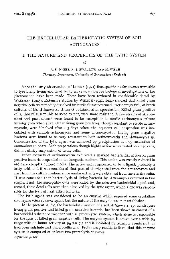
Fig. i. Lytic act ivi ty of Actinomyces culture filtrates against heat-killed Bact. lactis aerogenes at PH 7 .0
© O Lemco-peptone-glucose bro th [] . . . . . [] Lemco-peptone bro th ~ - - - A EVANS peptone glucose bro th
- - ¢ CZAPEK medium o . . . . . . o Mannitol synthet ic soil • • Glycerol-asparagine medium • • STANIER'S medium
T A B L E I
MEDIA FOR THE PRODUCTION OF THE Actinomyces LYTIC ENZYME SYSTEM
Synthetic media
I. STANIER'S (1942) inorganic medium (PH 7.0-7.3) 2. Mannitol synthet ic soil 3. GlyceroI-asparagine (CoNN and CONN 1941) 4" CzAPEK-sucrose-nitrate
Complex media
5. EVANS' peptone (2 %)-glucose bro th 6. LEMCO (I % )-peptone (I %) glucose bro th 7. LEMCO (I %)-peptone (I %)
After 15 and 35 days at 18 ° , cultures in each medium were filtered and the filtrates examined for lytic activity against a substrate of heat-killed, gram negative cells (Bact. laces aerogenes) at PH 7 .0 according to the method previously described (JoNEs, STACEY, AND WEBB 1948 ). The suspensions of the cells and enzyme were incubated at 37 ° in corked tubes with toluene (o.I ml). After 48 hours, the corks
were removed, the toluene allowed to evaporate, and the degree of lysis determined after a further 18 hours by comparison with a standard opacity scale. The lytic activities of the 35 day culture filtrates against the gram negative cells are shown in Fig. I. The 15 day culture filtrates possessed similar properties, though the enzyme concentration, particularly in each of the synthetic media, was considerably less. The results (Fig. I) revealed that the media most favourable for the production of the lytic enzyme system
References p. i32.
VOL. 2 ( I948) BACTERIOLYTIC SYSTEM OF SOIL ACTINOMYCES I 169
were complex and contained proteins and peptones. Such media also supported a more vigorous growth of the Actinomyces sp. Of the synthetic media, mannitol synthetic-soil was most favourable for the production of the enzyme system and for the growth of the organism. This medium was, in general, employed for the production of the enzyme system for chemical studies.
Lytic Activity o/oth,'r Actinomycetes
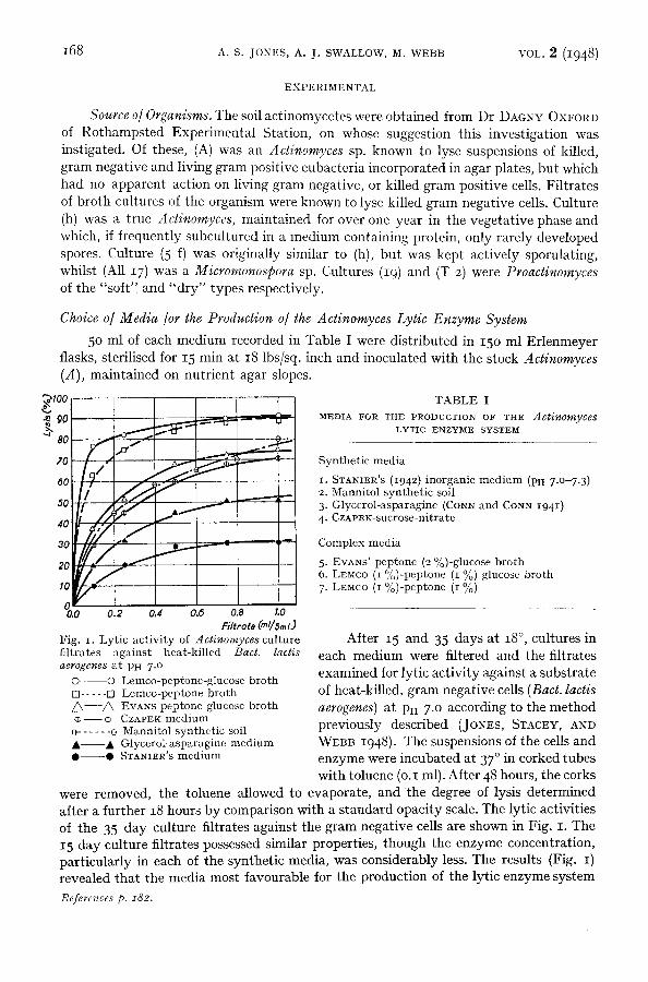
The non-sporulating (h) and sporulating (5 f) Actinomyces, Micromonospora (All I7) and Proactinomyces (19 and T 2) were inoculated into flasks of mannitol synthetic soil medium (50 ml). In these cultures (h) grew mainly throughout the medium with the production of a yellow pigment. However, possibly owing to the absence of protein from the culture medium, some surface growth, with the production of spores, occurred. The sporulating form (5 f) grew entirely at the surface of 9100 the medium with the formation of a light brown pig- ~. 9o ment. The remaining strains gave rise to submerged growth and produced little or no pigment.
The cultures were filtered after 5o days at 18 ° and the filtrates examined for lytic activity against heat-killed Bact. laces aerogenes at Pu 7 .0 according to the standard procedure. Filtrates of cultures of the Proactinomyces (19 and T 2) were without lytic activity against the killed gram negative cells while that of the Micromonospora sp. (All 17) was without activity in low concentrations. The sporulating Actinomyces (5 f) culture filtrate had considerably greater activity than the filtrate of the non-sporulating form (h) (Fig. 2).
As it has been reported (GoRYUNOVA 1944) that the lytic enzyme does not appear in broth cultures until the actinomyces have reached the sporulating stage, the following experiments, in which spore formation was respectively stimulated and reduced, were devised.
80
70
50
50
40
f /
3o / f
1o
oYT 0.0 0.2 0.4 0.~ 0.8
Filtl'tTte (=l/sin/)
Fig. 2. Lytic act ivi ty of noa-sporu- lat ing (e O) and sporulating (0 O) Actinomyces culture fil- t ra tes against heat-killed Bact. lactis aerogenes at PH 7 .o
Lytie Enzyme Production in Actinomyces cultures with Restricted Air Supply
700 ml of mannitol synthetic soil medium were placed in each of two 750 ml Erlen- meyer flasks, sterilised and inoculated immediately after cooling with Actinomyces (A). One culture flask was covered with a rubber cap which was then sealed with paraffin wax. The culture with the restricted air supply (A 22) was free from pigment, whereas the second culture (A 23) produced a light brown pigment which diffused into the medium. After 30 days at 18 ° , the cultures were filtered and increasing concentrations of the filtrates examined for lytic activity against killed Bact. lactis aerogenes at Pa 7.0. Solution (A 22) exhibited no action against the gram negative cells, whereas solution A 23 (0.5 ml) lysed a suspension of the killed cells (5 ml), of opacity corresponding to No. IO on MACFARLAND'S standard barium sulphate opacity scale, to the extent of 50 %.
Lytic Enzyme Production in Aerated Actinomyces Cultures
Actinomyces (A) was inoculated into each of two 3 1 bolt-head flasks containing man- References p. 182.

170 A.S . JONES, A. J. SWALLOW, M. WEBB VOL. 2 (1948)
, I I i - 9ol - ~ . . . . . r . . . . } . . . . . . t - - - F
4 0 t ~ J - 5 0 . . . . . i - - - J -
;;Wd ,og; o L ! 1 i I o.o o.¢ 0.2 o.a 0.4 0.5
F//trote (ml/sm!) Fig. 3. Lytic act ivi ty of aerated (O ©) and sta- t ionary (¢ ¢) Acti- nomyces culture filtrates against heat-killed Bact. lactis aerogenes at PH 7 .0
nitol synthetic-soil medium (1.5 1). One of the cultures was continuously aerated with sterile air to prevent a stationary surface and spore formation. The second culture was
allowed to stand undisturbed, when surface growtb with the production of spores took place. After 28 days at room tem- perature the cultures were centrifuged and the supernatants decanted. Determination of the lyric activity of these solu- tions against killed Bact. lactis aerogenes showed that the filtrate from the aerated culture contained the greater enzyme concentration (Fig. 3).
From these results it was concluded that the production of the lyric enzyme was dependent upon the strain of acti- nomyces and upon the free access of air, but not upon spore formation. Accordingly, all subsequent cultures were grown in shallow layers of the medium in penicillin pans. I t is to be noted that WELSCH (1941) in his studies on the actinomyces lyric system, found that the best results were obtained when the organism was cultivated in very shallow layers of ordinary broth.
Enzyme Production with time o/ Growth o/ the Actinomyees
Aliquot fractions of a culture of Actinomyces (A) in man- nitol synthetic soil medium at 18 ° were withdrawn under sterile conditions at weekly intervals, filtered and the filtrates (0.05, O.l-O.5 ml) examined for lyric activity against killed Bact. lactis aerogenes at PH 7 .0 under identical conditions. From the corresponding lysis-concentration curves thus obtained, the mean lyric activity of o.15 ml culture filtrate was determined for each interval (Fig. 4) shows that the production of the enzyme at first increases with the time of growth, becomes stationary and then after 7-8 weaks again increases. I t is considered that this increase in lytic activity may be in part due to autolysis of the Actinomyces, with the liberation of proteolytic enzymes, occurring when the culture reaches a certain age. Such proteinases, as for example those of the staphylococcus autolytic system (JONES, STACEY, AND WEBB 1948 ) are not species specific in their action and readily lyse killed Bact. lactis aerogenes.
In order to avoid the possible contami- nation of the exoeellular bacteriolytic system with the endocellular autolytic system, preparations of the former were made from 30-35 day cultures.
of time. The curve expressing these results
~9o - - - r - -
~ ~o
70 . . . . . .
"~ 60 - * - -
50 , - - -
- I
0 i 0 1 2 3 4 5 6 7 8 9 I0 11
hine (weeks) of 9rowth of act[nomyces culture
Fig. 4. Lyric enzyme production with t ime of growth of Actinornyces culture
The remarkable stability of the lytic enzymes of Actinomyces sp. is evidenced by the fact that cultures in both synthetic and complex media possessed considerable lyric activity against killed gram negative cells after 6 months or I year at room temperature.
References p. z82.

VOL. 2 (1948) BACTERIOLYTIC SYSTEM OF SOIL ACTINOMYCES I 171
Concentration o] the Actinomyces Lytic System
In preliminary experiments to ascertain the best procedure, 2 1 of a 35 day culture of Actinomyces (A) in Lemco-peptone medium were filtered through paper and the filtrate (A 2) fractionated as follows:
(I). The filtrate (650 ml) was saturated with solid ammonium sulphate and the resulting precipitate collected (filtration) after 15 minutes. A solution of the precipitate in distilled water (50 ml) was dialysed against tap-water until free from ammonium sulphate, centrifuged and the clear supernatant (A 3) diluted to IOO ml.
(II). Saturated .ammonium sulphate solution (3 1) adjusted to PH 8 with ammonium hydroxide was added to the culture filtrate (600 ml) (Final concentration, 0.8 saturation of ammonium sulphate). The precipitate was collected (filtration), dissolved in distilled water and dialysed. After centrifuging free from the insoluble material the clear solu- tion (A 4) was diluted to 92 ml.
/ / 5O
-- t / / / ~ 40 ~
20 ~ ~
lO f i
,, i -0.0 0.I 0.2 o.a 0.4
Enzyme soln (ml/~ml) Fig. 5. Lytic activities of Actinomyces cul- ture filtrate (A 2) and fractions separated therefrom by saturation with (NH4)2SO 4 (A 3), 0.8 saturation with (NH4)2SOa (A 4) and by ethanol precipitation (A 5)
,~ ~ 70
~ 40 - - ~
./,'z , /
oY t~O O.l 0.2 0.3 0.4
Enzyme *o/n (ml/sml.~
Fig. 6. Ly t ic ac t iv i t i es of Actinomyces e n z y m e s p r epa ra t i ons shown in Fig. 5 a f t e r s t a n d i n g for 5 ° days a t r o o m t em- pe r a tu r e
(III). Absolute ethanol (250 ml) cooled to o °, was added to the culture filtrate (250 ml). No precipitation occurred, and after 15 minutes at o ° a further 250 ml ethanol were added. The resulting precipitate was then separated, dialysed and the final volume of the crude enzyme solution (A 5) adjusted to 32 ml.
Thus the dilutions of A 3, A 4, and A 5 corresponded to 6.5 fold concentration of the initial filtrate (h 2).
The preparations were examined for Iytic activity against heat-killed Bact. lactis aerogenes immediately after isolation and again after standing for 50 days at room tem- perature. The results (Fig. 5) show that the enzyme was more completely precipitated by saturation with solid ammonium sulphate (A 3) than at 0.8 saturation of the salt (A 4) or with ethanol (A.5) and indicate that the enzyme(s) is of relatively small mole- cular size. The activity of A 3 was, however, relatively less than the activity of the initial filtrate (A 2) indicating that some inactivation of the enzyme occurred on fractionation. Activities of the preparations decreased on standing at room temperature and the form References p. I82. 12
~ a o I - ' -i I ~ 2... -I--- -- - - -
; ; - L
i/'i . . . . 1 -
';I:%_ 4.0 5.0 0.0 ZO 8.0 9.0 I0.0
Fig. 7. Optimum PH for the lysis of heat-killed 13acl. lactis aerogenes by the Acli- nomyces lyric enzyme system
]772 A.S. JONES, A. J. SWALLOW, M. WEBB VOL. 2 (I948)
of the concentration-activity curves (Fig. 6) would suggest that the lyric system consists of more than one enzyme, one of which is more stable than the other.
Although active against killed gram negative cells these preparations were without activity against heat-killed gram positive cells.
Large Scale Fractionalion o/Cul ture Filtrates
The Actinomyces (A) culture in complex Lemco-peptone medium or mannitol synthetic soil was filtered and the filtrate saturated with solid ammonium sulphate. The resulting precipitate collected at the liquid surface and was concentrated by means of a large tap-funnel. The suspension was then transferred to hardened paper on a Buchner funnel and filtered with suction. The solid was dissolved in distilled water and the solution dialysed against running tap-water for 18 hours. After centrifuging from the small inactive precipitate the clear solution was diluted to 50 ml for each litre of initial culture filtrate.
Refractionation of this solution by the addition of ammonium sulphate to 0.5 and I.O saturation yielded precipitates which possessed approximately the same lytic activity against heat-killed Bact. lactis aerogenes, and accordingly, in this preliminary investigation, concentration of the enzyme was not extended further than the initial precipitation by saturation with ammonium sulphate.
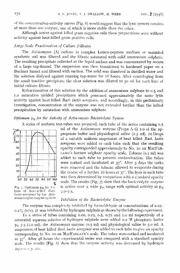
op t i m um Pi~ /or the Activity o /Act inomyces Bacteriolytic System
A series of uniform test-tubes was prepared, each tube of the series containing 0.5 ml of the Actinomyces enzyme (Prepn A 6) 2.0 of the ap- propriate buffer and physiological saline (2.5 ml). IO Drops of a suitable uniform suspension of heat killed Bact. lactis aerogenes were added to each tube such that the resulting opacity corresponded approximately to No. IO on MACFAR- LAND'S barium sulphate opacity scale. Toluene (o.I ml) was added to each tube to prevent contamination. The tubes were corked and incubated at 37 °. After 3 days the corks were removed and the toluene allowed to evaporate during the course of a further 16 hours at 37 °. The lysis in each tube was then deternfined by comparison with a standard opacity scale. The results (Fig. 7) show that the bacteriolytic enzyme is active over a wide Pn range with optimal activity at PH
7.O-7.5.
Inhibition o/ the Bacteriolytic Enzyme
The enzyme was completely inhibited by formaldehyde at concentrations of o.o5- o.1% (v/v). I t was inhibited by hydrogen sulphideas shown by the following experiment.
To a series of tubes containing o.oo, 0.25, 0.5, 0.75 and I.O ml respectively of a saturated aqueous solution of hydrogen sulphide were added 0.2 M phosphate buffer PH 7.5 (i.o ml), the Actinomyces enzyme (o.5 nil) and physiological saline to 5.0 ml. A suspension of heat-killed Bact. lactis aerogenes was added to each tube to give an opacity corresponding to No. IO on MACFARLAND'S scale. The tubes were corked and incubated at 37 °. After 48 hours the experimental series was compared with a standard opacity scale. The results (Fig. 8) show that the enzyme activity was decreased by hydrogen
R@:r<,:~ccs p. ±b>2.
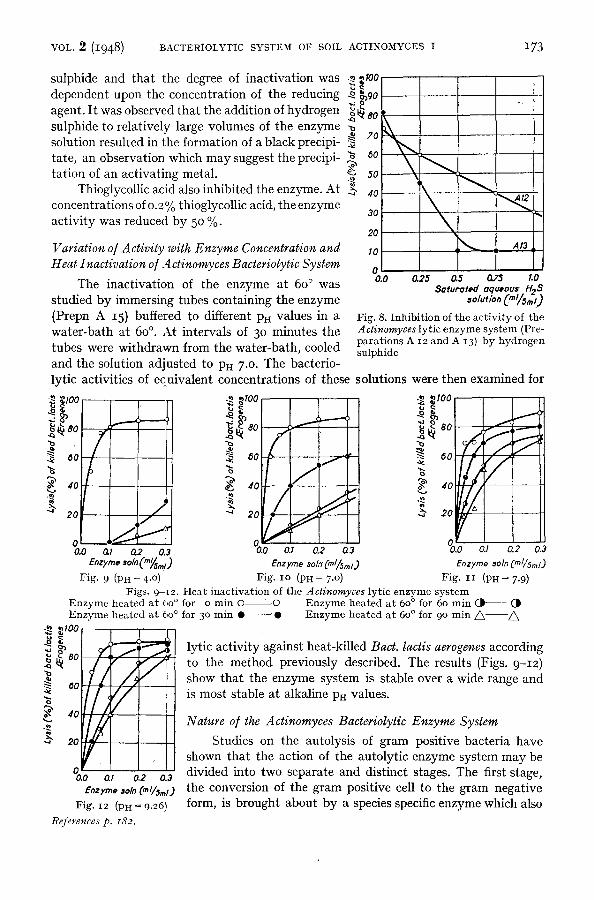
VOL. 2 (I948) BACTERIOLYTIC SYSTEM OF SOIL ACTINOMYCES I 173
sulphide and that the degree of inactivation was dependent upon the concentration of the reducing agent. I t was observed that the addition of hydrogen sulphide to relatively large volumes of the enzyme solution resulted in the formation of a black precipi- tate, an observation which may suggest the precipi- tation of an activating metal.
Thioglycollic acid also inhibited the enzyme. At concentrations of 0.2% thioglycollic acid, the enzyme activity was reduced by 50 %.
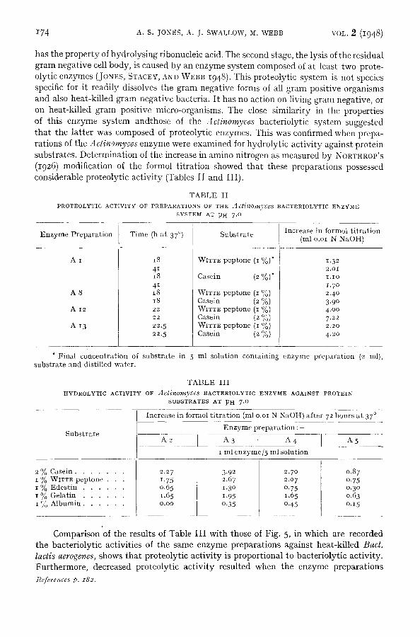
Variation o/A ctivity with Enzyme Concentration and Heat InactivaEon o/Actinomyces Bacteriolytic System
The inactivatk,n of the enzyme at 6o ° was studied by immersing tubes containing the enzyme (Prepn A 15) buffered to different p~ values in a water-bath at 60 ° . At intervals of 30 minutes the
.~. ~I00
po
"~ 80
50 x
3O
20
I0 ,
0 0.0 0.25 0.5 0.75 1.0
Seturoted equeous H~3
Fig. 8. Inhibi t ion of the act ivi ty of the A ctinomyces lytic enzyme sys tem (Pre- para t ions A 12 and A 13) by hydrogen
tubes were withdrawn from the water-bath, cooled sulphide
and the solution adjusted to P}t 7 .0. The bacterio- lyric activities of equivalent concentrations of these solutions were then examined for
80 80 ~ 80
~. oo ~ 8o / ~ 8o
40 40 / I , ,
~" 20 20 "~ 20 r = "
°o.o 02 o., 02 % o., 03 e.zrmo ,o/0(,.~,.~) E, zy,~e *oln" (ml/Sml.) Enzyme *•In (ml/5,~/.)
Fig. 9 (PH ~ 4 .°) Fig. IO (PH = 7 .o) Fig. IT (PH = 7.9) Figs. 9-[2. Heat inact ivat ion of the Actinomyces lytic enzyme sys tem
Enzyme heated a t 60 ° for o min O O Enzyme heated a t 60 ° for 60 min ( . | (J~ Enzyme heated a t 60 ° for 3 ° min • "0 Enzyme heated a t 60 ° for 90 m i n / ~ /~
~ ,~ ~ 80 ! . ~ ]ytic activity against heat-killed Bact. lactis aerogenes according ~ I / / / ~ t° the meth°d previ°usly described" The results (Figs" 9-I2)
show that the enzyme system is stable over a wide range and =o[////,1 is most stable at alkaline pH values.
~ 4 ! ~ ~ l
2 Studies on the autolysis of grain positive bacteria have shown that the action of the autolytic enzyme system may be
0.0 al 0.2 0.3 divided into two separate and distinct stages. The first stage, Enzrr~, ,oln (ml/5,. D the conversion of the gram positive cell to the gram negative
Fig. I2 (PH = 9.26) f o r m , is brought about by a species specific enzyme which also References p. z82.
I74 A.S. JONES, A. J. SWALLOW, M. WEBB VOL. 2 (1948)
has the p r o p e r t y of hydro lys ing r ibonucleic acid. The second stage, the lysis of the res idual g ram negat ive cell body, is caused by an enzyme sys tem composed of a t least two prote- olyt ic enzymes (JoNEs, Sr.aCEY, aND WEBB I948 ). This pro teo ly t ic sys tem is not species specific for i t readi ly dissolves the g ram negat ive forms of all g ram posi t ive organisms and also hea t -k i l led g ram negat ive bacter ia . I t has no act ion on living gram negative, or on hea t -k i l led g ram posi t ive micro-organisms. The close s imi la r i ty in the proper t ies of this enzyme sys tem andthose of the Actinomyces bac te r io ly t ic sys tem suggested t ha t the l a t t e r was composed of pro teo ly t ic enzymes. This was confirmed when prepa- ra t ions of the Aclinomyces enzyme were examined for hydro ly t i c ac t iv i ty agains t pro te in subst ra tes . De te rmina t ion of the increase in amino ni t rogen as measured by NORTHROP'S (1926) modif icat ion of the formol t i t r a t ion showed tha t these p repa ra t ions possessed considerable pro teo ly t ic ac t iv i ty (Tables I I and III).
TABLE II P R O T E O L Y T I C A C T I V I T Y O F P R E P A R A T I O N S O F T H E Actinomyces B A C T E R I O L Y T I C E N Z Y M E
S Y S T E M A T P H 7 .0
Increase in Iormol titration Enzyme Preparation Time (11 at 37 °) Substrate (ml o.oi N NaOH)
A I WIT:re peptone (I %)*
A 8
A I 2
A I 3
18 41 i8 4 I 18 18 2 2
2 2
22. 5 22.5
Casein (2 %)'~
WITTR peptone (i %) Casein (2 %) WIT:rE peptone (i %) Casein (2 %) WlTTE peptone (I %) Casein (2 %)
1 . 3 2 2.oi i . io 1.7 ° 2.40 3.9 ° 4.oo 7.22 2 . 2 0
4.20
* Final concentration of substrate in 5 ml solution containing enzyme preparation (2 ml), substrate and distilled water.
TABLE III H Y D R O L Y T I C A C T I V I T Y O F Actinomycgs B A C : r E R I O L Y T I C E N Z Y M E A G A I N S T P R O T E I N
S U B S T R A T E S A T PH 7 . 0
Substrate
_Increase in formoi titration (ml o.oi N NaOH) afte_r 72 hours at 37 °
] Enzyme preparation : -
i A5 A3 A4 I ml enzyme/5 ml solution
2 % Casein . . . . . . . 1 % WITTE peptone . . . i % Edestin . . . . . . 1% Gelatin . . . . . . i % Albumin . . . . . .
2,27 3.92 1.75 2.67 °'65 i 1"3° t .65 I 1.95
_ _ 0 . 7 _~ o.35
2.7o 2.o 7 0.75 1.65 0.45
0.87 o.75 o.3o o.63 o.15
Comparison of the results of Table I I I wi th those of Fig. 5, in which are recorded the bac te r io ly t ic act ivi t ies of the same enzyme prepara t ions agains t hea t -k i l led Bact. lactis aerogenes, shows t ha t p ro teo ly t ic ac t iv i ty is p ropor t iona l to bac te r io ly t i c ac t iv i ty . Fur the rmore , decreased pro teo ly t ic ac t iv i ty resul ted when the enzyme p repa ra t ions
]~cferences p. x82.

VOL. 2 (1948) BACTERIOLYTIC SYSTEM OF SOIL ACTINOMYCES I I75
(2 ml) in physiological saline (i ml) and 0.2 M phospha te buffer (I ml) were allowed to s t a n d for 18 hours at room t e m p e r a t u r e wi th I.O ml of a s a t u r a t e d aqueous solut ion of hydrogen sulphide (Table IV). I t has previous ly been shown (Fig. 8) t ha t the bac ter io- ly t ic ac t i v i t y of the enzyme prepara t ions is decreased b y hydrogen sulphide.
TABLE IV INHIBITION OF PROTEOLYTIC ACTIVITY OF dctinomyces LYTIC ENZYME SYSTEM WITH
HYDROGEN SULPHIDE
Enzyme Preparation
A 7 A 8 A I 2 A I 3
Time (H at 37 °)
66 41 2O
2 0
Increase in Formol Titration (ml o.oi N NaOH)
alone t Enzyme with hydrogen sulphide Enzyme
Substrate
2 % Casein
2.85 6.15 6.37 3.75
I ~o W I T T E p e p t o n e
1.45 3.15 2.30 1.6o
o, Casein 2 , ' 0
O.OO
3.3 ° 2.98 0.57
i % V¢ITTE peptone
o.55 2.3o i .5 ° 1.42
F u r t h e r correla t ion of the bac te r io ly t ic and pro teo ly t ic act ivi t ies of the A ctinomyces ly t ic sys tem was p rov ided b y the inac t iva t ion studies. 2 ml of the enzyme p repa ra t ions (A 15) buffered to PH 7 and hea ted a t 60 ° for the given t ime intervals , were examined b y the Fo rmo l t i t r a t ion for hyd ro ly t i c ac t iv i ty agains t pro te in subs t ra tes . The resul ts (Table V) show tha t hea t inac t iva t ion caused a decrease of p ro teo ly t ic ac t iv i ty paral le l to the decrease in bac te r io ly t ic ac t iv i ty .
TABLE V HEAT INACTIVATION OF THE PROTEoLYTIC ENZYMES OF THE dctinomyces LYTIC SYSTEM
(PREPARATION A 15) AT PH 7 .0
Substrate
2 % Casein . . . . . . . I ~o WITTE peptone . . .
o/ Edestin I /O . . . . . .
1% Gelatin . . . . . .
Time (h at 37 °)
23 23 51 23 51 23 51
Activity (increase in Formol titration ml o.oi 1% NaOH) of enzyme solution heated at 60 ° for
o min 3 ° mill
2.85 I.IO 1.75 0.78 1.13 1.65 2 . 0 0
I.O 5 0.60 I .OO
0.33 0.53 0.65 o.9o
60 min
o.78 o.4o 0.90 o.25 o.4o O.50
O .70
90 rnin
0.58 0.36 0.76 0 . 2 0
0.35 0.40 0.58
The resul ts of Table IV show tha t the ra t io : hydro lys i s of casein: hydro lys i s of pep tone for each enzyme p repa ra t ion was cons iderably a l tered b y the presence of hy- drogen sulphide. An a l te ra t ion in this ra t io from 1.81 immed ia t e ly af ter the p repa ra t ion of the enzyme (A 12) to 2.77 af ter 15 days also occurred when the solut ion was al lowed to s t and a t room tempera tu re .
Since the ra t io of the hydro lys i s of one subs t r a t e to the ra te of hydro lys i s of a second subs t ra te is cons tan t for a pa r t i cu la r enzyme under s t a n d a r d condi t ions (IRVING, FRtl- TON, AND BERGMANN 1941 ) these resul ts indicate t ha t the Actinomyces bac te r io ly t i c sys tem is composed of a t least two pro teo ly t ic enzymes. I t was found possible to a l te r References p. 182.
176 A.S. JONES, A. J. SWALLOW, M. WEBB VOL. 2 (1948)
considerably the ra t io of the enzymes b y f rac t ionat ion on foam according to the me thod of SCHOTZ (I937), bu t comple te sepa ra t ion was not achieved.
As a p re l iminary exper iment to de te rmine the s tab i l i ty of the enzyme on foam, a cur rent of n i t rogen was passed th rough the enzyme solut ion (prepara t ion A 19) for 18 hours. The ra te of the ni t rogen flow was ad jus t ed such t ha t foam was p roduced bu t did not pass into the receiver. Compar ison of the p ro teo ly t i c act ivi t ies of the ini t ia l solut ion (A 19, 0. 7 ml in 5 ml solution) and of the solut ion remain ing af ter foaming (A 19 a, 0. 7 ml in 5 ml solution) showed tha t no inac t iva t ion of the enzyme occurred on the foam (Table VI). The ra te of the n i t rogen flow was then increased unt i l foam was col- lected in the receiver. Af ter 2 hours the foam fract ion (A 19 b, 0.7 ml in 5 ml solution) and the residual solut ion (A 19 c, 0.7 ml in 5 ml solution) were examined for hydro ly t i c ac t iv i ty agains t casein and WITTE peptone. The a l te ra t ion in the ra t io of the hydro lys i s of the two subs t ra tes achieved b y this procedure is shown in Table VI. Ex tens ion of this me thod of separa t ion is being inves t iga ted further .
TABLE VI P A R T I A L S E P A R A T I O N O F T H E P R O T E O L Y T I C E N Z Y M E S O F T H E Actinon*yces B A C T E R I O L Y T I C
Enzyme Solution
A 1 9 . . . . . .
A I9a . . . . . A I9b . . . . . A i9c . . . . .
S Y S T E M O N F O A M
Increase in Formol titration (inl o.oi N NaOH) after 2~ hours at 37 °
2 % Casein Substrate
1.58 1.65 2.30 1.45
o/ W I T T E Peptone S u b s t r a t e I /o
I .OO
0.95 o.83 o.6o
Casein activity Ratio :
Peptone activity
1.58 1.74 2.77 2.42
Proteolytic Activity o/ Culture filtrates o/other Actinomycetes
Fi l t r a t e s of cul tures of the sporu la t ing (5 f) and non-sporula t ing (h) Actinomyces, Micromonospora sp. (All 17) and Proactinomyces (19 and T 2) in the manni to l - syn the t i c soil med ium which were examined for lyric ac t iv i ty against ki l led Bact. lactis aerogenes (p. I69), were also examined for hydro ly t i c ac t iv i ty agains t casein and WITTE peptone. The resul ts (Table vii) are of considerable in teres t as the Proactinomyces cul ture fi l trates, which exh ib i t ed no lyric ac t iv i ty agains t kil led g ram negat ive cells, were able to hy- drolyse peptone bu t not casein. By ana logy with the pro teoly t ic enzymes of the s tap-
TABLE VII P R O T E O L Y T I C A C T I V I T I E S O F C U L T U R E F I L T R A T E S O F O T H E R Acti~o~yce~es
Culture filtrate of
Actinomyces (h) . . . . . . . Actinomyces (5 f) . . . . . . Micromonospora (All 17) . . . Proactinomyces (I9) . . . . . Proactinomyces (T 2) . . . . .
Increase in formol titration (ml o.oi N NaOII) due to culture filtrate (2.o ml in 5 ml sohltion) after 48 hours at 37 °
. o/ Casein Substrate
0.90 0 . 7 0 0 , 2 0
O.OO
O.OO
I C~o W I T T E peptone S u b s t r a t e
o.81 o.4o o.31 O . I 5
0 . 5 2
References p. z82.
VOL. 2 (I948) BACTERIOLYTIC SYSTEM OF SOIL ACTINOMYCES I 177
hylococcus autolytic system it would therefore appear that the hydrolysis of casein on the one hand, and of peptone on the other, is due to two separate and distinct enzymes. The enzyme which hydrolyses casein is responsible for the initial disintegration of the killed, gram negative cells and the enzyme which hydrolyses peptone, and which itself has no action on the killed cells, is responsible for the further hydrolysis of the split products resulting from the action of the casein hydrolysing enzyme. The complete separation of these activities, a problem also of considerable importance in studies of the autolytic enzyme systems, is at present under investigation.
Action o~ the Actinomyces Lytic Enzyme System on the Culture Medium
As the media most favourable for the production of the Actinomyces lytic system contained proteins and peptides, it was of interest to determine whether the isolated system was able to hydrolyse these complex molecules into smaller units capable of being utilised by the cell. It was established that the enzyme system, concentrated by precipitation with ammonium sulphate, hydrolysed the proteins of the growth medium for when the enzyme preparation (A 27, 2.0 ml) was incubated at 37 ° with the Lemco peptone medium (I.O ml) diluted with distilled water (2.0 ml), an increase in the Formol titration value of 1.o5 ml o.oi N NaOH was obtained.
In a further experiment, the enzyme (A 27) and the Lemco peptone medium were dialysed for 72 horn's against running tap water. Each solution was analysed for total nitrogen by the micro KJELDAItL method and the enzyme (50 ml) added to the dialysed medium (ioo ml). The solution was incubated at 37 ° for 48 hours and then dialysed through cellophane in the concentrating dialyser. The results (Table viii) show that during this period ~2. 9 % of the total nitrogen of the substrate had been rendered dialysable.
TABLE v n I ACTION OF THE Actinomyces EACTERIOLYTIC SYSTEM (PREPARATION A 27) ON LEMCO~
PEPTONE CULTURE MEDIA
Total nitrogen (Micro ](jeldahl) of: ioo ml dialysed culture medium . . . . . . . 86.34 mg 5 ° ml dialysed enzyme solution . . . . . . . 3.71 mg
Systcm (enzyme + substrate) . . . . . . . . 90.o 5 ml
After 48 hours at 37 °, ~:otal nitrogen of: dialysed material . . . . . . . . . . . . . . 71.6o mg non-dialysable residue . . . . . . . . . . . . 18.68 mg
System (dialysate + residue) . . . . . . . . . 9o.28 mg
During the routine examination of actinomyces culture filtrates and the concen- trated enzyme preparations for lytic activity against killed cells, it was found that a culture filtrate and the corresponding enzyme preparation of a 35 day culture of the organism in Lemco-peptone medium were capable of lysing heat-killed gram positive Staph. albus (9238) as well as the killed gram negative organisms. The concentrated enzyme solutions (I ml/5 ml) completely lysed suspensions of heat-killed gram positive Sarcinae, Micrococci, Streptococcus/aecalis and Staphylococcus aureus (3661). At lower concentrations partial lysis was obtained (Fig. 13).
The experimental procedure was as follows: Increasing concentrations (see Fig. 13) of the enzyme preparation and 0.2 M phosphate buffer PH 7 .0 (I.0 ml) were added to a References p. z82.

178 A . s . JONES, A. J. SWALLOW, M. WEBB VOL. 2 (i948)
~ 90 ~o
70
oo
5o
40
30
2o
1o
o 0.0 0.25 0.50 0.75
Enzyme (ml/St, i. )
Fig. 13. Lysis of heat-killed, gram positive micro-organisms by the Actinomyces lytic system from 35 day ettltur~s in Lemco peptone
medium 0 0 Sarcina sp.
Z~ Staph. aureus (3661)
series of uniform test tubes. The total wflume in each tube was adjusted to 5.0 ml with physiological saline. io Drops of a uniform suspension of the heat killed, gram positive organism were added to each tube to- gether with toluene (o.I ml). The tubes were corked and incubated at 37 °. After 3 days the corks were removed, the toluene allowed to evaporate during the course of a further 16 hours and the degree of lysis in each tube determined by comparison with a standard opacity scale.
Examination of further filtrates of Lemco-peptone cultures of Actinomyces (A) showed that as the age of the culture increased the activity of the filtrate against gram positives decreased. Thus, a 43 day culture tilt rate produced only slight lysis of suspensions of heat- killed Staph. albus at PH 7 .0 and a 50 day culture filtrate failed to produce any lysis in suspensions of the killed gram positive organisms.
In view of the studies on the bacterial autolytic enzyme systems (JONES, STACEY, AND WEBB, 1948 ) and of the present demonstration that the proteolytic
enzymes of Actinomyces culture filtrate were capable of lysing gram negative organisms, it appeared that this activity against killed gram positives was due to the presence in the 35 day culture filtrate of a nucleinase capable of converting the killed gram positive organisms to the gram negative state. In this condition the cells would be readily lysed by the proteolytic enzyme system.
The existence of a nucleinase in such enzyme preparations was confirmed by following the hydrolysis of ribonucleic acid produced by the enzyme by the method of DAVlDSON AND WAYMOUTH (1944). The bacteriolytic system from 35 day cultures of Actinomyces (A) in Lemco peptone medium exhibited a relatively strong ribonucleinase activity (Fig. 14). This enzyme activity was considerably reduced in 43 day cultures and was not present in 50 day cultures. The
"~ .~, 7o decrease in ribonucleinase activity, with con- ~ ~,~ sequent decrease in lytic activity against heat "-- . . . . - - .~. killed gram positive cells, may possibly be ~ ~ 0 f / due to the digestion of the nucleinase by ~.~4o the enzymes of the proteolytic system. In s0 / , , ~ support of this is the experimental finding , I , that gram positive cells rendered gram nega- 2o / tive by the action of ribonucleinase were 1 o / completely lysed by the subsequent addition o t "
o.o to 3.o 5,o 8.o Io.o of the A ctinomyces proteolytic enzyme sys- r;me in hour, tern, but only slight lysis was obtained when , Fig. 14. Hydrolysis of ribonucleic acid by the these enzymes were added together to the Actinomyces lytic enzyme system from 35 day
cultures in Lemco-peptone medium suspension of heat-killed, gram positive cells.
Nucleinase activity could only be demonstrated in Actinomyces (A) cultures in Lemco peptone medium, Filtrates from 35 day cultures in synthetic media were without ]¢ffereuces p. I82.

VOL. 2 (1948) BACTERIOLYTIC SYSTEM OF SOIL ACTINOMYCES I 179
action upon heat killed, gram positive cells. I t was at first considered that the production of the nucleinase in the complex was due to the presence of ribonucleic acid in the con- stituents of the medium. The presence of ribonucleic acid in either Lab-Lemco or com- mercial peptones could not, however, be established. Furthermore, the production of ribonucleinase activity by A ctinomyces (A) could not be induced by continued subculture of the organism in STANIER'S (1942) inorganic salt medium containing, in addition, 0.7 % sodium ribonucleate. Filtrates from 35 day cultures of Actinomyces (A) in this medium exhibited considerable lytic activity against killed gram negative cells, but had no action on heat killed gram positive organisms.
ACTION OF Actinomyces CULTURE FILTRATES ON LIVING GRAM POSITIVE CELLS
Actinomyces (A) plated onto agar containing living gram positive organisms was known to bring about the lysis of the these cells. Sterile filtrates of cultures of A ctinomyces (A) in either complex or synthetic media were also found to lyse living gram positive cells, as shown by the following representative experiments, but had no action on living gram negative cells.
IO Drops of a uniform suspension of the cells from a 24 hour culture of Staph. citreus (B 9) on peptone-agar were added to each of a series of uni- form tubes containing increasing concentrations (o.o-I.O ml) of the Actinomyces culture filtrate (A 26), 0.2 M phosphate buffer PH 7.0 (I.O ml) and physiological saline r.o 5.o ml. The tubes were corked and incubated at 37 °. After 3 days the degree of lysis in each tube was estimated by comparison with a standard opacity scale. The results (Fig. 15) show that the clearing of the bacterial suspension in the presence of the Actinomyces culture filtrate was considerably in excess of that due to the spontaneous autolysis of the cells occurring at zero concentration of the culture filtrate.
Since lysis of the cell suspensions in excess of that due to autolysis was not observed in sus-
100
• ~. 90
.4 80
° S ~0
50
40
f f
30
20
I0
0 0.0 0.25 0.50 0.75
F//trate(ml/'Srn I ) Fig . 15. L y s i s of l i v i n g g r a m p o s i t i v e ce l l s (Staph. citreus B 9) b y A ctinomyces
c u l t u r e f i l t r a t e (A 26) a t PH 7 .0
pensions of living Staph. citreus containing the dctinomyces system concentrated by ammonium sulphate precipitation, it was concluded that some additional component
T A B L E I X
B A C T E R I O S T A T I C A C T I V [ T I E S O F E T H E R E X T R A C T S O F dctinomyces C E L L S A N D C U L T U R E F I L T R A T E S
A G A I N S T Staph. a u r e u s I N G L U C O S E P E P T O N E B R O T I ~ ( S E R I A L D I L U T I O N M E T H O D )
E t h e r E x t r a c t of
A ctinomyces ce l l s a n d m y c e l i u m , . . . . . . . . . . . . . Actinomyces c u l t u r e f i l t r a t e ( m a n n i t o l s y n t h e t i c so i l m e d i u m )
s a t u r a t e d w i t h (NH4)2SO 4 . . . . . . . . . . . . . . Actinorayces c u l t u r e f i l t r a t e ( L e m c o - p e p t o n e m e d i u m ) s a t u -
r a t e d w i t h (NH4)~SO 4 . . . . . . . . . . . . . . . .
B a c t e r i o s t a t i c A c t i v i t y a f t e r
2 4 h o u r s
I : 4000
I : IOOO
i : 2600
4.8 h o u r s
i : 4000
I : 500
i : 2 6 0 0
References p. z82.

180 A.S. JONES, A. J. SWALLOW, M. WEBB VOL. 2 (1948)
of the bac te r io ly t ic sys tem was present in cul ture fil trates. Fol lowing the me thod of WELSCH (1941) we were able to isolate, b y e ther ex t rac t ion of Actinomyces f i l t rates af ter sa tu ra t ion with ammonium sulphate or b y e ther ex t rac t ion of dr ied A ctinomyces, an an t ibac te r i a l substance which af ter removal of the solvent, formed a brown syrup and appea red to be a l ipoid or a f a t t y acid. Solutions of such ex t rac t s in o.o5 M phospha te buffer PH 8.2 were inact ive agains t g ram negat ive organisms, bu t were bac te r ios ta t ic (Table IX) and bac ter ic ida l (Table X) for g ram posi t ive organisms.
TABLE X BACTERICIDAL ACTIVITIES OF ETHER EXTRACTS OF Actinomyces CELLS AND CULTURE FILTRATES
AGAINST Slaph. a u r e u s ( R I D E A L - W A L K E R METHOD)
Ether Extract of
Actinomyces culture Actinomyces culture Period of filtrate (mannitol syn- filtrate (Lemco-
Incubation Actinomyces cells thetic soil medium) peptone medium) Dilution of Subcul- and mycelium of ether saturated with saturated with
tures at (NH4)2SO4 I (NH4)2SO4 extract 37 ° (h) Time (min) ether extract in contact with cells
; ,io 5 IO I 5 I 20 5 IO 15 20 [ 15 20
I 0 0
i 5oo
i 25o0
i 12500
24
24
24
24
+
+
+
+ +
+ +
+ +
r
+
+
+
+
+
+
+ +
+ +
+ +
- I + + t
+ + +
+ + +
+ +
+ +
- - no growth + growth in subcultures
Action o/the Actinomyces Bactericidal Fractions on Living gram positive Cells
Gram posi t ive organisms were inocula ted into nu t r ien t b ro th in steri le centr ifuge tubes and the cul tures centr i fuged af ter 18-24 hours at 37 ° . The cell deposi ts were suspended in steri le physiological saline (I ml) and incuba ted at 37 ° wi th an equal volume of a steri le 1 % solut ion of the Actinomyces bac te r ic ida l substance.
Af te r 3 days at 37 ° examina t ion of s ta ined smears p repared from the suspension revealed tha t in each case the cells were more or less comple te ly g ram negat ive (Table XI) .
I t was also observed t ha t this effect, namely the dea th of the organism followed by the change from gram posi t ive to g ram negat ive , occurred when l iving g ram posi t ive cells were incuba ted at 37 ° wi th solut ions of s t rep to thr ic in and s t r ep tomyc in (I ml / I ml), the an t ibac te r i a l subs tances p roduced b y Streptomyces lavendulae and Streptomyces griseus.
Since the change from gram posi t ive to g ram negat ive was not observed when suspensions of l iving cells conta in ing the A ctinomyces an t ibac te r ia l f ract ions were kep t a t o ° or 6o °, or when ki l led g ram posi t ive cells were incuba ted a t 37 ° with the act ive ext rac ts , it is concluded t ha t the change in the g ram staining react ion is not d i rec t ly due to the ac t ion of the bac ter ic ida l substance, bu t is a secondary effect, autolysis follow- ing the dea th of the cells. A similar conclusion has been reached to expla in the lysis which follows the act ion of tyroc id in on cer ta in bac te r ia l species (HEILMAN AN D HERRELL 1941; MANN, HEILMAN, AND HERRELL 1943).
References p. z82.

VOL. 9. (1948) BACTERIOLYTIC SYSTEM OF SOIL ACTINOMYCES I
T A B L E X I
A C T I O N O F E T H E R E X T R A C T S O F Actinomyces C E L L S A N D C U L T U R E F I L T R A T E S O N L I V I N G
G R A M P O S I T I V E C E L L S
I81
Organ i sm
Approx. Percen tage G r a m Nega t ive Cells
E t h e r Extract A ctinomyces
cells and mycelium
E t h e r E x t r a c t A clinomyces cul tu re fil-
trate (Manni- tol synthetic soil medium)*
E t h e r Extract dctinomyces cul ture f i l t ra te (Lemco-peptone
medium)*
Prepn I Prepn I I
Staph. citreus (B 9) •
Micrococcus 1o47o . .
Micrococcus 382 . . . .
C1. welchii . . . . . . .
9 °
80
IOO
IOO
90
l O O
6o
I O O
IOO
IOO
IOO
IOO
8 0 - 9 °
70-80
I O O
* Culture filtrates saturated with ammonium sulphate
Gram positive organisms, killed and rendered gram negative by the action by the bactericidal fractions from Actinomyces (A) (Table XI) were readily lysed by the sub- sequent addition of the Actinomyces bacteriolytic enzyme system. Furthermore, living gram positive cells were completely lysed after 3 days when incubated at 37 ° with a 1% solution of the bactericidal substance and the lytic enzyme system (I ml) at PH 7 .o.
DISCUSSION
In agreement with the work of WELSCH (1941, 1942 ) the foregoing experimental has shown that the bacteriolytic system of a soil A ctinomyces is composed of an anti- bacterial substance and a proteolytic enzyme system. The latter enzyme system lyses killed gram negative organisms and the gram negative forms of gram positives, but has no action on living gram positive or gram negative cells. The antibacterial substance has no action on living gram negative organisms, but gram positives are killed under con- ditions which are favourable for the action of the autolytic enzymes of the cell. The action of these enzymes results, in the first instance, in a change in the gram staining reaction of the cells which lherefore become susceptible to the action of the Actinomyces lytic enzyme system.
WELSCH (1941) was of the opinion that part of the antibacterial activity of Actino- myces culture filtrates was due to the presence in the medium of bactericidal fatty acids. Such substances have been isolated from the peptones which have been used in this work (WEBB, 1948 a) and have approximately the same bactericidal activity as the active fractious herein described. However, although gram positive organisms are killed by the action of these extracts, conditions are not favourable for the action of the auto- lytic enzymes and the cells do not become gram negative. Thus, such fatty acids in the complex culture media, though possibly increasing the bactericidal activity of Actino- myces cultures, do not themselves play any part in the bacteriolysis of living gram positive organisms.
Certain cultures of Actinomyces (A) in complex media contain a ribonucleinase and References p. z82.
182 A.S. JONES, A. J. SWALLOW, M. WEBB VOL. 2 (1948)
are able to lyse heat killed gram positive organisms. This enzyme is only produced in complex media, is relatively unstable and cannot be considered as forming part of the normal bacteriolytic system of the Aclinomyccs. WELSCrI (1941) has s ta ted that killed gram-posit ive cells, though more resistant, are to some extent susceptible to the Aciino- myces lyric system. I t may, however, be pointed out that , unless special precautions are taken, a variable percentage of gram negative cells result when gram positive bac- teria are killed by heat (WEBI~ 1948 b) and these are dissolved by proteolytic enzymes.
The bacteriolytic system herein described appears closely similar to tha t of myxo- cocci which has been shown to consist of a relatively unstable, e thanol soluble, ant i- bacterial agent and an exoeellular proteinase (OxFoRD, 1947). The proteolytic enzyme was found to lyse killed gram negative eubacteria, bu t had no action on living gram negative cells. The ant ibacter ia l substance was bactericidal for gram positive bu t had no action on gram negative bacteria. In view of the present work, it would be of interest to determine whether gram positive cells killed by this bactericidal substance subse- quent ly became gram negative.
Thanks are due to Professor M. STACEY for his interest, and to Dr DAGNY OXFORD for m a n y helpful discussions, suggestions and construct ive criticism, throughout this work.
SUMMARY
The bacteriolytic system of a soil Actinomyces has been shown to consist of a bactericidal sub- stance and a proteolytic enzyme system. The latter enzyme system lyses killed gram negative cells and killed gram positive cells rendered gram negative (with ribonucleinasc), but has no action on the intact gram positive cells. Under the influence of the bactericidal substance, gram positive cells are killed and become gram nega+ive. In this state they are susceptible to the action of the proteolytic enzymes.
R~SUM~
I1 a 6t6 d6montr6 que le syst~mc bact6riolytique d'un aclinomyces du sol se compose d'une substance bact6ricide et d'un complexe d'enzymcs prot6olytiqueS. Sous l'action de ce dernicr com- plexe d'enzymes, des cellules metres gram-n6gatives et des cellules mortes gram-positives rendus gram-n6gatives (par la ribonucI6inase) sent lys6es. Par centre des ccllules gram-positives intactes ne subissent pas cette action. Sous l'influence de Ia substance bact6ricide, des cellules gram-positives sent tu6cs et deviennent gram-n6gatives. Dans cet 6tat, elles sent sensibles a Faction des enzymes prot6olytiques.
ZUSAMMENFASSUNG
Das bakteriolytische System einer Erde-Actinomyces besteht aus einer bakterient6tenden Substanz und cinem proteolytischen Enzymsystem. Letzteres Enzymsystem 16st get6tete gram- negative Zdlen und 16st ebenfalls diejenigen grampositiven Zellcn, die (durch Ribonucleinase) in gramnegative umgewandelt worden sind, aber es ist ohne irgendeinen Einfluss auf die unbeschS.digten grampositiven Zellen. Die grampositiven Zellen werden durch Einwirkung der bakterient6tenden Substanz g~t6tet und in gramnegative fibergefflhrt. In diesem Stadium sind sie f/Jr die Einwirkung der proteolytischen Enzyme empfindlich.
REFERENCES
H. J. CONN AND JEAN E. CONN, Value of pigmentation in classifying actinomycetes, J. Bact., 42 (1941 ) 791.
J. N. DAVIDSON AND C. •VAYMOUTH, Tissue nucleic acids. II. The isolation and properties of liver nucleic acid, Biochem. dr., 38 (1944) 375.

VOL. 2 (1948) BACTERIOLYTIC SYSTEM OF SOIL ACTINOMYCES I 183
S. V. GORYUNOVA, Mechanism of lysis. I. Comparison of processes of lysis and autolysis in actinomyces of MAKHLIN strain, Microbiology (U.S.S.R), 13 (1944) 226.
HEILMAN, DOROTHY AND W. E. HERRELL, Mode of action of gramicidin, Proc. Soc. Exp. Biol. Med., 47 (1941) 48°.
G. W. IRVING, J. S. FRUrON, AND M. BERGMANN, Kinetics of protease action. Application to specificity problems, J. Biol. Chem., 138 (1941) 231
A. S. JONES, M. STACEY, AND ~I. WEBB, The autolytic enzyme system of gram positive micro-orga- nisms. The proteotytic system of staphylococci, Proc. Roy. Soc. B., in the press (1948).
R. LIESKA, Morphologie und Biologie der Strahlenpilze, Leipzig (I92I), p. 138. F. C. MANN, DOROTHY I-IEILMAN, AND W. E. HERRELL, Effect of serum on haemolysis by grami-
cidiI1 and tyrocidin, Proc., Soc. Exp. Biol. l~1ed., 52 (1943) 31. J. H. NORTHROp, A convenient method for the formol t i t rat ion, J. Gen. Physiol., 9 (I926) 767. A. E. OXFORD, Observal ions concerning the growth and metabolic activities of myxococci ill a simple,
protein-free liquid medium, J. Bact., 53 (1947) 129. F. SCHOTZ, Adsorption on Foam, Nature, 139 (1937) 629. R. Y. STANIER, Agar decomposing strains of the actinomyces-coelicolor species group, J. BacL,
44 (1942 ) 555. S. A. WAKSMAN, Microbial antagonists and antibiotic substances, Commonwealth Fund, New York
(1945) Chap. 6. M. WESB, The influence, of magnesium on cell division. I. The growth of C1. welchii in complex media
deficient in magnesium, J. Gen. Microbiol., in the press (i948, a). M. WEBB, The action of lysozyme on gram positive micro-organisms, J. Gen. Microbiol., in the press
(1948, b). 5f. WELSCH, Bactericidal substances from sterile culture media and bacterial cultures, J. Bact., 42
(1941) 8Ol. M. WELSCH, Bacteriostatic and bacteriolytic properties oI actinomycetes, J. Bact., 44 (1942) 571.
R e c e i v e d J a n u a r y 3 o t h , 1948