the effect of protein deprivation on alimentary healing
TRANSCRIPT
THE EFFECT OF PROTEIN DEPRIVATION ON ALIMENTARY HEALING
P. MuKERJEE, M.B., B.S., F.R.c.s. (ENG. AND ED.),*
J. A. MEPHAM, A.I.M.L.T., S. WAPNICK, M.B., CH.B., F.R.C.S.,
B. N. DATTA, M.D.,f AND ALAN G. COX, M.D., F.R.C.S. (ED.)
SEVERAL STUDIES HAVE SHOWN that the rate of healing of skin wounds is de- creased in protein deficiency [l, 3, 4, 6, 71. However, surprisingly little work has been done to examine the effect of protein defici- ency on the healing of a wound in the ali- mentary tract. This is the subject of the ex- periments described in this paper. We have used an incision in the cecum of the rat as a model to examine the effect of a low protein diet on the rate of healing. The opportunity was also taken to study the effect of giving methionine supplements to rats receiving low- protein diets, since this amino acid has been found to accelerate the healing of skin wounds in protein deficiency [4].
Measurement of strength of skin wounds
From the Departments of Surgery and Morbid Anatomy, Royal Postgraduate Medical School, Du- cane Road, London, W. 12, England.
*Present address: 32A Lady Hardinge Road, New Delhi 1, India.
fPresent address: Department of Pathology, Insti- tute of Postgraduate Medical Education and Research, Chandigarh, Punjab, India.
The authors gratefully acknowledge the following: Prof. R. B. Welboum for advice and criticism, the Ministry of Overseas Development for financial as- sistance, Mr. W. M. Brackenbury for the photo- micrographs, and Mrs. Delores Rigby for the type- script.
Submitted for publication Aug. 24, 1968.
is relatively easy but there are special prob- lems associated with the determination of strength of intestinal wounds. With the small experimental animal used, only small wounds could be made and measurement of tensile strength by distraction was extremely difh- cult and it was decided to measure bursting pressure; i.e., the pressure at which the intes- tinal wound disrupts when the viscus is sub- jected to distention by fluid at a constant rate. Bursting pressure gives an indirect measure of the actual tension in the wall of the viscus at the moment of bursting. This tension de- pends on the size of the viscus and is calcu- lated from the law of Laplace which defines the relationship between the pressure in a hollow viscus, its size and the tension in its wall. Thus, for a sphere:
wall tension = bursting pressure x radius x G
where: wall tension is expressed in dynes/cm. and bursting pressure measured in mm. Hg is converted to dynes/sq. cm. by multiplying by 1,330 (5).
Since it was impossible to measure the radius in the viscus after bursting, two experiments were carried out. In the first, the effect of different diets on the size of the cecum was determined. This value was then used as a
283
JOURNAL OF SURGICAL RESEARCH VOL. 9 NO. 5, MAY 1969
correction factor in the second experiment in which the effect of the same diets on burst- ing pressure was measured.
FIRST EXPERIMENT
Materials and Methods Growing Wistar rats weighing between 150
and 250 gm. were given one of the following isocaloric diets ad libitum for 3 weeks.
Nomull diet. Ten rats were fed a 41B pellet diet containing 25 mg./gm. of nitrogen and 34 mg./gm. of fat.
Low protein diet. Nine rats received a diet containing 0.02 mg./gm. of nitrogen and 89 mg./gm. of fat.
Low protein diet and methionine. Nine rats were given a diet similar to the low protein diet with the addition of 0.147 mg./gm. of methionine.
At the end of the 3-week period, the rats were killed with ether. The abdomen was opened and the cecum removed and emptied. The cecum was then gently flattened on pho- tographic paper which was exposed to light to give a permanent record of the outline of the viscus. This outline was traced onto graph paper and the distance between the lesser and greater curvatures measured. This value (half the circumference) was used to calculate the radius. All rats were weighed at the begin- ning and end of the experiment.
Results
The mean cecal radius in the rats on the normal diet was significantly (P < O.OQl) greater than the mean radius in the rats re- ceiving the other two diets (Table 1). The mean radius of the cecum in the rats receiving
Table 1. E@ect of Different Diets on Cecal Radius
Diet
Normal Low protein Low protein and
methionine
No. of Radius in cm. Rats (mean 2 S.D.) 10 0.88 f 0.095 9 0.52 ?I 0.075 9 0.61 " 0.082
methionine supplements to a low protein diet was also significantly (P < 0.05) greater than the mean radius in the rats on a low protein diet but significantly (P < 0.001) smaller than that in the rats on a normal diet. Therefore, the effect of a low protein diet was to cause a significant reduction in the size of the rat cecum. This effect was less when methionine was added to the diet.
The rats fed a normal diet gained an aver- age of 94.9 gm. during the 3-week experiment. This was significantly different (P < 0.01) from the weight change in the rats on a low protein diet (mean loss of 35.5 gm.) and in those receiving methionine supplements ( mean loss of 55.5 gm. ) .
SECOND EXPERIMENT
Materials and Methods
Rats of a similar age and weight to those used in the first experiment were divided into three groups and given the different diets described above for 3 weeks. They were then operated upon under general anesthesia using an aseptic technique. The cecum was deliv- ered through a midline abdominal incision. A 1.5cm. longitudinal incision was made on the anterior surface of the cecum and closed in two layers with 5-O atraumatic black silk, using continuous through-and-through sutures for the hemostatic layer and interrupted su- tures for the seromuscular layer. The abdo- men was then closed. The next day the rats continued with the diet they had received during the previous three weeks. On post- operative days 5, 10, and 15 one-third of the animals in each dietary group was killed. The bursting pressure of the cecum and the his- tological appearances of the healing wound were determined as follows:
BURSTING PRESSURE. The cecum was emptied and the distal end clamped with artery for- ceps. A cannula was tied into the proximal end through the ileocecal valve. This cannula was attached via a Y connection to a constant- rate infusion pump and to a mercury manom- eter. Water was infused at a rate of ~2 ml./
MUKERJEE ET AL.: PROTEIN DEPRIVATION AND HEALING
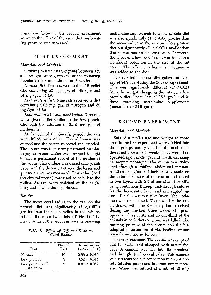
minute. The pressure at which leakage oc- EFFECT OF DIFFERENT OIETS curred was recorded in millimeters Hg. ON INTESTINAL WOUND STRENGTH
WALL TENSION. Wall tension in each rat was calculated by substituting the mean ra- dius measured in the first experiment and the bursting pressure measured in the second ex- periment in the equation:
Nurrml dirt
Low proteln det
wall tension = bursting pressure X radius X 6
HISTOLOGY. These studies were made to examine the progress of the inflammatory reaction and healing process in the neighbor- hood of the wound. The stains used were: hematoxylin and eosin, and van Gieson.
Days after wounding
Results
BURSTING PRESSURE. In all three groups of rats there was a steady rise in bursting pres- sure during the 15 days after making the wound (Table 2). Analysis of variance of the mean values in each diet group showed that the rise in bursting pressure was significant (P < 0.01). However, the mean values in the rats on different diets did not differ signif- icantly from each other on postoperative days 5, 10, and 15.
Fig. 1. Changes in strength of an incision in the cecum of rats receiving a normal diet, a low protein diet, and a low protein diet supplemented with small quantities of methionine.
mean wall tension in the rats fed the low protein and low protein plus methionine diets. The mean wall tension in the rats receiving methionine supplements was also significantly (P < 0.01) greater than in those receiving a low protein diet without methionine.
WALL TENSION. The results showed a steady rise in wall tension in each group of rats (Table 3 and Fig. 1) . Analysis of variance of the mean values in each diet group re- vealed that the rise in wall tension during the 15 postoperative days was significant (P < 0.01). At each stage of the experiment (i.e., postoperative days 5, 10, and 15) the mean wall tension in the rats fed a normal diet was significantly (P < 0.01) greater than the
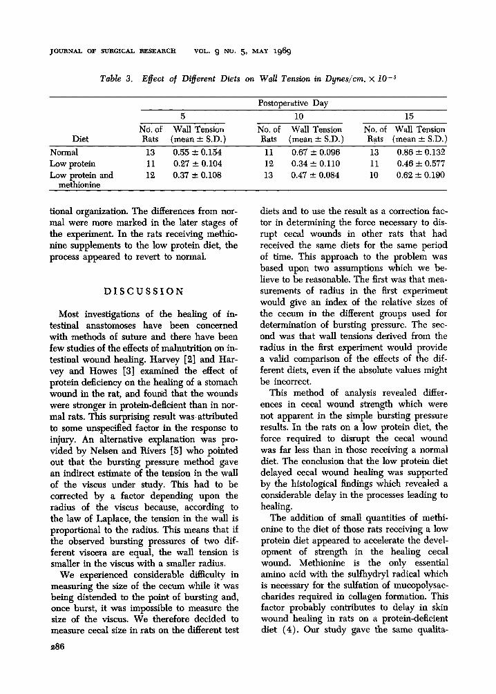
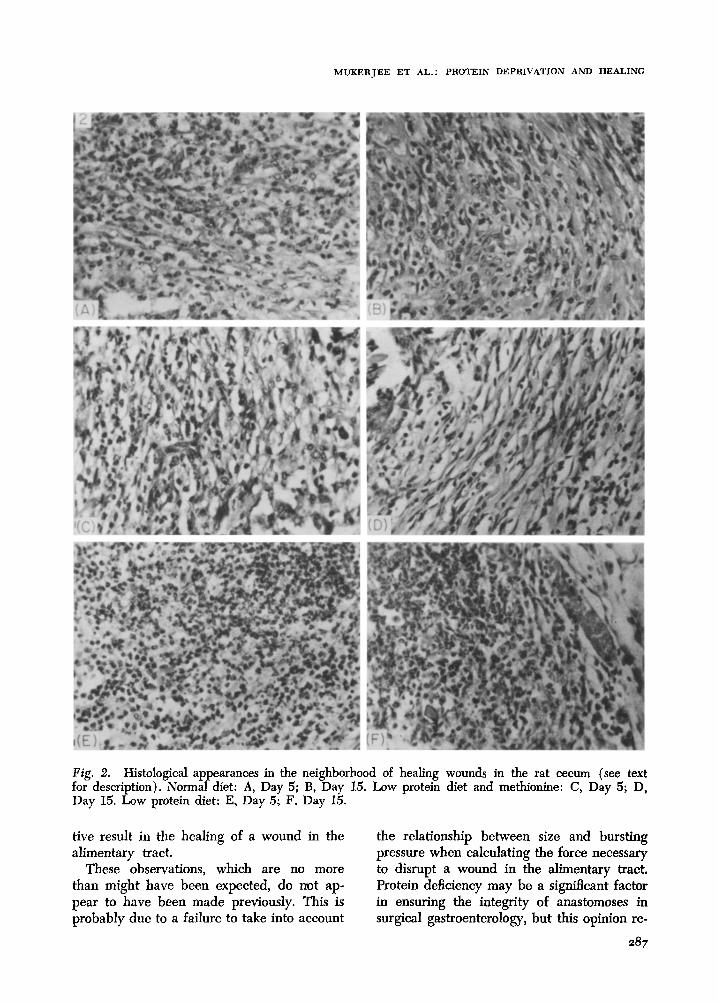
HISTOLOGY. The normal animals showed an orderly progression of the inflammatory reaction through an exudative phase (Day 5), a proliferative phase of organizing granu- lation tissue (Day 10) to healing and the formation of fibrous tissue on (Day 15) (Fig. 2). In the rats receiving a low protein diet, there were three important deviations from the normal. First, the acute phase persisted longer. Second, the proliferative phase was less markedly productive with a lack of proper granulation tissue. Third, the fibrous tissue displayed a lack of orderly and direc-
Table 2. Effect of Diflerent Diets on Bursting Pressure in mm. Hg
Diet
Normal Low protein Low protein and
methionine
Postoperative Day
5 10 15 Bursting
No. of Pressure No. of Bursting Pressure
Bursting No. of Pressure
Rats (mean & S.D.) Rats (mean + SD.) Rats (mean f S.D.)
13 92.7 -c 26.19 11 114.2 -’ 16.30 13 146.8 2 22.46 11 79.9 + 30.75 12 101.6 2 32.4 11 135.0 + 16.88 12 88.8 f 25.04 13 130.7 _e 38.83 10 151.5 I!Z 46.19
JOURNAL OF SURGICAL RESEARCH VOL. 9 NO. 5, MAY 1969
Table 3. Efect of Different Diets on Wall Tendon in Dynes/cm, x 1O-5
Diet
Normal Low protein Low protein and
methionine
5 Nd. of Wall Tension Rats (mean + S.D.)
13 0.55 f 0.154 11 0.27 r 0.104 12 0.37 r 0.108
Postoperative Day
10 No. of Wall Tension Rats (mean -C S.D.)
11 0.67 f 0.096 12 0.34 f 0.110 13 0.47 f 0.084
15 No. of Wall Tension Rats (mean f S.D.)
13 0.86 * 0.132 11 0.46 + 0.577 10 0.62 + 0.190
tional organization. The differences from nor- mal were more marked in the later stages of the experiment. In the rats receiving methio- nine supplements to the low protein diet, the process appeared to revert to normal.
DISCUSSION
Most investigations of the healing of in- testinal anastomoses have been concerned with methods of suture and there have been few studies of the effects of malnutrition on in- testinal wound healing. Harvey [2] and Har- vey and Howes [3] examined the effect of protein deficiency on the healing of a stomach wound in the rat, and found that the wounds were stronger in protein-deficient than in nor- mal rats. This surprising result was attributed to some unspecified factor in the response to injury. An alternative explanation was pro- vided by Nelsen and Rivers [S] who pointed out that the bursting pressure method gave an indirect estimate of the tension in the wall of the viscus under study. This had to be corrected by a factor depending upon the radius of the viscus because, according to the law of Laplace, the tension in the wall is proportional to the radius. This means that if the observed bursting pressures of two dif- ferent viscera are equal, the wall tension is smaller in the viscus with a smaller radius.
We experienced considerable difhculty in measuring the size of the cecum while it was being distended to the point of bursting and, once burst, it was impossible to measure the size of the viscus. We therefore decided to measure cecal size in rats on the different test
286
diets and to use the result as a correction fac- tor in determining the force necessary to dis- rupt cecal wounds in other rats that had received the same diets for the same period of time. This approach to the problem was based upon two assumptions which we be- lieve to be reasonable, The first was that mea- surements of radius in the first experiment would give an index of the relative sizes of the cecum in the different groups used for determination of bursting pressure. The sec- ond was that wall tensions derived from the radius in the first experiment would provide a valid comparison of the effects of the dif- ferent diets, even if the absolute values might be incorrect.
This method of analysis revealed differ- ences in cecal wound strength which were not apparent in the simple bursting pressure results. In the rats on a low protein diet, the force required to disrupt the cecal wound was far less than in those receiving a normal diet. The conclusion that the low protein diet delayed cecal wound healing was supported by the histological findings which revealed a considerable delay in the processes leading to healing.
The addition of small quantities of methi- onine to the diet of those rats receiving a low protein diet appeared to accelerate the devel- opment of strength in the healing cecal wound. Methionine is the only essential amino acid with the sulfhydryl radical which is necessary for the sulfation of mucopolysac- charides required in collagen formation. This factor probably contributes to delay in skin wound healing in rats on a protein-deficient diet (4). Our study gave the same qualita-
MUKERJEE ET AL.: PROTEIN DEPRIVATION AND HEALING
Fig. 2. Histological appearances in the neighborhood of healing wounds in the rat cecum (see text for description). Normal diet: A, Day 5; B, Day 15. Low protein diet and methionine: C, Day 5; D, Day 15. Low protein diet: E, Day 5; F, Day 15.
tive result in the healing of a wound in the the relationship between size and bursting alimentary tract. pressure when calculating the force necessary
These observations, which are no more to disrupt a wound in the alimentary tract, than might have been expected, do not ap- Protein deficiency may be a significant factor pear to have been made previously. This is in ensuring the integrity of anastomoses in probably due to a failure to take into account surgical gastroenterology, but this opinion re-
JOURNAL OF SURGICAL RESEARCH VOL. 9 NO. 5, MAY 1969
quires the confirmation of clinical observa- tion.
SUMMARY
Experiments were carried out to evaluate the effects of different diets on the strength and histological appearances of a wound in the cecum of a rat. Low protein diet caused a significant reduction in the strength and de- lay in the appearance of normal scar tissue. These features were less prominent in rats re- ceiving a low protein diet with supplements of small quantities of methionine.
REFERENCES
1. Chamey, J., Williamson, M. B., and Bernhard, F. W. An apparatus for the determination of the
2.
3.
4.
5.
6.
7.
tensile strength of healing wounds. Science 105: 396, 1947. Harvey, S. C. The velocity of the growth of fibroblasts in the healing wound. Ash. Surg. (Chicago) 18:1227, 1929. Harvey, S. C., and Howes, E. L. The effect of high protein diet on the velocity of fibroblasts in the healing wound. Ann. Sung. 91:641, 1930. Localio, S. A., Morgan, M. E., and Hinton, J. W. The biological chemistry of wound healing. Surg. Gynec. Obstet. 86~582, 1948. Nelsen, T. S., and Anders, C. J. Dynamic aspects of small intestinal rupture with special considera- tion of anastomotic strength. Awh. Surg. (Chi- cago) 93:309, 1966. Varco, R. L. Nutritional preparation for sub- standard risk patients. Surg. Gynec. Obstet. 84: 611, 1947. Williamson, M. B., McCarthy, T. H., and Fromm, H. J. Relation of protein nutrition to the heal- ing of experimental wounds. Proc. Sot. Exxp. Biol. Med. 77:302, 1951.
288