the effect of nitric oxide on ischemia–reperfusion injury in rat liver
TRANSCRIPT
Clinica Chimica Acta 288 (1999) 55–62www.elsevier.com/ locate /clinchim
The effect of nitric oxide on ischemia–reperfusioninjury in rat liver
~ *¨ ¨Tulay Koken, Mine Inal
Department of Biochemistry, The Medical School, Osmangazi University, 26480, Eskisehir, Turkey
Received 18 April 1997; received in revised form 4 January 1999; accepted 30 June 1999
Abstract
A dual role for nitric oxide (NO) in ischemia–reperfusion (I /R) injury is still controversial.This study aims to investigate the role of NO in rat hepatic reperfusion injury. Ischemia wasinduced by total occlusion of hepatic artery and portal vein for 30 min, then the tissue was
Greperfused for 30 min. The animals in the L-NAME group (n 5 10) received N nitro-L-argininemethyl ester (L-NAME) (15 mg/kg) intraperitoneally 60 min before ischemia. The ischemia group(n 5 10) was given an equal volume of saline solution. The control group comprised eight healthyrats which were not exposed to ischemia or reperfusion. An indicator of hepatic injury, plasmaalanine amino transferase (ALT) enzyme activities, were increased in the L-NAME group ascompared with the ischemia group ( p , 0.001). The level of serum nitrite, an index of NOproduction, and hepatic reduced glutathione (GSH) concentration were lower in the L-NAMEgroup than in the ischemia group ( p , 0.001, p , 0.01, respectively). Hepatic levels ofmalondialdehyde (MDA) and conjugated dienes (CD) were significantly increased in the L-NAMEgroup as compared to the ischemia group ( p , 0.05, p , 0.001, respectively). Our results confirmthat L-NAME, an inhibitor of the enzyme NO synthase, increased the lipid peroxidation andpossibly tissue injury, due to the inhibition of cytoprotective effects of NO in a rat hepatic I /Rmodel. 1999 Published by Elsevier Science B.V. All rights reserved.
Keywords: Ischemia / reperfusion; Nitric oxide; Free radicals
*Corresponding author. Tel.: 1 90-222-2393-750.
0009-8981/99/$ – see front matter 1999 Published by Elsevier Science B.V. All rights reserved.PI I : S0009-8981( 99 )00138-2
~¨56 T. Koken, M. Inal / Clinica Chimica Acta 288 (1999) 55 –62
1. Introduction
Studies have demonstrated that oxygen free radicals play a major role in thepathogenesis of many diseases [1]. One of these is ischemia–reperfusion, whichis a deficiency in the transport of O to the tissue. Complete interruption of2
blood flow to the liver is often necessary during operations, for extensive injuryto the liver is inevitably associated with periods of complete ischemia. Hepaticcirculation is also reduced during hemorragic shock, after severe trauma and inlate sepsis. Ligation of the hepatic artery, which was advocated in the treatmentof certain tumors of the liver, vascular lesions or injury to the liver, reduces totalhepatic blood flow, at least initially [2,3].
During ischemia, adenine nucleotides are rapidly broken down to formhypoxanthine concomitantly, the enzyme xanthine dehydrogenase is convertedto xanthine oxidase. When blood flow is re-established to the ischemic organ,hypoxanthine is oxidized to xanthine and, during this reaction which is regulatedby xanthine oxidase, oxygen free radicals are formed. Oxygen radicals damagecell membranes by peroxidation of fatty acids within the phospholipid structureof the membrane. During this process, lipid peroxide radicals, lipid hydro-peroxides and other lipid fragmentation products, that are themselves activeoxidizing agents, are formed. Thus, the free radical reactivity has a tendency togenerate a chain reaction of radical species production, which enhances theultimate destructive effect [4,5].
To protect an organ from reperfusion injury, the production of oxygen radicalsis reduced or radical scavengers are administrated. The best known scavangersare reduced glutathione, glutathione peroxidase(GSH-Px), superoxide dismutase(SOD) and catalase. In particular, reduced glutathione (GSH) is present inmillimolar concentrations in the liver and plays a key role in its protectionagainst oxidative damage by acting as a scavenger of radicals and as a substratefor GSHPx [6–8].
Nitric oxide is a biologically active compound, which may have an impact onleukocyte endothelial cell interactions, in addition to causing smooth musclerelaxation [9].
There are many positive [10–12] and negative [13,14] data relating toefficiency of NO. Studies show that NO is both beneficial and ineffective.Inhibition of NO synthesis using various analogues of L-arginine, includingNO-nitro-L-arginine-methyl ester, promotes leukocyte adhesion to postcapillaryvenules [15]. Although many studies suggest that NO may function as anendogenous antiadhesion molecule, the mechanism underling this importantproperty of nitric oxide remains unknown.
Superoxide promotes leukocyte-adhesion in postcapillary venules, and it has[16] been implicated as a mediator of ischemia–reperfusion induced leukocyteadhesion [17]. Nitric oxide and superoxide are known to interact rapidly toproduce nitrates and nitrites, compounds without the biological activity of either
~¨T. Koken, M. Inal / Clinica Chimica Acta 288 (1999) 55 –62 57
free radical [18]. Therefore, nitric oxide may act as a biological scavenger orinactivator of the proadhesive oxygen free radical.
2. Material and methods
Male Spraque-Dawley rats, weighing 250–300 g, were used in all experi-ments. The animals were not fed for 12 h prior to the experiments. Twenty-eightrats were divided into three groups.
(1) Control group (n 5 8): General anesthesia was induced with urethane, amidline abdominal incision was made and blood samples were obtained bycardiac puncture for determination of serum ALT and nitrite. The rats were thenkilled, and the liver was removed from each animal for determination of liverGSH and lipid peroxidation products.
(2) Ischemial–reperfusion (I /R) group (n 5 10): The animals were pretreatedwith saline solution (4 ml /kg body wt.) intraperitoneally (i.p.) 1 h beforeischemia. An in vivo hepatic ischemia / reperfusion model in the rat was studiedin which the hepaticoduodenal ligament (a. hepatica, v. porta, bile duct) wastotally occluded for 30 min using an atraumatic clamp. Reperfusion wasestablished by removal of the clamp. Thirty min after removal of the vascularclamp, blood and tissue samples were taken.
(3) I /R 1 L-NAME group (n 5 10): All surgical procedures were done as inthe I /R group. Instead of saline solution, these animals received 15 mg/kg bodywt. L-NAME intra-peritoneally.
The liver tissues were washed with cold saline and stored at 2 208C untilassay. The tissues were thawed and homogenized in potassium chloride buffer(0.15 mol / l) for GSH and malondialdehyde (MIDA) analysis, and in distilledwater for conjugated diene (CD) analysis with an Ultra-Turrax. Serum nitritelevels were determined according to Hortelano et al. [19]. The measurement ofGSH levels was carried out according to the method of Beutler [20]. MDA wasdetermined by the method of Okhawa [21]. CD was assessed according to themethod of Ward et al. [22]. Protein concentrations were analysed using acommercial Lowry protein kit. The alanine aminotransferase (ALT) and lactatedehydrogenase (LDH) levels were measured by BM-Hitachi 911 automatedanalyzer on the same day.
Experimental data were analyzed and differences established using Studentst-test. Results were expressed as mean6standard error of the mean (SEM).
3. Results
As shown in Table 1, the ALT enzyme activities in the I /R group weresignificantly higher as compared to the control group ( p , 0.001). Also these
~¨58 T. Koken, M. Inal / Clinica Chimica Acta 288 (1999) 55 –62
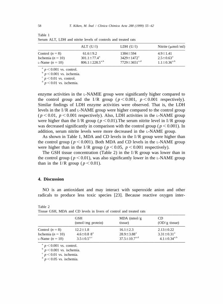
Table 1Serum ALT, LDH and nitrite levels of controls and treated rats
ALT (U/ l) LDH (U/ l) Nitrite (mmol /ml)
Control (n 5 8) 61.669.2 13846594 4.961.41a c aIschemia (n 5 10) 301.1677.4 342961472 2.560.63
a,b a,d a,bL-Name (n 5 10) 806.16228.5 772963651 1.160.36
a p , 0.001 vs. control.b p , 0.001 vs. ischemia.c p , 0.01 vs. control.d p , 0.01 vs. ischemia.
enzyme activities in the L-NAMIE group were significantly higher compared tothe control group and the I /R group ( p , 0.001, p , 0.001 respectively).Similar findings of LDH enzyme activities were observed. That is, the LDHlevels in the I /R and L-NAME group were higher compared to the control group( p , 0.01, p , 0.001 respectively). Also, LDH activities in the L-NAME groupwere higher than the I /R group ( p , 0.01).The serum nitrite level in I /R groupwas decreased significantly in comparison with the control group ( p , 0.001). Inaddition, serum nitrite levels were more decreased in the L-NAME group.
As shown in Table 1, MDA and CD levels in the I /R group were higher thanthe control group ( p , 0.001). Both MDA and CD levels in the L-NAME groupwere higher than in the I /R group ( p , 0.05, p , 0.001 respectively).
The GSH tissue concentration (Table 2) in the I /R group was lower than inthe control group ( p , 0.01), was also significantly lower in the L-NAME groupthan in the I /R group ( p , 0.01).
4. Discussion
NO is an antioxidant and may interact with superoxide anion and otherradicals to produce less toxic species [23]. Because reactive oxygen inter-
Table 2Tissue GSH, MDA and CD levels in livers of control and treated rats
GSH MDA (nmol /g CD(nmol /mg protein) tissue) (OD/g tissue)
Control (n 5 8) 12.261.8 16.162.3 2.1360.22a a aIschemia (n 5 10) 4.660.8 8 28.963.88 3.3160.31
a,c a,d a,bL-Name (n 5 10) 3.560.5 37.5610.7 4.160.34
a p , 0.001 vs. control.b p , 0.001 vs. ischemia.c p , 0.01 vs. ischemia.d p , 0.05 vs. ischemia.
~¨T. Koken, M. Inal / Clinica Chimica Acta 288 (1999) 55 –62 59
mediates may mediate liver injury induced by macrophages and endotoxin [24],an antioxidant function of NO might explain the apparent protective effect ofNO synthesis in the murine model of endotoxin-induced hepatic necrosis [25]. Incontrast, other evidence suggests that NO may interact with reactive oxygenintermediates to form more toxic species. The reaction of NO with superoxideanion can produce the peroxynitrite anion, which can decompose to generate astrong oxidant with reactivity similar to the hydroxyl radical [26]. Peroxynitritecan induce sulfhydryl oxidation [27] and lipid peroxidation [28] suggesting thatNO may have cytotoxic potential through its interaction with superoxide anion.
In this study, serum ALT and LDH enzyme activities were measured as theindex of hepatic injury. ALT and LDH levels were statistically increasedsignificantly ( p , 0.001, p , 0.01 respectively) with the application of I /R tothe liver. The increased enzyme levels suggest that 30 min of I /R caused liverinjury. Goto et al. have also indicated that such enzyme activities were increasedin their study [29]. Administration of L-NAME resulted in an increase in ALTand LDH enzyme activities, perhaps as a result of eliminating a cytoprotectiveeffect by the inhibition of NO synthesis.
The glutathione (GSH) system is an important endogenous antioxidant that isfound in particularly high concentration in the liver [30]. Many studies haveshown the protective effect of GSH has been indicated on the I /R mediatedinjury [31]. The GSH redox cycle, catalyzed by the enzyme glutathioneperoxidase, reduces H O , thus breaking the chain reaction leading from the2 2
superoxide radical to the highly reactive hydroxyl radical. In addition, GSH actsas a natural scavenger for the superoxide anion and protects protein thiol groups,essential for maintaining cellular integrity, against oxidation. GSH also has amajor role in restoring other free radical scavengers and antioxidants such asvitamin E and vitamin C to their reduced state [32].
Stein et al. have demonstrated that I /R mediated injury is greater in GSH-depleted animals [33]. Kobayashi et al. have also shown that liver tissue GSHconcentrations were decreased in the ischemia–reperfusion process [31]. Theseinvestigators have also shown that GSH itself acts as a natural scavenger forsuperoxide anion. The GSH redox cycle, catalyzed by the enzyme GSHperoxidase, also contributes to radical scavenging, i.e. it reduces hydroperoxide,thus breaking the chain reaction leading from the superoxide radical to thehighly toxic hydroxyl radical.
In our study, liver tissue GSH concentrations were also decreased after I /R( p , 0.001). The application of L-NAME after I /R decreases GSH concen-trations ( p , 0.01). It has recently been pointed out that the effect of NO onhepatocyte GSH metabolism may be the result of activation of a component inthe glutathione recyclic pathway, such as GSH reductase; GSH synthesase,g-glutamylcysteine synthetase; or plasma membrane transport of GSH synthesisprecursors.
In our study,the inhibition of NO synthesis caused tissue-damage by leading
~¨60 T. Koken, M. Inal / Clinica Chimica Acta 288 (1999) 55 –62
to reduction of GSH. Our results corroborate the of cytoprotective effect of NO.As is known, free-radical-mediated peroxidation occurs formed during the I /R
process and causes tissue-injury. In this study, levels of MIDA and CD, an end-product of lipid–peroxidation, were measured. These products increase inparallel with tissue-injury. It was observed that both MDA and CD levels wereincreased after I /R ( p , 0.001). MDA and CD levels were found to be highlysignificantly higher in the L-NAME application group compared to both controland I /R groups ( p , 0.05, p , 0.001 respectively). These results show thatL-NAME increased lipid peroxidation, after I /R; the increase of lipid–peroxida-tion was accompanied by a decrease in nitrite level, which is a metabolite ofNO, suggesting a deficiency of NO.
Johnson et al. have suggested that cardiac injury after I /R was dependent ondepletion of NO release [34]. Administration of L-NAME to rats accelerated theinhibition of NO synthesis and I /R-mediated-injury would be expected as aresult. In this study, serum nitrite level was decreased significantly by givingL-NAME (P , 0.001). The depletion of serum nitrite was parallel by increasedserum AST and LDH enzyme levels, which are indicators of liver injury, and theenhanced tissue MDA and CD levels. This also makes us think that the tissueinjury was increased, that is to say, NO may have a protective effect after I /R.
References
[1] Halliwell B. Free radicals, antioxidants, and human disease: curiosity, cause, or consequence.Free Rad Antiox 1994;344:721–4.
[2] Ferrari R. Oxygen free radicals at myocardial level: effects of ischemia and reperfusion. In:Armstrong D, editor, Free radicals in diagnostic medicine, New York: Plenum Press, 1994,pp. 99–111.
[3] Hasselgren PO. Prevention and treatment of ischemia of the liver. Surg Gyn Obst1987;164:187–96.
[4] Zimmerman BJ, Granger DN. Reperfusion injury. Surg Clin North Am 1992;72(1):65–83.[5] Thurman RG, Marzi I, Seitz G, Thies J, Lemasters J, Zimmerman F. Hepatic reperfusion
injury following orthotopic liver transplantation in the rat. Transplantation 1988;46:502–6.[6] Duval DL, Sieg DJ, Billings RE. Regulation of hepatic nitric oxide synthase by reactive
oxygen intermediates and glutathione. Arch Biochem Biophys 1995;316:699–706.[7] Erden M, Bor NM. Changes of reduced glutathione reductase, and glutathione peroxidase
after radiation in guinea pigs. Biochem Med 1984;31:217–27.[8] Okuda M, Lee HC, Chance B, Kumar C. Glutathione and ischemia–reperfiision injury in the
perfused rat liver. Free Rad Biol Med 1992;12:271–9.[9] Gaboury J, Woodman RC, Granger DN, Reinhart P, Kubes P. Nitric oxide prevents leukocyte
adherence: Role of superoxide. Am Physiol 1993;265:H862–7.[10] Hoshida S, Yamashita N, Igarashi J, Nishida M, Hori M, Kamada T et al. Nitric oxide
synthase protects the heart against ischemia–reperfusion injury in rabbits. J Pharmacol ExpTher 1995;274(1):413–8.
~¨T. Koken, M. Inal / Clinica Chimica Acta 288 (1999) 55 –62 61
[11] Kuo PC, Slivka A. Nitric oxide decreases oxidant-mediated hepatocyte injury. J Surg Res1994;56:594–600.
[12] Parks DA, Granger DN. Ischemia-reperfusion injury: A radical view. Hepatology1988;8(3):680–2.
[13] Kubes P. Ischemia reperfusion in feline small intestine: A role for nitric oxide. Am J Physiol1993;264:G143–9.
[14] Loscalzo J, Vita JA. Ischemia, hyperemia, exercise, nitric oxide. Complex physiology andcomplex molecular adaptations. Circulation 1994;90(5):2556–9.
[15] Kubes P, Suzuki M, Granger DN. Nitric oxide: an endogenous modulator of leukocyteadhesion. Proc Natl Acad Sci 1991;88:4651–5.
[16] Del Maestro RF, Planker M, Arfors KE. Evidence for the participation of superoxide anionradical in altering the adhesive interaction between granulocytes and endothelium, in vivo.Int J Microcirc Clin Exp 1982;1:105–20.
[17] Suzuki M, Inauen W, Kvietya PR, Grisham MB, Meininger C, Schelling ME et al.Superoxide mediates reperfusion-induced leukocyte-endothelial cell interactions. Am JPhysiol 1989;257:H1740–1745.
[18] Ignarro LJ. Biosynthesis and metabolism of endothelium-derived nitric oxide. Annu RevPharmacol Toxicol 1990;30:535–60.
[19] Hortelano S, Dewez B, Genaro AN. Nitric oxide is released in regenerating liver after partialhepatectomy. Hepatology 1995;21:776–86.
[20] Beutler E, Robson MJ, Buttenweiser E. The glutathione instability of drug sensivite red cells.J Lab Clin Med 1957;49:84–95.
[21] Okhawa H, Ohishi N, Yagi K. Assay for lipid peroxides imal tissues by thiobarbituric acidreaction. Anal Biochem 1978;95:351–8.
[22] Ward PA, Till GO, Hatherill JR, Annesley TM, Kunkel RG. Systemic complement activation,lung injury, and products of lipid peroxidation. J Clin Invest 1985;76:517–27.
[23] Stark ME, Szurszewski JH. Role of nitric oxide in gastrointestinal and hepatic function anddisease. Gastroenterology 1992;103:1928–49.
[24] Arthur MJP, Bentley IS, Tanner AR, Kowalski SP, MIillward–Sadler GH, Wright R.Oxygen-derived free radicals promote hepatic injury in the rat. Gastroenterology1985;89:1114–22.
[25] Billiar TR, Curran RD, Harbrecht BG, Stuehr DJ, Demetris AJ, Simmons RL. Modulation ofGnitrogen oxide synthesis in vivo: N -monomethyl-L-arginine inhibits endotoxin-induced
nitrite /nitrate biosynthesis while promoting hepatic damage. J Leukoc Biol 1990;48:565–9.[26] Beckman JS, Beckman TW, Chen J, Marshall PA, Freeman BA. Apparent hydroxyl radical
production by peroxynitrite: Implications for endothelial injury from nitric oxide andsuperoxide. Proc Natl Acad Sci 1995;87:1620–4.
[27] Radi R, Beckman JS, Bush KM, Freeman BA. Peroxynitrite oxidation of sulfhydryls: thecytotoxic potential of superoxide and nitric oxide. J Biol Chem 1991;266:4244–50.
[28] Radi R, Beckman JS, Bush KM, Freeman BA. Peroxynitrite-induced membrane lipidperoxidation: the cytotoxic potential of superoxide and nitric oxide. Arch Biochem Biophys1991;288(2):481–7.
[29] Goto M, Kawano S, Yoshihar H, Takei Y, Hijioka T, Fukui H et al. Hepatic tissueoxygenation as a protective indicator of ischemia–reperflision liver injury. Hepatology1992;15(3):432–7.
[30] Okuda M, Lee HC, Chance B, Kumar C. Glutathione and ischemia / reperfusion injury in theperfused rat liver. Free Rad Biol Med 1992;12:271–9.
[31] Kobayashi H, Kurokawa T, Kitahara S, Nonami T, Harada A, Nakao A et al. The effects ofg-glutamylcysteine ethyl ester, a prodrug of glutathione, on ischemia-reperfusion inducedliver injury in rats. Transplantation 1992;54(3):414–8.
~¨62 T. Koken, M. Inal / Clinica Chimica Acta 288 (1999) 55 –62
[32] Gruetter CA, Gruetter DY, Lyon JE, Kadowitz PJ, Ignarro U. Relationship between cyclicguanosine 39:59-monophosphate formation and relaxation of coronary arterial smooth muscleby glyceryl trinitrate, nitroprusside, nitrite and nitric oxide: Effect of methylene blue andmethemoglobin. J Pharmacol Exp Ther 1981;219:181–6.
[33] Stein HJ, Oosthuizen MJ, Hinder RA, Lamprechts H. Oxygen free radicals and glutathione inhepatic ischemia / reperfusion injury. J Surg Res 1991;50:398–402.
[34] Johnson G, Tsao PC, Lefer AM. Cardioprotective effect of authentic nitric oxide inmyocardial ischemia with reperfusion. Crit Care Med 1991;19:244–52.