the effect of bmp-2 on the osteoconductive properties of β-tricalcium phosphate in rat calvaria...
TRANSCRIPT
lable at ScienceDirect
Biomaterials 32 (2011) 3855e3861
Contents lists avai
Biomaterials
journal homepage: www.elsevier .com/locate/biomater ia ls
The effect of BMP-2 on the osteoconductive properties of b-tricalcium phosphatein rat calvaria defects
Eloa R. Luvizuto a,d, Stefan Tangl a,b, Gerald Zanoni c, Tetuo Okamoto d,e, Celso K. Sonoda d,Reinhard Gruber a,b,*, Roberta Okamoto d,e
aDepartment of Oral Surgery, Medical University of Vienna, AustriabAustrian Cluster for Tissue Regeneration, Austriac Ludwig Boltzmann Institute for Experimental and Clinical Traumatology, Vienna, AustriadDepartment of Surgery and Integrated Clinic, Araçatuba Dental School, UNESP-Univ Estadual Paulista, BrazileDepartment of Research and Post-Graduation, Universidade do Sagrado Coração, Bauru, Brazil
a r t i c l e i n f o
Article history:Received 17 January 2011Accepted 31 January 2011Available online 3 March 2011
Keywords:Animal modelBone regenerationBMP (bone morphogenetic protein)Calcium phosphate cementPolylactic acidBone tissue engineering
* Corresponding author. Department of Oral SurVienna, Waehringerstrasse 25a, A-1090 Vienna, Austfax: þ43 1 400 70 4109.
E-mail address: [email protected]
0142-9612/$ e see front matter � 2011 Elsevier Ltd.doi:10.1016/j.biomaterials.2011.01.076
a b s t r a c t
Bone formation in critical-sized calvaria defects is strongly dependent on the osteoconductive propertiesof grafts. It remains a matter of controversy whether biomaterials can replace autografts and whether thesupplementation of biomaterials with Bone Morphogenetic Proteins (BMPs) is necessary to enhancebone formation. We examined rat calvaria critical-sized defects (5-mm-diameter) treated with b-tri-calcium phosphate (TCP; Cerasorb� M), polylactic and polyglycolic acid gel (PLA/PGA; Fisiograft�) andcalcium phosphate cement (CPC; Norian� CRS�), either alone or in the presence of 5 mg of BMP-2 after 45days. Autografts and untreated defects served as controls. Bone formation was evaluated based on mCTanalysis, histomorphometric analysis and fluorescence analysis. We report that TCP supported boneformation more efficiently than did autografts. Bone formation in the presence of TCP alone reacheda maximal level, as BMP-2 supplementation failed to enhance bone formation. By contrast, no significantdifference in bone formation was observed when PLA/PGA and CPC were compared to autografts.Moreover, the presence of BMP-2 did not substantially change the osteoconductive properties of PLA/PGA or CPC. We conclude that the osteoconductive properties of TCP are superior to those of autograftsand that TCP does not require BMP-2 supplementation. Our findings also show that the decreasedosteoconductive properties of PLA/PGA and CPC cannot be overcome by BMP-2 supplementation in ratcalvaria defects.
� 2011 Elsevier Ltd. All rights reserved.
1. Introduction
The use of biomaterials instead of autografts is a long-awaitedhope in reconstructive surgery [1]. Biomaterials can provide anosteoconductive matrix that guides new bone into the defect oraugmented area. Improving the osteoconductive properties ofbiomaterials is a major task in biomaterial research and develop-ment. It is not surprising that biomaterials are supplemented withgrowth factors to enhance the process of bone formation and, thus,graft consolidation. The major task, however, lies in providinga biomaterial with favorable osteoconductive properties that aresimilar or even superior to autografts and avoiding growth factorsupplementation.
gery, Medical University ofria. Tel.: þ43 1 400 70 2623;
t (R. Gruber).
All rights reserved.
Growth factors, including bone morphogenetic proteins (BMPs),can support the process of bone formation; however, concernsabout their use have been raised. For example, it is not always clearthat the possible enhancement in bone formation justifies the costsand possible side effects associated with the use of growth factors.The FDA has approved a combination of BMPs and collagen fororthopedic [2] and dentistry [3] applications. However, combina-tions of BMPs andother biomaterials havenotbeen approved [1,4,5].
Initial attempts to identify appropriate carriers for BMPs wereperformed with high expectations. For example, TCP [6,7], PLA/PGA[8,9] and calcium phosphate cement [10,11] have been tested fortheir capacities to support the osteoinductive properties of BMP-2in preclinical models. However, the findings of these studies are notconvincing because bone formation is not necessarily enhanced byBMP-2 supplementation [7,9,11,12]. Thus, not all biomaterialssupport the osteoinductive properties of BMP-2. Currently, itremains unclear which of the three commercially available carriers(TCP, Cerasorb� M; PLA/PGA; Fisiograft�; CPC, Norian� CRS�) allow
E.R. Luvizuto et al. / Biomaterials 32 (2011) 3855e38613856
maximum bone formation and if their supplementation withBMP-2 causes a substantial enhancement in bone formation ata defect site.
Animal models have been the basis of studying the impact ofbiomaterials loaded with BMP-2 on bone formation [13]. There isa consensus that proof-of principle studies should follow the phy-logenic tree, starting with rodent models [14]. The rat calvarium isexemplary for use in a critical size defect model inwhich BMP-2 canexert its osteoinductive capacity [9,15,16]. Even though this is anorthotopic defect model, the small borders between the biomate-rial and residual calvaria bone provides a challenging situation fornew bone to span the entire defect site. Another advantage of therat calvaria model is its reproducibility and the ability to evaluatethe study outcome based on mCT and histomorphometric analysesin a standardizedmanner, keeping the inner experimental variationlow [14]. These arguments support the use of the rat calvaria defectmodel for our study.
In the present study, we compared the osteoconductive prop-erties of autografts with those of three commercially availablebiomaterials, which were tested alone or when supplemented withBMP-2. We chose surgically-created defects in rat calvaria to eval-uate bone formation by means of mCT, histomorphometry andfluorescence microscopy analyses.
2. Material and methods
2.1. Study design and ethics
Twenty Wistar rats (90 days old) that were acquired from the Animal Center ofSão Paulo State University were maintained at a temperature of 22 �C in a 12-h light/12-h dark cycle with free access to water and rodent food. A total of 40 calvariadefects (5-mm-diameter) were randomly divided into 8 treatment groups, witha total of 5 defects per treatment group (n ¼ 5). The treatment groups were asfollows: [1] 500e1000 mm b-tricalcium phosphate (TCP) (Cerasorb�M, Curassan Ltd,Germany); [2] TCP plus 5 mg BMP-2 (R&D Systems, Inc., Minneapolis, MN, USA); [3]polylactic and polyglycolic acid (PLA/PGA) gel (Fisiograft�, Ghimas SPA, Italy); [4]PLA/PGA plus BMP-2; [5] calcium phosphate cement (CPC) (Norian� CRS�, Cranio-facial Repair System�, Germany); and [6] CPC plus BMP-2. The other two treatmentgroups were the empty control (untreated) [7] and the autograft control [8]. Thepresent study complied with the principles of laboratory animal care and nationallaws on animal use, and the study was authorized by the Animal Research EthicsCommittee of the São Paulo State University, Brazil (protocol #2008-004517).
2.2. Surgical procedures and fluorochrome labeling
After general anesthesia with xylazine (0.03 ml/100 g body weight [bw]/intra-peritoneal [ip]; Dopaser� Laboratories Calier SA, Barcelona, Spain) and ketamine(7 ml/kg bw/ip; Fort Dodge Saúde Animal Ltd, Brazil), the animal skulls were shavedand disinfected with polyvinylpyrrolidone iodide (Indústria Química e FarmacêuticaRioquímica Ltd, Brazil). Using aseptic technique, an incision was made through theskin of the calvaria and periosteum, and full-thickness flaps were reflected. Undercopious sterile saline irrigation, two 5-mm-diameter bone defects were preparedwith a trephine bur (3i Implant Innovations, Inc., Palm Beach Gardens, USA) in eachanimal, one in each parietal region. The defects were treated as described abovebefore the periosteum was repositioned and sutured using polylactic acid sutures(Vycril 5.0, Ethicon, Johnson Prod., São José dos Campos, Brazil). The skin was closedusing nylon sutures (Nylon 5.0, Mononylon, Ethicon). All animals received a singledose of 20.000 UI of penicillin G benzathine (Pentabiótico, Veterinário PequenoPorte, Fort Dodge Saúde Animal Ltd, Campinas, Brazil) by intramuscular injection.Rats received 20 mg/kg body weight of calcein and alizarin (Sigma Chemical,St. Louis, MO, USA) at the 15th and 30th postoperative day, respectively. The ratswere euthanized by anesthetic overdose after the 45th postoperative day.
2.3. Sample processing
The relevant part of the skull was removed and fixed in neutral buffered 4%formalin solution. The calvarias were divided in half using a high-precision saw(Exakt Apparatebau, Norderstedt, Germany). The samples were dehydrated inascending grades of alcohol and embedded in light-curing resin (Technovit 7200VLC þ BPO, Kulzer & Co., Hanau, Germany). The blocks were scanned with a mCTsystem (vivaCT 75, ScancoMedical AG, Basserdorf, Switzerland) at 70 kV/114 mAwithan integration time of 1 � 380 ms at high resolution (pixels ¼ 21 � 21 mm). Theblocks were processed with the Exakt Cutting and Grinding Equipment (ExaktApparatebau) according to a standardized method [17]. Sections were prepared in
parallel to the sagittal suture and reduced to a thickness of approximately 40 mmbefore being stained with Levai-Laczko stain.
2.4. Histomorphometric analysis
Blinded analyses were performed. Stained sections were examined by lightmicroscopy (Nikon Microphot-FXA, Nikon, Tokyo, Japan), and images were obtainedwith adigital camera (NikonDXM1200) coupledwith amotorized stage (MärzhäuserWetzlar GmbH&Co KG,Wetzlar Steindorf, Germany). Single pictureswere assembledto generate large overview images (Lucia G 4.71, Laboratory Imaging Ltd, Brno, CzechRepublic) at a resolution of 890 pxl/mm. Analysis was restricted to the gap bridgedbetween both edges of the drilled hole. Based on interactively drawn false colorimages (Adobe Photoshop, Adobe, San Jose, CA, USA), bone tissue and bone substi-tutes were measured with histomorphometry software (Definiens Developer 7,Munich, Germany). Two-dimensional measurements of the defect closure (mCT DC)and three-dimensional measurements of the amount of mineralized volume pertissue volume (mCT MV/TV) were calculated from the mCT samples. Mineralizedvolume per tissue volume (MV/TV), bone volume in the defect [bone volume (BV) inmm2, bone volume per tissue volume (BV/TV)], bone substitute volume in the defect[bone substitute volume (SV) in mm2 and bone substitute volume per tissue volume(SV/TV)] were calculated from the histological samples. Fluorochrome qualitativeanalyses were performed with the epifluorescence mode of a Nikon Microphot-FXAmicroscope (Nikon, Tokyo, Japan).
2.5. Statistics
Data were analyzed with Multiple Comparisons of Means by Tukey Contrasts.Significant difference was assigned at the level of P < 0.05.
3. Results
3.1. mCT analysis
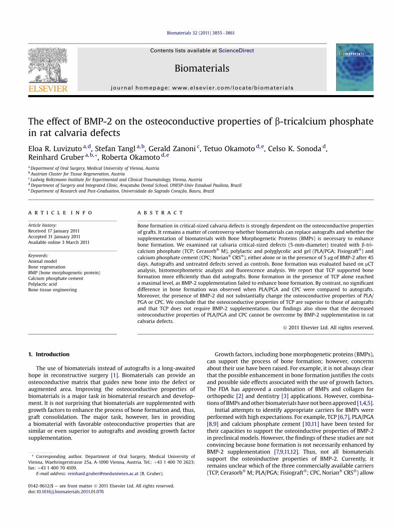
The 2-dimensional view from above provides information aboutoverall defect closure. Complete defect closure was observed onlywith TCP alone and TCP supplemented with BMP-2. Autografts,PLA/PGA and CPC failed to provide sufficient support for the bone tocompletely close the defect. In addition, in the samples in whichPLA/PGA and CPC were supplemented with BMP-2, no defectclosure was observed (Fig. 1; Fig. 1 Supplement).
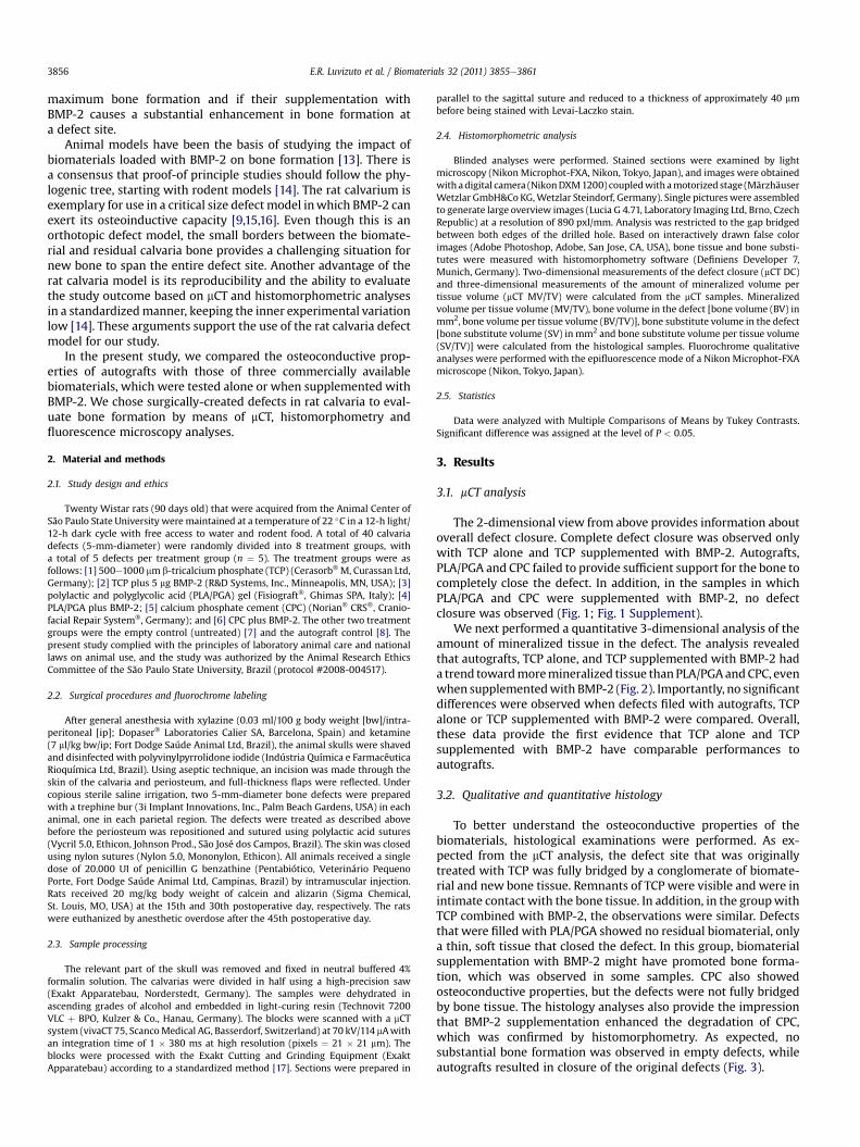
We next performed a quantitative 3-dimensional analysis of theamount of mineralized tissue in the defect. The analysis revealedthat autografts, TCP alone, and TCP supplemented with BMP-2 hada trend towardmoremineralized tissue thanPLA/PGA andCPC, evenwhen supplementedwith BMP-2 (Fig. 2). Importantly, no significantdifferences were observed when defects filed with autografts, TCPalone or TCP supplemented with BMP-2 were compared. Overall,these data provide the first evidence that TCP alone and TCPsupplemented with BMP-2 have comparable performances toautografts.
3.2. Qualitative and quantitative histology
To better understand the osteoconductive properties of thebiomaterials, histological examinations were performed. As ex-pected from the mCT analysis, the defect site that was originallytreated with TCP was fully bridged by a conglomerate of biomate-rial and new bone tissue. Remnants of TCP were visible and were inintimate contact with the bone tissue. In addition, in the groupwithTCP combined with BMP-2, the observations were similar. Defectsthat were filled with PLA/PGA showed no residual biomaterial, onlya thin, soft tissue that closed the defect. In this group, biomaterialsupplementation with BMP-2 might have promoted bone forma-tion, which was observed in some samples. CPC also showedosteoconductive properties, but the defects were not fully bridgedby bone tissue. The histology analyses also provide the impressionthat BMP-2 supplementation enhanced the degradation of CPC,which was confirmed by histomorphometry. As expected, nosubstantial bone formation was observed in empty defects, whileautografts resulted in closure of the original defects (Fig. 3).
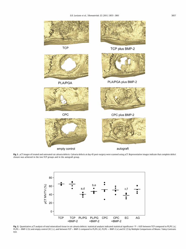
Fig. 1. mCT images of treated and untreated rat calvaria defects: Calvaria defects at day 45 post-surgery were scanned using mCT. Representative images indicate that complete defectclosure was achieved in the two TCP groups and in the autograft group.
Fig. 2. Quantitative mCT analysis of total mineralized tissue in rat calvaria defects: statistical analysis indicated statistical significance *P < 0.05 between TCP compared to PL/PG (a),PL/PG þ BMP-2 (b) and empty control (EC) (c), and between TCP þ BMP-2 compared to PL/PG (d), PL/PG þ BMP-2 (e) and EC (f) by Multiple Comparisons of Means: Tukey Contraststest.
E.R. Luvizuto et al. / Biomaterials 32 (2011) 3855e3861 3857
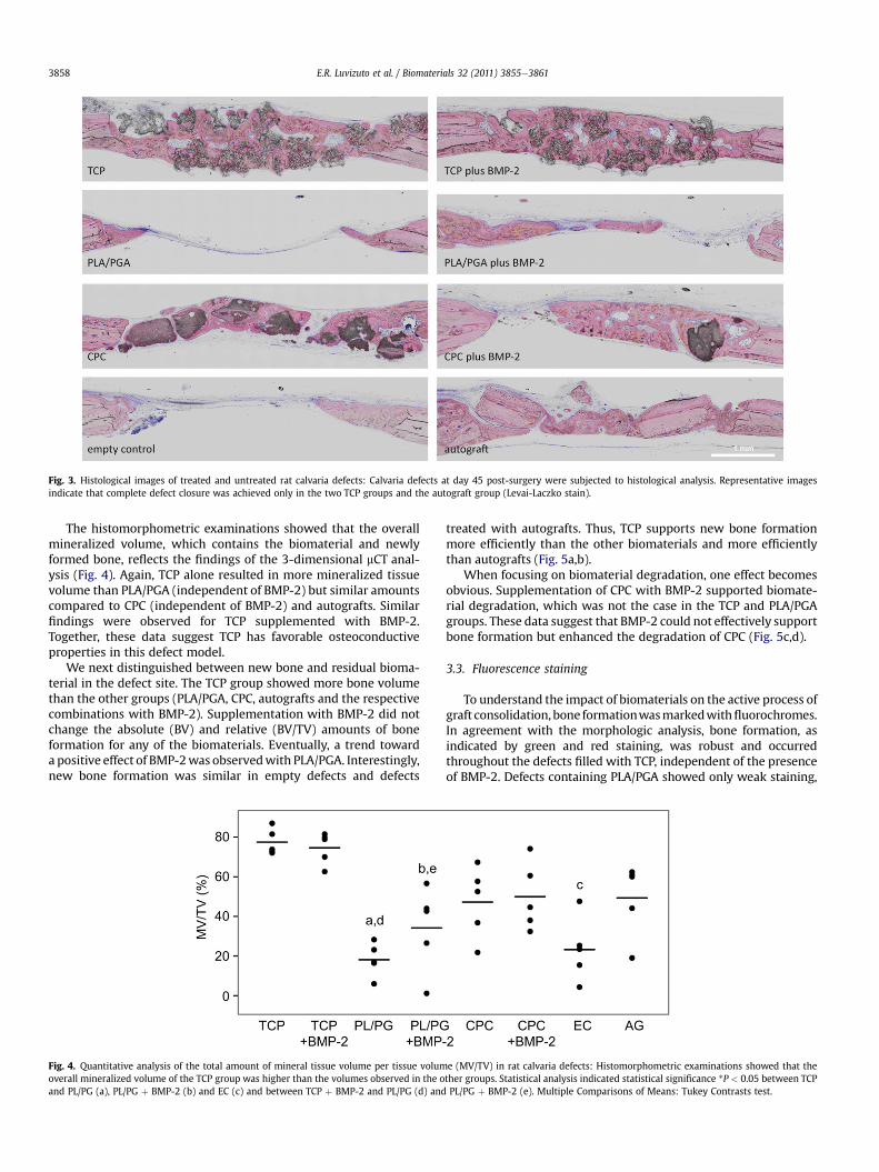
Fig. 3. Histological images of treated and untreated rat calvaria defects: Calvaria defects at day 45 post-surgery were subjected to histological analysis. Representative imagesindicate that complete defect closure was achieved only in the two TCP groups and the autograft group (Levai-Laczko stain).
E.R. Luvizuto et al. / Biomaterials 32 (2011) 3855e38613858
The histomorphometric examinations showed that the overallmineralized volume, which contains the biomaterial and newlyformed bone, reflects the findings of the 3-dimensional mCT anal-ysis (Fig. 4). Again, TCP alone resulted in more mineralized tissuevolume than PLA/PGA (independent of BMP-2) but similar amountscompared to CPC (independent of BMP-2) and autografts. Similarfindings were observed for TCP supplemented with BMP-2.Together, these data suggest TCP has favorable osteoconductiveproperties in this defect model.
We next distinguished between new bone and residual bioma-terial in the defect site. The TCP group showed more bone volumethan the other groups (PLA/PGA, CPC, autografts and the respectivecombinations with BMP-2). Supplementation with BMP-2 did notchange the absolute (BV) and relative (BV/TV) amounts of boneformation for any of the biomaterials. Eventually, a trend towarda positive effect of BMP-2was observedwith PLA/PGA. Interestingly,new bone formation was similar in empty defects and defects
Fig. 4. Quantitative analysis of the total amount of mineral tissue volume per tissue volumoverall mineralized volume of the TCP group was higher than the volumes observed in the oand PL/PG (a), PL/PG þ BMP-2 (b) and EC (c) and between TCP þ BMP-2 and PL/PG (d) and
treated with autografts. Thus, TCP supports new bone formationmore efficiently than the other biomaterials and more efficientlythan autografts (Fig. 5a,b).
When focusing on biomaterial degradation, one effect becomesobvious. Supplementation of CPC with BMP-2 supported biomate-rial degradation, which was not the case in the TCP and PLA/PGAgroups. These data suggest that BMP-2 could not effectively supportbone formation but enhanced the degradation of CPC (Fig. 5c,d).
3.3. Fluorescence staining
To understand the impact of biomaterials on the active process ofgraft consolidation, bone formationwasmarkedwithfluorochromes.In agreement with the morphologic analysis, bone formation, asindicated by green and red staining, was robust and occurredthroughout the defects filled with TCP, independent of the presenceof BMP-2. Defects containing PLA/PGA showed only weak staining,
e (MV/TV) in rat calvaria defects: Histomorphometric examinations showed that thether groups. Statistical analysis indicated statistical significance *P < 0.05 between TCPPL/PG þ BMP-2 (e). Multiple Comparisons of Means: Tukey Contrasts test.
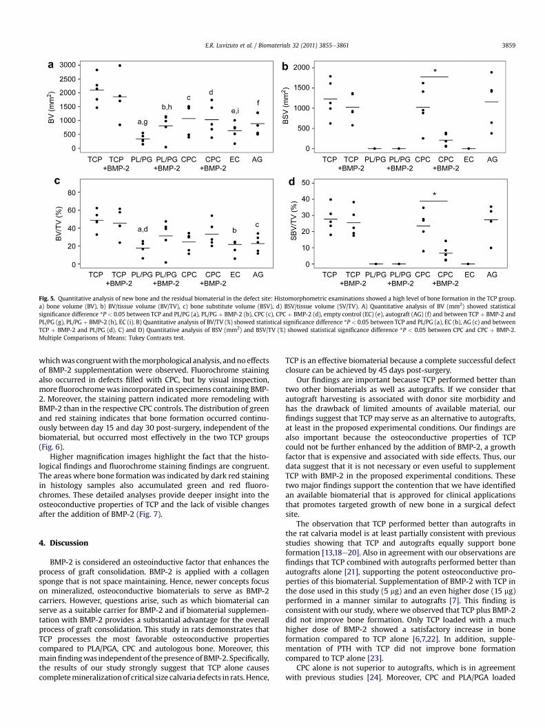
Fig. 5. Quantitative analysis of new bone and the residual biomaterial in the defect site: Histomorphometric examinations showed a high level of bone formation in the TCP group.a) bone volume (BV), b) BV/tissue volume (BV/TV), c) bone substitute volume (BSV), d) BSV/tissue volume (SV/TV). A) Quantitative analysis of BV (mm2) showed statisticalsignificance difference *P < 0.05 between TCP and PL/PG (a), PL/PG þ BMP-2 (b), CPC (c), CPC þ BMP-2 (d), empty control (EC) (e), autograft (AG) (f) and between TCP þ BMP-2 andPL/PG (g), PL/PG þ BMP-2 (h), EC (i). B) Quantitative analysis of BV/TV (%) showed statistical significance difference *P < 0.05 between TCP and PL/PG (a), EC (b), AG (c) and betweenTCP þ BMP-2 and PL/PG (d). C) and D) Quantitative analysis of BSV (mm2) and BSV/TV (%) showed statistical significance difference *P < 0.05 between CPC and CPC þ BMP-2.Multiple Comparisons of Means: Tukey Contrasts test.
E.R. Luvizuto et al. / Biomaterials 32 (2011) 3855e3861 3859
whichwas congruentwith themorphological analysis, andnoeffectsof BMP-2 supplementation were observed. Fluorochrome stainingalso occurred in defects filled with CPC, but by visual inspection,more fluorochromewas incorporated in specimens containing BMP-2. Moreover, the staining pattern indicated more remodeling withBMP-2 than in the respective CPC controls. The distribution of greenand red staining indicates that bone formation occurred continu-ously between day 15 and day 30 post-surgery, independent of thebiomaterial, but occurred most effectively in the two TCP groups(Fig. 6).
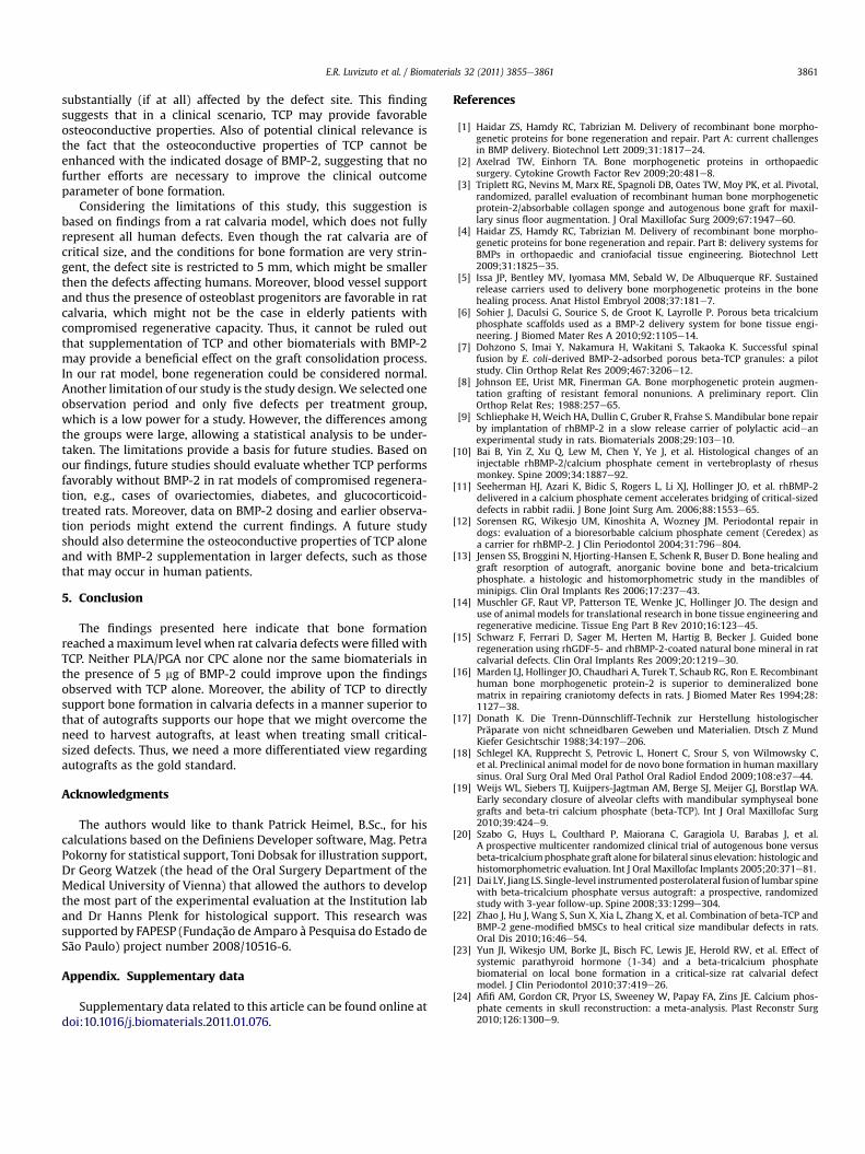
Higher magnification images highlight the fact that the histo-logical findings and fluorochrome staining findings are congruent.The areas where bone formationwas indicated by dark red stainingin histology samples also accumulated green and red fluoro-chromes. These detailed analyses provide deeper insight into theosteoconductive properties of TCP and the lack of visible changesafter the addition of BMP-2 (Fig. 7).
4. Discussion
BMP-2 is considered an osteoinductive factor that enhances theprocess of graft consolidation. BMP-2 is applied with a collagensponge that is not space maintaining. Hence, newer concepts focuson mineralized, osteoconductive biomaterials to serve as BMP-2carriers. However, questions arise, such as which biomaterial canserve as a suitable carrier for BMP-2 and if biomaterial supplemen-tation with BMP-2 provides a substantial advantage for the overallprocess of graft consolidation. This study in rats demonstrates thatTCP processes the most favorable osteoconductive propertiescompared to PLA/PGA, CPC and autologous bone. Moreover, thismainfindingwas independentof thepresence of BMP-2. Specifically,the results of our study strongly suggest that TCP alone causescompletemineralizationof critical size calvariadefects in rats. Hence,
TCP is an effective biomaterial because a complete successful defectclosure can be achieved by 45 days post-surgery.
Our findings are important because TCP performed better thantwo other biomaterials as well as autografts. If we consider thatautograft harvesting is associated with donor site morbidity andhas the drawback of limited amounts of available material, ourfindings suggest that TCP may serve as an alternative to autografts,at least in the proposed experimental conditions. Our findings arealso important because the osteoconductive properties of TCPcould not be further enhanced by the addition of BMP-2, a growthfactor that is expensive and associated with side effects. Thus, ourdata suggest that it is not necessary or even useful to supplementTCP with BMP-2 in the proposed experimental conditions. Thesetwo major findings support the contention that we have identifiedan available biomaterial that is approved for clinical applicationsthat promotes targeted growth of new bone in a surgical defectsite.
The observation that TCP performed better than autografts inthe rat calvaria model is at least partially consistent with previousstudies showing that TCP and autografts equally support boneformation [13,18e20]. Also in agreement with our observations arefindings that TCP combined with autografts performed better thanautografts alone [21], supporting the potent osteoconductive pro-perties of this biomaterial. Supplementation of BMP-2 with TCP inthe dose used in this study (5 mg) and an even higher dose (15 mg)performed in a manner similar to autografts [7]. This finding isconsistent with our study, where we observed that TCP plus BMP-2did not improve bone formation. Only TCP loaded with a muchhigher dose of BMP-2 showed a satisfactory increase in boneformation compared to TCP alone [6,7,22]. In addition, supple-mentation of PTH with TCP did not improve bone formationcompared to TCP alone [23].
CPC alone is not superior to autografts, which is in agreementwith previous studies [24]. Moreover, CPC and PLA/PGA loaded
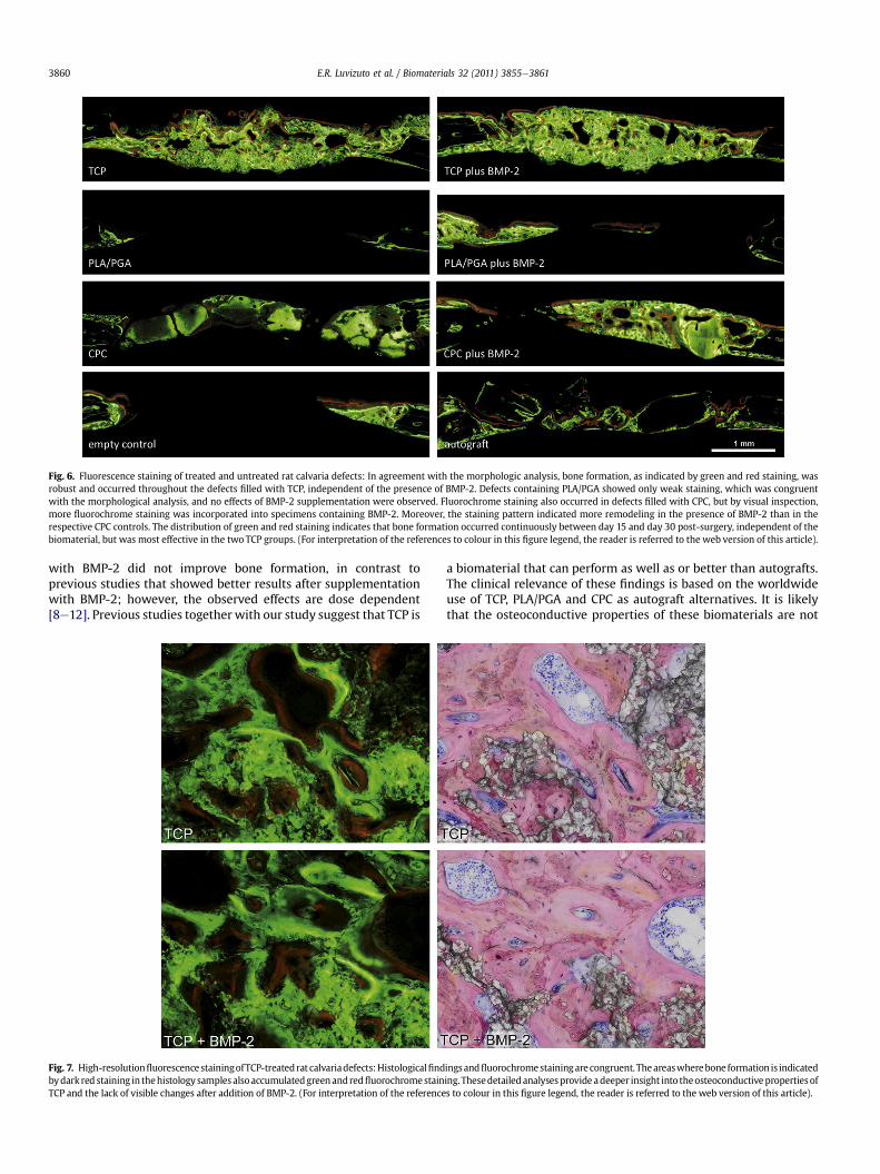
Fig. 6. Fluorescence staining of treated and untreated rat calvaria defects: In agreement with the morphologic analysis, bone formation, as indicated by green and red staining, wasrobust and occurred throughout the defects filled with TCP, independent of the presence of BMP-2. Defects containing PLA/PGA showed only weak staining, which was congruentwith the morphological analysis, and no effects of BMP-2 supplementation were observed. Fluorochrome staining also occurred in defects filled with CPC, but by visual inspection,more fluorochrome staining was incorporated into specimens containing BMP-2. Moreover, the staining pattern indicated more remodeling in the presence of BMP-2 than in therespective CPC controls. The distribution of green and red staining indicates that bone formation occurred continuously between day 15 and day 30 post-surgery, independent of thebiomaterial, but was most effective in the two TCP groups. (For interpretation of the references to colour in this figure legend, the reader is referred to the web version of this article).
E.R. Luvizuto et al. / Biomaterials 32 (2011) 3855e38613860
with BMP-2 did not improve bone formation, in contrast toprevious studies that showed better results after supplementationwith BMP-2; however, the observed effects are dose dependent[8e12]. Previous studies together with our study suggest that TCP is
Fig. 7. High-resolutionfluorescence stainingofTCP-treatedrat calvariadefects:Histologicalfindbydarkred staining in thehistology samples alsoaccumulatedgreenandredfluorochromestainTCP and the lack of visible changes after addition of BMP-2. (For interpretation of the reference
a biomaterial that can perform as well as or better than autografts.The clinical relevance of these findings is based on the worldwideuse of TCP, PLA/PGA and CPC as autograft alternatives. It is likelythat the osteoconductive properties of these biomaterials are not
ingsandfluorochromestainingarecongruent. Theareaswherebone formation is indicateding.Thesedetailedanalysesprovide adeeper insight into theosteoconductivepropertiesofs to colour in this figure legend, the reader is referred to the web version of this article).
E.R. Luvizuto et al. / Biomaterials 32 (2011) 3855e3861 3861
substantially (if at all) affected by the defect site. This findingsuggests that in a clinical scenario, TCP may provide favorableosteoconductive properties. Also of potential clinical relevance isthe fact that the osteoconductive properties of TCP cannot beenhanced with the indicated dosage of BMP-2, suggesting that nofurther efforts are necessary to improve the clinical outcomeparameter of bone formation.
Considering the limitations of this study, this suggestion isbased on findings from a rat calvaria model, which does not fullyrepresent all human defects. Even though the rat calvaria are ofcritical size, and the conditions for bone formation are very strin-gent, the defect site is restricted to 5 mm, which might be smallerthen the defects affecting humans. Moreover, blood vessel supportand thus the presence of osteoblast progenitors are favorable in ratcalvaria, which might not be the case in elderly patients withcompromised regenerative capacity. Thus, it cannot be ruled outthat supplementation of TCP and other biomaterials with BMP-2may provide a beneficial effect on the graft consolidation process.In our rat model, bone regeneration could be considered normal.Another limitation of our study is the study design.We selected oneobservation period and only five defects per treatment group,which is a low power for a study. However, the differences amongthe groups were large, allowing a statistical analysis to be under-taken. The limitations provide a basis for future studies. Based onour findings, future studies should evaluate whether TCP performsfavorably without BMP-2 in rat models of compromised regenera-tion, e.g., cases of ovariectomies, diabetes, and glucocorticoid-treated rats. Moreover, data on BMP-2 dosing and earlier observa-tion periods might extend the current findings. A future studyshould also determine the osteoconductive properties of TCP aloneand with BMP-2 supplementation in larger defects, such as thosethat may occur in human patients.
5. Conclusion
The findings presented here indicate that bone formationreached amaximum level when rat calvaria defects were filled withTCP. Neither PLA/PGA nor CPC alone nor the same biomaterials inthe presence of 5 mg of BMP-2 could improve upon the findingsobserved with TCP alone. Moreover, the ability of TCP to directlysupport bone formation in calvaria defects in a manner superior tothat of autografts supports our hope that we might overcome theneed to harvest autografts, at least when treating small critical-sized defects. Thus, we need a more differentiated view regardingautografts as the gold standard.
Acknowledgments
The authors would like to thank Patrick Heimel, B.Sc., for hiscalculations based on the Definiens Developer software, Mag. PetraPokorny for statistical support, Toni Dobsak for illustration support,Dr Georg Watzek (the head of the Oral Surgery Department of theMedical University of Vienna) that allowed the authors to developthe most part of the experimental evaluation at the Institution laband Dr Hanns Plenk for histological support. This research wassupported by FAPESP (Fundação de Amparo à Pesquisa do Estado deSão Paulo) project number 2008/10516-6.
Appendix. Supplementary data
Supplementary data related to this article can be found online atdoi:10.1016/j.biomaterials.2011.01.076.
References
[1] Haidar ZS, Hamdy RC, Tabrizian M. Delivery of recombinant bone morpho-genetic proteins for bone regeneration and repair. Part A: current challengesin BMP delivery. Biotechnol Lett 2009;31:1817e24.
[2] Axelrad TW, Einhorn TA. Bone morphogenetic proteins in orthopaedicsurgery. Cytokine Growth Factor Rev 2009;20:481e8.
[3] Triplett RG, Nevins M, Marx RE, Spagnoli DB, Oates TW, Moy PK, et al. Pivotal,randomized, parallel evaluation of recombinant human bone morphogeneticprotein-2/absorbable collagen sponge and autogenous bone graft for maxil-lary sinus floor augmentation. J Oral Maxillofac Surg 2009;67:1947e60.
[4] Haidar ZS, Hamdy RC, Tabrizian M. Delivery of recombinant bone morpho-genetic proteins for bone regeneration and repair. Part B: delivery systems forBMPs in orthopaedic and craniofacial tissue engineering. Biotechnol Lett2009;31:1825e35.
[5] Issa JP, Bentley MV, Iyomasa MM, Sebald W, De Albuquerque RF. Sustainedrelease carriers used to delivery bone morphogenetic proteins in the bonehealing process. Anat Histol Embryol 2008;37:181e7.
[6] Sohier J, Daculsi G, Sourice S, de Groot K, Layrolle P. Porous beta tricalciumphosphate scaffolds used as a BMP-2 delivery system for bone tissue engi-neering. J Biomed Mater Res A 2010;92:1105e14.
[7] Dohzono S, Imai Y, Nakamura H, Wakitani S, Takaoka K. Successful spinalfusion by E. coli-derived BMP-2-adsorbed porous beta-TCP granules: a pilotstudy. Clin Orthop Relat Res 2009;467:3206e12.
[8] Johnson EE, Urist MR, Finerman GA. Bone morphogenetic protein augmen-tation grafting of resistant femoral nonunions. A preliminary report. ClinOrthop Relat Res; 1988:257e65.
[9] Schliephake H, Weich HA, Dullin C, Gruber R, Frahse S. Mandibular bone repairby implantation of rhBMP-2 in a slow release carrier of polylactic acideanexperimental study in rats. Biomaterials 2008;29:103e10.
[10] Bai B, Yin Z, Xu Q, Lew M, Chen Y, Ye J, et al. Histological changes of aninjectable rhBMP-2/calcium phosphate cement in vertebroplasty of rhesusmonkey. Spine 2009;34:1887e92.
[11] Seeherman HJ, Azari K, Bidic S, Rogers L, Li XJ, Hollinger JO, et al. rhBMP-2delivered in a calcium phosphate cement accelerates bridging of critical-sizeddefects in rabbit radii. J Bone Joint Surg Am. 2006;88:1553e65.
[12] Sorensen RG, Wikesjo UM, Kinoshita A, Wozney JM. Periodontal repair indogs: evaluation of a bioresorbable calcium phosphate cement (Ceredex) asa carrier for rhBMP-2. J Clin Periodontol 2004;31:796e804.
[13] Jensen SS, Broggini N, Hjorting-Hansen E, Schenk R, Buser D. Bone healing andgraft resorption of autograft, anorganic bovine bone and beta-tricalciumphosphate. a histologic and histomorphometric study in the mandibles ofminipigs. Clin Oral Implants Res 2006;17:237e43.
[14] Muschler GF, Raut VP, Patterson TE, Wenke JC, Hollinger JO. The design anduse of animal models for translational research in bone tissue engineering andregenerative medicine. Tissue Eng Part B Rev 2010;16:123e45.
[15] Schwarz F, Ferrari D, Sager M, Herten M, Hartig B, Becker J. Guided boneregeneration using rhGDF-5- and rhBMP-2-coated natural bone mineral in ratcalvarial defects. Clin Oral Implants Res 2009;20:1219e30.
[16] Marden LJ, Hollinger JO, Chaudhari A, Turek T, Schaub RG, Ron E. Recombinanthuman bone morphogenetic protein-2 is superior to demineralized bonematrix in repairing craniotomy defects in rats. J Biomed Mater Res 1994;28:1127e38.
[17] Donath K. Die Trenn-Dünnschliff-Technik zur Herstellung histologischerPräparate von nicht schneidbaren Geweben und Materialien. Dtsch Z MundKiefer Gesichtschir 1988;34:197e206.
[18] Schlegel KA, Rupprecht S, Petrovic L, Honert C, Srour S, von Wilmowsky C,et al. Preclinical animal model for de novo bone formation in human maxillarysinus. Oral Surg Oral Med Oral Pathol Oral Radiol Endod 2009;108:e37e44.
[19] Weijs WL, Siebers TJ, Kuijpers-Jagtman AM, Berge SJ, Meijer GJ, Borstlap WA.Early secondary closure of alveolar clefts with mandibular symphyseal bonegrafts and beta-tri calcium phosphate (beta-TCP). Int J Oral Maxillofac Surg2010;39:424e9.
[20] Szabo G, Huys L, Coulthard P, Maiorana C, Garagiola U, Barabas J, et al.A prospective multicenter randomized clinical trial of autogenous bone versusbeta-tricalciumphosphate graft alone for bilateral sinus elevation: histologic andhistomorphometric evaluation. Int J Oral Maxillofac Implants 2005;20:371e81.
[21] Dai LY, Jiang LS. Single-level instrumented posterolateral fusion of lumbar spinewith beta-tricalcium phosphate versus autograft: a prospective, randomizedstudy with 3-year follow-up. Spine 2008;33:1299e304.
[22] Zhao J, Hu J, Wang S, Sun X, Xia L, Zhang X, et al. Combination of beta-TCP andBMP-2 gene-modified bMSCs to heal critical size mandibular defects in rats.Oral Dis 2010;16:46e54.
[23] Yun JI, Wikesjo UM, Borke JL, Bisch FC, Lewis JE, Herold RW, et al. Effect ofsystemic parathyroid hormone (1-34) and a beta-tricalcium phosphatebiomaterial on local bone formation in a critical-size rat calvarial defectmodel. J Clin Periodontol 2010;37:419e26.
[24] Afifi AM, Gordon CR, Pryor LS, Sweeney W, Papay FA, Zins JE. Calcium phos-phate cements in skull reconstruction: a meta-analysis. Plast Reconstr Surg2010;126:1300e9.