the echinoderm adhesome - massachusetts institute of ...web.mit.edu/ccrhq/hyneslab/2nd floor/website...
TRANSCRIPT
The echinoderm adhesome
Charles A. Whittaker a, Karl-Frederik Bergeron e, James Whittle c, Bruce P. Brandhorst e,Robert D. Burke d, Richard O. Hynes a,b,c,⁎
a Center for Cancer Research, Massachusetts Institute of Technology, Cambridge, MA 02139, USAb Howard Hughes Medical Institute, Massachusetts Institute of Technology, Cambridge, MA 02139, USA
c Department of Biology, Massachusetts Institute of Technology, Cambridge, MA 02139, USAd Department of Biology, Biochemistry and Microbiology, University of Victoria, Victoria, BC, Canada V8W 3N5e Department of Molecular Biology and Biochemistry, Simon Fraser University, Burnaby, BC, Canada V5A1S6
Received for publication 27 May 2006; revised 19 July 2006; accepted 31 July 2006Available online 7 August 2006
Abstract
Although the development of sea urchin embryos has been studied extensively and clearly involves both cell adhesion and cell migration,rather little is known about the adhesion receptors and extracellular matrix molecules involved. The completion of the genome of Strongylo-centrotus purpuratus allows a comprehensive survey of the complement of cell–cell and cell–matrix adhesion molecules in this organism.Furthermore, the phylogenetic position of echinoderms offers the opportunity to compare the complement of adhesion proteins betweenprotostome and deuterostome invertebrates and between invertebrate and vertebrate deuterostomes. Many aspects of development and cellinteractions differ among these different taxa and it is likely that analysis of the spectrum of adhesion receptors and extracellular matrix proteinscan open up new insights into which molecules have evolved to suit particular developmental processes. In this paper, we report the results of aninitial analysis along these lines. The echinoderm adhesome (complement of adhesion-related genes/proteins) is similar overall to that of otherinvertebrates although there are significant deuterostome-specific innovations and some interesting features previously thought to be chordate orvertebrate specific.© 2006 Elsevier Inc. All rights reserved.
Keywords: Integrin; Cadherin; Laminin; Cell adhesion; Extracellular matrix
Introduction
Cell–cell and cell–matrix adhesion play essential roles inmany aspects of development. The genes and proteinsmediating cell–cell and cell–matrix adhesion are among themost rapidly evolving in deuterostome genomes. Vertebrateshave many more cell adhesion genes than do protostomeinvertebrates—a few of the more obvious examples include theimmunoglobulin superfamily (IgSF) genes involved in interac-tions among cells of the immune and nervous systems and thecadherin/protocadherin superfamily. There are also many moreextracellular matrix molecules in vertebrates, including novelprotein architectures not found in protostomes such as insectsand nematodes. It is therefore of interest to explore when these
genes evolved during the deuterostome lineage and echino-derms, as invertebrate deuterostomes, whose development hasbeen extensively studied over many years, offer an excellentopportunity for such an exploration.
Cell adhesion proteins have characteristic domains anddomain combinations (Copley et al., 1999; Hynes and Zhao,2000; Hohenester and Engel, 2002; Whittaker and Hynes,2002). Because most of these domains are shared among manygenes, simple homology searches such as BLAST are not veryinformative, giving many “hits.” Therefore, one of our mainstrategies was to search for diagnostic domains and domainorganizations as an initial basis for gene identification. This wasthen supplemented by extensive BLAST and BLAT compar-isons using adhesion sequences of known adhesion proteinsfrom other invertebrates or from vertebrates and, in a few cases,previously described echinoderm sequences. Because manyadhesion genes are very large with many exons, the originalgene predictions were frequently incomplete. More complete
Developmental Biology 300 (2006) 252–266www.elsevier.com/locate/ydbio
⁎ Corresponding author. Center for Cancer Research, Massachusetts Instituteof Technology, Cambridge, MA 02139, USA. Fax: +1 617 253 8357.
E-mail address: [email protected] (R.O. Hynes).
0012-1606/$ - see front matter © 2006 Elsevier Inc. All rights reserved.doi:10.1016/j.ydbio.2006.07.044
gene predictions were developed for many genes (although byno means all) by exploring proximity among gene predictionsthat might comprise fragments of a complete adhesion gene andby Genewise gene predictions around the original predictedgene fragments. We concentrated on obtaining as complete apicture as possible for relatively small (dozens) gene families(integrins, cadherins, collagens, laminins and several others)including phylogenetic analyses. Larger families (hundreds) ofadhesion receptors and extracellular matrix proteins such asthose containing IgSF, FN3, EGF or LRR domains, could not beanalyzed in the same detail but we were able to extractreasonable estimates of the extent and complexity of thosefamilies together with more detailed analyses of some of themore interesting members.
Most adhesion genes common to protostomes and verte-brates are, as expected, also present in sea urchins and wediscuss those genes here. We were particularly interested inexploring the presence of adhesion genes that have previouslybeen detected only in chordates or only in vertebrates. Many ofthose could not be found, although there are some veryintriguing exceptions to that generalization. Given the currentstate of the genome assembly, it is clear that some genes maywell have been missed and any statements as to the absence of aparticular class of genes/proteins may be subject to revisionbased on future data. Nonetheless, it seems clear from theanalyses that we will discuss below, that sea urchins lack manyadhesion genes that are present in chordates and we discuss theimplications of these apparent absences as well as of thecomplement of adhesion genes (the echinoderm adhesome) thatwe were able to annotate.
Methods
Gene model improvement methods
When possible, we used the interim BAC-based assembly (BAC-WGS) toimprove upon original GLEAN3 gene models. The original GLEAN3 modelswere mapped to the BAC-WGS using blat. Scaffolds containing GLEAN3models of interest were used as query sequence in a BLAST search of a databasecontaining all known proteins related to the one being annotated. New genemodels based on BAC-WGS were created using Geneid (Human3isoparameters; Parra et al., 2000) and Genewise (using best BLAST hits as proteinsubstrate; Birney et al., 2004). All these data were visualized by SMART(Letunic et al., 2006) and/or pfam (Finn et al., 2006) and plausible gene modelswere created using argo (http://www.broad.mit.edu/annotation/argo/). In gen-eral, an attempt was made to incorporate into the final gene model all exonspredicted by the various gene-finding algorithms that were supported by relevantBLAST hits. Non-canonical splice junctions were allowed in order to repairopen reading frames. The protein sequences of substantially modified genemodels are provided in Supplementary Material 18.
Phylogenetic analysis
Sequences were aligned using clustalX (Chenna et al., 2003) with theGonnet250 protein weight matrix. Alignments were manually inspected andonly regions of high quality alignment were considered for downstreamanalysis. Positions containing gaps were stripped from the alignments usingGAPSTREEZE (http://hiv-web.lanl.gov/content/hiv-db/GAPSTREEZE/gap.html). For the ! and " integrins, phylogenetic analysis was performed usingthe proml, protpars and fitch programs from the PHYLIP package (Felsenstein,2005). Other phylogenetic analyses were done with the Neighbor Joining
method included in clustalX, gaps were excluded and the Kimura correction formultiple substitution was employed. Cladograms were created using N-J plot(Perriere and Gouy, 1996) and unrooted trees were created using the PHYLIPprogram Drawtree. Reported bootstrap values correspond to the number out of100 bootstrap trials that support the indicated branch.
Analysis of embryonic expression
Samanta et al. (2006) used a whole-genome tiling array to identify regions ofthe sea urchin genome likely to be transcribed during embryonic development.Probes that overlapped with gene models annotated by our group were identifiedand an average intensity value for each gene was obtained by dividing the sumof the signals from all probes overlapping a gene by the total number ofoverlapping probes. These data are presented in Supplementary Table 17.Background hybridization yields a signal of approximately 1.5 or less and anygene with an average value over 4 is likely to be expressed in embryos.
Results
Integrins
! integrin subunitsThe sea urchin genome has predictions for 4 " subunits; "C,
"G, "L and "D (Supplementary Table 1). Three of thesesubunits were known from cDNA cloning (Marsden and Burke,1997; Murray et al., 2000); "D is novel. All of these " subunitsare expressed in embryos and are reported from EST projects onadult coelomocytes. Embryonic expression of "C and "Dbegins during cleavage, and the "C and "D proteins areexpressed by blastodermal cells (Burke et al., 2004; andunpublished). The "G and "L subunits are expressed duringgastrulation in a range of cell types including mesenchyme(Marsden and Burke, 1997). The majority of integrin " subunitshave at least one cytoplasmic NPxY motif that supportsintracellular binding to talin and the actin cytoskeleton. Seaurchin "C, "D, "G and "L each have two of these motifssuggesting that they all bind talin via their cytoplasmic domains.
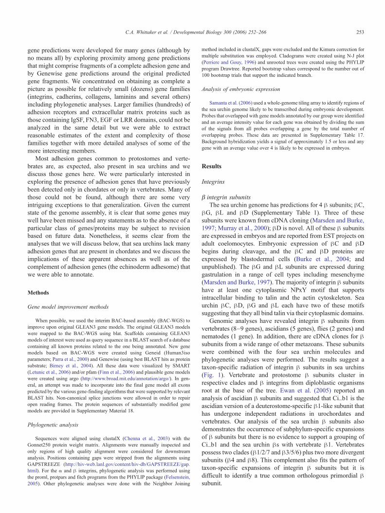
Genomic analyses have revealed integrin " subunits fromvertebrates (8–9 genes), ascidians (5 genes), flies (2 genes) andnematodes (1 gene). In addition, there are cDNA clones for "subunits from a wide range of other metazoans. These subunitswere combined with the four sea urchin molecules andphylogenetic analyses were performed. The results suggest ataxon-specific radiation of integrin " subunits in sea urchins(Fig. 1). Vertebrate and protostome " subunits cluster inrespective clades and " integrins from diploblastic organismsroot at the base of the tree. Ewan et al. (2005) reported ananalysis of ascidian " subunits and suggested that Ci_b1 is theascidian version of a deuterostome-specific "1-like subunit thathas undergone independent radiations in urochordates andvertebrates. Our analysis of the sea urchin " subunits alsodemonstrates the occurrence of subphylum-specific expansionsof " subunits but there is no evidence to support a grouping ofCi_b1 and the sea urchin "s with vertebrate "1. Vertebratespossess two clades ("1/2/7 and "3/5/6) plus two more divergentsubunits ("4 and "8). This complement also fits the pattern oftaxon-specific expansions of integrin " subunits but it isdifficult to identify a true common orthologous primordial "subunit.
253C.A. Whittaker et al. / Developmental Biology 300 (2006) 252–266
The vertebrate "4 subunit is distinctive structurally in havinga large cytoplasmic domain that associates with intermediatefilaments. The ascidian genome reveals that this is a sharedfeature of chordates (Ewan et al., 2005; our unpublishedanalyses). There is no similar subunit predicted from the seaurchin genome, suggesting that the "4 subunit is a derived,shared feature of chordates.
" integrin subunitsThere are 8 gene predictions for ! integrin subunits from
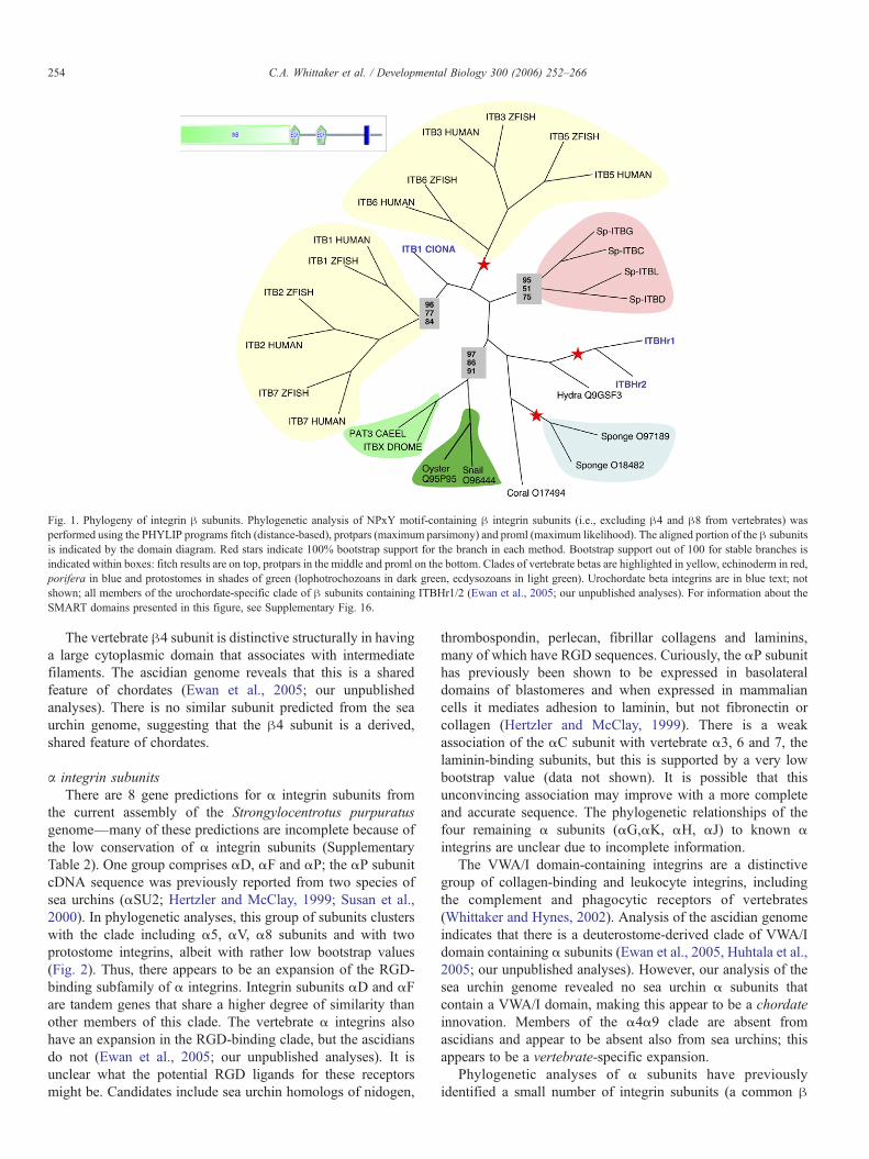
the current assembly of the Strongylocentrotus purpuratusgenome—many of these predictions are incomplete because ofthe low conservation of ! integrin subunits (SupplementaryTable 2). One group comprises !D, !F and !P; the !P subunitcDNA sequence was previously reported from two species ofsea urchins (!SU2; Hertzler and McClay, 1999; Susan et al.,2000). In phylogenetic analyses, this group of subunits clusterswith the clade including !5, !V, !8 subunits and with twoprotostome integrins, albeit with rather low bootstrap values(Fig. 2). Thus, there appears to be an expansion of the RGD-binding subfamily of ! integrins. Integrin subunits !D and !Fare tandem genes that share a higher degree of similarity thanother members of this clade. The vertebrate ! integrins alsohave an expansion in the RGD-binding clade, but the ascidiansdo not (Ewan et al., 2005; our unpublished analyses). It isunclear what the potential RGD ligands for these receptorsmight be. Candidates include sea urchin homologs of nidogen,
thrombospondin, perlecan, fibrillar collagens and laminins,many of which have RGD sequences. Curiously, the !P subunithas previously been shown to be expressed in basolateraldomains of blastomeres and when expressed in mammaliancells it mediates adhesion to laminin, but not fibronectin orcollagen (Hertzler and McClay, 1999). There is a weakassociation of the !C subunit with vertebrate !3, 6 and 7, thelaminin-binding subunits, but this is supported by a very lowbootstrap value (data not shown). It is possible that thisunconvincing association may improve with a more completeand accurate sequence. The phylogenetic relationships of thefour remaining ! subunits (!G,!K, !H, !J) to known !integrins are unclear due to incomplete information.
The VWA/I domain-containing integrins are a distinctivegroup of collagen-binding and leukocyte integrins, includingthe complement and phagocytic receptors of vertebrates(Whittaker and Hynes, 2002). Analysis of the ascidian genomeindicates that there is a deuterostome-derived clade of VWA/Idomain containing ! subunits (Ewan et al., 2005, Huhtala et al.,2005; our unpublished analyses). However, our analysis of thesea urchin genome revealed no sea urchin ! subunits thatcontain a VWA/I domain, making this appear to be a chordateinnovation. Members of the !4!9 clade are absent fromascidians and appear to be absent also from sea urchins; thisappears to be a vertebrate-specific expansion.
Phylogenetic analyses of ! subunits have previouslyidentified a small number of integrin subunits (a common "
Fig. 1. Phylogeny of integrin " subunits. Phylogenetic analysis of NPxY motif-containing " integrin subunits (i.e., excluding "4 and "8 from vertebrates) wasperformed using the PHYLIP programs fitch (distance-based), protpars (maximum parsimony) and proml (maximum likelihood). The aligned portion of the " subunitsis indicated by the domain diagram. Red stars indicate 100% bootstrap support for the branch in each method. Bootstrap support out of 100 for stable branches isindicated within boxes: fitch results are on top, protpars in the middle and proml on the bottom. Clades of vertebrate betas are highlighted in yellow, echinoderm in red,porifera in blue and protostomes in shades of green (lophotrochozoans in dark green, ecdysozoans in light green). Urochordate beta integrins are in blue text; notshown; all members of the urochordate-specific clade of " subunits containing ITBHr1/2 (Ewan et al., 2005; our unpublished analyses). For information about theSMART domains presented in this figure, see Supplementary Fig. 16.
254 C.A. Whittaker et al. / Developmental Biology 300 (2006) 252–266
subunit and two ! subunits, one apparently RGD-specific andone laminin binding) that appear widely shared in the bilaterianmetazoans (Hynes and Zhao, 2000; Hynes, 2002). Theseevolutionarily ancient subunits appear to have been supple-mented by diversification of taxon-specific clades. The additionof the ! subunit predictions from the ascidian and sea urchingenomes has made this analysis somewhat less clear. Whereasadditional taxon-specific clades occur in both, clear members ofthe presumed basal RGD- and laminin-binding families are lesswell defined in sea urchins. As mentioned, this could be aconsequence of the low homology and incomplete nature ofsome of the gene predictions and further research usingcomplete cDNA sequences will be needed to clarify thesituation.
Cadherins
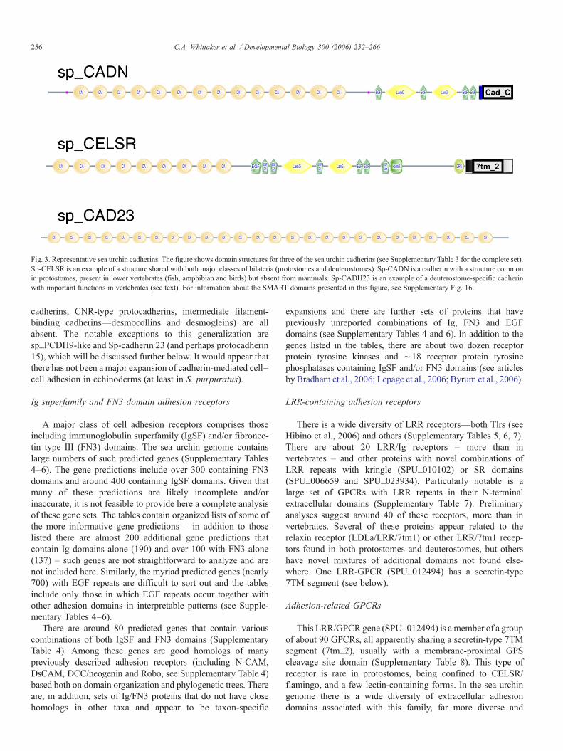
The complement of cadherins in the sea urchin genome issurprisingly small—fewer than a dozen well-defined represen-tatives; fewer even than flies or worms (Supplementary Table3). This limited set of cadherins has some surprising features.There are only two genes encoding catenin-binding cytoplas-mic domains characteristic of typical classical type I andatypical type II cadherins of vertebrates and neither of the seaurchin genes encodes such a typical vertebrate-type classicalcadherin. One of these two sea urchin catenin-bindingcadherins (Sp-CADN) is orthologous to Lytechinus variegates
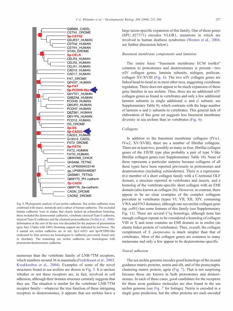
G-cadherin (Miller and McClay, 1997) and is most likeprotostome-type cadherins, containing LamG and EGFdomains as well as cadherin domains (Fig. 3). It turns outthat non-mammalian vertebrates (fish, amphibians and birds)do have such cadherins, termed type III cadherins by Tanabe etal. (2004). Therefore, Sp-CADN represents a class of cadherinsfound in both protostomes and deuterostomes but missing inmammals—an unusual phylogenetic distribution. The secondsea urchin catenin-binding cadherin appears to be an echino-derm ortholog of Dachsous/protocadherin 16 (found inbilateria). The sea urchin genome also contains apparentorthologs of other large cadherin superfamily moleculescommon to protostomes and deuterostomes (bilateria); namely,fat, fatH, flamingo/CELSR and calsyntenin (Figs. 3 and 4). Incontrast, two sea urchin cadherins, sp_CAD23 and sp_PCDH9-like, both good homologs of vertebrate counterparts, werepreviously known only in chordates (Fig. 4, blue arrows).Sp_PCDH9-like is homologous with the vertebrate protocad-herins 1, 7, 9, 11 and 20. Four other gene predictions withcadherin domains, several with only one or two domains, aredifficult to assign clear orthologies, although several giveprotocadherin 15 as their top BLAST hits (see below). Fig. 3shows some representative examples of the sea urchin cadheringene set and Fig. 4 shows the phylogenetic analysis.
Overall, this complement of cadherins is more invertebratethan vertebrate in character and major vertebrate-specificexpansions (classical type I and atypical type II catenin-binding
Fig. 2. Phylogeny of integrin ! subunits. Phylogenetic analysis of selected sea urchin and other non-VWA domain-containing ! integrin subunits was performed usingthe PHYLIP programs fitch (distance-based), protpars (maximum parsimony) and proml (maximum likelihood). The aligned portion of the ! subunits is indicated bythe domain diagram. Only three out of the eight sea urchin alpha gene predictions are included in these analyses because it was not possible to obtain sufficientlyaccurate gene models representing the other five. Red stars indicate 100% bootstrap support for the branch in each method. Bootstrap support out of 100 for stablebranches is indicated within boxes: fitch results are on top, protpars in the middle and proml on the bottom. Note the echinoderm-specific clade (red), distinct from, butproximal with, the vertebrate RGD-specific clade (5/8/IIb). Other clades are the laminin-specific (3/6/7) and vertebrate-specific (4/9) subunits. For information aboutthe SMART domains presented in this figure, see Supplementary Fig. 16.
255C.A. Whittaker et al. / Developmental Biology 300 (2006) 252–266
cadherins, CNR-type protocadherins, intermediate filament-binding cadherins—desmocollins and desmogleins) are allabsent. The notable exceptions to this generalization aresp_PCDH9-like and Sp-cadherin 23 (and perhaps protocadherin15), which will be discussed further below. It would appear thatthere has not been a major expansion of cadherin-mediated cell–cell adhesion in echinoderms (at least in S. purpuratus).
Ig superfamily and FN3 domain adhesion receptors
A major class of cell adhesion receptors comprises thoseincluding immunoglobulin superfamily (IgSF) and/or fibronec-tin type III (FN3) domains. The sea urchin genome containslarge numbers of such predicted genes (Supplementary Tables4–6). The gene predictions include over 300 containing FN3domains and around 400 containing IgSF domains. Given thatmany of these predictions are likely incomplete and/orinaccurate, it is not feasible to provide here a complete analysisof these gene sets. The tables contain organized lists of some ofthe more informative gene predictions – in addition to thoselisted there are almost 200 additional gene predictions thatcontain Ig domains alone (190) and over 100 with FN3 alone(137) – such genes are not straightforward to analyze and arenot included here. Similarly, the myriad predicted genes (nearly700) with EGF repeats are difficult to sort out and the tablesinclude only those in which EGF repeats occur together withother adhesion domains in interpretable patterns (see Supple-mentary Tables 4–6).
There are around 80 predicted genes that contain variouscombinations of both IgSF and FN3 domains (SupplementaryTable 4). Among these genes are good homologs of manypreviously described adhesion receptors (including N-CAM,DsCAM, DCC/neogenin and Robo, see Supplementary Table 4)based both on domain organization and phylogenetic trees. Thereare, in addition, sets of Ig/FN3 proteins that do not have closehomologs in other taxa and appear to be taxon-specific
expansions and there are further sets of proteins that havepreviously unreported combinations of Ig, FN3 and EGFdomains (see Supplementary Tables 4 and 6). In addition to thegenes listed in the tables, there are about two dozen receptorprotein tyrosine kinases and !18 receptor protein tyrosinephosphatases containing IgSF and/or FN3 domains (see articlesby Bradham et al., 2006; Lepage et al., 2006; Byrum et al., 2006).
LRR-containing adhesion receptors
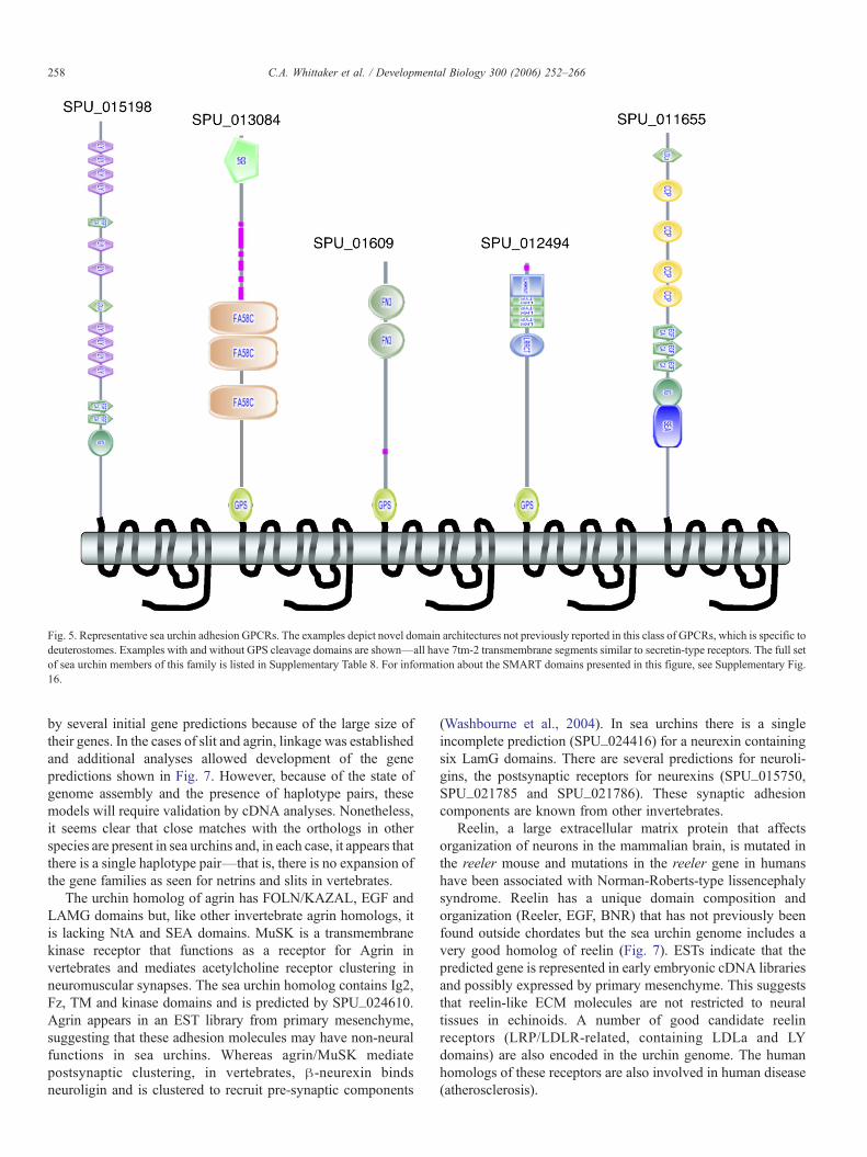
There is a wide diversity of LRR receptors—both Tlrs (seeHibino et al., 2006) and others (Supplementary Tables 5, 6, 7).There are about 20 LRR/Ig receptors – more than invertebrates – and other proteins with novel combinations ofLRR repeats with kringle (SPU_010102) or SR domains(SPU_006659 and SPU_023934). Particularly notable is alarge set of GPCRs with LRR repeats in their N-terminalextracellular domains (Supplementary Table 7). Preliminaryanalyses suggest around 40 of these receptors, more than invertebrates. Several of these proteins appear related to therelaxin receptor (LDLa/LRR/7tm1) or other LRR/7tm1 recep-tors found in both protostomes and deuterostomes, but othershave novel mixtures of additional domains not found else-where. One LRR-GPCR (SPU_012494) has a secretin-type7TM segment (see below).
Adhesion-related GPCRs
This LRR/GPCR gene (SPU_012494) is a member of a groupof about 90 GPCRs, all apparently sharing a secretin-type 7TMsegment (7tm_2), usually with a membrane-proximal GPScleavage site domain (Supplementary Table 8). This type ofreceptor is rare in protostomes, being confined to CELSR/flamingo, and a few lectin-containing forms. In the sea urchingenome there is a wide diversity of extracellular adhesiondomains associated with this family, far more diverse and
Fig. 3. Representative sea urchin cadherins. The figure shows domain structures for three of the sea urchin cadherins (see Supplementary Table 3 for the complete set).Sp-CELSR is an example of a structure shared with both major classes of bilateria (protostomes and deuterostomes). Sp-CADN is a cadherin with a structure commonin protostomes, present in lower vertebrates (fish, amphibian and birds) but absent from mammals. Sp-CADH23 is an example of a deuterostome-specific cadherinwith important functions in vertebrates (see text). For information about the SMART domains presented in this figure, see Supplementary Fig. 16.
256 C.A. Whittaker et al. / Developmental Biology 300 (2006) 252–266
numerous than the vertebrate family of LNB-7TM receptors,which numbers around 36 in mammals (Fredriksson et al., 2003;Kwakkenbos et al., 2004). Examples of some of the novelstructures found in sea urchins are shown in Fig. 5. It is unclearwhether or not these receptors are, in fact, involved in celladhesion, although their domain structure certainly suggests thatthey are. The situation is similar for the vertebrate LNB-7TMreceptor family—whatever the true function of these intriguingreceptors in deuterostomes, it appears that sea urchins have a
large taxon-specific expansion of this family. One of these genes(SPU_027371) encodes VLGR1, mutations in which areinvolved in human deafness syndromes (Weston et al., 2004;see further discussion below).
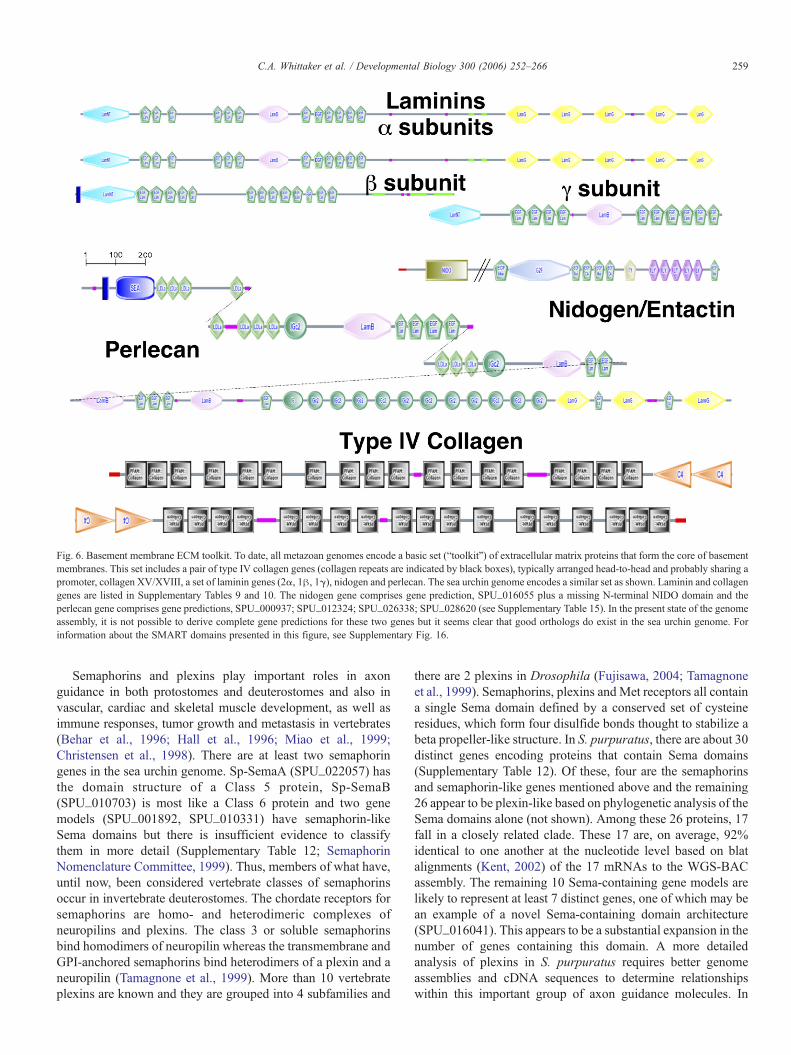
Basement membrane components and laminins
The entire basic “basement membrane ECM toolkit”common to protostomes and deuterostomes is present—two!IV collagen genes, laminin subunits, nidogen, perlecan,collagen XV/XVIII (Fig. 6). The two !IV collagen genes arelinked head-to-head as in most other taxa, suggesting coordinateregulation. There does not appear to be much expansion of thesegene families in sea urchins. Thus, there are no additional !IVcollagen genes as found in vertebrates and only a few additionallaminin subunits (a single additional ! and " subunit; seeSupplementary Table 9), which contrasts with the large numberof laminin ! and " subunits in vertebrates. This general lack ofelaboration of this gene set suggests less basement membranediversity in sea urchins than in vertebrates (Fig. 6).
Collagens
In addition to the basement membrane collagens (IV!1,IV!2, XV/XVIII), there are a number of fibrillar collagens.There are at least two, possibly as many as four, fibrillar collagengenes of the I/II/III type plus probably a pair of type V-likefibrillar collagen genes (see Supplementary Table 10). None ofthese represents a particular surprise because collagens of allthese types have been reported previously in protostomes anddeuterostomes (including echinoderms). There is a representa-tive member of a short collagen family with a C-terminal OLFdomain, a structure reported in vertebrates and insects, and ahomolog of the vertebrate-specific short collagen with an EMIdomain (also known as collagen 26). However, in contrast, thereappear to be no clear examples of the complex collagensprevalent in vertebrates (types VI, VII, XII, XIV, containingVWA and FN3 domains), although one sea urchin collagen gene(sp_col91) has some features of this family (see SupplementaryFig. 11). There are several C1q homologs, although none hasenough collagen repeats to be considered a homolog of collagenVIII or X and none contains an EMI domain as in emilin (anelastin linker protein of vertebrates). Thus, overall, the collagencomplement of S. purpuratus is much simpler than that ofvertebrates. Most of the collagen genes are common to manymetazoans and only a few appear to be deuterostome-specific.
Neural adhesion
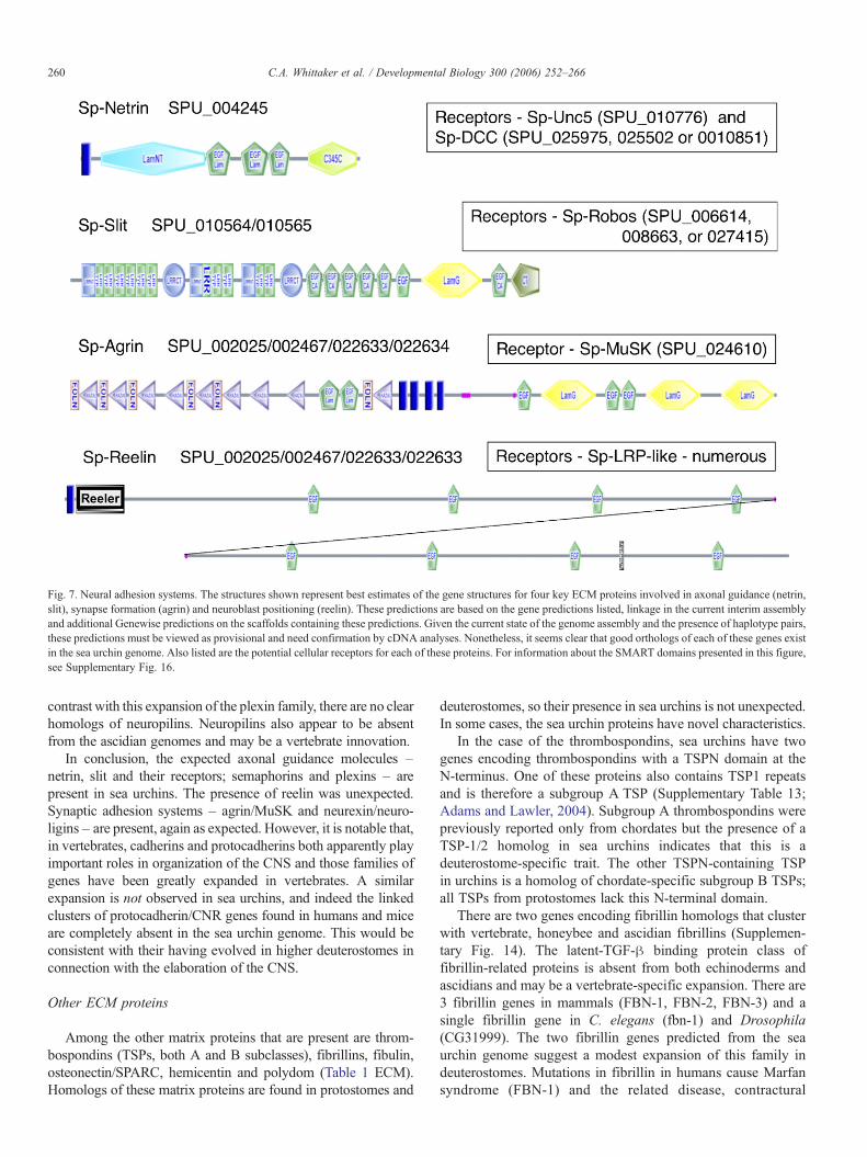
The sea urchin genome encodes good homologs of the axonalguidance matrix proteins, netrin and slit, and of the postsynapticclustering matrix protein, agrin (Fig. 7). That is not surprisingbecause those are known in both protostomes and deutero-stomes. In each of these cases, good candidates for the receptorsfor these axon guidance molecules are also found in the seaurchin genome (see Fig. 7 for listings). Netrin is encoded in asingle gene prediction, but the other proteins are each encoded
Fig. 4. Phylogenetic analysis of sea urchin cadherins. Sea urchin cadherins werecombined with insect, nematode and a subset of human cadherins. The excludedhuman cadherins were in clades that clearly lacked an echinoderm homolog;these included the desmosomal cadherins, vertebrate classical/Type-I cadherins,atypical/Type-II cadherins and the clustered protocadherins (Nollet et al., 2000).Information at the root of the tree was discarded for the purpose of presentation(grey bar). Clades with 100% bootstrap support are indicated by red boxes. The8 named sea urchin cadherins are in red. Sp-CAD23 and Sp-PCDH9-like(indicated by blue arrows) are homologous to cadherins previously found onlyin chordates. The remaining sea urchin cadherins are homologous withprotostome/deuterostome cadherins.
257C.A. Whittaker et al. / Developmental Biology 300 (2006) 252–266
by several initial gene predictions because of the large size oftheir genes. In the cases of slit and agrin, linkage was establishedand additional analyses allowed development of the genepredictions shown in Fig. 7. However, because of the state ofgenome assembly and the presence of haplotype pairs, thesemodels will require validation by cDNA analyses. Nonetheless,it seems clear that close matches with the orthologs in otherspecies are present in sea urchins and, in each case, it appears thatthere is a single haplotype pair—that is, there is no expansion ofthe gene families as seen for netrins and slits in vertebrates.
The urchin homolog of agrin has FOLN/KAZAL, EGF andLAMG domains but, like other invertebrate agrin homologs, itis lacking NtA and SEA domains. MuSK is a transmembranekinase receptor that functions as a receptor for Agrin invertebrates and mediates acetylcholine receptor clustering inneuromuscular synapses. The sea urchin homolog contains Ig2,Fz, TM and kinase domains and is predicted by SPU_024610.Agrin appears in an EST library from primary mesenchyme,suggesting that these adhesion molecules may have non-neuralfunctions in sea urchins. Whereas agrin/MuSK mediatepostsynaptic clustering, in vertebrates, "-neurexin bindsneuroligin and is clustered to recruit pre-synaptic components
(Washbourne et al., 2004). In sea urchins there is a singleincomplete prediction (SPU_024416) for a neurexin containingsix LamG domains. There are several predictions for neuroli-gins, the postsynaptic receptors for neurexins (SPU_015750,SPU_021785 and SPU_021786). These synaptic adhesioncomponents are known from other invertebrates.
Reelin, a large extracellular matrix protein that affectsorganization of neurons in the mammalian brain, is mutated inthe reeler mouse and mutations in the reeler gene in humanshave been associated with Norman-Roberts-type lissencephalysyndrome. Reelin has a unique domain composition andorganization (Reeler, EGF, BNR) that has not previously beenfound outside chordates but the sea urchin genome includes avery good homolog of reelin (Fig. 7). ESTs indicate that thepredicted gene is represented in early embryonic cDNA librariesand possibly expressed by primary mesenchyme. This suggeststhat reelin-like ECM molecules are not restricted to neuraltissues in echinoids. A number of good candidate reelinreceptors (LRP/LDLR-related, containing LDLa and LYdomains) are also encoded in the urchin genome. The humanhomologs of these receptors are also involved in human disease(atherosclerosis).
Fig. 5. Representative sea urchin adhesion GPCRs. The examples depict novel domain architectures not previously reported in this class of GPCRs, which is specific todeuterostomes. Examples with and without GPS cleavage domains are shown—all have 7tm-2 transmembrane segments similar to secretin-type receptors. The full setof sea urchin members of this family is listed in Supplementary Table 8. For information about the SMART domains presented in this figure, see Supplementary Fig.16.
258 C.A. Whittaker et al. / Developmental Biology 300 (2006) 252–266
Semaphorins and plexins play important roles in axonguidance in both protostomes and deuterostomes and also invascular, cardiac and skeletal muscle development, as well asimmune responses, tumor growth and metastasis in vertebrates(Behar et al., 1996; Hall et al., 1996; Miao et al., 1999;Christensen et al., 1998). There are at least two semaphoringenes in the sea urchin genome. Sp-SemaA (SPU_022057) hasthe domain structure of a Class 5 protein, Sp-SemaB(SPU_010703) is most like a Class 6 protein and two genemodels (SPU_001892, SPU_010331) have semaphorin-likeSema domains but there is insufficient evidence to classifythem in more detail (Supplementary Table 12; SemaphorinNomenclature Committee, 1999). Thus, members of what have,until now, been considered vertebrate classes of semaphorinsoccur in invertebrate deuterostomes. The chordate receptors forsemaphorins are homo- and heterodimeric complexes ofneuropilins and plexins. The class 3 or soluble semaphorinsbind homodimers of neuropilin whereas the transmembrane andGPI-anchored semaphorins bind heterodimers of a plexin and aneuropilin (Tamagnone et al., 1999). More than 10 vertebrateplexins are known and they are grouped into 4 subfamilies and
there are 2 plexins in Drosophila (Fujisawa, 2004; Tamagnoneet al., 1999). Semaphorins, plexins and Met receptors all containa single Sema domain defined by a conserved set of cysteineresidues, which form four disulfide bonds thought to stabilize abeta propeller-like structure. In S. purpuratus, there are about 30distinct genes encoding proteins that contain Sema domains(Supplementary Table 12). Of these, four are the semaphorinsand semaphorin-like genes mentioned above and the remaining26 appear to be plexin-like based on phylogenetic analysis of theSema domains alone (not shown). Among these 26 proteins, 17fall in a closely related clade. These 17 are, on average, 92%identical to one another at the nucleotide level based on blatalignments (Kent, 2002) of the 17 mRNAs to the WGS-BACassembly. The remaining 10 Sema-containing gene models arelikely to represent at least 7 distinct genes, one of which may bean example of a novel Sema-containing domain architecture(SPU_016041). This appears to be a substantial expansion in thenumber of genes containing this domain. A more detailedanalysis of plexins in S. purpuratus requires better genomeassemblies and cDNA sequences to determine relationshipswithin this important group of axon guidance molecules. In
Fig. 6. Basement membrane ECM toolkit. To date, all metazoan genomes encode a basic set (“toolkit”) of extracellular matrix proteins that form the core of basementmembranes. This set includes a pair of type IV collagen genes (collagen repeats are indicated by black boxes), typically arranged head-to-head and probably sharing apromoter, collagen XV/XVIII, a set of laminin genes (2!, 1", 1#), nidogen and perlecan. The sea urchin genome encodes a similar set as shown. Laminin and collagengenes are listed in Supplementary Tables 9 and 10. The nidogen gene comprises gene prediction, SPU_016055 plus a missing N-terminal NIDO domain and theperlecan gene comprises gene predictions, SPU_000937; SPU_012324; SPU_026338; SPU_028620 (see Supplementary Table 15). In the present state of the genomeassembly, it is not possible to derive complete gene predictions for these two genes but it seems clear that good orthologs do exist in the sea urchin genome. Forinformation about the SMART domains presented in this figure, see Supplementary Fig. 16.
259C.A. Whittaker et al. / Developmental Biology 300 (2006) 252–266
contrast with this expansion of the plexin family, there are no clearhomologs of neuropilins. Neuropilins also appear to be absentfrom the ascidian genomes and may be a vertebrate innovation.
In conclusion, the expected axonal guidance molecules –netrin, slit and their receptors; semaphorins and plexins – arepresent in sea urchins. The presence of reelin was unexpected.Synaptic adhesion systems – agrin/MuSK and neurexin/neuro-ligins – are present, again as expected. However, it is notable that,in vertebrates, cadherins and protocadherins both apparently playimportant roles in organization of the CNS and those families ofgenes have been greatly expanded in vertebrates. A similarexpansion is not observed in sea urchins, and indeed the linkedclusters of protocadherin/CNR genes found in humans and miceare completely absent in the sea urchin genome. This would beconsistent with their having evolved in higher deuterostomes inconnection with the elaboration of the CNS.
Other ECM proteins
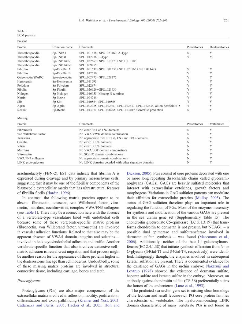
Among the other matrix proteins that are present are throm-bospondins (TSPs, both A and B subclasses), fibrillins, fibulin,osteonectin/SPARC, hemicentin and polydom (Table 1 ECM).Homologs of these matrix proteins are found in protostomes and
deuterostomes, so their presence in sea urchins is not unexpected.In some cases, the sea urchin proteins have novel characteristics.
In the case of the thrombospondins, sea urchins have twogenes encoding thrombospondins with a TSPN domain at theN-terminus. One of these proteins also contains TSP1 repeatsand is therefore a subgroup A TSP (Supplementary Table 13;Adams and Lawler, 2004). Subgroup A thrombospondins werepreviously reported only from chordates but the presence of aTSP-1/2 homolog in sea urchins indicates that this is adeuterostome-specific trait. The other TSPN-containing TSPin urchins is a homolog of chordate-specific subgroup B TSPs;all TSPs from protostomes lack this N-terminal domain.
There are two genes encoding fibrillin homologs that clusterwith vertebrate, honeybee and ascidian fibrillins (Supplemen-tary Fig. 14). The latent-TGF-" binding protein class offibrillin-related proteins is absent from both echinoderms andascidians and may be a vertebrate-specific expansion. There are3 fibrillin genes in mammals (FBN-1, FBN-2, FBN-3) and asingle fibrillin gene in C. elegans (fbn-1) and Drosophila(CG31999). The two fibrillin genes predicted from the seaurchin genome suggest a modest expansion of this family indeuterostomes. Mutations in fibrillin in humans cause Marfansyndrome (FBN-1) and the related disease, contractural
Fig. 7. Neural adhesion systems. The structures shown represent best estimates of the gene structures for four key ECM proteins involved in axonal guidance (netrin,slit), synapse formation (agrin) and neuroblast positioning (reelin). These predictions are based on the gene predictions listed, linkage in the current interim assemblyand additional Genewise predictions on the scaffolds containing these predictions. Given the current state of the genome assembly and the presence of haplotype pairs,these predictions must be viewed as provisional and need confirmation by cDNA analyses. Nonetheless, it seems clear that good orthologs of each of these genes existin the sea urchin genome. Also listed are the potential cellular receptors for each of these proteins. For information about the SMART domains presented in this figure,see Supplementary Fig. 16.
260 C.A. Whittaker et al. / Developmental Biology 300 (2006) 252–266
arachnodactyly (FBN-2). EST data indicate that fibrillin A isexpressed during cleavage and by primary mesenchyme cells,suggesting that it may be one of the fibrillar components of theblastocoele extracellular matrix that has ultrastructural featuresof fibrillin fibrils (Hardin, 1996).
In contrast, the following matrix proteins appear to beabsent—fibronectin, tenascins, von Willebrand factor, vitro-nectin, matrilins, cochlin/vitrin, complex VWA/FN3 collagens(see Table 1). There may be a connection here with the absenceof a vertebrate-type vasculature lined with endothelial cellsbecause some of these vertebrate-specific matrix proteins(fibronectin, von Willebrand factor, vitronectin) are involvedin vascular adhesion functions. Related to that also may be theapparent absence of VWA/I domain integrins and selectins—involved in leukocyte/endothelial adhesion and traffic. Anothervertebrate-specific function that also involves extensive cell–matrix adhesion is neural crest migration and that process mightbe another reason for the appearance of these proteins higher inthe deuterostome lineage than echinoderms. Undoubtedly, someof these missing matrix proteins are involved in structuralconnective tissue, including cartilage, bones and teeth.
Proteoglycans
Proteoglycans (PGs) are also major components of theextracellular matrix involved in adhesion, motility, proliferation,differentiation and axon pathfinding (Kramer and Yost, 2003;Cattaruzza and Perris, 2005; Hacker et al., 2005; Holt and
Dickson, 2005). PGs consist of core proteins decorated with oneor more long repeating disaccharide chains called glycosami-noglycans (GAGs). GAGs are heavily sulfated molecules thatinteract with extracellular cytokines, growth factors andmorphogens. Variations in GAG sulfation patterns can modulatetheir affinities for extracellular proteins (Mulloy, 2005). Thestatus of GAG sulfation therefore plays an important role inregulating the function of PGs. Most of the enzymes necessaryfor synthesis and modification of the various GAGs are presentin the sea urchin gene set (Supplementary Table 15). Thechondroitin glucuronate C5-epimerase (EC 5.1.3.19) that trans-forms chondroitin to dermatan is not present, but NCAG1 – apossible dual epimerase and sulfotransferase involved indermatan sulfate synthesis – was found (Maccarana et al.,2006). Additionally, neither of the beta-1,4-galactosyltrans-ferases (EC 2.4.1.38) that initiate synthesis of keratan from N- orO-glycans ("4Gal-T1 and "4Gal-T4, respectively) were identi-fied. Intriguingly though, the enzymes involved in subsequentkeratan sulfation are present. There is documented evidence forthe existence of GAGs in the urchin embryo; Nakatsuji andLovtrup (1978) showed the existence of dermatan sulfate,heparan sulfate and keratan sulfate in the embryo. Moreover, anantibody against chondroitin sulfate (CS-56) preferentially stainsthe lumen of the archenteron (Lane et al., 1993).
The predicted sea urchin gene set is missing clear homologsof the lectican and small leucine-rich PG core protein familiescharacteristic of vertebrates. The hyaluronan-binding LINKdomain characteristic of many vertebrate PGs is not found in
Table 1ECM proteins
Present
Protein Common name Comments Protostomes Deuterostomes
Thrombospondin Sp-TSPA1 SPU_001638+SPU_023469; A-Type N YThrombospondin Sp-TSPB1 SPU_012936; B-Type Y YThrombospondin Sp-TSP_like-1 SPU_022667+SPU_017370+SPU_013106Thrombospondin Sp-TSP_like-2 SPU_009755Fibrillin Sp-Fibrillin A SPU_001532+SPU_001533+SPU_020166+SPU_021495 Y YFibrillin Sp-Fibrillin B SPU_012550 Y YOsteonectin/SPARC Sp-osteonectin SPU_002473+SPU_028275 Y YHemicentin Sp-Hemicentin SPU_011693 Y YPolydom Sp-Polydom SPU_022974 Y YFibulin Sp-Fibulin SPU_026629+SPU_021630 Y YNidogen Sp-Nidogen SPU_016055; Missing N-terminus Y YNetrin Sp-Netrin SPU_004245 Y YSlit Sp-Slit SPU_010564, SPU_010565 Y YAgrin Sp-Agrin SPU_002025, SPU_002467, SPU_022633, SPU_022634, all on Scaffold 675 Y YReelin Sp-Reelin SPU_013071, SPU_008268, SPU_023409; Genewise prediction N Y
Missing
Protein Comments Protostomes Vertebrates
Fibronectin No clear FN1 or FN2 domains N Yvon Willebrand factor No VWA/VWD domain combination N YTenascins No appropriate mix of EGF, FN3 and FBG domains N YCochlin No clear LCCL domains N YVitrin No clear LCCL domains N YMatrilins No VWA/EGF domain combinations N YVitronectin No SO/HX domain combinations N YVWA/FN3 collagens No appropriate domain combinations N YLINK proteoglycans No LINK domains coupled with other signature domains N Y
261C.A. Whittaker et al. / Developmental Biology 300 (2006) 252–266
protostomes and is apparently very rare in the sea urchin genome(two gene predictions only) and is not found in domaincombinations diagnostic of proteoglycans. Moreover, SPOCK/testican (a SPARC-related PG) seems to be absent from theechinoderm genome while homologs of SPARC and SMOC(another SPARC-related PG) are present. SPOCK/testican is alsoabsent from the cnidarian, protostome and urochordate genomessequenced to date, suggesting it is a vertebrate-specific gene.However, basement membrane PGs (perlecan, bamacan, agrinand SMOC) and membrane-associated PGs (betaglycan, dystro-glycan, syndecan and glypicans) are present (SupplementaryTable 15). The sea urchin has one syndecan and two glypicangenes, one of the 1/2/4/6 class and one of the 3/5 class. These areexpressed in distinct but overlapping radially symmetrical
patterns in the S. purpuratus gastrula embryo as determined byin situ hybridization (K.F.B., unpublished). A similar pattern ofsyndecan expression was observed in Anthocidaris crassispinaurchin blastula and gastrula embryos (Tomita et al., 2000).
Thus PGs with GAGs are present and expressed in seaurchins as in other bilateria – in basement membranes and aspart of signal transduction systems – but large structural PGs areabsent. This presumably correlates with the absence of hyalinecartilage and bone.
Echinoderm homologues of human deaf/blindness genes
One of the more surprising and striking observations toemerge from the analysis of potential adhesion genes/proteins
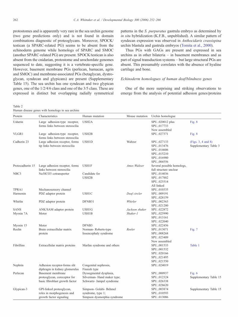
Table 2Human disease genes with homologs in sea urchins
Protein Characteristics Human mutation Mouse mutation Urchin homologue
Usherin Large adhesion-type receptor,forms links between stereocilia
USH2A SPU_020012 plus Fig. 8SPU_017733Now assembled
VLGR1 Large adhesion-type receptor,forms links between stereocilia
USH2B SPU_027371 Fig. 8
Cadherin 23 Large adhesion receptor, formstip links between stereocilia
USH1D Waltzer SPU_027133 (Figs. 3, 4 and 8)SPU_013476 Supplementary Table 3SPU_014606SPU_015210SPU_016980SPU_004556
Protocadherin 15 Large adhesion receptor, formslinks between stereocilia
USH1F Ames Waltzer Several possible homologs,full structure unclear
NBC3 Na/HCO3 cotransporter Candidate forUSH2B
SPU_014036SPU_017882SPU_025514All linked
TPRA1 Mechanosensory channel SPU_010335Harmonin PDZ adaptor protein USH1C Deaf circler SPU_009191
SPU_028159Whirlin PDZ adaptor protein DFNB31 Whirler SPU_002363
SPU_021200SANS ANK/SAM adaptor protein USH1G Jackson shaker SPU_022872Myosin 7A Motor USH1B Shaker-1 SPU_025990
SPU_013161SPU_022040
Myosin 15 Motor DFNB3 SPU_022456Reelin Brain extracellular matrix
proteinNorman- Roberts-typelissencephaly syndrome
Reeler SPU_013071 Fig. 7SPU_008268SPU_023409New assembled
Fibrillins Extracellular matrix proteins Marfan syndrome and others SPU_001533 Table 1SPU_001532SPU_020166SPU_021495SPU_021550
Nephrin Adhesion receptor-forms slitdiphragm in kidney glomerulus
Congenital nephrosis,Finnish type
SPU_024019
Perlecan Basement membraneproteoglycan, coreceptor forbasic fibroblast growth factor
Dyssegmental dysplasia,Silverman- Hand maker type;Schwartz- Jampel syndrome
SPU_000937 Fig. 6SPU_012324 Supplementary Table 15SPU_026338SPU_028620
Glypican-3 GPI-linked proteoglycan,roles in morphogenesis andgrowth factor signaling
Simpson- Golabi- Behmelsyndrome, type 1;Simpson dysmorphia syndrome
SPU_003874 Supplementary Table 15SPU_010593SPU_013086
262 C.A. Whittaker et al. / Developmental Biology 300 (2006) 252–266
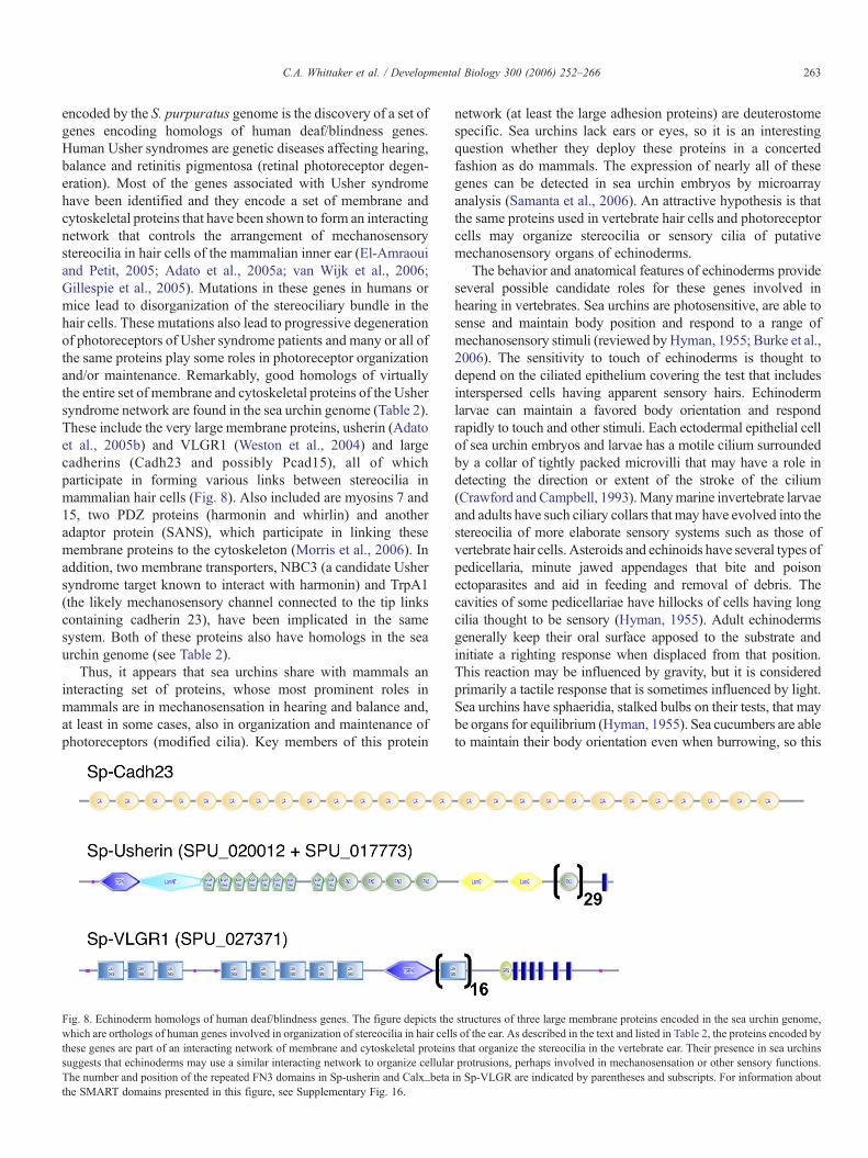
encoded by the S. purpuratus genome is the discovery of a set ofgenes encoding homologs of human deaf/blindness genes.Human Usher syndromes are genetic diseases affecting hearing,balance and retinitis pigmentosa (retinal photoreceptor degen-eration). Most of the genes associated with Usher syndromehave been identified and they encode a set of membrane andcytoskeletal proteins that have been shown to form an interactingnetwork that controls the arrangement of mechanosensorystereocilia in hair cells of the mammalian inner ear (El-Amraouiand Petit, 2005; Adato et al., 2005a; van Wijk et al., 2006;Gillespie et al., 2005). Mutations in these genes in humans ormice lead to disorganization of the stereociliary bundle in thehair cells. These mutations also lead to progressive degenerationof photoreceptors of Usher syndrome patients and many or all ofthe same proteins play some roles in photoreceptor organizationand/or maintenance. Remarkably, good homologs of virtuallythe entire set of membrane and cytoskeletal proteins of the Ushersyndrome network are found in the sea urchin genome (Table 2).These include the very large membrane proteins, usherin (Adatoet al., 2005b) and VLGR1 (Weston et al., 2004) and largecadherins (Cadh23 and possibly Pcad15), all of whichparticipate in forming various links between stereocilia inmammalian hair cells (Fig. 8). Also included are myosins 7 and15, two PDZ proteins (harmonin and whirlin) and anotheradaptor protein (SANS), which participate in linking thesemembrane proteins to the cytoskeleton (Morris et al., 2006). Inaddition, two membrane transporters, NBC3 (a candidate Ushersyndrome target known to interact with harmonin) and TrpA1(the likely mechanosensory channel connected to the tip linkscontaining cadherin 23), have been implicated in the samesystem. Both of these proteins also have homologs in the seaurchin genome (see Table 2).
Thus, it appears that sea urchins share with mammals aninteracting set of proteins, whose most prominent roles inmammals are in mechanosensation in hearing and balance and,at least in some cases, also in organization and maintenance ofphotoreceptors (modified cilia). Key members of this protein
network (at least the large adhesion proteins) are deuterostomespecific. Sea urchins lack ears or eyes, so it is an interestingquestion whether they deploy these proteins in a concertedfashion as do mammals. The expression of nearly all of thesegenes can be detected in sea urchin embryos by microarrayanalysis (Samanta et al., 2006). An attractive hypothesis is thatthe same proteins used in vertebrate hair cells and photoreceptorcells may organize stereocilia or sensory cilia of putativemechanosensory organs of echinoderms.
The behavior and anatomical features of echinoderms provideseveral possible candidate roles for these genes involved inhearing in vertebrates. Sea urchins are photosensitive, are able tosense and maintain body position and respond to a range ofmechanosensory stimuli (reviewed byHyman, 1955; Burke et al.,2006). The sensitivity to touch of echinoderms is thought todepend on the ciliated epithelium covering the test that includesinterspersed cells having apparent sensory hairs. Echinodermlarvae can maintain a favored body orientation and respondrapidly to touch and other stimuli. Each ectodermal epithelial cellof sea urchin embryos and larvae has a motile cilium surroundedby a collar of tightly packed microvilli that may have a role indetecting the direction or extent of the stroke of the cilium(Crawford and Campbell, 1993).Manymarine invertebrate larvaeand adults have such ciliary collars that may have evolved into thestereocilia of more elaborate sensory systems such as those ofvertebrate hair cells. Asteroids and echinoids have several types ofpedicellaria, minute jawed appendages that bite and poisonectoparasites and aid in feeding and removal of debris. Thecavities of some pedicellariae have hillocks of cells having longcilia thought to be sensory (Hyman, 1955). Adult echinodermsgenerally keep their oral surface apposed to the substrate andinitiate a righting response when displaced from that position.This reaction may be influenced by gravity, but it is consideredprimarily a tactile response that is sometimes influenced by light.Sea urchins have sphaeridia, stalked bulbs on their tests, that maybe organs for equilibrium (Hyman, 1955). Sea cucumbers are ableto maintain their body orientation even when burrowing, so this
Fig. 8. Echinoderm homologs of human deaf/blindness genes. The figure depicts the structures of three large membrane proteins encoded in the sea urchin genome,which are orthologs of human genes involved in organization of stereocilia in hair cells of the ear. As described in the text and listed in Table 2, the proteins encoded bythese genes are part of an interacting network of membrane and cytoskeletal proteins that organize the stereocilia in the vertebrate ear. Their presence in sea urchinssuggests that echinoderms may use a similar interacting network to organize cellular protrusions, perhaps involved in mechanosensation or other sensory functions.The number and position of the repeated FN3 domains in Sp-usherin and Calx_beta in Sp-VLGR are indicated by parentheses and subscripts. For information aboutthe SMART domains presented in this figure, see Supplementary Fig. 16.
263C.A. Whittaker et al. / Developmental Biology 300 (2006) 252–266
cannot depend only on tactile stimuli. They have statocystsincluding statoliths in vesicular cavities that are thought to beinvolved in maintaining their equilibrium in relation to gravity(Hyman, 1955). Many invertebrates have statocysts/statoliths thathave a role in geotaxis or body orientation and may have evolvedinto the macula of the vertebrate vestibular apparatus andeventually into the cochlea involved in hearing. It is therefore aplausible hypothesis that echinoderms have mechanosensorystereocilia that may be organized using the same proteins found invertebrate hair cells or other cells having modified cilia and itwould be interesting to test these hypotheses using theinformation from the genome annotation.
Conclusions
The echinoderm adhesome includes many hundreds of geneslikely to be involved in cell–cell and cell–matrix adhesion. Seaurchins have the basic metazoan adhesion gene set but there areseveral apparently taxon-specific expansions of cell adhesionmolecules. Overall, the echinoderm adhesome has more of an“invertebrate character”—that is, it is more similar to theadhesion gene sets of protostome invertebrates than to that ofvertebrates. That said, there are some clear examples of genes/proteins previously “chordate specific.”
Basic adhesion toolkit
The main classes of adhesion receptors (integrins, cadherins,Ig superfamily, FN3, LRR, C-type lectins) are all represented inthe S. purpuratus genome. The integrin family is larger thanthat in insects or nematodes but lacks several chordate-specificexpansions (see below). The cadherin gene set is surprisinglysmall and more like that of protostome invertebrates. Thebasement membrane “toolkit” (collagens IV, XV/XVIII,laminins, nidogen, perlecan) is present as in all metazoawhose genomes have been analyzed but it is little expanded.The basic set of neural guidance matrix proteins and theirreceptors is present, as are homologs of many known adhesionreceptors used in development of the nervous system (N-CAM,DsCAM, neogenin etc), much as in protostomes.
Taxon-specific expansions
Several families of adhesion proteins show expansions. Bothintegrin subunit families include what appear to be echinoderm-specific expansions, as is characteristic of other taxa. There aresubsets of Ig/FN3 adhesion receptors that appear to beechinoderm-specific expansions. Various families of LRR-domain-containing receptors appear expanded, including butnot limited to the Tlrs involved in innate immune defenses (seeHibino et al., 2006). It could be that the LRR domains have beenused in other families of adhesion receptors for purposes ofdefense against pathogens. The scavenger receptor (SR) domainfamily is also expanded (Hibino et al., 2006). One notableexpansion is of GPCRs with extended extracellular domains,many of them containing adhesion domains. This class ofproteins is significantly larger in S. purpuratus than in
vertebrates and it will be interesting to investigate the functionsof these enigmatic receptors.
Interesting absences
S. purpuratus lacks specific subfamilies of integrins,particularly those with VWA domains, including those that areinvolved in adhesion to collagen. This could be related to theabsence of complex collagens (see below). There is a paucity ofcadherins and many vertebrate cadherin subfamilies are missing—interestingly there are neither integrins nor cadherins of theintermediate-filament-linked subclasses although S. purpuratusdoes have intermediate filaments (Morris et al., 2006).
The absence of themore complex vertebrate-type collagens andproteoglycans is perhaps to be expected, given absence of cartilageand bones. Echinoderms are known to have collagenous tissuesthat undergo rapid, neurally mediated changes in mechanicalproperties (Motokawa, 1984; Wilkie, 2002). The stiffening of seaurchin spines in response to mechanical stimulation is a well-studied example of this (Wilkie, 1996; Takemae and Motokawa,2005). The low diversity of collagen types and similarity to thecollagens of other metazoans are consistent with hypotheses thatthe changes in mechanical properties of collagenous connectivetissues are mediated through accessory molecules in the matrixrather than the collagens themselves (Motokawa, 1984; Wilkie,2002; Trotter et al., 1996). Many vertebrate accessory ECMproteins are not found in S. purpuratus but others are present.
Other absences may reflect the absence of endothelial-linedvasculature. Many vertebrate ECM proteins are missing,including matrix proteins involved in assembly, maintenanceor functions of a high-pressure, high-shear, lined vascularsystem. Examples include fibronectin and von Willebrandfactor, both involved in adhesion of vascular cells duringhemostasis; fibronectin is also involved in angiogenesis.Thrombospondins are also involved in regulating angiogenesisin vertebrates but are present in sea urchins consistent withtheir having additional functions. Echinoderm coelomocytesare macrophage-like cells that appear to be mediators of theinnate immune responses of sea urchins. The biology of thesecells is poorly understood, but they are probably the equivalentof vertebrate thrombocytes and leukocytes. It would beinteresting to know which adhesion receptors are expressedon those cells – there are none of the characteristic vertebrateleukocyte integrins ("2 or !4 subclasses) and no selectins –presumably a low-shear circulation would impose feweradhesion demands. However, echinoderm coelomocytes likelyexpress some or many of the candidate innate immuneadhesion receptors.
Another potential reason for the absence of many adhesiveECM proteins may be the lack of neural crest migration. Lack ofexpansion of the laminin and type IV collagen families suggestsa lack of basement membrane diversity. As mentioned, some ofthe basic neural adhesion systems are present but it is notablethat there is very little expansion of the cadherin superfamily,believed to play major roles in establishing vertebrate nervoussystems. There are few protocadherins and none of the clusteredprotocadherin multigene loci found in vertebrates.
264 C.A. Whittaker et al. / Developmental Biology 300 (2006) 252–266
Surprising presences
In contrast with these absent adhesion proteins, the presenceof reelin mentioned earlier is something of a surprise becausethis gene is involved in controlling organization of neuronswithin mammalian brains. Its presence and embryonic expres-sion in sea urchins suggest that it plays some different role herebut it will be of interest to explore its expression in the seaurchin nervous system.
Other surprises include several genes known in vertebratesfor their involvement in genetic diseases leading to loss ofhearing—not generally a skill attributed to echinoderms. Theseinclude usherin, a large ECM protein and VLGR1, a very largeGPCR as well as cadherin 23 and several cytoskeletal proteinsinvolved in a network of protein interactions in stereocilia andphotoreceptors. As discussed earlier, it will be interesting toexplore whether this set of proteins is expressed in sea urchinsensory organelles (stereocilia, sensory cilia). These sea urchinhomologs of mammalian deaf/blindness genes and reelinrepresent only some of the adhesion genes involved in humandisease that have homologs in echinoderms (see Table 2). Giventhe experimental tractability of the sea urchin system, it isreasonable to anticipate that studies of these “disease genehomologs” in this system will yield new insights.
In conclusion, the sea urchin adhesome includes somesurprises but overall reflects the phylogenetic position ofechinoderms. The complexity of the adhesome is somewhatlarger than that of protostome invertebrates and contains somegenes characteristic of deuterostomes. There are intriguingtaxon-specific expansions of some gene families that meritfurther investigation. However, many of the elaborations ofcell adhesion receptors and extracellular matrix seen invertebrates are absent, many of them possibly reflecting theabsence of processes requiring cell adhesion and migrationsuch a neural crest development and leukocyte traffic in ahigh-shear vasculature and the absence of a complex centralnervous system. Some but not all of those vertebrateexpansions are seen in urochordates. It will soon be possibleto compare the adhesomes of all the major deuterostomesubphyla, which should yield additional hypotheses as to theevolutionary consequences of the appearance of new membersand families of adhesion-related genes. The sea urchin willprovide one of the experimental systems in which to test thosehypotheses.
Acknowledgments
Grants from the Howard Hughes Medical Institute (ROH),the National Cancer Institute (CAW), the Science andEngineering Canada and Canadian Institutes of HealthResearch (RDB), the NSERC to BPB and the NSF Fellowshipto JW.
Appendix A. Supplementary data
Supplementary data associated with this article can be found,in the online version, at doi:10.1016/j.ydbio.2006.07.044.
References
Adams, J.C., Lawler, J., 2004. The thrombospondins. Int. J. Biochem. Cell Biol.36, 961–968.
Adato, A., Lefevre, G., Delprat, B., Michel, V., Michalski, N., Chardenoux, S.,Weil, D., El-Amraoui, A., Petit, C., 2005a. Usherin, the defective protein inUsher syndrome type IIA, is likely to be a component of interstereo-cilia ankle links in the inner ear sensory cells. Hum. Mol. Genet. 14,3921–3932.
Adato, A., Michel, V., Kikkawa, Y., Reiners, J., Alagramam, K.N., Weil, D.,Yonekawa, H., Wolfrum, U., El-Amraoui, A., Petit, C., 2005b. Interactionsin the network of Usher syndrome type 1 proteins. Hum. Mol. Genet. 14,347–356.
Behar, O., Golden, J.A., Mashimo, H., Schoen, F.J., Fishman, M.C., 1996.Semaphorin III is needed for normal patterning and growth of nerves, bonesand heart. Nature 383, 525–528.
Birney, E., Clamp, M., Durbin, R., 2004. GeneWise and Genomewise. GenomeRes. 14, 988–995.
Bradham, C., Foltz, K.R., Beane, W.S., Arnone, M.I., Rizzo, F., Coffman, J.A.,Mushegian, A., Goel, M., Morales, J., Geneviere, A., Lapraz, F., Robertson,A.J., Kelkar, H., Loza-Coll, M., Townley, I.K., Raisch, M., Roux, M.M.,Lapage, T., Gache, C., McClay, D.R., Manning, G., 2006. The sea urchinkinome: A first look. Dev. Biol. 300, 180–193.
Burke, R.D., Murray, G., Rise, M., Wang, D., 2004. Integrins on eggs: ThebetaC subunit is essential for formation of the cortical actin cytoskeleton insea urchin eggs. Dev. Biol. 265, 53–60.
Burke, R.D., Angerer, L.M., Elphick, M.R., Humphrey, G.W., Yaguchi, S.S.,Kiyama, T., Liang, S., Mu, X., Agca, C., Klein, W.H., Brandhorst, B.P.,Rowe, M., Wilson, K., Churcher, A.M., Taylor, J.S., Chen, N., Murray,G., Wang, D.Y., Mellott, D., Hallbook, F., Olinski, R., Thorndyke, M.C.,2006. A genomic view of the sea urchin nervous system. Dev. Biol. 300,434–460.
Byrum, C.A., Walton, K.D., Robertson, A.J., Carbonneau, S., Thomason, R.T.,Coffman, J.A., McClay, D.R., 2006. Protein tyrosine and serine-threoninephosphatases in the sea urchin, Strongylocentrotus purpuratus: Identifica-tion and potential functions. Dev. Biol. 300, 194–218.
Cattaruzza, S., Perris, R., 2005. Proteoglycan control of cell movement duringwound healing and cancer spreading. Matrix Biol. 24, 400–417.
Chenna, R., Sugawara, H., Koike, T., Lopez, R., Gibson, T.J., Higgins, D.G.,Thompson, J.D., 2003. Multiple sequence alignment with the clustal seriesof programs. Nucleic Acids Res. 31, 3497–3500.
Christensen, C.R., Klingelhofer, J., Tarabykina, S., Hulgaard, E.F., Kramerov,D., Lukanidin, E., 1998. Transcription of a novel mouse semaphorin gene,M-semaH, correlates with the metastatic ability of mouse tumor cell lines.Cancer Res. 58, 1238–1244.
Committee, S.N., 1999. Unified nomenclature for the semaphorins/collapsins.Cell 97, 551–552.
Copley, R.R., Schultz, J., Ponting, C.P., Bork, P., 1999. Protein families inmulticellular organisms. Curr. Opin. Struct. Biol. 9, 408–415.
Crawford, B.J., Campbell, S.S., 1993. The microvilli and hyaline layer ofembryonic asteroid epithelial collar cells: a sensory structure to determinethe position of locomotory cilia? Anat. Rec. 236, 697–709.
El-Amraoui, A., Petit, C., 2005. Usher I syndrome: Unravelling the mechanismsthat underlie the cohesion of the growing hair bundle in inner ear sensorycells. J. Cell Sci. 118, 4593–4603.
Ewan, R., Huxley-Jones, J., Mould, A.P., Humphries, M.J., Robertson, D.L.,Boot-Handford, R.P., 2005. The integrins of the urochordate Cionaintestinalis provide novel insights into the molecular evolution of thevertebrate integrin family. BMC Evol. Biol. 5, 31.
Felsenstein, J., 2005. PHYLIP (phylogeny inference package).Finn, R.D., Mistry, J., Schuster-Bockler, B., Griffiths-Jones, S., Hollich, V.,
Lassmann, T., Moxon, S., Marshall, M., Khanna, A., Durbin, R., Eddy, S.R.,Sonnhammer, E.L., Bateman, A., 2006. Pfam: Clans, web tools and services.Nucleic Acids Res. 34, D247–D251.
Fredriksson, R., Gloriam, D.E., Hoglund, P.J., Lagerstrom, M.C., Schioth, H.B.,2003. There exist at least 30 human G-protein-coupled receptors withlong Ser/Thr-rich N-termini. Biochem. Biophys. Res. Commun. 301,725–734.
265C.A. Whittaker et al. / Developmental Biology 300 (2006) 252–266
Fujisawa, H., 2004. Discovery of semaphorin receptors, neuropilin and plexin,and their functions in neural development. J. Neurobiol. 59, 24–33.
Gillespie, P.G., Dumont, R.A., Kachar, B., 2005. Have we found the tip link,transduction channel, and gating spring of the hair cell? Curr. Opin.Neurobiol. 15, 389–396.
Hacker, U., Nybakken, K., Perrimon, N., 2005. Heparan sulphate proteoglycans:The sweet side of development. Nat. Rev., Mol. Cell Biol. 6, 530–541.
Hall, K.T., Boumsell, L., Schultze, J.L., Boussiotis, V.A., Dorfman, D.M.,Cardoso, A.A., Bensussan, A., Nadler, L.M., Freeman, G.J., 1996. HumanCD100, a novel leukocyte semaphorin that promotes B-cell aggregation anddifferentiation. Proc. Natl. Acad. Sci. U. S. A. 93, 11780–11785.
Hardin, J., 1996. The cellular basis of sea urchin gastrulation. Curr. Top Dev.Biol. 33, 159–262.
Hertzler, P.L., McClay, D.R., 1999. alphaSU2, an epithelial integrin that bindslaminin in the sea urchin embryo. Dev. Biol. 207, 1–13.
Hibino, T., Loza-Coll, M., Messiera, C., Majeske, A.J., Cohen, A., Terwilliger,D.P., Buckley, K.M., Brockton, V., Nair, S.V., Berney, K., Fugmann, S.D.,Anderson, M.K., Pancer, Z., Cameron, R.A., Smith, L.C., Rast, J.P., 2006.The immune gene repertoire encoded in the purple sea urchin genome. Dev.Biol. 300, 249–365.
Hohenester, E., Engel, J., 2002. Domain structure and organisation inextracellular matrix proteins. Matrix Biol. 21, 115–128.
Holt, C.E., Dickson, B.J., 2005. Sugar codes for axons? Neuron 46, 169–172.Huhtala, M., Heino, J., Casciari, D., de Luise, A., Johnson, M.S., 2005. Integrin
evolution: Insights from ascidian and teleost fish genomes. Matrix Biol. 24,83–95.
Hyman, L.H., 1955. The Invertebrates Vol. IV Echinodermata. McGraw-Hill,New York.
Hynes, R.O., 2002. Integrins: Bidirectional, allosteric signaling machines. Cell110, 673–687.
Hynes, R.O., Zhao, Q., 2000. The evolution of cell adhesion. J. Cell Biol. 150,F89–F96.
Kent, W.J., 2002. BLAT-the BLAST-like alignment tool. Genome Res. 12,656–664.
Kramer, K.L., Yost, H.J., 2003. Heparan sulfate core proteins in cell–cellsignaling. Annu. Rev. Genet. 37, 461–484.
Kwakkenbos, M.J., Kop, E.N., Stacey, M., Matmati, M., Gordon, S., Lin, H.H.,Hamann, J., 2004. The EGF-TM7 family: A postgenomic view. Immuno-genetics 55, 655–666.
Lane, M.C., Koehl, M.A., Wilt, F., Keller, R., 1993. A role for regulatedsecretion of apical extracellular matrix during epithelial invagination in thesea urchin. Development 117, 1049–1060.
Lepage, T., Lapraz, F., Rottinger, E., Duboc, V., Range, R., Duloquin, L.,Walton, K., Wu, S., Bradham, C., Wittaker, C., Loza, M., Hibino, T., Wilson,K., Poustka, A., Mc Clay, D.R., Angerer, L.M., Gache, C., 2006. Genes forreceptors tyrosine kinases and TGF-b signaling pathways encoded in the seaurchin genome. Dev. Biol. (This issue).
Letunic, I., Copley, R.R., Pils, B., Pinkert, S., Schultz, J., Bork, P., 2006.SMART 5: Domains in the context of genomes and networks. Nucleic AcidsRes. 34, D257–D260.
Maccarana, M., Olander, B., Malmstrom, J., Tiedemann, K., Aebersold, R.,Lindahl, U., Li, J.P., Malmstrom, A., 2006. Biosynthesis of dermatan sulfate:Chondroitin-glucuronate C5-epimerase is identical to SART2. J. Biol.Chem. 281, 11560–11568.
Marsden, M., Burke, R.D., 1997. Cloning and characterization of novel betaintegrin subunits from a sea urchin. Dev. Biol. 181, 234–245.
Miao, H.Q., Soker, S., Feiner, L., Alonso, J.L., Raper, J.A., Klagsbrun, M.,1999. Neuropilin-1 mediates collapsin-1/semaphorin III inhibition ofendothelial cell motility: Functional competition of collapsin-1 and vascularendothelial growth factor-165. J. Cell Biol. 146, 233–242.
Miller, J.R., McClay, D.R., 1997. Characterization of the role of cadherin inregulating cell adhesion during sea urchin development. Dev. Biol. 192,323–339.
Morris, R.L., Hoffman, M.P., Obar, R.A., McCafferty, S.S., Gibbons, I.R.,Leone, A.D., Cool, J., Allgood, E.L., Musante, A.M., Judkins, K.M.,
Rossetti, B.J., Rawson, A.P., Burgess, D.R., 2006. Analysis of cytoskeletaland motility proteins in the sea urchin genome assembly. Dev. Biol. 300,219–237.
Motokawa, T., 1984. Connective-tissue catch in echinoderms. BiologicalReviews of the Cambridge Philosophical Society 59, 255–270.
Mulloy, B., 2005. The specificity of interactions between proteins and sulfatedpolysaccharides. An Acad. Bras. Cienc. 77, 651–664.
Murray, G., Reed, C., Marsden, M., Rise, M., Wang, D., Burke, R.D., 2000. ThealphaBbetaC integrin is expressed on the surface of the sea urchin egg andremoved at fertilization. Dev. Biol. 227, 633–647.
Nakatsuji, N., Lovtrup, S., 1978. Preliminary observations on the synthesis ofglycosaminoglycans in the sea urchin embryo. Arch. Anat. Microsc.Morphol. Exp. 67, 185–189.
Nollet, F., Kools, P., van Roy, F., 2000. Phylogenetic analysis of the cadherinsuperfamily allows identification of six major subfamilies besides severalsolitary members. J. Mol. Biol. 299, 551–572.
Parra, G., Blanco, E., Guigo, R., 2000. GeneID in Drosophila. Genome Res. 10,511–515.
Perriere, G., Gouy, M., 1996. WWW-query: An on-line retrieval system forbiological sequence banks. Biochimie 78, 364–369.
Samanta, M., Tongprasit, W., Istrail, S., Cameron, A., Tu, Q., Davidson, E.H.,Stolc, V., 2006. A high resolution transcriptome map of the sea urchinembryo. Dev. Biol. (This issue).
Susan, J.M., Just, M.L., Lennarz, W.J., 2000. Cloning and characterization ofalphaP integrin in embryos of the sea urchin Strongylocentrotus purpuratus.Biochem. Biophys. Res. Commun. 272, 929–935.
Takemae, N., Motokawa, T., 2005. Mechanical properties of the isolated catchapparatus of the sea urchin spine joint: muscle fibers do not contribute topassive stiffness changes. Biol. Bull. 208, 29–35.
Tamagnone, L., Artigiani, S., Chen, H., He, Z., Ming, G.I., Song, H., Chedotal,A., Winberg, M.L., Goodman, C.S., Poo, M., Tessier-Lavigne, M.,Comoglio, P.M., 1999. Plexins are a large family of receptors fortransmembrane, secreted, and GPI-anchored semaphorins in vertebrates.Cell 99, 71–80.
Tanabe, K., Takeichi, M., Nakagawa, S., 2004. Identification of a nonchordate-type classic cadherin in vertebrates: Chicken Hz-cadherin is expressed inhorizontal cells of the neural retina and contains a nonchordate-specificdomain complex. Dev. Dyn. 229, 899–906.
Tomita, K., Yamasu, K., Suyemitsu, T., 2000. Cloning and characterization ofcDNA for syndecan core protein in sea urchin embryos. Dev. Growth Differ.42, 449–458.
Trotter, J.A., Lyons-Levy, G., Luna, D., Koob, T.J., Keene, D.R., Atkinson,M.A., 1996. Stiparin: A glycoprotein from sea cucumber dermis thataggregates collagen fibrils. Matrix Biol. 15, 99–110.
van Wijk, E., van der Zwaag, B., Peters, T., Zimmermann, U., Te Brinke, H.,Kersten, F.F., Marker, T., Aller, E., Hoefsloot, L.H., Cremers, C.W.,Cremers, F.P., Wolfrum, U., Knipper, M., Roepman, R., Kremer, H., 2006.The DFNB31 gene product whirlin connects to the Usher protein network inthe cochlea and retina by direct association with USH2A and VLGR1. Hum.Mol. Genet. 15, 751–765.
Washbourne, P., Dityatev, A., Scheiffele, P., Biederer, T., Weiner, J.A.,Christopherson, K.S., El-Husseini, A., 2004. Cell adhesion molecules insynapse formation. J. Neurosci. 24, 9244–9249.
Weston, M.D., Luijendijk, M.W., Humphrey, K.D., Moller, C., Kimberling,W.J., 2004. Mutations in the VLGR1 gene implicate G-protein signalingin the pathogenesis of Usher syndrome type II. Am. J. Hum. Genet. 74,357–366.
Whittaker, C.A., Hynes, R.O., 2002. Distribution and evolution of vonWillebrand/integrin A domains: Widely dispersed domains with roles incell adhesion and elsewhere. Mol. Biol. Cell 13, 3369–3387.
Wilkie, I.C., 1996. Mutable collagenous structure or not? A comment on the re-interpretation by del Castillo et al of the catch mechanism in the sea urchinspine ligament. Biol. Bull. 190, 237–242.
Wilkie, I.C., 2002. Is muscle involved in the mechanical adaptability ofechinoderm mutable collagenous tissue? J. Exp. Biol. 205, 159–165.
266 C.A. Whittaker et al. / Developmental Biology 300 (2006) 252–266