the difficult airway || the role of awake intubation
TRANSCRIPT

33D.B. Glick et al. (eds.), The Difficult Airway: An Atlas of Tools and Techniques for Clinical Management,DOI 10.1007/978-0-387-92849-4_3, © Springer Science+Business Media New York 2013
I n t r o d u c t i o n
In 1993 the American Society of Anesthesiologists (ASA) Task Force on Management of the Dif fi cult Airway published its fi rst set of guidelines for management of the dif fi cult airway 1 . These guidelines brought consideration of awake intubation (intubation of the trachea under topical anesthesia with or without sedation) to the forefront of airway management. In the updated guidelines published in 2003, one of the fi rst recommendations is that the
3 The Role of Awake
Intubation P. Allan Klock Jr.
P. A. Klock Jr. (�) Department of Anesthesiology and Critical Care , University of Chicago Medical Center , Chicago , IL 60637 , USA e-mail: [email protected]
Introduction .................................................................................................................. 33Indications for Awake Intubation................................................................................. 35Contraindications to Awake Intubation ...................................................................... 36Advantages of Awake Tracheal Intubation .................................................................. 36Keys to Successful Awake Intubation ........................................................................... 37
Psychological Preparation .......................................................................................... 37Dry the Airway ........................................................................................................... 37Supplemental Oxygen ................................................................................................ 37Anxiolysis and Sedation ............................................................................................. 38Anesthesia of the Airway ........................................................................................... 38Applied Airway Anatomy .......................................................................................... 39Topical Anesthesia of the Tongue .............................................................................. 39Intubating the Trachea ............................................................................................... 45
References ..................................................................................................................... 46
34
The Difficult Airway
patient be evaluated to determine if the patient can safely be rendered unconscious and apneic prior to securing the airway or if spontaneous ventilation should be preserved 2 . The principles discussed herein generally apply to a wide variety of clinical settings apart from anesthesia, requiring management of a suspected or known dif fi cult airway.
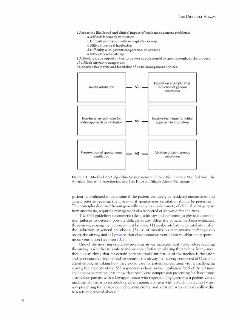
The 2003 guidelines recommend taking a history and performing a physical examina-tion tailored to detect a possible dif fi cult airway. After the patient has been evaluated, three airway management choices must be made: (1) awake intubation vs. intubation after the induction of general anesthesia, (2) use of invasive vs. noninvasive techniques to secure the airway, and (3) preservation of spontaneous ventilation vs. ablation of sponta-neous ventilation (see Figure 3.1 ).
One of the most important decisions an airway manager must make before securing the airway is whether it is safe to induce apnea before intubating the trachea. Many anes-thesiologists think that for certain patients awake intubation of the trachea is the safest and most conservative method for securing the airway. In a survey conducted of Canadian anesthesiologists asking how they would care for patients presenting with a challenging airway, the majority of the 833 respondents chose awake intubation for 5 of the 10 most challenging scenarios: a patient with cervical cord compression presenting for discectomy; a stridulous patient with a laryngeal tumor who requires a laryngectomy; a patient with a mediastinal mass who is stridulous when supine; a patient with a Mallampati class IV air-way presenting for laparoscopic cholecystectomy; and a patient who cannot swallow due to a retropharyngeal abscess 3 .
Figure 3.1. Modi fi ed ASA algorithm for management of the dif fi cult airway. Modi fi ed from The American Society of Anesthesiologists Task Force on Dif fi cult Airway Management. 2

35
The Role of Awake Intubation
In many areas of medicine, no randomized controlled trials de fi ne the superiority of a current standard of care over earlier techniques. Studying the management of a dif fi cult airway is challenging because many anesthesiologists believe that it would be unethical to randomly allocate patients to another treatment group. Although we may never have studies that demonstrate the superiority of awake intubation over intubation after induc-tion of general anesthesia, it is clear in several settings the standard of care is to secure the airway before induction of general anesthesia.
I n d i c a t i o n s f o r A w a k e I n t u b a t i o n
A dif fi cult airway can be de fi ned as dif fi culty with (1) mask ventilation, (2) tracheal intu-bation, (3) securing a surgical airway, or (4) an uncooperative patient 4 . For the fi rst three situations, awake intubation may be the best option (see Table 3.1 ).
In order to develop the appropriate management plan, it is helpful to know the prob-ability of a dif fi cult airway. A proper evaluation can help detect the presence of a dif fi cult airway. The ASA standards for preanesthesia care require review of the available medical record, obtaining a pertinent medical history, and performing a focused physical examina-tion. Prior anesthesia records are reviewed with careful attention to the airway techniques employed and their outcomes. Patient imaging studies should be reviewed with special attention paid to radiographs of the cervical spine and MRI and CT studies of the head, neck, and thorax. A patient who has a history consistent with a previous dif fi cult mask ventilation or dif fi cult intubation has a high likelihood of a dif fi cult airway 5 .
A dif fi cult airway can be the result of many conditions. These are discussed in detail in Chap. 2 .
Aside from an anticipated dif fi cult airway, there are three other situations in which awake intubation may be desirable. First, if the patient has a tenuous neurologic status due to cervical spine pathology, it might be desirable to have the patient intubated prior to induction of general anesthesia. The patient can be intubated and positioned for surgery while awake or with minimal sedation, thereby protecting his own spinal integrity. The patient can then have a neurological assessment prior to induction of general anesthesia.
In addition to indications for awake intubation driven by patient anatomy or pathol-ogy, there are two physiologic indications for awake intubation. First, the patient who is hypotensive or in shock may not respond well to sedative or anesthetic induction agents. These agents may reduce sympathetic tone or have a deleterious effect on blood pressure due to negative inotropic or vasodilating properties. Often, a patient in extremis will toler-ate laryngoscopy and intubation with minimal or no topical anesthesia and no sedation. The second physiologic indication for awake intubation is respiratory failure. The patient with minimal respiratory reserve may not tolerate even a brief period of apnea without suffering signi fi cant hypoxia. In this setting it may be most prudent to allow the patient to breathe spontaneously throughout the intubation procedure. Blockade of the nerves innervating the airway and/or topical anesthesia can be provided while the patient breathes with a well-sealed face mask with very brief interruptions to apply topical agents.
Table 3.1. Indications for awake intubation.
Previous history of dif fi cult intubation Anticipated dif fi cult mask ventilation Physical examination consistent with dif fi cult intubation Trauma to the face, neck, upper airway, cervical spine High risk of aspiration Cervical spine disease Hypotension, shock Respiratory failure

36
The Difficult Airway
C o n t r a i n d i c a t i o n s t o A w a k e I n t u b a t i o n
There are few absolute contraindications to awake intubation. If patient refusal or reluc-tance is encountered initially, with proper counseling and reassurance most patients will agree to awake intubation (see “psychological preparation” below). Uncooperative patients such as children and intoxicated adults may present special challenges. A documented allergy to all local anesthetics means a patient is not a candidate for awake intubation. Most patients who report an allergy to local anesthetics have had an intravascular injec-tion of local anesthetic before a dental procedure. The signs and symptoms they report are usually consistent with systemic toxicity from local anesthetics or epinephrine. These patients may safely receive local anesthetics as part of preparation for awake intubation.
A d v a n t a g e s o f A w a k e Tr a c h e a l I n t u b a t i o n
There are several advantages to awake intubation over asleep intubation. First, an awake patient maintains ventilation and oxygenation. There is truth to the aphorism, “It is hard to kill a spontaneously breathing patient.” Because the patient is breathing and oxygenat-ing, they will not desaturate as quickly, eliminating the time pressure associated with intu-bating an apneic patient. If the initial technique is unsuccessful, then alternative techniques can be attempted in a deliberate and methodical manner. In the unlikely event that none of the attempted intubation techniques works, elective surgery and anesthesia can be rescheduled without harming the patient. Unfortunately, this may not be the case when airway management is required for resuscitation purposes.
The second advantage of awake intubation is that the normal anatomic architecture of the upper airway is maintained. In an unconscious patient, the normal muscular tone of the tongue and upper airway structures relaxes 6 .
This collapse can render ineffective the patient’s efforts to inspire as seen in patients with obstructive sleep apnea. Airway collapse also can make efforts to assist spontaneous ventilation less effective and may make positive pressure ventilation with a face mask impossible leading to a cannot ventilate scenario.
When airway structures collapse, intubation of the trachea may become dif fi cult, especially when a fl exible bronchoscope is used to guide a tube into the trachea. Since unlike during direct or video laryngoscopy where a lifting force is applied to the anterior structures of the airway creating an open space, a fl exible bronchoscope cannot create an open space and must navigate an open channel from the nose or mouth to the trachea.
The third advantage of awake intubation is that the risk for aspiration is reduced because the airway is secured while the patient is conscious. First, the lower esophageal sphincter and cricopharyngeal muscle tone are maintained. Second, if the patient does regurgitate, gastric contents that reach the mouth can be expelled. Finally, if the carina is not anesthetized with a topical local anesthetic, the patient will cough vigorously if gastric contents do enter the distal trachea. Upper airway re fl exes are blunted by many anesthetic drugs, so it is important that the patient not be deeply sedated if maintenance of airway re fl exes is desired 7– 9 .
Finally, the neurologic status of the patient can be monitored during the intubation and positioning process. Evaluating the neurologic status is especially important in patients with cervical spine pathology. Because tracheal intubation of an anesthetized patient often involves manipulation of the cervical spine, there is some degree of uncertainty about the integrity of the central and peripheral nervous system if the patient is anesthetized before the airway is secured. It is very reassuring to the patient and the care providers if the patient is intubated and positioned for surgery while awake. This is especially important for surgery that will be performed with the neck extended or with the patient in the lateral or prone position.
37
The Role of Awake Intubation
K e y s t o S u c c e s s f u l A w a k e I n t u b a t i o n
There is an adage, “The key to successful awake fi beroptic intubation is good preparation, while the key to successful asleep fi beroptic intubation is good help.” Good preparation starts by establishing a good rapport with the patient and obtaining excellent topical anesthesia.
Psychological Preparation
Probably one of the most underappreciated aspects of a successful awake intubation is the development of good rapport with the patient. Patients should be told that the awake intu-bation is in their best interest. A patient’s mood will often mirror the mood of the anesthe-siologist. If an anesthesiologist is calm and matter of fact, the patient will be calm. It is important for the anesthesiologist to listen to and address the patient’s concerns. Patients are often worried about gagging and loss of control. They can be comforted by assurances that topical anesthesia will block the gag re fl ex. It is also helpful to tell patients that they will be appropriately sedated and can retain control by having the ability to pause the intu-bation process by holding up a hand. Patients are told that they will be given more local anesthetic or anxiolytics if they experience discomfort or anxiety during the procedure.
Dry the Airway
A patient’s airway should be dry for two reasons. The fi rst reason is that oral secretions hinder the ability of the anesthesiologist to view anatomic structures through fi beroptic devices. This is more of a concern for fl exible bronchoscopes than other intubation devices because bronchoscopes traverse the most dependent portion of the airway where fl uids tend to pool. The second reason for drying the mucous membranes is that a topically applied local anesthetic is much more effective when applied to dry mucous membranes. Oral secretions decrease the effectiveness of topical local anesthetics in several ways. First, saliva will dilute the local anesthetic. Second, the local anesthetic will need to diffuse through a layer of viscous saliva, reducing the concentration at the tissue surface and lengthening the onset time of the local anesthetic. Finally, if the mouth is wet, a larger portion of the topical local anesthetic may be swallowed increasing the risk for nausea and systemic toxicity 10 .
Glycopyrrolate 0.2 mg i.v. is administered to reduce oral secretions. Glycopyrrolate causes less tachycardia than atropine and does not cross the blood–brain barrier, so the risk of delirium from the central anticholinergic effects of drugs like atropine and scopolamine is avoided. Intravenous glycopyrrolate has an onset time of 2–4 min and an antisialogogue effect lasting over 2 h 11 .
If the mucous membranes are not dry when it is time to apply topical anesthesia, then manual drying techniques should be used. Gauze can be wrapped around a tongue depres-sor and then applied to the tongue to dry it. Alternatively, the patient can be given gauze to wrap around his or her fi nger for insertion into his or her mouth to dry the mucous membranes. Insertion of one’s fi nger into the mouth of a patient who is not anesthetized can elicit a gag or bite re fl ex and is therefore not advised.
Supplemental Oxygen
Supplemental oxygen is applied to prevent hypoxemia if the patient hypoventilates as a result of sedation. A larger fraction of oxygen in the lungs will provide a margin of safety if the patient becomes hypopneic or apneic or if the airway becomes completely obstructed. Clearly, one should take every precaution to prevent apnea or airway obstruction, but if either should occur there will be more time before the patient desaturates if the patient has been breathing supplemental oxygen.
38
The Difficult Airway
Anxiolysis and Sedation
Ideally the patient should be cooperative, calm, and not anxious. The patient should need little or no analgesia if local anesthesia is working well. Minimal or moderate sedation-analgesia as de fi ned by the American Society of Anesthesiologists is the appropriate depth for most patients 12 . The patient should be responsive to verbal or light tactile stimulation, airway architecture will remain unchanged, and the airway will remain patent. The patient should have normal or near-normal ventilatory drive, and cardiovascular function is largely unaffected.
The cooperative patient will be able to control his airway if he vomits. He will also be able to inspire deeply when requested to do so. Deep inspiration has three desirable effects. First, if the patient’s oxygen level is falling, one or two vital capacity maneuvers are usually enough to raise the oxygen saturation to an acceptable level. Second, active inspiration makes it easier for the endoscopist to identify the glottic opening. Third, the vocal cords abduct during deep inspiration easing the passage of the endotracheal tube into the trachea.
Several drugs may be used individually or in combination to produce the desired level of sedation. Midazolam is used frequently because it is an anxiolytic with a rapid onset of action, a relatively short duration of action, and minimal effect on respiratory or cardio-vascular physiology. Midazolam’s effects are reversed with fl umazenil. The amnestic prop-erties of midazolam are also bene fi cial, particularly if the patient has a condition that requires multiple anesthetics with awake intubation. Often fentanyl or remifentanil are used in conjunction with midazolam for two reasons. The opiates are a potent antitussive that reduces coughing associated with translaryngeal injection or instrumentation of the trachea with a bronchoscope, endotracheal tube, or other device. If nerve blocks are per-formed before the intubation, fentanyl reduces the discomfort associated with the injec-tions. It is important to realize, however, that there is a synergistic interaction between benzodiazepines and opiates with respect to sedation/hypnosis and hypoventilation, hypoxemia, and apnea 13, 14 .
Dexmedetomidine also has several desirable properties for airway management. Patients sedated with dexmedetomidine are calm and cooperative. In a study that compared dexmedetomidine and midazolam, patient satisfaction was superior and hemodynamics were smoother in patients sedated with dexmedetomidine and a small amount of midazo-lam than with properly titrated doses of midazolam alone 15 . In patients who received dex-medetomidine, hypercarbic and hypoxic ventilatory drive were nearly normal. A bene fi cial effect of dexmedetomidine sedation is a very dry airway. The disadvantages of dexmedeto-midine are its cost, slow onset, and the need for an infusion pump for most applications. In addition, there is no reversal agent for dexmedetomidine.
Anesthesia of the Airway
It is important to provide excellent anesthesia of the airway before attempting laryngos-copy or intubation. At a minimum, the posterior portion of the tongue, the hypopharynx, the larynx, and the proximal trachea must be anesthetized. If the plan is to intubate the trachea via the nose, then the nasopharynx also must be anesthetized.
For topical anesthesia, the right drug must be selected in the right concentration, and suf fi cient time must be allowed for it to work. Benzocaine produces anesthesia of the mucous membranes quickly, but is associated with methemoglobinemia and has been removed from the formulary of several hospitals 16 . Lidocaine is safe and effective for pro-viding topical anesthesia of the airway. It is possible to provide adequate anesthesia with 2 % lidocaine, but the onset of action with this low concentration takes approximately 20 min. For this reason, 4 or 5 % lidocaine is preferred for topical anesthesia of the airway. With the higher concentrations, the airway is ready for instrumentation within 2 min 17 .
39
The Role of Awake Intubation
Applied Airway Anatomy
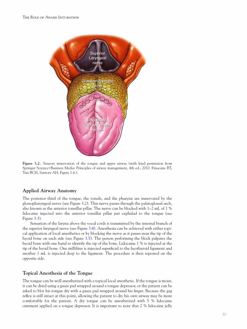
The posterior third of the tongue, the tonsils, and the pharynx are innervated by the glossopharyngeal nerve (see Figure 3.2 ). This nerve passes through the palatoglossal arch, also known as the anterior tonsillar pillar. The nerve can be blocked with 1–2 mL of 1 % lidocaine injected into the anterior tonsillar pillar just cephalad to the tongue (see Figure 3.3 ).
Sensation of the larynx above the vocal cords is transmitted by the internal branch of the superior laryngeal nerve (see Figure 3.4 ). Anesthesia can be achieved with either topi-cal application of local anesthetics or by blocking the nerve as it passes near the tip of the hyoid bone on each side (see Figure 3.5 ). The person performing the block palpates the hyoid bone with one hand to identify the tip of the bone. Lidocaine 1 % is injected at the tip of the hyoid bone. One milliliter is injected super fi cial to the hyothyroid ligament and another 1 mL is injected deep to the ligament. The procedure is then repeated on the opposite side.
Topical Anesthesia of the Tongue
The tongue can be well anesthetized with a topical local anesthetic. If the tongue is moist, it can be dried using a gauze pad wrapped around a tongue depressor, or the patient can be asked to blot his tongue dry with a gauze pad wrapped around his fi nger. Because the gag re fl ex is still intact at this point, allowing the patient to dry his own airway may be more comfortable for the patient. A dry tongue can be anesthetized with 5 % lidocaine ointment applied on a tongue depressor. It is important to note that 2 % lidocaine jelly
Figure 3.2. Sensory innervation of the tongue and upper airway (with kind permission from Springer Science+Business Media: Principles of airway management, 4th ed.; 2010. Finucane BT, Tsui BCH, Santora AH, Figure 1.6.).

40
The Difficult Airway
Figure 3.3. Injection site for glossopharyngeal nerve block. Local anesthetic is injected into the anterior tonsillar pillar 2 mm cephalad to the tongue at the spot indicated by the X.
Figure 3.4. Innervation of the larynx. Sensory innervation of the larynx is provided by the internal branch of the superior laryngeal nerve (with kind permission from Springer Science+Business Media: Principles of airway management, 4th ed.; 2010. Finucane BT, Tsui BCH, Santora AH, Figure 1.19).

41
The Role of Awake Intubation
frequently does not produce adequate anesthesia of the tongue. To anesthetize the tongue, the operator starts in the middle of the tongue and gently applies the ointment with a painting side-to-side motion on the tongue (see Figure 3.6a, b ). The tongue blade is slowly moved to the posterior aspect of the tongue (see Figure 3.6c ). The goal is to deliver enough ointment to the posterior part of the tongue that some will melt and drip down into the hypopharynx, affecting the pyriform sinuses bilaterally and the vallecula.
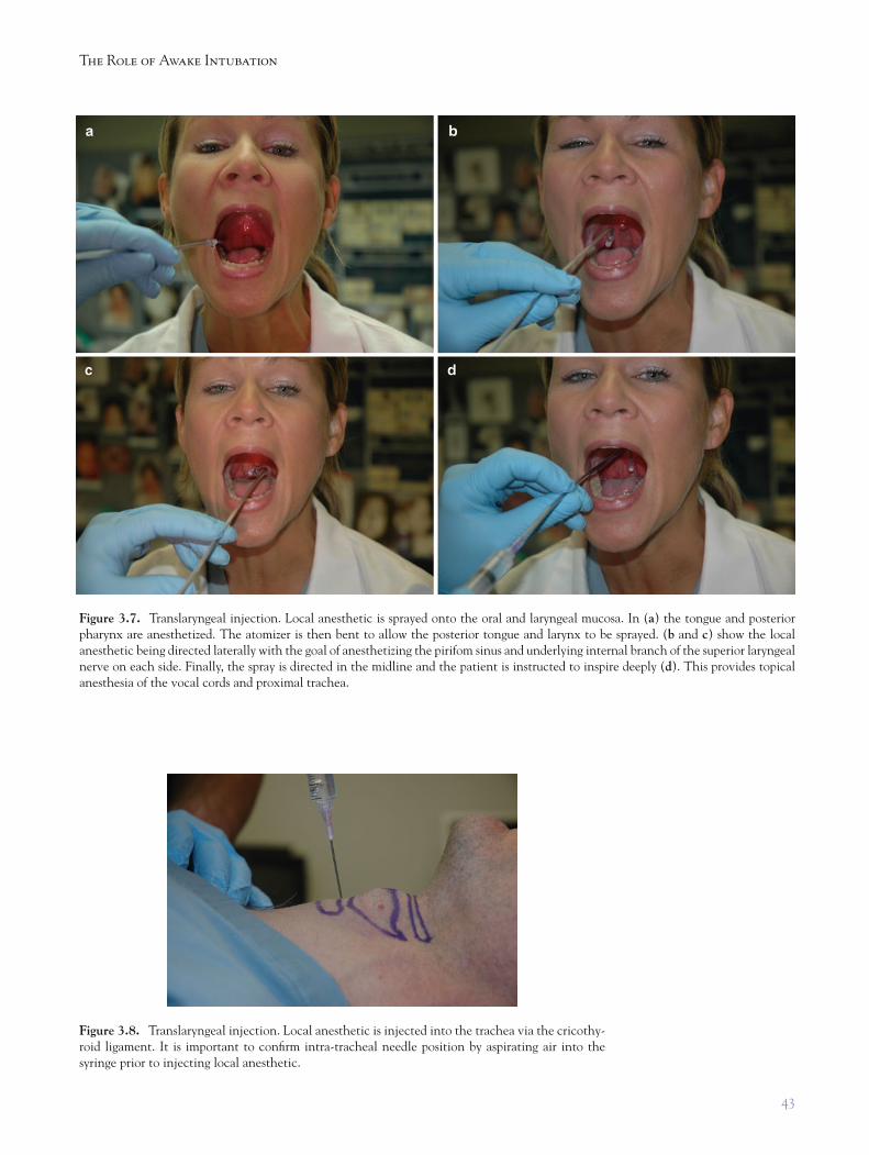
After the lidocaine ointment has had 2–3 min to take effect, the application of topi-cal anesthesia is completed by spraying the tongue, posterior pharynx, and larynx with 4 % lidocaine. An atomizer is used to deliver a directable spray. First the tongue and pos-terior pharynx are sprayed with approximately 1 mL (see Figure 3.7a ). Then the atomizer is de fl ected to allow the spray to be directed to the pyriform sinus, applying approximately 0.5 mL on each side (see Figure 3.7b, c ). Finally, the atomizer is placed in the posterior pharynx and directed in the midline toward the vocal cords. The patient is instructed to inspire deeply while 1 mL of lidocaine is injected quickly (see Figure 3.7d ). This provides topical anesthesia of the epiglottis, vocal cords, distal larynx, and proximal trachea.
The trachea distal to the vocal cords is supplied by the recurrent laryngeal branch of the vagus nerve. While it is essential to thoroughly anesthetize this area, the recurrent laryngeal nerve is not amenable to direct neural blockade. To properly anesthetize the airway below the cords, topical anesthetic must be applied via one of two techniques; a translaryngeal injection or via a “spray as you go” technique 18 .
To perform a translaryngeal block, the operator fi rst identi fi es the cricothyroid liga-ment. The larynx is stabilized with the thumb and middle fi nger, while the ligament is palpated with the index fi nger. The other hand introduces a 20-gauge catheter or 22-gauge needle attached to a syringe containing 2–4 mL of 4 % lidocaine through the ligament into the airway. The plunger of the syringe is withdrawn, aspirating air into the lidocaine (see Figure 3.8 ). This “bubble contrast” technique is important to con fi rm that the needle is in the airway and not in tissue before injecting the local anesthetic. Once proper placement of the needle is con fi rmed, the patient is informed that he is likely to cough, and the local anesthetic is injected briskly into the airway. If a needle is used for the injection, it is removed immediately after the injection. If a catheter is used, it may be left in place or removed.
After an injection of local anesthetic through the cricothyroid ligament, the patient usually coughs, sometimes quite vigorously. Coughing spreads the local anesthetic proxi-mally and distally in the airway, helping to anesthetize the larynx and trachea.
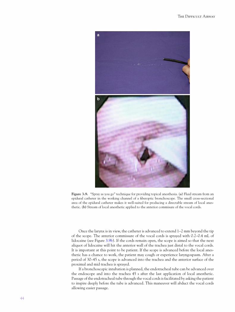
The “spray as you go” technique works well and is especially advantageous when it is undesirable to place a needle through the neck. In this technique the working channel of a fi beroptic bronchoscope is used to deliver local anesthetic to the larynx and trachea.
Figure 3.5. Blocking the superior laryngeal nerve. Local anesthetic is injected 2–3 mm caudad of the hyoid bone tip.

42
The Difficult Airway
For the technique to work, the velocity of the local anesthetic must be high enough to create a directable stream of fl uid out of the end of the scope. Most adult bronchoscopes have a large (2.0–2.5 mm) working channel to allow for effective suctioning and passage of brushes and biopsy forceps. The large cross-sectional area of these channels produces a poor stream of fl uid. This problem can be overcome by introducing an epidural catheter through the working channel. The small internal diameter of the catheter generates good fl ow characteristics (see Figure 3.9a ). The epidural catheter is fed into the channel so that its tip is just inside the bronchoscope. At the same time, the scope is introduced in the airway and positioned to allow visualization of the larynx.
Figure 3.6. Application of Local anesthetic ointment. ( a ) Local anesthetic ointment is applied to the middle third of the tongue. The tongue blade is then moved side-to-side to anesthetize the entire tongue as seen in ( b ). The patient is then instructed to hold the local anesthetic-covered tongue blade over the posterior tongue in a manner similar to a lollipop ( c ),
43
The Role of Awake Intubation
Figure 3.7. Translaryngeal injection. Local anesthetic is sprayed onto the oral and laryngeal mucosa. In ( a ) the tongue and posterior pharynx are anesthetized. The atomizer is then bent to allow the posterior tongue and larynx to be sprayed. ( b and c ) show the local anesthetic being directed laterally with the goal of anesthetizing the pirifom sinus and underlying internal branch of the superior laryngeal nerve on each side. Finally, the spray is directed in the midline and the patient is instructed to inspire deeply ( d ). This provides topical anesthesia of the vocal cords and proximal trachea.
Figure 3.8. Translaryngeal injection. Local anesthetic is injected into the trachea via the cricothy-roid ligament. It is important to con fi rm intra-tracheal needle position by aspirating air into the syringe prior to injecting local anesthetic.
44
The Difficult Airway
Once the larynx is in view, the catheter is advanced to extend 1–2 mm beyond the tip of the scope. The anterior commissure of the vocal cords is sprayed with 0.2–0.4 mL of lidocaine (see Figure 3.9b ). If the cords remain open, the scope is aimed so that the next aliquot of lidocaine will hit the anterior wall of the trachea just distal to the vocal cords. It is important at this point to be patient. If the scope is advanced before the local anes-thetic has a chance to work, the patient may cough or experience laryngospasm. After a period of 30–45 s, the scope is advanced into the trachea and the anterior surface of the proximal and mid trachea is sprayed.
If a bronchoscopic intubation is planned, the endotracheal tube can be advanced over the endoscope and into the trachea 45 s after the last application of local anesthetic. Passage of the endotracheal tube through the vocal cords is facilitated by asking the patient to inspire deeply before the tube is advanced. This maneuver will abduct the vocal cords allowing easier passage.
Figure 3.9. “Spray as you go” technique for providing topical anesthesia. ( a ) Fluid stream from an epidural catheter in the working channel of a fi beroptic bronchoscope. The small cross-sectional area of the epidural catheter makes it well-suited for producing a directable stream of local anes-thetic. ( b) Stream of local anesthetic applied to the anterior commisure of the vocal cords.

45
The Role of Awake Intubation
Intubating the Trachea
After proper intubating conditions have been created with airway anesthesia and appro-priate sedation, the trachea can be intubated. In order from least to most stimulating, the common modes of tracheal intubation of the awake patient are: intubation over a fl exible bronchoscope, video laryngoscopy, and direct laryngoscopy.
Flexible bronchoscopic intubation (FBI) allows the device to be gently steered through the open space in the airway with minimal contact and pressure on the base of the tongue and other airway structures. The one caveat to intubation with a fl exible broncho-scope is that the scope must be passed through the vocal cords before the endotracheal tube is passed through the cords. If the trachea is not adequately anesthetized when the bronchoscope is passed through the cords, the patient may cough vigorously and/or develop laryngospasm.
Video laryngoscopes (VL) place more pressure on the base of the tongue than a fl exible bronchoscope, thereby increasing the amount of stimulation, particularly pressure sensa-tion on the base of the tongue. The shape of the blades used for video laryngoscopes vary considerably from one manufacturer to another, so the response to one device may be dif-ferent than to another. Direct laryngoscopes (DL) need to create a straight line of sight from the upper incisors to the vocal cords exerting more pressure on the airway tissues than other techniques. VLs with the same shape as traditional DL blades (e.g., the Storz C-MAC) presumably produce similar amounts of pressure as the DL blades upon which they are based. VLs with highly curved blades (e.g., the Pentax AWS or the GlideScope) allow the operator to “see around the corner” of the airway and presumably produce less stimulation on the airway.
Other intubating techniques can be used with the awake patient. The Intubating LMA (LMA North America, San Diego) or C-Trach has been used for a hybrid “awake-asleep” intubation technique 19 . In this technique the oro-pharynx and hypopharynx are anesthetized with topical agents, while the larynx and trachea are not anesthetized. The C-Trach device is inserted and the video monitor applied. If the monitor shows a clear view of the vocal cords, general anesthesia is induced and the trachea is intubated through the device. This technique is especially good for patients when translaryngeal injection of lidocaine or tracheal anesthesia is undesirable.
Finally, surgical access to the trachea may be obtained in the awake patient with or without sedation. While surgical access to the airway is the technique of last resort in most dif fi cult airway algorithms, there are times when it may be best to proceed with cricothy-roidotomy or tracheostomy without anesthetizing or sedating the patient. Awake surgical techniques may be indicated in cases of massive airway hemorrhage, massive regurgitation, trauma to the face or neck, and cases of obstruction due to edema, infection, or foreign body. If the patient’s airway is so tenuous that surgery is required prior to induction of general anesthesia, then extreme caution should be exercised with sedation. Often patients undergoing awake surgical airway access receive little or no sedatives or analgesia until the airway is secured. As always, the risk of supplemental oxygen must be recognized if the surgeon is to use electrocautery after the airway has been entered.
It is important for anesthesia providers to understand the role of awake airway man-agement in the delivery of clinical anesthesia. Providers must be comfortable and compe-tent in intubating the trachea prior to induction of general anesthesia. Establishing patient rapport to develop trust, providing appropriate antisialogogues and sedative agents to opti-mize conditions and cooperation, and applying appropriate techniques to anesthetize the airway are essential elements of successful awake intubation. As with all skills, these must be practiced regularly to maintain top pro fi ciency. With proper technique and regular use, one should be able to perform an awake intubation in an ef fi cient manner with little stress for the patient and provider.

46
The Difficult Airway
R E F E R E N C E S
1. The American Society of Anesthesiologists Task Force on Dif fi cult Airway Management. Practice guidelines for management of the dif fi cult airway. Anesthesiology. 1993;78(3):597–602.
2. The American Society of Anesthesiologists Task Force on Dif fi cult Airway Management. Practice guidelines for management of the dif fi cult airway: an updated report by the American Society of anesthesiologists task force on management of the dif fi cult airway. Anesthesiology. 2003;98(5):1269–77.
3. Jenkins K, Wong DT, Correa R. Management choices for the dif fi cult airway by anesthesiologists in Canada. Can J Anaesth. 2002;49(8):850–6.
4. Klock PA, Benumof J. De fi nition and incidence of the dif fi cult airway. In: Hagberg CA, editor. Benumof’s dif fi cult airway management. 2nd ed. Mosby Elsevier: Philadelphia; 2007. p. 215–20.
5. Lundstrøm LH, Møller AM, Rosenstock C, Astrup G, Gätke MR, Wetterslev J. A documented previous dif fi cult tracheal intubation as a prognostic test for a subsequent dif fi cult tracheal intu-bation in adults. Anaesthesia. 2009;64:1081–8.
6. Eastwood PR, Szollosi I, Platt PR, Hillman DR. Collapsibility of the upper airway during anes-thesia with iso fl urane. Anesthesiology. 2002;97:786–93.
7. Drummond GB. In fl uence of thiopentone on upper airway muscles. Br J Anaesth. 1989;63:12–21.
8. Murphy PJ, Langton JA, Barker P, Smith G. Effect of oral diazepam on the sensitivity of upper airway re fl exes. Br J Anaesth. 1993;70:131.
9. Nishino T, Hiraga K, Sugimori K. Effects of i.v. lignocaine on airway re fl exes elicited by irrita-tion of the tracheal mucosa in humans anaesthetized with en fl urane. Br J Anaesth. 1990;64:682–7.
10. Benumof JL, Feroe D. Swallowing topically administered 4 % lidocaine results in nausea and vomiting. Am J Anesthesiol. 1998;25(4):150–3.
11. Reed AP. Preparation of the patient for awake fl exible fi beroptic bronchoscopy. Chest. 1992;101:244–53.
12. Practice Guidelines for Sedation and Analgesia by Non-Anesthesiologists. An updated report by the American Society of Anesthesiologists Task Force on Sedation and Analgesia by Non-Anesthesiologists. Anesthesiology. 2002; 96:1004–17.
13. Bailey PL, Pace NL, Ashburn MA, Moll JW, East KA, Stanley T. Frequent hypoxemia and apnea after sedation with midazolam and fentanyl. Anesthesiology. 1990;73(5):826–30.
14. Vinik HR, Bradley EL, Kissin I. Triple anesthetic combination: propofol-midazolam-alfentanil. Anesth Analg. 1994;78(2):354–8.
15. Bergese SD, Bender SP, McSweeney TD, Fernandez S, Dzwonczyk R, Sage K. A comparative study of dexmedetomidine with midazolam and midazolam alone for sedation during elective awake fi beroptic intubation. J Clin Anesth. 2010;22(1):35–40.
16. Nguyen ST, Cabrales RE, Bashour CA, Rosenberger TE, Michener JA, Yared JP, et al. Benzocaine-induced methemoglobinemia. Anesth Analg. 2000;90(2):369–71.
17. Takita K, Morimoto Y, Kemmotsu O. Tracheal lidocaine attenuates the cardiovascular response to endotracheal intubation. Can J Anesth. 2001;48(8):732–6.
18. Webb AR, Fernando SS, Dalton HR, Arrowsmith JE, Woodhead MA, Cummin AR. Local anaesthesia for fi breoptic bronchoscopy: transcricoid injection or the “spray as you go” tech-nique? Thorax. 1990;45(6):474–7.
19. Ovassapian A. Awake-asleep sequential intubation using CTrach LMA. Anesthesiology. 2006;105:870.