the diagnosis and management of sinusitis: a practice ... · the diagnosis and management of...
TRANSCRIPT
The diagnosis and management of sinusitis:A practice parameter update
Chief Editors: Raymond G. Slavin, MD, Sheldon L. Spector, MD, and
I. Leonard Bernstein, MD
SinusitisUpdateWorkgroup: Chairman—RaymondG. Slavin, MD;Members—Michael
A. Kaliner, MD, David W. Kennedy, MD, Frank S. Virant, MD, and Ellen R. Wald, MD
Joint Task Force Reviewers: David A. Khan, MD, Joann Blessing-Moore, MD,
David M. Lang, MD, Richard A. Nicklas, MD,* John J. Oppenheimer, MD,
Jay M. Portnoy, MD, Diane E. Schuller, MD, and Stephen A. Tilles, MD
Reviewers: Larry Borish, MD, Robert A. Nathan, MD, Brian A. Smart, MD,
and Mark L. Vandewalker, MD
These parameters were developed by the Joint Task Force on Practice
Parameters, representing the American Academy of Allergy, Asthma
and Immunology; the American College of Allergy, Asthma and
Immunology; and the Joint Council of Allergy, Asthma and
Immunology.
The American Academy of Allergy, Asthma and Immunology (AAAAI)
and the American College of Allergy, Asthma and Immunology
(ACAAI) have jointly accepted responsibility for establishing ‘‘The
diagnosis and management of sinusitis: a practice parameter
update.’’ This is a complete and comprehensive document at the
current time. The medical environment is a changing environment,
and not all recommendations will be appropriate for all patients.
Because this document incorporated the efforts of many
*This parameter was edited by Dr Nicklas in his private capacity and not in
his capacity as a medical officer with the Food and Drug Administration.
No official support or endorsement by the Food and Drug Administration
is intended or should be inferred.
Disclosure of potential conflict of interest: E. Wald has received grants from
GlaxoSmithKline, MedImmune, and Sanofi Pasteur. F. Virant has received
grants from Abbott, AstraZeneca, Boehringer Ingelheim, Bristol-Myers
Squibb, Dey Labs, Genentech, GlaxoSmithKline, Hoffman LaRoche, Immu-
nex, Key, Lederle, Lilly Research, Merck, Novartis, Pfizer, Purdue Fredrick,
Sandofi, Schering, Sepracor, TAP Pharmaceuticals, 3M Pharmaceuticals,
UCN Pharma, Upjohn Laboratories, and Med Point Pharmaceuticals; has
consultant arrangements with NeoRex; and is on the speakers’ bureau for
GlaxoSmithKline,Aventis,Merck, Pfizer, Schering,AstraZeneca, and IDEC.
S. Tilles has received grants from GlaxoSmithKline, Aventis, and Novartis
and is on the speakers’ bureau for GlaxoSmithKline, Aventis, and Pfizer.
J. Oppenheim has consultant arrangements with Sepracor, GlaxoSmithKline,
AstraZeneca, and Roche; has received grants from Boehringer Ingelheim,
Schering, GlaxoSmithKline, Merck, Sepracor, AstraZeneca, Novartis and
Altana; and is on the speakers’ bureau for Sepracor, GlaxoSmithKline, Astra-
Zeneca, Novartis, and Merck. D. Khan has consultant arrangements with
Pfizer; has received grants from AstraZeneca; and is on the speakers’ bureau
forMerck, Pfizer, GlaxoSmithKline, and Aventis. D. Kennedy has consultant
arrangements with Medtronic-Xomed and Schering-Plough; has received
grants from Novartis; and is on the speakers’ bureau for Merck. M. Kaliner
has consultant arrangements with Aventis, Medpoint, Glaxo, Gasser, Adams,
and King; has received grants from numerous pharmaceutical companies
that are researching allergies; and is on the speakers’ bureau for Aventis,
Medpoint, GlaxoSmithKline, Gasser, and Abbot.
Reprint requests: Joint Council of Allergy, Asthma and Immunology, 50 N
Brockway St, #3-3, Palatine, IL 60067.
J Allergy Clin Immunol 2005;116:S13-47.
0091-6749/$30.00
� 2005 American Academy of Allergy, Asthma and Immunology
doi:10.1016/j.jaci.2005.09.048
participants, no single individual, including those who served on
the Joint Task force, is authorized to provide an official AAAAI or
ACAAI interpretation of these practice parameters. Any request for
information about or an interpretation of these practice parameters
by the AAAAI or the ACAAI should be directed to the Executive
Offices of the AAAAI, the ACAAI, and the Joint Council of Allergy,
Asthma and Immunology. These parameters are not designed for
use by pharmaceutical companies in drug promotion.
Published practice parameters of the Joint Task Forceon Practice Parameters for Allergy and Immunologyinclude the following:
1. Practice parameters for the diagnosis and treatmentof asthma. J Allergy Clin Immunol 1995;96(suppl):S707-S870.
2. Practice parameters for allergy diagnostic testing.Ann Allergy 1995;75:543-625.
3. Practice parameters for the diagnosis andmanagementof immunodeficiency. Ann Allergy 1996;76:282-94.
4. Practice parameters for allergen immunotherapy.J Allergy Clin Immunol 1996;98:1001-11.
5. Disease management of atopic dermatitis: a practiceparameter. Ann Allergy 1997;79:197-211.
6. The diagnosis and management of anaphylaxis. J Al-lergy Clin Immunol 1998;101(suppl):S465-S528.
7. Algorithm for the diagnosis and management ofasthma: a practice parameter update. Ann Allergy1998;81:415-20.
8. Diagnosis and management of rhinitis: parameterdocuments of the Joint Task Force on Practice param-eters in Allergy, Asthma and Immunology. AnnAllergy 1998;81(suppl):S463-S518.
9. Parameters for the diagnosis and management ofsinusitis. J Allergy Clin Immunol 1998;102(suppl):S107-S144.
10. Stinging insect hypersensitivity: a practice param-eter. J Allergy Clin Immunol 1999;103:963-80.
11. Disease management of drug hypersensitivity: apractice parameter. Ann Allergy 1999;83(suppl):S665-S700.
12. Diagnosis and management of urticaria: a practiceparameter. Ann Allergy 2000;85(suppl):S521-S544.
S13
J ALLERGY CLIN IMMUNOL
DECEMBER 2005
S14 Slavin et al
13. Allergen immunotherapy: a practice parameter. AnnAllergy 2003;90(suppl):SI-S540.
14. Symptom severity assessment of allergic rhinitis:part I. Ann Allergy 2003;91:105-14.
15. Disease management of atopic dermatitis: an updatedpracticeparameter.AnnAllergy2004;93(suppl):S1-S21.
16. Stinging insect hypersensitivity: a practice parameterupdate. J Allergy Clin Immunol 2004;114:869-86.
17. The diagnosis and management of anaphylaxis: anupdated practice parameter. J Allergy Clin Immunol2005;115(suppl):S483-S523.
18. Practice parameter for the diagnosis and managementof primary immunodeficiency. Ann Allergy 2005;94(suppl):S1-63.
These parameters are also available on the internet at:http://www.jcaai.org.
CONTRIBUTORS
The Joint Task Force has made a concerted effort toacknowledge all contributors to this parameter. If anycontributors have been excluded inadvertently, the TaskForce will ensure that appropriate recognition of suchcontributions is made subsequently.
CHIEF EDITORS
Raymond G. Slavin, MDDepartments of Internal Medicine, Molecular
Microbiology and ImmunologySaint Louis University, Health Science CenterSt Louis, MissouriSheldon L. Spector, MDDepartment of MedicineUCLA School of MedicineDirector, California Allergy & Asthma Medical GroupLos Angeles, CaliforniaI. Leonard Bernstein, MDDepartment of Medicine and Environmental HealthUniversity of Cincinnati College of MedicineCincinnati, Ohio
WORKGROUP MEMBERS
Michael A. Kaliner, MDDepartment of MedicineGeorge Washington University School of MedicineWashington, DCDavid W. Kennedy, MDRhinology Professor and Vice DeanUniversity of Pennsylvania Medical CenterPhiladelphia, PennsylvaniaFrank S. Virant, MDDepartment of PediatricsUniversity of WashingtonSeattle, Washington
Ellen R. Wald, MDDepartment of PediatricsUniversity of Pittsburgh School of MedicineChief, Division of Allergy, Immunology and
Infectious DiseasesPittsburgh, Pennsylvania
JOINT TASK FORCE REVIEWERS
David A. Khan, MDDepartment of Internal MedicineUniversity of Texas Southwestern Medical CenterDallas, TexasJoann Blessing-Moore, MDDepartments of Medicine and PediatricsStanford University Medical CenterDepartment of ImmunologyStanford, CaliforniaDavid M. Lang, MDAllergy/Immunology SectionDivision of MedicineDirector, Allergy and Immunology Fellowship
Training ProgramCleveland Clinic FoundationCleveland, OhioRichard A. Nicklas, MDDepartment of MedicineGeorge Washington Medical CenterWashington, DCJohn Oppenheimer, MDDepartment of Internal MedicineNew Jersey Medical SchoolPulmonary and Allergy AssociatesMorristown, New JerseyJay M. Portnoy, MDSection of Allergy, Asthma & ImmunologyThe Children’s Mercy HospitalProfessor of PediatricsUniversity of Missouri-Kansas City School of
MedicineKansas City, MissouriDiane E. Schuller, MDDepartment of PediatricsPennsylvania State University Milton S. Hershey
Medical CollegeHershey, PennsylvaniaStephen A. Tilles, MDDepartment of MedicineUniversity of Washington School of MedicineRedmond, Washington
REVIEWERS
Larry Borish, MD, Charlottesville, VirginiaRobert A. Nathan, MD, Colorado Springs, ColoradoBrian A. Smart, MD, Chicago, IllinoisMark L. Vandewalker, MD, Columbia, Missouri
J ALLERGY CLIN IMMUNOL
VOLUME 116, NUMBER 6
Slavin et al S15
CLASSIFICATION OF RECOMMENDATIONSAND EVIDENCE
Category of evidence
Ia Evidence from meta-analysis of randomized con-trolled trials
Ib Evidence from at least one randomized controlled trialIIa Evidence from at least one controlled study without
randomizationIIb Evidence from at least one other type of quasiexper-
imental studyIII Evidence from nonexperimental descriptive studies,
such as comparative studiesIV Evidence from expert committee reports or opinions,
clinical experience of respected authorities, or both
Strength of recommendation
A Directly based on category I evidenceB Directly based on category II evidence or extrapolated
recommendation from category I evidenceC Directly based on category III evidence or extrapo-
lated recommendation from category I or II evidenceD Directly based on category IV evidence or extrapolated
recommendation from category I, II, or III evidence
NR Not rated
PREFACE
Sinusitis is one of the most commonly diagnoseddiseases in the United States, affecting an estimated 16%of the adult population annually. It extracts an overall di-rect annual health care cost of $5.8 billion. Total restricted
The diagnosis and management ofsinusitis: A practice parameter update
Preface S15Executive summary S16Algorithm of sinusitis practice parameters (Fig 1)S17Annotations to the algorithm S17Complete guidelines and references S21
Definitions, anatomic considerations, sinusphysiology, and microbiology S21
Definitions S21Anatomic considerations S21Sinus physiology S22Microbiology S22
Clinical diagnosis S24History S24Physical examination S25Imaging studies S26Laboratory tests S28
Predisposing factors S29Viral infections S29Allergic rhinitis S30Nonallergic rhinitis S30GERD S31
activity days increased from 50 million per year during1986 through 1988 to 73 million per year during 1990through 1992.1 Sinusitis also significantly affects qualityof life in some symptom domains even more than otherchronic diseases, such as chronic obstructive pulmonarydisease, angina, and back pain.2
Because of the importance of sinusitis, the Joint TaskForce on Practice Parameters, representing the AmericanAcademy of Allergy, Asthma, and Immunology, theAmerican College of Allergy, Asthma, and Immunology,and the Joint Council of Allergy, Asthma and Immunol-ogy, developed the first set of ‘‘Parameters for the diag-nosis and management of sinusitis,’’ which was publishedin 1998.3 Much has happened since then with respect tonew concepts in diagnosis and management and new in-sights into pathogenesis. For these reasons, it was decidedthat a revision-update was indicated.
Four documents comprise this present practice param-eter on sinusitis: (1) an executive summary that reviews, innarrative format, the key clinical issues considered in theparameter documents; (2) a management algorithm withnarrative annotations designed to assist clinical decisionmaking; (3) a document listing only numbered summarystatements that is intended to promote rapid review andidentification ofmaterial comprehensively discussed in thefinal document; and (4) the complete guidelines docu-ment, which is organized so that the numbered key sum-mary statements precede relevant supporting text andcitations of evidence-based publications. This format pro-vides a ready reference for any physician who evaluatesand treats a patient with suspected sinusitis. In particular,the algorithm and its accompanying annotations aredesigned to present a global and useful approach to bothdiagnosis and management. Clinical decision points areclearly shown, and each of these proceeds stepwise tological implementation strategies. If further justification isrequired at any step in the algorithm, the evidentiary-basedguidelines text can and should be consulted. In addition,
Immunodeficiency S32Cystic fibrosis S33Ciliary dysfunction S34
Associated conditions S34Otitis media S34Asthma S35
Treatment S35Medical S35
Antibiotics S35Antihistamines S37a-Adrenergic decongestants S38Glucocorticosteroids S38Adjunctive therapies: Saline, S39mucolytics, and expectorants
IVIG S39Aspirin desensitization S40
Surgical S40Indications for referral S41
J ALLERGY CLIN IMMUNOL
DECEMBER 2005
S16 Slavin et al
guidance about appropriate referral of refractory cases,either because of treatment failure or for further investi-gation of possible associated conditions, is provided.
The great majority of patients with sinusitis seek carefrom their primary care physician. Various subspecialists(allergists and otolaryngologists) also see patients withsinusitis, especially patients who aremore difficult to treat.It is incumbent on all physicians treating sinusitis to beknowledgeable concerning the latest information on path-ophysiology, diagnosis, and management, especially inlight of the rapidity with which infective organisms areable to change their character.
This practice parameter includes anatomic, allergic,immunologic, and physiologic considerations, as well asclinical diagnosis, differential diagnosis, diagnostic test-ing, and treatment. Predisposing factors, such as allergy,upper respiratory tract infections, anatomic abnormalities,immotile cilia syndrome, cystic fibrosis (CF), immunedefi-ciencies, and environmental factors, will be addressed.Medical and surgical therapies will be discussed.
An initial draft of parameters was prepared by a workgroup of experts in the field who carefully reviewed thecurrent medical literature. This material then underwentextensive peer review, revision, and annotation by exter-nal reviewers and by the Joint Task Force on PracticeParameters for Allergy and Immunology, a national panelof allergist-immunologists appointed by its cosponsoringorganizations: the AmericanAcademy of Allergy, Asthmaand Immunology; the American College of Allergy,Asthma and Immunology; and the Joint Council of Al-lergy, Asthma and Immunology. The parameters were re-viewed and approved by the cosponsoring organizationsand thereby represent an evidence-based, broadly ac-cepted consensus opinion.
The Joint Task Force is grateful for the cosponsoringorganizations’ financial support and encouragement. TheJoint Task Force would especially like to thank the manyindividuals who have donated substantial time and effortin producing this document that is intended to improve thequality of care of many millions of patients with sinusitis.
EXECUTIVE SUMMARY
Sinusitis, defined as inflammation of one or more of theparanasal sinuses, is characterized as acute when lastingless than 4 weeks, subacute when lasting 4 to 8 weeks, andchronic when lasting longer than 8 weeks. Recurrentsinusitis consists of 3 or more episodes of acute sinusitisper year. A noninfectious form of chronic sinusitis istermed chronic hyperplastic eosinophilic sinusitis. Viralupper respiratory tract infections frequently precede sub-sequent bacterial invasion of the sinuses by Streptococcuspneumoniae, Haemophilus influenzae, and Moraxellacatarrhalis. These organisms can also be found in chronicsinusitis, as well as Staphylococcus aureus, Pseudomonasaeruginosa, and certain anaerobes. Fungi are being recog-nized increasingly as a factor in chronic sinusitis, particu-larly in the southeast and southwest parts of the country.
Prominent symptoms of acute sinusitis include nasalcongestion, purulent rhinorrhea, facial-dental pain, post-nasal drainage, headache, and cough. Chronic sinusitissymptoms are similar but might be even more subtle. Painis much less a feature of chronic sinusitis. Clinical signs ofboth acute and chronic sinusitis include sinus tendernesson palpation, mucosal erythema, purulent nasal secretions,increased pharyngeal secretions, and periorbital edema.There is an overlap in these symptoms with those of peren-nial rhinitis, and there is a frequent need to perform imagingprocedures to confirm the diagnosis. Because of this over-lap, some have suggested the use of the term rhinosinusitis.
Imaging techniques can provide confirmatory evidenceof sinusitis when symptoms are vague, physical findingsare equivocal, or clinical disease persists despite optimalmedical therapy. The imaging technique of choice is com-puted tomography (CT) because it can demonstrate abnor-malities both in the ostiomeatal complex and the sinuscavities.
Laboratory evaluation of acute, chronic, or recurrentsinusitis might include nasal cytology, nasal-sinus biopsy,or tests for immunodeficiency, CF, or ciliary dysfunction.Nasal cytology is useful in the clinical evaluation ofconditions associated with sinusitis, including allergicrhinitis (AR), eosinophilic nonallergic rhinitis (NAR),neutrophilic rhinitis, and vasomotor rhinitis (VMR). Sinussecretions can be obtained for culture in adults bymeans ofeither an aspiration of the maxillary sinus or an endoscop-ically directed catheter placed at the middle meatus. Forchildren, sinus secretions should be obtained by means ofaspiration only.
A number of factors associated with sinusitis shouldbe considered. Probably the most common is viral upperrespiratory tract infections. There is both clinical and ex-perimental evidence that ongoing AR might ultimatelylead to or augment acute bacterial sinusitis. NAR wasfound in 26% of patients with chronic sinusitis. Recently,gastroesophageal reflux disease (GERD) has been sug-gested as a cause of sinusitis, and there are several studiesin children and adults indicating that medical treatmentof GERD results in significant improvement in sinusitissymptoms. Tests for immunodeficiency, including quan-titative immunoglobulin measurement, functional anti-body tests, and HIV testing, might be useful if eithercongenital or acquired immunodeficiency is suspected incases of recurrent sinusitis. Quantitative sweat chloridetests and genetic testing for diagnosis of CF should beconsidered in children with nasal polyps, colonization ofthe nose and sinuses with Pseudomonas species, or bothand in those who had chronic sinusitis at an early age.
Diseases associated with sinusitis are otitis media andbronchial asthma. Although no direct causal factor be-tween sinusitis and asthma has been found, a number ofstudies in both children and adults suggest that medicalmanagement, surgical management, or both of sinusitis re-sults in objective and subjective improvement of asthma.
The primary therapy for acute bacterial sinusitis isantibiotics. The choice is based on predicted efficacy,cost, and side effects. A 10- to 14-day course is generally
J ALLERGY CLIN IMMUNOL
VOLUME 116, NUMBER 6
Slavin et al S17
adequate for acute disease, although shorter courses areindicated for newer antibiotics. If there is no improvementin 3 to 5 days, then an alternative antibiotic should beconsidered. The role of antibiotics in chronic sinusitis iscontroversial. For chronic infectious sinusitis, a longerduration of therapy might be required, with possible atten-tion to anaerobic pathogens. In the case of chronic non-infectious sinusitis, sometimes referred to as chronichyperplastic sinusitis, consideration should be given tosystemic corticosteroids.
Concern has been raised about the overdiagnosis ofsinusitis and unnecessary treatment with antibiotics. Ap-propriate criteria for the use of antibiotics are symptomsof sinusitis for 10 to 14 days or severe symptoms of acutesinus infection, including fever with purulent nasal dis-charge, facial pain or tenderness, and periorbital swelling.
Intranasal corticosteroids as an adjunct to antibiotictherapy might be helpful in treating recurrent acute andchronic sinusitis. Other adjunctive therapy, such as anti-histamines, decongestants, saline irrigation, mucolytics,and expectorants, might provide symptomatic benefit inselected cases. The use of intravenous immunoglobulin(IVIG) is indicated only in patients with proved functionalimpairment of humoral immunity. The beneficial effectsof aspirin desensitization on aspirin-sensitive patients withsinusitis and asthma have been reported.
Medically resistant sinusitis might respond to appro-priate nasal-sinus surgery. In instances of localized per-sistent disease within the ostiomeatal complex, functionalendoscopic sinus surgery might result in significantimprovement.
Consultation with a specialist should be sought when(1) there is a need to clarify the allergic or immunologicbasis for sinusitis, (2) sinusitis is refractory to the usualantibiotic treatment, (3) sinusitis is recurrent, (4) sinusitisis associated with unusual opportunistic infections, and (5)sinusitis significantly affects performance and quality oflife. Consultation is also appropriate when concomitantconditions are present that complicate assessment or treat-ment, including chronic otitis media, bronchial asthma,nasal polyps, recurrent pneumonia, immunodeficiencies,aspirin sensitivity, allergic fungal disease, granulomas,and multiple antibiotic sensitivities.
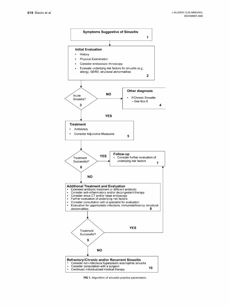
ALGORITHM OF SINUSITIS PRACTICEPARAMETERS (Fig 1)
Annotations to the algorithm
1. Symptoms suggestive of acute sinusitisd Acute sinusitis typically presents as a persistentupper respiratory tract infection (10-14 days with-out improvement).
d In adults prominent symptoms include nasal conges-tion, purulent rhinorrhea, postnasal drainage, facialor dental pain, headache, and cough, frequently witha more severe nocturnal component.
d Any patient with orbital swelling or pain, swellingof the forehead, and/or diplopia should be urgentlyscheduled for evaluation.
d Children with acute sinusitis might also exhibit in-creased irritability and vomiting occurring in asso-ciation with gagging on mucus, prolonged cough,or both.
d In all age groups less frequent symptoms associatedwith acute sinusitis include fever, nausea, malaise,irritability, fatigue, halitosis, hyposmia, and sorethroat.
2. Office visitd Review medical history for diagnosis of sinusitisand underlying risk factors.
d General examination includes an evaluation forsigns of upper airway and sinus inflammation asso-ciated with nasal mucosal edema, purulent secre-tions, and increased localized blood flow. Typicalclinical signs include tenderness overlying thesinuses, dark circles beneath the eyes, and/or peri-orbital edema. Pharyngeal erythema, lymphoid hy-perplasia, and purulent material in the posteriorpharynx are also frequently observed.
d Nasal examination in patients with acute sinusitismight reveal mucosal erythema and purulent secre-tions. Nasal endoscopy, whether performed with arigid or fiberoptic instrument, offers a significantlybetter view than a nasal speculum. Nasal polypsmight contribute to nasal congestion and can be asource of recurrent sinusitis by obstructing the sinusostia. In adults nasal polypsmight be associated withnonsteroidal anti-inflammatory drug sensitivity andasthma. Nasal polyps are relatively uncommon inchildren, and their presence should prompt evalua-tion for possible CF. Ear examination in patientswith suspected acute sinusitis frequently will revealmiddle ear effusions and associated eustachian tubedysfunction.
d Acute or chronic sinusitis might initiate or worsenasthma and bronchial hyperresponsiveness. Accord-ingly, chest auscultation and other objective mea-surements of airflow obstruction, such as officespirometry, should be considered in any patientwith possible sinusitis and cough.
d Patients with obvious acute sinusitis should be care-fully reviewed for any possible evidence of com-plicating factors, including the presence of facialswelling–erythema over an involved sinus, visualchanges, abnormal extraocular movements, propto-sis, periorbital inflammation–edema–erythema, anysuggestion of intracranial involvement, or centralnervous system involvement manifested as abnor-mal neurologic signs.
d In general, radiographs are not necessary in makingthe diagnosis of acute sinusitis, and plain radio-graphs have significant false-positive and false-negative results. Occasionally, imaging studiesmight be useful to support the diagnosis or provideevidence of the degree of mucosal involvement,thereby guiding more aggressive therapy. Plainradiographic signs compatible with sinusitis includegreater than 6 mm of mucosal thickening in the
J ALLERGY CLIN IMMUNOL
DECEMBER 2005
S18 Slavin et al
FIG 1. Algorithm of sinusitis practice parameters.
J ALLERGY CLIN IMMUNOL
VOLUME 116, NUMBER 6
Slavin et al S19
maxillary sinuses in adults (>4 mm in children),greater than 33% loss of air space volume withinthe maxillary sinuses, or opacification–air-fluidlevels in any of the paranasal sinuses. Occipitomen-tal view radiographs might be helpful in screeningadults and children older than 1 year of age buthave inadequate sensitivity. A limited coronal sinusCT scan, with a focus on the ostiomeatal complex,might be helpful and should be considered if imag-ing is deemed necessary. Axial and coronal sinusCT is indicated in suspected orbital involvement,and sinus magnetic resonance imaging (MRI) canprovide useful information with related soft tissueinvolvement.
d Nasal cultures are not reliable for establishing thediagnosis of sinusitis or for determining a specificcausative microorganism. Maxillary antrum aspira-tion for culture is definitive but is indicated onlywhen precise microbial identification is essential.Obtaining cultures of the middle meatus throughendoscopically directed culture has shown promisein adults but not in children.
3. Acute sinusitisd Acute sinusitis is defined as symptoms and signsfor less than 4 weeks. The diagnosis of acute sinus-itis is based primarily on the clinical history, thephysical examination, and possibly other ancillaryevaluations, including nasal cytology or radio-graphic imaging. In most instances the diagnosisis made presumptively, and treatment is initiated.Clinical improvement usually occurs promptly;complete resolution of symptoms might require10 to 14 days.
4. Other diagnosesDifferential diagnoses include the following:
d AR and NAR;d viral upper respiratory tract infection;d nasal polyps;d sinonasal tumors;d nasopharyngeal tumor, granulomata, dental infections;d enlarged or infected adenoids in children.
5. Treatment
Antibiotics:d Amoxicillin often is the drug of choice for childrenand adults. It is generally effective, inexpensive,and well tolerated. Trimethoprim-sulfamethoxazolecan be used as an alternative drug in adults. Resis-tance is more commonly seen in children, and it isrecommended that the clinician refer to their localbiogram profile of antibiotic resistance. For patientswho do not respond to amoxicillin, high-doseamoxicillin-clavulanate (90 mg/kg amoxicillin and6.4 mg/kg clavulanate, not to exceed 2 g every12 hours) is recommended. For patients allergicto or intolerant of amoxicillin, alternatives includecephalosporins, macrolides, or quinolones.
d Acute sinusitis generally responds to treatment for10 to 14 days. Some physicians continue treatment
for 7 days after the patient is well to ensure com-plete eradication of the organism and prevent re-lapse. It is important to instruct the patient tocomplete the course of antibiotics.
d A reasonable approach would be to start the patienton amoxicillin for 3 to 5 days and determinewhether the signs and symptoms are improving.If the patients symptoms are improving, continuethis treatment until the patient is well for 7 days(generally a 10- to 14-day course). If after 3 to 5days the patient has not shown improvement,switch to a different antibiotic, such as high-doseamoxicillin-clavulanate or cefuroxime axetil.
Corticosteroids:d The use of nasal corticosteroids might be helpful inpatients with acute and chronic sinusitis.
d Although efficacy has not yet been proved, theshort-term use of oral corticosteroids as an adjunctin treating patients with acute sinusitis is reasonablewhen the patient fails to respond to initial treat-ment, demonstrates nasal polyposis, or has demon-strated marked mucosal edema.
Saline-mucolytics:d Saline nasal sprays or lavage might be a useful ad-junct by liquefying secretions and decreasing therisk of crusting near the sinus ostia.
d There is no conclusive evidence that mucolytics,such as guaifenesin, are useful adjuncts in treatingacute sinusitis.
a-Adrenergic decongestants:d Topicaldecongestants (eg,oxymetazolineandphenyl-ephrine) and oral decongestants (eg, pseudoephe-drine) reduce mucosal blood flow, decrease tissueedema and nasal resistance, and might enhancedrainage of secretions from the sinus ostia.
d The use of topical decongestants beyond 3 to 5days might induce rhinitis medicamentosa, withassociated increased congestion and refractorinessto subsequent decongestant therapy.
Education:d The following comfort measures might be helpful:adequate rest, adequate hydration, analgesics asneeded, warm facial packs, steamy showers, andsleeping with the head of the bed elevated.
d Prevention measures might include appropriatetreatment of allergies and viral upper respiratorytract infections and avoidance of adverse environ-mental factors, such as relevant allergens, cigarettesmoke, pollution, and barotrauma.
d Patients should be instructed to phone if symptomsworsen (eg, especially with headache or high fever)or if symptoms have not improved within 3 to 5days of treatment (see annotation 10).
6. Treatment successful?Complete response:
d Patient is improved symptomatically to near normal.
J ALLERGY CLIN IMMUNOL
DECEMBER 2005
S20 Slavin et al
Partial response:d Patient is symptomatically improved but not backto normal at the end of the first course of antibiotics.
Poor response:d Patient has little or no symptomatic improvementafter the first course of antibiotic therapy.
7. Follow-upd No further evaluation for resolved uncomplicatedsinusitis.
d Consider further evaluation of underlying riskfactors, such as allergic rhinitis (AR) and NARand structural abnormalities.
8. Additional treatment and evaluationd For partial response, continue antibiotic treatmentfor another 10 to 14 days or consider antibioticchoices listed under ‘‘poor responses.’’
d For poor response to treatment with amoxicillin ortrimethoprim-sulfamethoxazole or in regions with ahigh incidence of antibiotic resistance, an antibioticshould be prescribed that covers resistant bacteria.Appropriate choices include high-dose amoxicillin-potassium clavulanate, cefuroxime, cefpodoxime,cefprozil, and cefdinir. Quinolones, macrolides,and ketolides might also be a consideration.
d Sinusitis that fails to improve after 21 to 28 daysof initial antibiotic treatment might be caused bypathogens not adequately covered by prior antibio-tics, the presence of nasal polyps, or noncompli-ance. The use of broader-spectrum single agents,such as high-dose amoxicillin-potassium clavula-nate, cefuroxime, or cefpodoxime should be con-sidered with or without the addition of anaerobiccoverage with clindamycin or metronidazole.
d Reinforce the comfort and prevention measuresoutlined in Annotation 5.
d Consider sinus CT scan if not already done.d Underlying risk factors should be evaluated in amore detailed manner.
d Consider consultation with an allergist-immunolo-gist for treatment of underlying allergic factorsand evaluation of unusual pathogens and immuno-deficiency. For structural abnormalities, consulta-tion should be sought with an otolaryngologist.
Recurrent sinusitis:d Repeated episodes of acute sinusitis typically 3 ormore times per year.
d Patients with chronic or recurrent sinusitis shouldbe evaluated for underlying inflammation, allergy,immunodeficiency, and anatomic abnormalities.
Rhinitis:d Patients with suspected AR in conjunction withsinusitis should be evaluated for the presence ofIgE sensitization to inhalant allergens.
d Emphasis of therapy for AR includes environ-mental control, pharmacotherapy, and, in selectedpatients, allergen immunotherapy.
d Other rhinitic conditions (vasomotor, nonallergicrhinitis–eosinophilia syndrome [NARES], and rhi-nitis medicamentosa) might also lead to sinusitis,
and the consultant must be capable of differentiat-ing these conditions and initiating an appropriatecourse of therapy.
Immunodeficiency:d Referral to an allergist-immunologist is particularlyindicated in patients with chronic or recurrentsinusitis associated with such conditions as otitismedia, bronchitis, bronchiectasis, or pneumonia andin patients who have undergone prior surgical pro-cedures and continue to experience sinusitis. Thisevaluation might include measurement of quantita-tive serum IgG, IgA, and IgM level and assessmentof specific antibody responses to protein andpolysaccharide antigens, such as tetanus toxoid orpneumococcal polysaccharide vaccine.
9. Treatment successful?d See Annotation 6.
Follow-upd See Annotation 7.
10. Chronic sinusitisd Signs and symptoms compatible with sinusitispersisting 8 weeks or longer.
d Consider a noninfectious form of sinusitis. Chronichyperplastic eosinophilic rhinosinusitis does notrespond to antibiotics and is marked by a prepon-derance of eosinophils and mixed mononuclearcells, with a relative paucity of neutrophils. A courseof systemic corticosteroids might have to beconsidered.
d If the patient has a significant nasal septal deviationthat compresses the middle turbinate into the ostio-meatal complex or obstruction of the sinus outflowtracts caused by middle turbinate deformity or thepresence of accessory structures that block sinusdrainage, consider consultation with an otolaryn-gologist. The presence of obstructing nasal polyps,after an appropriate course of treatment that mightinclude a trial of oral corticosteroids, is also anindication for referral. Finally, a patient with recur-rent or chronic symptoms and radiographic evidenceof ostiomeatal obstruction despite aggressive medi-cal management might also benefit from surgicalintervention.
d Evaluation should include coronal sinus CT withextra cuts through the ostiomeatal complex toclarify the extent of disease and specific locationor locations.
d Evaluation might also include nasal-sinus biopsy insuspected cases of neoplasia, fungal disease, gran-ulomatous disease, or tracheal biopsy for evaluat-ing ciliary structures, function, or both.
d In general, every effort should be made to maxi-mize medical treatment for underlying rhinitis be-fore proceeding with surgical intervention.
d Contemporary surgical therapy involves chieflyfunctional endoscopic sinus surgery.
d Most patients benefit from continued individualizedmedical therapy, including, when indicated, allergymanagement, after surgery.
J ALLERGY CLIN IMMUNOL
VOLUME 116, NUMBER 6
Slavin et al S21
COMPLETE GUIDELINES AND REFERENCES
Definitions, anatomic considerations, sinusphysiology, and microbiology
Definitions
Summary Statements
Summary Statement 1: It has been suggested that theterm sinusitis be replaced by rhinosinusitis. NRSummary Statement 2: Sinusitis is defined as inflamma-tion of one or more of the paranasal sinuses. The mostcommon cause of sinusitis is infection. Classification ofsinusitis is frequently based on duration of symptoms,the specific sinus involved, or both. NRSummary Statement 3: The most commonly used classi-fication is as follows (NR):
a. Acute sinusitis: symptoms for less than 4 weeksconsisting of some or all of the following: persis-tent symptoms of an upper respiratory tract in-fection, purulent rhinorrhea, postnasal drainage,anosmia, nasal congestion, facial pain, headache,fever, cough, and purulent discharge.
b. Subacute sinusitis: (unresolved acute) symptomsfrom 4 to 8 weeks.
c. Chronic sinusitis: symptoms for 8 weeks or longerof varying severity consisting of the same symp-toms as seen in acute sinusitis. In chronic sinusitisthere should be abnormal findings on CT or MRI.Some patients with chronic sinusitis might presentwith vague or insidious symptoms.
d. Recurrent sinusitis: 3 or more episodes of acute si-nusitis per year. Patients with recurrent sinusitismight be infected by different organisms at differenttimes.
Summary Statement 4: A noninfectious form of chronicsinusitis is termed chronic hyperplastic eosinophilicsinusitis. NR
Although it is not universally accepted, the suggestionhas been made that the term rhinosinusitis might be moreapplicable than sinusitis for the following reasons4-6: rhi-nitis typically precedes sinusitis; sinusitis without rhinitisis rare; the mucosa of the nose and sinuses are contiguous;and symptoms of nasal obstruction and nasal discharge areprominent in sinusitis. Rhinitis associated with sinusitiscan be allergic, bacterial, viral, or perennial nonallergic.Sinusitis is classified as acute, subacute, chronic, and recur-rent. It should be emphasized that this classification isentirely arbitrary, and the medical literature is unclear onthis point. Most treatment modalities are directed to acuteand chronic sinusitis. In acute sinusitis symptoms arepresent for less than 4 weeks. Symptoms consist ofsome or all of the following: persistent symptoms of an up-per respiratory tract infection, purulent rhinorrhea, postna-sal drainage, anosmia, nasal congestion, facial pain,headache, fever, and cough. Subacute sinusitis, also re-ferred to as unresolved acute sinusitis is the developmentof manifestations of minimal-to-moderate signs of sinusinflammation without an overt upper respiratory tract
infection or abrupt onset of symptoms. It might speak toinadequate or partial therapy of acute sinusitis or simpleinattention to acute problems. Chronic sinusitis is definedas persistent sinus inflammation for greater than 8 weeks.An operational definition of chronic sinusitis is persistentinflammation documented with imaging techniques atleast 4 weeks after initiation of appropriate medical ther-apy in the absence of an intervening acute episode.7 Incontrast to acute sinusitis, the role of bacterial infectionin chronic sinusitis is less certain.8 Recurrent sinusitis in-cludes 3 or more episodes of acute sinusitis per year.
The symptom-based definition of chronic sinusitis hasbeen recently brought into question. In one study morethan 50% of patients with a strong history of chronicsinusitis had a normal CT scan. However, it is still debatedwhether CT also provides the definitive diagnosis of thisdisorder. A reevaluation of the way chronic sinusitisis defined and diagnosed appears necessary.9 Chronicsinusitis can be divided into infectious and hyperplasticcategories. Chronic infectious sinusitis might be due toanaerobic bacteria, such as gram-positive streptococcus,bacteroides, and Fusobacterium species or S aureus. It isgenerally associated with a significant influx of neutro-phils. A noninfectious form of chronic sinusitis, some-times termed chronic hyperplastic eosinophilic sinusitis,is marked by a preponderance of eosinophils and mixedmononuclear cells, with a relative paucity of neutrophils.It is often associated with nasal polyps, asthma, and aspi-rin sensitivity.10
Anatomic considerations
Summary Statements
Summary Statement 5: The sinuses develop at differentages during childhood. BSummary Statement 6: The optic nerve, cavernous sinus,and carotid artery are adjacent to the sphenoid sinus.Tumors and infection in the sphenoid sinus might presentwith involvement of these structures. BSummary Statement 7: Because of the location of the fron-tal and sphenoid sinuses, infection of these sinuses has agreater propensity to cause intracranial complications. ASummary Statement 8: The maxillary, anterior ethmoid,and frontal sinuses drain through the ostiomeatal com-plex and are dependent on this region for normal ventila-tion and mucociliary clearance. BSummary Statement 9: The anterior ethmoid sinuses andmiddle meatus (ostiomeatal complex), as a result of theirlocation, are most frequently involved in sinusitis. DSummary Statement 10: Anatomic abnormalities of thenasal septum or within the ostiomeatal complex mightpredispose toward the development of sinusitis. B
Development. The maxillary sinus is the first to beginsignificant pneumatization between birth and 12 months.The floor of the maxillary sinus reaches the level of thefloor of the nose by approximately 12 years of age.Rudimentary ethmoid sinuses are present at birth andreach adult size at 12 to 14 years of age.11 Development of
J ALLERGY CLIN IMMUNOL
DECEMBER 2005
S22 Slavin et al
the frontal and sphenoid sinuses begins later than that ofthe ethmoid sinuses, and complete pneumatization is notachieved until mid to late adolescence.12 Although the de-gree of pneumatization of all sinuses is highly variable, thevariability in pneumatization of the frontal and sphenoidsinuses is greater than that of the ethmoid and maxillarysinuses.13 The frontal sinus is hypoplastic or completelyabsent in about 15% of the population, and the sphenoidsinus is rudimentary (conchal or presellar pneumatization)in 26% of patients. There is evidence to suggest thatsinusitis in childhood might inhibit sinus development.14
Because of their later development, frontal or sphenoiddisease is uncommon in childhood.Anatomy. The anterior ethmoid, frontal, and maxillary si-nuses all drain into the middle meatus through a relativelyconvoluted and narrow drainage pathway (ostiomeatalcomplex) rather than by simple ostia. The ethmoid sinusesthemselves consist of a honeycomb of cells lyingmedial tothe orbital structures and varying between 4 and 17 air cellsin number. Theymight also pneumatize to a variable extentabove (supraorbital) or below (infraorbital) the orbit. Theethmoid sinus is divided into an anterior group of cells(draining into the middle meatus) and a posterior group ofcells (draining into the superior meatus).15 The maxillarysinus lies between the teeth and the orbit on both sidesand drains into the middle meatus through a channel inits supramedial aspect. The paired frontal sinuses arisefrom the region of the anterior ethmoid and extend superi-orly into the forehead. Valveless veins that pass throughthe posterior wall of the frontal sinus might allow thespread of frontal sinus infection intracranially, particularlyin acute infection. The sphenoid sinuses are also paired andlie posterior and slightly inferior to the posterior ethmoidcells. They drain by separate ostia into the sphenoethmoi-dal recess on either side of the nasal septum posteriorly.The optic nerve courses over the sinus laterally and supe-riorly. The carotid artery indents the sinus laterally, and thesinus has an intimate relationshipwith the cavernous sinus,as well as the dura of both the anterior and middle cranialfossa.
The anatomic arrangement of the sinuses makes thefrontal anterior ethmoid and maxillary sinuses dependenton the ostiomeatal complex for their ventilation and mu-ciliary clearance. Significant obstruction of this complexcan predispose to the development of sinusitis. Becauseethmoid anatomy is extremely variable and dependent, tosome extent, on the position of the nasal septum, there isa potential for anatomic variations to cause ostiomeatalobstruction, although the importance of anatomic varia-tions in predisposing to chronic sinusitis either by meansof redirection of airflow or by means of direct compres-sion is still debated.16,17 In some situations the ethmoidcells might pneumatize into the head of the middle tur-binate (concha bullosa), and extreme middle turbinateaeration might narrow the ostiomeatal complex. The loca-tion of the anterior ethmoid sinuses and middle meatusmakes the ostiomeatal complex particularly at risk fromenvironmental exposures, and this region is typically thefirst and the most frequently involved region in chronic
sinusitis. Indeed, low-grade edema and inflammation canpersist within this region, resulting in intermittent episodesof inflammation in the dependent sinuses. When suchedema does not respond to medical therapy, endoscopicsurgical intervention might be required.
Sinus physiology
d The sinuses are air-filled cavities with classical, pseu-dostratified, ciliated columnar epithelium interspersedwith goblet cells. The cilia sweep mucus toward theostial opening. Obstruction of sinus ostia might leadto mucous impaction and decreased oxygenation inthe sinus cavities.
d During obstruction of the ostia, the pressure in thesinus cavity can decrease, which in turn causes thesymptom of pain, particularly in the frontal region.
The sinus cavities are air filled, with classical, pseudos-tratified, ciliated columnar epithelium interspersed withgoblet cells.Thecilia sweepmucus toward theostial opening.Blood flow in the maxillary sinus is roughly estimated tobe 100mL/100 g tissue per minute, which is similar to thatfound in the nose but higher than that found in the brain.Obstruction of the ostia can lead to mucous impaction anddecrease oxygenation in the sinus cavities. This in turnmight lead to further complications (discussed in furthersections). There is limited gas exchange in the sinuseswhen there is ostial obstruction, and then the oxygenconcentrations can decrease to close to 0% with purulentsecretions but notwith nonpurulent secretions. The growthof bacteria is facilitated in this anaerobic environment.
During obstruction of the ostia, the pressure in the sinuscavity can decrease, which in turn causes the symptoms ofpain, particularly in the frontal region.18 This pressure de-crease can range from 20 to 30 mm H2O, with the lowestpressure being 266 mm H2O. Transudation might startwhen the pressure is less than 20 to 30 mm H2O below0. This decrease in pressure is preceded by a transient pres-sure increase caused by the increase in CO2, whereas thedecrease in pressure is principally caused by O2 absorp-tion.19 However, in acute purulent sinusitis the pressurecan sometimes be as high as 100 mm H2O.
20 Purulentsecretions have a low oxygen content, and the pain mightbe due to a combination of inflammation originating fromthe mucosa and pressure from the secretions on the insidewalls of the sinus.
During deep sea diving, the change in sinus pressure canbe very high, causing transudation, bleeding, and edema,especially when pressures exceed 350 to 500 mm H2O.During flying, there is usually less change in pressurethan diving.When there is obstruction of the ostia, changesin sinus pressure similar to those of diving can occur.
Microbiology
Summary Statements
BacterialSummary Statement 11: In acute sinus disease viralupper respiratory tract infections frequently precede
J ALLERGY CLIN IMMUNOL
VOLUME 116, NUMBER 6
Slavin et al S23
bacterial superinfection by S pneumoniae, H influenzae,and M catarrhalis. Both M catarrhalis and H influenzaecan produce b-lactamase and thereby be resistant to pen-icillin and its derivatives. ASummary Statement 12: The prevalence of penicillin-resistant S pneumoniae is increasing. Twenty-five per-cent to 50% of respiratory isolates of S pneumoniae areresistant to penicillin. (A) There is wide geographicvariation.Summary Statement 13: In addition to the organismsmentioned above, the most common bacterial speciesin chronic sinusitis are S aureus, gram-negative entericorganisms (including P aeruginosa), and anaerobes,such as Prevotella species and fusobacteria. (A) Therole of infection in most patients with this disorder iscontroversial, and these culture results are similarly con-troversial and might reflect colonization only.Summary Statement 14: In contrast to community-acquired sinusitis, the usual pathogens in nosocomialsinusitis are gram-negative enterics (eg, P aeruginosa,Klebsiella pneumoniae, Enterobacter species, Proteusmirabilis, Serratia marcescens, and bacteroides) andgram-positive cocci (occasionally streptococci and staph-ylococci). A
FungalSummary Statement 15: Fungal sinusitis can take one of3 forms: allergic fungal sinusitis, mycetoma, or fulminantinvasive disease. ASummary Statement 16: Common causes of allergic fun-gal sinusitis are Bipolaris species, Curvularia species,Aspergillus species, and Dreschlera species. A
To determine the microbiology of sinus infectioninvolving the maxillary sinus, the best measure, or goldstandard, is to perform an aspirate of the maxillarysinus.8,21 The nasal mucosa should be sterilized in thearea beneath the inferior turbinate through which thetrocar will be passed to insure that contamination is elim-inated or reduced. Quantitative cultures should be per-formed or at least a Gram stain should be prepared toestimate the density of infection. Infection is documentedwhen a bacterial species is recovered in a density of at least103 to 104 cfu/mL.
Recently, there has been interest in and enthusiasm forobtaining cultures of the middle meatus endoscopically asa surrogate for cultures of a sinus aspirate. Although somestudies have suggested good correlation between thepathogenic organisms isolated in the middle meatus andthose in the maxillary sinus, further verification of thisapproach is warranted. In healthy children the middlemeatus is colonized with the same bacterial species thatare commonly recovered from children with sinus infec-tions.22 Accordingly, the recovery of such organisms in asymptomatic child cannot confirm the presence of infec-tion. In adults the correlation between cultures of the mid-dle meatus and the sinus aspirate has been reasonable.23
The bacterial species recovered from the middle meatalsamples of normal adults were coagulase-negative staph-ylococci in 35%, Corynebacterium species from 23%
and S aureus from 8%.24 The presence of S aureus as apresumed pathogen, as well as a common colonizer, canbe confusing.Bacterial. The microbiology of paranasal sinus infectionscan be anticipated according to the age of the patient,clinical presentation, and immunocompetency of thehost.25-37 In acute sinus disease viral upper respiratorytract infections frequently precede bacterial superinfectionby S pneumoniae, H influenzae, and M catarrhalis.25-27
BothM catarrhalis and H influenzae can produce b-lacta-mase and thereby be resistant to amoxicillin. Increasingly,S pneumoniae is resistant to penicillin; 25% to 50% ofrespiratory isolates of S pneumoniae will be intermediateor highly resistant to penicillin.38,39 In adults with acutesinusitis, S aureus is another isolate from the paranasalsinuses.
Assessing the microbiology of chronic sinusitis hasbeen particularly difficult. Aspirates are frequentlyperformed immediately after or during a course ofantimicrobials that has failed to eliminate the patient’ssymptoms. Furthermore, sterilization of the mucosa andquantitation of isolates are infrequently performed. Thefrequent recovery of coagulase-negative staphylococci,viridans streptococci, and diphtheroids are good exam-ples of the dilemma. Often, patients are evaluated duringacute exacerbations of chronic sinusitis, which explainsthe frequent recovery of S pneumoniae, H influenzae,and M catarrhalis. A large multicenter study to assessbacteriologic findings in adults with chronic bacterialmaxillary sinusitis was recently reported.37 The mostcommonly isolated anaerobes were Prevotella species(31%), anaerobic streptococci (22%), and Fusobacteriumspecies (16%). The aerobes most frequently recoveredincluded Streptococcus species (21%), H influenzae(16%), P aeruginosa (16%), S aureus (10%), and Mcatarrhalis (10%). In 2 small but recent series of patientswith chronic sinusitis, the commonly recovered bacterialspecies included S aureus, b-hemolytic streptococci,H influenzae, and various gram-negative enterics, in-cluding P aeruginosa, S marcescens, and Klebsiellaoxytoca.23,33
Three recent series of chronic sinusitis in pediatricpatients have been reported.34-36 Methods of obtainingmaterial for culture were either an aspirate of the maxillarysinus34-36 or an irrigation of the sinus cavity.35 The micro-biology of this disorder was described to include primarilyH influenzae, S pneumoniae, M catarrhalis, coagulase-negative staphylococci, and a-hemolytic streptococci.Anaerobes were recovered from 40% and 13%of the spec-imens in 2 of these studies.34,36
In contrast to community-acquired sinusitis, the usualpathogens in nosocomial sinusitis are gram-negativeenterics (eg, P aeruginosa, K pneumoniae, Enterobacterspecies, P mirabilis, and S marcescens) and gram-positivecocci (occasionally streptococci and staphylococci).40-42
It has been suggested that staphylococcal enterotoxinacting as a superantigen might trigger an enhancedimmune response, resulting in polypoid formation andchronic sinusitis.43
J ALLERGY CLIN IMMUNOL
DECEMBER 2005
S24 Slavin et al
Fungal. Fungal sinusitis can take one of 3 forms: allergicfungal sinusitis, fungus ball, or fulminant invasive fungalsinusitis. Both allergic fungal sinusitis and fungus ball areconsidered to be noninvasive forms of sinus infection.Classical allergic fungal sinusitis invariably occurs inimmunocompetent patients with atopic disease, usuallywith nasal polyps and chronic nasal congestion andobstruction. The symptoms can be longstanding. In chil-dren there is frequently unilateral disease, and there mighteven be some facial deformity.44 The material in the si-nuses has a peanut butter–like consistency, and histologicexamination shows fungal elements and abundant muci-nous material. The pathogenesis is an immunologicallymediated reaction to the presence of the fungal spores,which are acquired through inhalation of these commonsaprophytes.45 A positive skin test response to the relevantorganism is seen with an increase in total serum IgElevels.46 There might be dramatic encroachment on adja-cent structures (including the orbit and central nervoussystem) on the basis of expansion of the intrasinal con-tents.46,47 True invasion of surrounding structures veryrarely occurs. This clinical syndrome is observed in certaingeographic areas more commonly than in others. In theUnited States it is most common in the south, southwest,and western regions of the country. Treatment involvesa very complete surgical exenteration, with mucosal pres-ervation and steroid therapy. The role of postsurgical anti-fungal therapy in this disorder has not yet been proved,but anecdotal reports suggest that it might be adjunctive.The most common fungi to cause this clinical syndromeare Bipolaris, Curvularia, Aspergillus, and Dreschleraspecies, although a wide variety of other fungi havebeen observed in a myriad of case reports.
Fungus ball typically occurs in the maxillary or sphe-noid sinuses and is usually unilateral.48 The symptoms ofsinus infection are again chronic and might lead to nasalobstruction and headache. Although pressure necrosiscan occur as the mass impinges on surrounding structures,invasion is rare. The principal way in which this entity isdistinguished from allergic fungal sinusitis is by meansof histologic examination, which shows dense accumula-tions of hyphae in concentric layers forming a fungusball.48 Eosinophilic mucin is not present. Surgical removalis indicated.
Invasive fungal sinusitis, a fulminant disseminateddisease, is usually observed in immunocompromisedpatients, including diabetic patients, patients with leuke-mia or solid malignancies who are febrile and neutropenic(most of whom will have received broad-spectrum anti-microbial therapy), patients receiving high-dose steroidtherapy (eg, patients with connective tissue disease ortransplant recipients), and patients with severe impairmentof cell-mediated immunity (transplant recipients or per-sons with congenital or acquired T-cell immunodeficien-cies). Common clinical signs include fever, headache,epistaxis, and mental status changes. The patient mighthave insensate nasal ulcers. This symptom complex wasformerly called mucormycosis.48 Aggressive debridementand systemic antifungal therapy is warranted.49,50
There is an evolving literature of the potential fornon-IgE immunologically mediated reactions to fungi toresult in an inflammatory process in the sinuses.51
Clinical diagnosis
History
Summary Statements
Summary Statement 17: Acute bacterial sinusitis is sus-pected in patients inwhomupper respiratory tract infectionpersists beyond 10 to 14 days. A history of persistentpurulent rhinorrhea, postnasal drainage, and facial paincorrelates with increased likelihood of bacterial disease.ASummary Statement 18: Prominent symptoms of acutebacterial sinusitis include nasal congestion, purulent rhi-norrhea, facial-dental pain, postnasal drainage, headache,and cough. CSummary Statement 19: Predisposing factors for sinusitisinclude environmental exposures, genetic predisposition,AR, eosinophilic NAR, VMR, rhinitis medicamentosa,nasal polyps and other causes of ostiomeatal obstruction,CF, ciliary dyskinesia, cocaine abuse, and immunodefi-ciency. DSummary Statement 20: The diagnosis of sinusitis isbased on a combination of clinical history, physical ex-amination, imaging studies, and/or laboratory tests. DSummary Statement 21: The differential diagnosis ofsinusitis includes AR, eosinophilic NAR, VMR, andvascular headaches-migraines. D
The diagnosis of sinusitis is based on a combination ofclinical history, physical examination, imaging studies,and/or laboratory tests. Acute bacterial sinusitis is sus-pected in patients whose upper respiratory tract infectionhas persisted beyond 10 to 14 days.52,53 Prominent symp-toms in adults include nasal congestion, purulent rhinor-rhea, facial-dental pain, postnasal drainage, headache,and cough.54 Although all of these symptoms are nonspe-cific,55-58 a history of persistent purulent rhinorrhea andfacial pain appear to have some correlation with increasedlikelihood of bacterial disease.54,55 Symptoms of acutebacterial sinusitis are similar in children but often also in-clude increased irritability, even more prolonged cough,and vomiting that occurs in association with gagging onmucus.59-62
Less frequent symptoms include fever, nausea, malaise,fatigue, halitosis, or sore throat.
Chronic sinusitis symptoms are similar to those of acutesinusitis but might be even more subtle.63,64 In fact, pa-tients with rhinitis might not even perceive that theyhave chronic sinusitis; instead, they might only sense amild increase in a subset of symptoms (eg, congestionand fatigue). Rather than expressing concern about anew problem, such a patient might simply complain thattheir usual medications for rhinitis are not effective. It isprobable that headache attributed to chronic sinusitiscould be a migraine equivalent.65,66
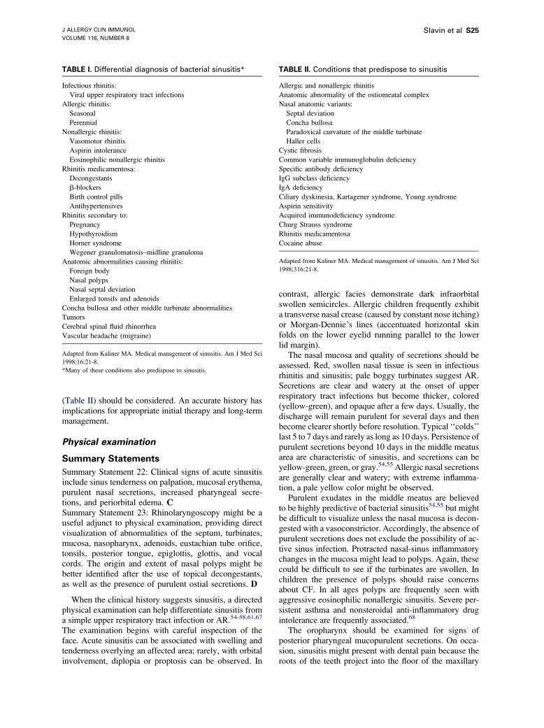
In reviewing a patient’s history, the differential diag-nosis of sinusitis (Table I) and predisposing conditions
J ALLERGY CLIN IMMUNOL
VOLUME 116, NUMBER 6
Slavin et al S25
(Table II) should be considered. An accurate history hasimplications for appropriate initial therapy and long-termmanagement.
Physical examination
Summary Statements
Summary Statement 22: Clinical signs of acute sinusitisinclude sinus tenderness on palpation, mucosal erythema,purulent nasal secretions, increased pharyngeal secre-tions, and periorbital edema. CSummary Statement 23: Rhinolaryngoscopy might be auseful adjunct to physical examination, providing directvisualization of abnormalities of the septum, turbinates,mucosa, nasopharynx, adenoids, eustachian tube orifice,tonsils, posterior tongue, epiglottis, glottis, and vocalcords. The origin and extent of nasal polyps might bebetter identified after the use of topical decongestants,as well as the presence of purulent ostial secretions. D
When the clinical history suggests sinusitis, a directedphysical examination can help differentiate sinusitis froma simple upper respiratory tract infection or AR.54-58,61,67
The examination begins with careful inspection of theface. Acute sinusitis can be associated with swelling andtenderness overlying an affected area; rarely, with orbitalinvolvement, diplopia or proptosis can be observed. In
TABLE I. Differential diagnosis of bacterial sinusitis*
Infectious rhinitis:
Viral upper respiratory tract infections
Allergic rhinitis:
Seasonal
Perennial
Nonallergic rhinitis:
Vasomotor rhinitis
Aspirin intolerance
Eosinophilic nonallergic rhinitis
Rhinitis medicamentosa:
Decongestants
b-blockers
Birth control pills
Antihypertensives
Rhinitis secondary to:
Pregnancy
Hypothyroidism
Horner syndrome
Wegener granulomatosis–midline granuloma
Anatomic abnormalities causing rhinitis:
Foreign body
Nasal polyps
Nasal septal deviation
Enlarged tonsils and adenoids
Concha bullosa and other middle turbinate abnormalities
Tumors
Cerebral spinal fluid rhinorrhea
Vascular headache (migraine)
Adapted from Kaliner MA. Medical management of sinusitis. Am J Med Sci
1998;16:21-8.
*Many of these conditions also predispose to sinusitis.
contrast, allergic facies demonstrate dark infraorbitalswollen semicircles. Allergic children frequently exhibita transverse nasal crease (caused by constant nose itching)or Morgan-Dennie’s lines (accentuated horizontal skinfolds on the lower eyelid running parallel to the lowerlid margin).
The nasal mucosa and quality of secretions should beassessed. Red, swollen nasal tissue is seen in infectiousrhinitis and sinusitis; pale boggy turbinates suggest AR.Secretions are clear and watery at the onset of upperrespiratory tract infections but become thicker, colored(yellow-green), and opaque after a few days. Usually, thedischarge will remain purulent for several days and thenbecome clearer shortly before resolution. Typical ‘‘colds’’last 5 to 7 days and rarely as long as 10 days. Persistence ofpurulent secretions beyond 10 days in the middle meatusarea are characteristic of sinusitis, and secretions can beyellow-green, green, or gray.54,55 Allergic nasal secretionsare generally clear and watery; with extreme inflamma-tion, a pale yellow color might be observed.
Purulent exudates in the middle meatus are believedto be highly predictive of bacterial sinusitis54,55 but mightbe difficult to visualize unless the nasal mucosa is decon-gested with a vasoconstrictor. Accordingly, the absence ofpurulent secretions does not exclude the possibility of ac-tive sinus infection. Protracted nasal-sinus inflammatorychanges in the mucosa might lead to polyps. Again, thesecould be difficult to see if the turbinates are swollen. Inchildren the presence of polyps should raise concernsabout CF. In all ages polyps are frequently seen withaggressive eosinophilic nonallergic sinusitis. Severe per-sistent asthma and nonsteroidal anti-inflammatory drugintolerance are frequently associated.68
The oropharynx should be examined for signs ofposterior pharyngeal mucopurulent secretions. On occa-sion, sinusitis might present with dental pain because theroots of the teeth project into the floor of the maxillary
TABLE II. Conditions that predispose to sinusitis
Allergic and nonallergic rhinitis
Anatomic abnormality of the ostiomeatal complex
Nasal anatomic variants:
Septal deviation
Concha bullosa
Paradoxical curvature of the middle turbinate
Haller cells
Cystic fibrosis
Common variable immunoglobulin deficiency
Specific antibody deficiency
IgG subclass deficiency
IgA deficiency
Ciliary dyskinesia, Kartagener syndrome, Young syndrome
Aspirin sensitivity
Acquired immunodeficiency syndrome
Churg Strauss syndrome
Rhinitis medicamentosa
Cocaine abuse
Adapted from Kaliner MA. Medical management of sinusitis. Am J Med Sci
1998;316:21-8.
J ALLERGY CLIN IMMUNOL
DECEMBER 2005
S26 Slavin et al
sinus. In fact, some cases of maxillary sinusitis are secon-dary to a dental root infection that erodes through the boneinto the adjacent sinus. Examination of the ears mightreveal otitis media, particularly in children with sinusitis;in fact, unresolved persistent bacterial sinusitis might leadto recurrent otitis media.61,67 Auscultation of the chestfor wheezing might disclose an asthmatic component ofa patient’s cough. The absence of audible wheezing doesnot exclude the possibility of asthma; subtle abnormalitiesmight only be apparent on spirometry (see the ‘‘Labora-tory tests’’ section).
Transillumination of the maxillary sinuses might bereported as opaque (no transmission), dull (reduced trans-mission), or normal, but the sensitivity and specificity ofthis technique alone is poor.69 Unless the clinician has ad-ditional knowledge of a patient’s sinus anatomy (eg, froma CT scan), it is impossible to differentiate active diseasefrom a congenitally small sinus or bilateral mild involve-ment of larger sinuses from normal size sinuses.70-73 Inone prospective study, however, abnormal transillumina-tion combined with purulent nasal secretions and a historyof maxillary pain, poor response to decongestants, andcolored rhinorrhea was the best predictor of acute bacterialsinusitis.71
In reassessing patients with recurrent sinusitis, thephysical examination should be modified to evaluate forsigns of immunodeficiency (associated recurrent otitismedia, bronchiectasis, or pneumonia), complications ofprimary infections (eg, mastoiditis or orbital cellulitis),poor growth in children, unexplained dermatitis, absenceof lymphoid tissue, CF (poor growth, nasal polyps, barrelchest, digital clubbing, and diffuse chest abnormalities onauscultation), ciliary dysfunction (nasal polyps, digitalclubbing, dextrocardia, and situs inversus), and anatomicabnormalities (eg, septal deviation, nasal polyps, foreignbodies, and tumors).
In selected patients with chronic or recurrent sinusitis,nasal endoscopy should be considered. This procedureprovides ideal direct visualization of abnormalities of theseptum, turbinates, mucosa, nasopharynx, adenoids, eu-stachian tube orifice, tonsils, posterior tongue, epiglottis,glottis, and vocal cords. The origin and extent of nasalpolyps can be identified, as well as the presence ofpurulent ostial secretions.73,74
Imaging studies
Summary Statements
Summary Statement 24: Imaging techniques can provideconfirmatory evidence when symptoms are vague, phys-ical findings are equivocal, or clinical disease persists de-spite optimal medical therapy. BSummary Statement 25: Ultrasonography has limitedutility but might be useful in pregnant women or for de-termining amounts of retained sinus secretions. CSummary Statement 26: Standard radiographs might beused to detect acute sinusitis, but they are not sensitive,particularly for ethmoid disease. C
Summary Statement 27: CT is the optimal technique forevaluating the ethmoid sinuses and for preoperative eval-uation of the nose and paranasal sinuses, including as-sessment of the ostiomeatal complex areas. CSummary Statement 28: MRI is a sensitive techniquefor evaluating suspected fungal sinusitis and for differenti-ating between inflammatory disease andmalignant tumors.MRI is limited in its ability to define bony anatomy. C
The diagnosis of acute sinusitis is generallymade on thebasis of history and physical examination.54,55 Occasion-ally, it might be difficult clinically to distinguish betweensinusitis and rhinitis, and it can be a challenge to documentthe extent of disease. As previously stated, the term rhino-sinusitis has been suggested in part to reflect this confu-sion. In this context imaging procedures, particularly CTand MRI, are the generally used imaging techniques forconfirmatory evidence in patients with both acute andchronic sinusitis.75 The clinical utility, relative safety,ease of performance, availability, and cost of these imag-ing techniques vary considerably, and all have inherentlimitations.58,76-79
Imaging procedures are often useful when symptomsare vague, physical findings are equivocal, or the responseto initial management is poor. The possibility of sinusitiscan also be confirmed by imaging patients with atypicalpresentations (eg, a child with refractory conjunctivitis ora patient with acute severe eye or occipital pain).
Some unusual causes of persistent disease, such as al-lergic fungal sinusitis and Wegener granulomatosis mightbe suggested by newer and more sophisticated imagingtechniques, such as MRI.80
Ultrasonography. A-mode ultrasonography is a safe,rapid, and noninvasive technique for evaluating the max-illary and frontal sinuses. Unfortunately, several studiescomparing ultrasonography with radiographic techniques(as the gold standard) suggest that the sensitivity andspecificity of ultrasonography is poor, ranging from 39%to 61% and 42% to 53%, respectively.81-84 Another limita-tion is that ultrasonography can only be used to evaluatethe frontal and maxillary sinuses.85 Despite these issues,ultrasonography might have some utility as a sinus diag-nostic screen in pregnant women to avoid risks of ionizingradiation.Standard radiography. Caldwell (anterior-posterior) andWaters (occipito-mental) standard radiographs demon-strate the frontal and maxillary sinuses, respectively.86
Lateral views visualize the sphenoid sinus and adenoidsin children. The fine bony anatomy of the ethmoid sinusesis not well seen on any standard radiographs becauseof problems of normal structural superimposition.Although other sinuses might also be involved, acute si-nusitis commonly involves the maxillary sinuses. Accord-ingly, a singleWaters view is a viable diagnostic substitutefor a 4-view sinus series in many patients.87 However,ethmoid involvement without maxillary sinus infectionoccurs in approximately 20% of patients.
The interpretation of standard radiographs remains con-troversial. It is generally believed that mild-to-moderatemucosal thickening is a nonspecific finding. In contrast,
J ALLERGY CLIN IMMUNOL
VOLUME 116, NUMBER 6
Slavin et al S27
sinus opacification, air-fluid level, or severe mucosalthickening in adults is more likely to reflect meaningfulpathology.88 Extrapolating from more recent CT studies,however, the correlation between radiographic extentof disease and likelihood of resolution without medicaltherapy is poor.76-78 The decision to treat with antibioticsshould therefore be made on clinical grounds alone.
Standard radiographs are of limited value in the eval-uation of chronic sinusitis; they are also inadequate forclarifying the need for endoscopic surgery and the preciseareas that need surgical attention.89
Computed tomography. High-resolution CT scans dem-onstrate the extent of disease and the degree of ostiomeatalcomplex obstruction; they might also delineate pathologicvariations and anatomic structures that are not apparent onphysical examination or rhinoscopy.89,90 Sinus CT is theimaging technique of choice in preparation for endoscopicsurgery.
Coronal scans provide a road map for surgery, bestdemonstrating the ostiomeatal drainage areas and intricaterelationships between the brain, fovea ethmoidalis, andethmoid sinuses. However, increasingly, surgeons areusing computer-assisted surgical navigation, particularlyin revision or other cases of complicated anatomy.Computer-assisted surgical navigation requires axialscans with scanning parameters specific for the computerdevice used, as well as standard coronal views.
The amount of radiation in standard sinus CTs is notnegligible. An alternative approach is a low-dose CT witha standard dose in 1 or 2 sections in which more intricatedetail is required (eg, in the ostiomeatal complex).91
In the past, a series of standard radiographs has beenfavored in preference to CT because of cost and timeconsiderations. More recently, however, some medicalcenters offer a limited 4- to 5-cut coronal sinus CT scan ata similar cost to a set of standard radiographs. Thistypically takes only 15 to 20 minutes and provides muchbetter high-resolution bone and soft tissue detail, includ-ing the ethmoids (which are poorly visualized on anystandard radiograph).92 This field is progressing rapidly,and current scanners take only 17 seconds for a completesinus set.
High-resolution sinus CT is considered a mandatorypreoperative study and is important for the planning andsafe performance of functional endoscopic sinus surgery.This is particularly crucial when the ostiomeatal complexand ethmoids are involved.93
Sinus CT is particularly helpful in the diagnosis offungal sinusitis. Classic findings for fungal disease are acombination of unilateral lesions of one or more sinuses,nodular mucoperiosteal thickening, focal areas of bonedestruction, and/or dense intrasinus concretions.94
Magnetic resonance imaging. MRI provides ideal imag-ing of soft tissues; it is less suited to imaging bonyanatomy because bone and air yield similar signal inten-sities onMRI. The value of MRI is further limited becausethe signal intensity from normal edematous mucosa isindistinguishable from extensively inflamed and diseasedtissue.
The differential diagnosis between inflammatory dis-eases and malignant tumor is aided byMRI.95 MRI assistsin clarifying the degree of orbital or intracranial extensionin complicated sinusitis. T2-weighted image intensity isuseful: high signal with bacterial and viral inflammation,intermediate bright signal with neoplastic processes (eg,squamous cell carcinoma), and very low signal with fun-gal concretions, similar to that of air.86
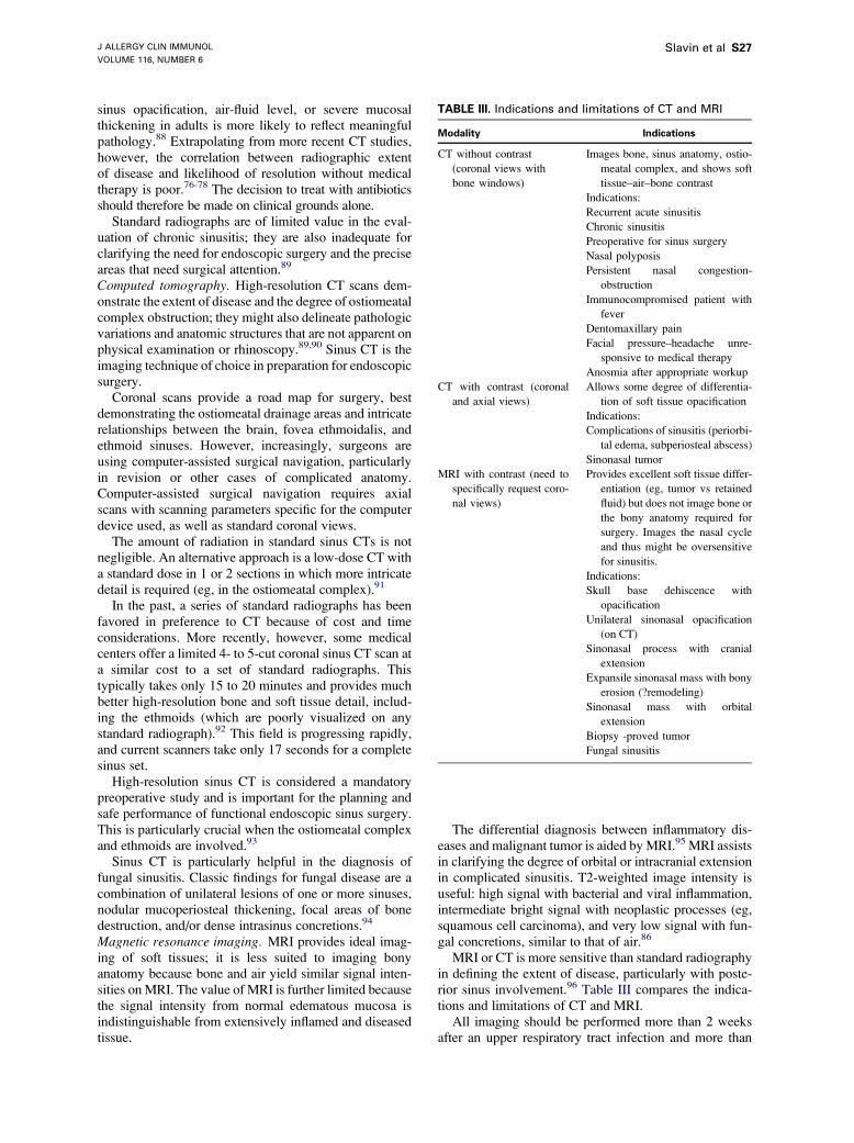
MRI or CT is more sensitive than standard radiographyin defining the extent of disease, particularly with poste-rior sinus involvement.96 Table III compares the indica-tions and limitations of CT and MRI.
All imaging should be performed more than 2 weeksafter an upper respiratory tract infection and more than
TABLE III. Indications and limitations of CT and MRI
Modality Indications
CT without contrast
(coronal views with
bone windows)
Images bone, sinus anatomy, ostio-
meatal complex, and shows soft
tissue–air–bone contrast
Indications:
Recurrent acute sinusitis
Chronic sinusitis
Preoperative for sinus surgery
Nasal polyposis
Persistent nasal congestion-
obstruction
Immunocompromised patient with
fever
Dentomaxillary pain
Facial pressure–headache unre-
sponsive to medical therapy
Anosmia after appropriate workup
CT with contrast (coronal
and axial views)
Allows some degree of differentia-
tion of soft tissue opacification
Indications:
Complications of sinusitis (periorbi-
tal edema, subperiosteal abscess)
Sinonasal tumor
MRI with contrast (need to
specifically request coro-
nal views)
Provides excellent soft tissue differ-
entiation (eg, tumor vs retained
fluid) but does not image bone or
the bony anatomy required for
surgery. Images the nasal cycle
and thus might be oversensitive
for sinusitis.
Indications:
Skull base dehiscence with
opacification
Unilateral sinonasal opacification
(on CT)
Sinonasal process with cranial
extension
Expansile sinonasal mass with bony
erosion (?remodeling)
Sinonasal mass with orbital
extension
Biopsy -proved tumor
Fungal sinusitis
J ALLERGY CLIN IMMUNOL
DECEMBER 2005
S28 Slavin et al
4 weeks after acute bacterial sinusitis and after medicalmanagement if the aim is to identify the underlying extentof chronic sinusitis or potential causes of recurrent acutesinusitis.
Laboratory tests
Summary Statements
Summary Statement 29: Laboratory evaluation of acute,chronic, or recurrent sinusitis might include the follow-ing: nasal cytology, nasal-sinus biopsy, or tests for im-munodeficiency, CF, or ciliary dysfunction. NRSummary Statement 30: Nasal cytology is useful in theevaluation of conditions associated with sinusitis, in-cluding AR, eosinophilic NAR, neutrophilic rhinitis,and VMR. CSummary Statement 31: Nasal-sinus biopsy is useful inseveral clinical situations: determining whether a lesionis neoplastic and, if so, its nature; confirming the presenceof fungal disease; confirming the presence of granuloma-tous disease; and determining the ultrastructure of cilia. C
Laboratory tests might provide additional diagnosticinformation in patients with acute or chronic sinusitis.Nasal cytology can provide data that support or refute theclinical diagnosis of acute infectious sinusitis; resultsmight suggest the presence of underlying disease (eg,eosinophilic rhinitis). Antral puncture and endoscopicallyobtained culture were discussed in the ‘‘Microbiology’’section. Nasal-sinus biopsy is useful in suspected cases ofneoplasia, fungal disease, or granulomatous disease; thistechnique is also used in patients with ciliary dysmotilityto define the ultrastructural nature of the defect. Other testsare used to assess disorders that might be causallyassociated with recurrent or chronic sinusitis (eg, immu-nodeficiency, CF, or ciliary dysfunction); these tests arediscussed in separate sections (see below).Nasal cytology. Nasal cytology demonstrates the type ofcellular inflammation in patients with acute or chronicsinusitis. This can provide useful adjunctive informationto the clinical history and examination with regard tolikely infection or associated eosinophilic disease. Sam-ples can be obtained by having the patient blow secretionsinto transparent plastic wrap or by the use of a cytologybrush or Rhinoprobe (Synbiotics Corp, San Diego, Calif).Samples collected by means of the blowing techniqueoften contain less cellular material but are still useful fordetecting eosinophils and neutrophils.1
Nasal secretions are transferred to a glass slide, fixed,and then treated with Hansel stain, a stain that highlightseosinophil granular contents. Additional stains (eg,Papanicolaou, hematoxylin and eosin, acidified toluidineblue, orMay-Grunwald-Giemsa) might be used to identifyother elements, including mast cells, basophils, neutro-phils, nonciliated epithelial cells, squamous cells, or evenpollens or mold spores.
Nasal cytology results can be consistent with a varietyof syndromes, including AR, eosinophilic NAR, VMR,bacterial sinusitis, neutrophilic nonbacterial sinusitis, and
metaplasia caused by environmental exposures.97,98 Thepresence of eosinophils correlates with AR or eosinophilicNAR, a condition often associated with chronic sinusitis,nasal polyposis, asthma, and nonsteroidal anti-inflamma-tory drug sensitivity.99
Unfortunately, the clinical value of nasal cytology inthe assessment of sinusitis is limited, with specificityranging from 40% to 90% and sensitivity from 67% to80%.100,101 This occurs because acute viral infections caninduce nasal neutrophilia and transient abnormal imagingstudies.101 In addition, patients with AR and acute bacte-rial sinusitis will demonstrate a mixture of nasal eosino-philia and neutrophilia, potentially creating diagnosticconfusion. At the same time, nasal cytology can providesome useful information: the absence of neutrophils (as-suming an adequate sample) argues against an infectiouscomponent, and the presence of more than 10% eosino-phils in a patient with chronic or recurrent disease suggeststhe potential for underlying AR or NARES.Nasal-sinus biopsy. Nasal-sinus biopsy should be con-sidered to aid in the diagnosis of nasal or paranasal lesionsthat obstruct sinus cavities and give rise to chronic orrecurrent sinus disease. Clinical reasons to obtain a biopsyare (1) to determine whether a lesion is neoplastic and, ifso, to clarify the nature of the neoplasm; (2) to confirmthe presence of suspected invasive fungal disease; (3) toconfirm the presence of granulomatous disease when thediagnosis is unclear on the basis of clinical or radiographicgrounds; and (4) to provide an initial evaluation of theultrastructure of cilia in patients with known or suspectedciliary dysfunction. In the presence of chronic inflamma-tion, a tracheal biopsy is typically required for confirmation.Nasal and paranasal sinus neoplasms. Early signs ofsinus neoplasia are nonspecific and include nasal obstruc-tion, anosmia, rhinorrhea, and pain. Accordingly, clinicalsuspicion of tumor is often delayed until epistaxis, prop-tosis, trismus, facial swelling, or cranial nerve (I/VI)dysfunction develop. Early diagnosis is enhanced by theuse of CT or MRI. The most common tumor is an invertedpapilloma, which is characterized by a polypoid appear-ance and unilateral location seen on both physical exam-ination and imaging.
Tumors within the nose or sinuses are histologicallydiverse and both benign and malignant. Generally, aspecific diagnosis is not reliable on the basis of clinicalor radiographic findings, making a tissue biopsy invalu-able.102 Juvenile angiofibroma is a notable exception.Diagnosis is made on the basis of finding a vascular pos-terior nasal or nasopharyngeal mass in an adolescent orpreadolescent male. Biopsy should not be performedbecause of the risk of significant hemorrhage.103
Invasive fungal infections. Fungal infection produced byphycomycetes (Absidia, Mucor, and Rhizopus species)can progress from the sinuses to the central nervous sys-tem and is potentially fatal. Similar rapidly fulminant in-fections can be caused by Aspergillus species. Thesefulminant diseases occur essentially only in the immuno-compromised (eg, patients with diabetes mellitus, hemato-logic malignancies, adrenal suppression, chronic renal
J ALLERGY CLIN IMMUNOL
VOLUME 116, NUMBER 6
Slavin et al S29
failure, hematologic dyscrasia or overt immunodeficiency[eg, patients undergoing chemotherapy, intravenous drugusers, and patients with HIV]) However, less fulminantand more indolent invasive disease might occasionallyoccur in the immunocompetent patient.
Patients with fulminant invasive fungal sinusitis oftenpresent with fever, which rapidly progresses to facial pain,proptosis, ophthalmoplegia, and/or facial necrosis. Lateradiographic findings can include sinus opacification,mucoperiosteal thickening, bone erosion, and cavernoussinus thrombosis. Endoscopically, early lesions mightappear brick red or as black necrotic areas. Suspiciouslesions should be biopsied immediately, with the samplesent for fungal staining and culture. Early diagnosis can becritical in determining patient prognosis.104
Granulomatous inflammatory disease. Sinusitis can beassociated with several granulomatous diseases, bothinfectious and noninfectious. Sarcoidosis is by far themost common of these disorders identified within theUnited States. Another noninfectious cause is midlinegranuloma–Wegener granulomatosis. Infectious causesinclude Klebsiella species–induced rhinoscleroma, tuber-culosis, leprosy, syphilis, and fungal infections. Lesionscan cause ulceration, necrosis, or hyperplastic mucosa.When such lesions are apparent, biopsy should beobtained to exclude neoplasia, and special staining andcultures should be done for mycobacteria and fungi.105,106
Cilia ultrastructure. When ciliary dysfunction is sus-pected in patients (eg, with recurrent or persistent infec-tious disease in the absence of underlying rhinitis,anatomic defects, or immunodeficiency), biopsy shouldbe considered for ultrastructural analysis. Reported resultsinclude the type of defect (dynein defects are mostspecific), the percentage of cilia having the defect, thedistribution of the defect (bronchial versus nasal if bothareas are assessed), and the number of cilia present.107,108
Because acquired ciliary defects can sometimes mimicprimary ciliary dyskinesia in the presence of chronic in-flammation, tracheal biopsy is frequently required forconfirmation of this diagnosis.Immunodeficiency tests. See the ‘‘Immunodeficiency’’section.Cystic fibrosis tests. See the ‘‘Cystic fibrosis’’ section.Ciliary tests. See the ‘‘Ciliary dysfunction’’ section.Miscellaneous tests. The role of nasopharyngeal culturein the diagnosis of acute sinusitis remains controversial.In the past, studies have suggested a poor correlationbetween the presence of nasal bacteria and the likelihoodof active bacterial sinusitis; even in patients with culture-proved sinusitis, the bacteria present in the sinus wasdifferent from bacteria in the nasophayrnx. More recently,it has been suggested that the presence of pathogenicorganisms (S pneumoniae,H influenzae, orM catarrhalis)in the nasopharynx of patients presenting with upperrespiratory tract infection symptoms might define patientsmore likely to have bacterial disease.109
The value of routine hematologic tests (eg, erythrocytesedimentation rate, white blood cell count, and C-reactiveprotein) was recently evaluated in 176 patients with acute
maxillary sinusitis. In general, these tests were not sensi-tive indicators of disease or specific cause (82% of testresults were normal). Increased C-reactive protein (>40mg/L) was associated with likely infection with Strepto-coccus pyogenes or S pneumoniae, a fact that could influ-ence choice and duration of therapy.110
In suspected Wegener granulomatosis, an antineutro-phil cytoplasmic antibody test might be a useful adjunctto tissue biopsy.111 Similarly, increases of angiotensin-converting enzyme and soluble IL-2 receptor levels mightbe of help in clarifying a suspected tissue diagnosis ofsarcoidosis.112
Predisposing factors
Viral infections
Summary Statements
Summary Statement 32: Acute viral upper respiratorytract infections often precede acute bacterial sinusitis. BSummary Statement 33: Viral upper respiratory tract in-fections are often (40% to 90% of the time) associatedwith CT evidence of sinusitis. Viral sinusitis appears toresolve within 21 days without the need for antibiotics. BSummary Statement 34: There is minimal evidence thatviruses play a role in chronic sinusitis. B
The average child in the United States has 3 to 8 viralupper respiratory tract infections per year; the averageadult has 2 to 3 such infections. Up to 90% of these upperrespiratory tract infections are associated with CT changesconsistent with mucosal involvement within the si-nuses.113-115 Thus these subjects have acute viral sinusitis.Acute bacterial sinusitis is thought to follow 0.5% to 13%of these acute upper respiratory tract infections,113,116,117
resulting in approximately 20 million bacterial infec-tions.118 The symptoms of patients with a viral upper res-piratory tract infection and radiologic evidence of sinusitisare no different than those of subjects experiencing a colduncomplicated byCT abnormalities.119 In one study of CTabnormalities in patients with colds, 39% of patients witha cold had CT evidence of viral sinusitis, and all resolvedwithin 21 days without the need for antibiotics.119 Thisobservation has been confirmed.115
Because many acute sinus infections resolve withoutthe need for antibiotics, it has always been thought (butnot proved) that viruses might be responsible for thesymptoms in such patients. The technique of PCR allowsamplification of small quantities of viral genomes to thelevel of detection. PCR analysis of the sinus mucosaobtained at surgery for chronic sinusitis in 20 patientsrevealed that 20% had positive results for respiratorysyncytial virus and none for adenovirus. Thus a number ofpatients with chronic sinusitis might have had a predis-posing viral infection.120 By contrast, biopsy of the max-illary sinus mucosa in patients with acute sinusitisrevealed that 50% had RNA evidence of rhinovirus.121
The weight of the evidence supports the clinical observa-tions that a sizeable number of acute sinusitis infectionsare caused by viruses but that viruses play a more limited
J ALLERGY CLIN IMMUNOL
DECEMBER 2005
S30 Slavin et al
role in chronic sinusitis, except to predispose to possiblesuperimposed acute bacterial sinusitis.
Allergic rhinitis
Summary Statements
Summary Statement 35: AR commonly precedes the de-velopment of recurrent or chronic sinusitis. The nasal ob-struction and inflammation associated with AR interruptsnormal mucociliary clearance and leads to retention ofsecretions within the sinus cavities. BSummary Statement 36: Patients with recurrent orchronic sinusitis should be evaluated for the presenceof underlying allergy. B
AR is one of the most common chronic diseases,accounting for approximately 2.5% of all physician visits.The incidence is estimated at 10% to 14% of the popu-lation at any time, with a cumulative prevalence rangingup to 20%. AR usually develops during childhood at theaverage age of 10 years, although 30% of patients havetheir onset of symptoms after age 30 years.122-124
Allergic inflammation leads to nasal congestion andswelling of the mucous membrane, which can obstruct orimpede normal sinus drainage. Obstructed sinuses par-tially fill with secretions, use trapped oxygen, and becomeacidotic, leading to even more impaired mucociliaryfunction and impaction of secretions. Bacteria, eitheralready in the sinuses or gaining access because ofabnormal ciliary flow, multiply, infecting the mucosallining. Subsequent inflammatory responses in the epithe-lium lead to an influx of granulocytes, with swelling andpain from the mucosa and thickened secretions.
A number of observations support the association ofAR and sinusitis both in adults and children. In childrenevidence of AR has been found in 36% to 60% of patientswith chronic sinusitis.125-127 Young adults with acutemaxillary sinusitis had a 25% to 31% incidence ofAR,128,129 whereas chronic sinusitis was associated withAR in 40% to 84% of adult patients.130-132 Newmanet al133 found an association between extensive sinus dis-ease, quantified by means of CT, and allergy in 78% of pa-tients and asthma in 71% of patients. Moreover, sinusitisin patients with AR is associated with more extensive ab-normalities on CT scans.134 About twice as many patientswith AR, compared with normal subjects, have abnormalCT scans (67% versus 33%).135 Although these studieswould suggest a uniform association between allergyand sinusitis, other studies have failed to confirm differ-ences between allergic and nonallergic subjects withsinusitis by means of CT scan.136,137
In a careful retrospective study of 200 patients withchronic sinusitis, McNally et al138 found that more thanhalf the patients hadAR, and this diagnosiswas consideredthemost important underlying cause of sinusitis. In one ad-ditional study, 43% of acute sinusitis was noted to be sea-sonal, the cause of which was thought to be allergic.139
A suggestive experimental link is derived from a studyin which rhinomanometry was performed and maxillary
sinus x-ray films were taken before and after nasalprovocation tests.140 Of 73 separate provocation tests,41 led to early-phase responses only, 18 led to late-phaseresponses alone, and 10 yielded both early and lateresponses. Thirty-two of these patients experienced in-creased sinus mucosal edema and opacification of theparanasal sinuses, as revealed on sinus x-ray films.Concomitantly, increased pressure in the maxillary sinus,acute headaches, and associated otalgia were reportedby patients. In addition, 3 of the patients who did nothave an obvious clinical response had thickened mucosaand subjective symptoms. In view of the difficulty in per-forming direct paranasal sinus challenges, these nasalchallenges provide an experimental link between ARand sinusitis. However, in contrast to this study involvingexperimental provocations, examination of ragweed-sensitive subjects in the midst of their allergy season dem-onstrated no consistent abnormalities on sinus CT.141
Does allergen enter the sinus through the nasal ostia andinitiate an allergic response within the sinus cavity? Asnoted above,140 experimentally instilled allergen in thenose can cause sinus edema and changes on CT scans,whereas other studies using radiolabeled pollen couldshow no evidence that pollen enters the sinus cavity.142
Collection of fluid from patients with chronic sinusitis re-veals the presence of allergic mediators, including hista-mine and leukotrienes,143 inflammatory cells (includingeosinophils),143,144 and cytokines,145,146 suggesting thatallergic responses, inflammatory responses, or both partic-ipate in sinusitis.
In one recent study higher levels of eosinophil-derivedneurotoxin and decreased levels of lysozyme were foundin nasal fluids from patients with perennial AR withrecurrent sinusitis compared with patients with eitherperennial AR alone or control subjects.147
Management of AR is focused on environmental con-trol, pharmacologic management, and immunotherapy asan immunomodulating approach. The effective treatmentof AR might decrease the frequency of sinusitis by re-ducing the inflammation and swelling in the nasal mucosathat compromises the sinus outflow tract, although at thistime there are few or no studies that have focused on thisend point. Patients with sinusitis, especially of a chronic orrecurrent nature, should have an allergy evaluation.
Nonallergic rhinitis
Summary Statements
Summary Statement 37: NAR has a much higher preva-lence than usually suspected. BSummary Statement 38: The primary symptoms associ-ated with NAR, congestion and increased secretions,are often found in patients with sinusitis. BSummary Statement 39: NAR is one of the most frequentdiseases occurring in patients with chronic sinusitis andmight predispose toward the development of sinusitis. B
NAR, by itself or in combination with AR, occurs morefrequently than commonly recognized. Recent data
J ALLERGY CLIN IMMUNOL
VOLUME 116, NUMBER 6
Slavin et al S31
suggest that about 40 million Americans have either pureNAR or mixed rhinitis, in which both NAR and ARcontribute to the symptoms.148 In one study 52% of 142patients with rhinitis seen in an allergy clinic hadNAR,149 whereas in another study 17% of patients withrhinitis in an academic setting had NAR.150 TheEuropean Community Respiratory Health Survey of1412 subjects found 25% with NAR.151 A recent retro-spective survey conducted by the National RhinitisClassification Task Force of 975 patients with rhinitisfound 43% to have AR, 23% to have NAR, and 34% tohave both diseases (mixed rhinitis).148 The few studiesthat have reported incidence by sex suggest that womenare disproportionately affected by NAR or mixed rhini-tis.152 Diagnosing NAR can be confusing because the pri-mary symptoms (ie, rhinorrhea, nasal congestion, andpostnatal drip) can be indistinguishable from those ofAR. Diagnosis of NAR is made by a combination of care-ful history taking and exclusion: negative or irrelevantskin test responses rule out IgE-mediated rhinitis, and ahistory of symptoms triggered by environmental irritantssuggests a diagnosis of NAR. Positive skin test responsesdo not, however, rule out NAR.152,153 Patients withNAR often have normal nasal examination results, and ep-ithelial scrapings are usually normal as well.
There are limited data on the incidence or frequency ofdistinct subtypes of NAR. A 1985 study of 78 patientswith NAR showed that VMR was the major type of NAR,affecting about two thirds of the patients.154 In anotherstudy VMR was present in 37% of 142 patients withrhinitis followed prospectively. Fifty-two percent of thesepatients were given diagnoses of NAR.148 Some have es-timated that more than 65% of all NAR is vasomotor innature.155
VMR deserves special attention because of its highincidence in clinical practice. It is estimated that theprevalence of VMR in all patients is 5% to 10%, whereasthe prevalence of AR is 15% to 17%.155,156 VMR shouldbe suspected in patients presenting with symptoms ofchronic nasal congestion, rhinorrhea, or postnasal drip.These classic symptoms are sometimes accompanied bylack of taste or smell, sinus headache, chronic cough,and throat clearing.152,153,156 A patient describing nasalsymptoms in response to cold air; strong smells (eg, per-fumes, tobacco, paint, or cleaning solutions); ingestionof alcohol; changes in temperature, humidity, and/or baro-metric pressure; or emotional stress has vasomotor symp-toms. It should be emphasized that patients with AR orNAR might also respond to these triggers. Some havesuggested that VMR be reserved for subjects in whomthe nasal symptoms are more chronic in nature.155,156 Inrecent years, the use of CT has revealed that many patientsgiven diagnoses of VMR in fact have chronic sinusitis orother disorders. Thus the diagnosis of VMR has come un-der question.
NAR can be associated with sinusitis. In the only studyof chronic sinusitis in which NAR was evaluated, 26% ofpatients had NAR. In the same study, more than half thepatients had AR.138 There have been no other studies in
which NAR was estimated with respect to its relationshipwith sinusitis.
One study157 described a series of patients with sneez-ing, watery rhinorrhea, nasal pruritus, and negative skintest and RAST results. Smears of secretions revealed eo-sinophilia, and this syndrome was identified as NARES.It is estimated that 15% to 20% of patients with NARhave NARES. NARES is found as an isolated syndromeor in conjunction with aspirin sensitivity. In general,patients with NARES have rather profound nasal symp-toms and respond well to treatment with nasal corticoste-roids. Some observers think that NARES can beassociated with an increased risk of nasal polyps, andpolyps are often found in patients with NARES with aspi-rin sensitivity.
GERD
Summary Statements
Summary Statement 40: GERD has been suggested as acause of sinusitis. DSummary Statement 41: pH probe monitoring of bothchildren and adults with chronic sinusitis shows a high in-cidence of both esophageal and nasopharyngeal reflux. BSummary Statement 42: Medical treatment of GERD inchildren and adults has been shown to result in signifi-cant improvement in sinusitis symptoms. BSummary Statement 43: In patients with sinusitis refrac-tory to medical therapy, treatment of associated GERDshould be considered before surgical intervention. B
GERD has been suggested as a cause of sinusitis. Themechanism is thought to be direct reflux of gastric acidinto the pharynx and subsequently to the nasopharynx,causing inflammation of the sinus ostium and leading tosinusitis.158
A study in 30 children with chronic sinusitis was doneusing 24-hour monitoring with dual-pH probes, one in thenasopharynx and one in the distal esophagus. Nineteen(63%) showed gastroesophageal reflux, well above theexpected prevalence of 5% in the healthy general popu-lation. Of these 19, 6 (32%) demonstrated nasopharyngealreflux. Seventy-nine percent improved their sinusitissymptoms after treatment of GERD. The recommendationof the authors was that children with chronic sinus diseaserefractory to medical therapy be evaluated for GERD andtreated before sinus surgery is considered.159
A study in adults evaluated the prevalence of gastro-esophagopharyngeal reflux in 11 patients with CT scan–confirmed chronic sinusitis who had not responded toconventional therapy and 11 healthy control subjects. A3-site ambulatory esophagopharyngeal pH monitoringtechnique was used. Ambulatory pH monitoring docu-mented gastroesophagopharyngeal reflux in 7 of 11patients and 2 of 11 healthy volunteers.160
Another uncontrolled study involved 19 adult patientswith chronic sinusitis, 18 of whom had undergone sinussurgery. Sixty-eight percent had classic GERD symptoms,and 78% had abnormal results on an esophageal pH probe.
J ALLERGY CLIN IMMUNOL
DECEMBER 2005
S32 Slavin et al
Twelve were treated with proton-pump inhibitors, 4 weretreated with proton-pump inhibitors and pyrokinetics, and2 had repeat surgery. Six months later, 12 (67%) hadimprovement in sinus symptoms, with 4 having dramaticimprovement. The authors suggest that medical therapy asa treatment for adults with chronic sinusitis be confined topatients with abnormal pH results.161 Although an associ-ation between GERD and sinusitis has been suggested,a definitive causal relationship has not been proved ina well-performed controlled study. However, in patientswith sinusitis refractory to medical therapy, treatment ofassociated GERD should be considered before surgicalintervention.
Immunodeficiency
Summary Statements
Summary Statement 44: Immune deficiency should beconsidered in cases of sinusitis resistant to usual medicaltherapy. BSummary Statement 45: The majority of immunodefi-cient patients with recurrent sinusitis have defects inhumoral immunity. However, other types of immunode-ficiencies might present with recurrent sinusitis as one oftheir clinical features, including AIDS. BSummary Statement 46: The most common primary im-munodeficiency disorders with recurrent sinusitis as aclinical feature are humoral immunodeficiencies, suchas selective IgA deficiency and common variable immu-nodeficiency. Other primary immunodeficiencies thatmight present with recurrent sinusitis among otherfeatures include Wiskott-Aldrich syndrome, ataxia telan-giectasia, warts, hypogammaglobulinemia, infections,myelokathexis syndrome, and caspase-8 deficiency. CSummary Statement 47: Appropriate laboratory studiesin patients with recurrent or chronic sinusitis might in-clude quantitative immunoglobulin measurement (IgG,IgA, and IgM), specific antibody responses (tetanus tox-oid and pneumococcal vaccine), and measurement ofT-cell number and function (delayed hypersensitivityskin tests and flow cytometric enumeration of T cells). B
Immunodeficiency should be considered in any patientwith recurrent or chronic sinusitis, particularly in patientsin whom aggressive prior medical and surgical manage-ment has failed.162 One study of medically diagnosedpatients with therapy-refractory recurrent sinusitis foundonly 8 (3%) of 245 patients to have hypogammablobuline-mia, impaired pneumococcal vaccine responses, orboth.163 However, a more rigorous study of 79 patientswith sinusitis diagnosed radiographically and refractoryto medical and surgical therapy revealed 10% of patientsto have common variable immunodeficiency and 6% tohave IgA deficiency.162 Suspicion is heightened whenthe patient also has a history of recurrent otitis media,bronchitis, and/or bronchiectasis. When congenital immu-nodeficiency is suspected, the physical examinationshould focus on findings associated with specific diseases:absence of tonsillar tissue, ocular telangiectasia, skin and
mucous membrane infections, eczema, clubbing, rales,rhonchi, petechiae, and purpura.164 Themajority of immu-nodeficient patients with recurrent sinusitis have defects inhumoral immunity; however, patients with other types ofimmune defects might present with recurrent sinusitis,such as patients with AIDS who have combined humoraland cellular impairments.165-168
The indications for pursuing an immunodeficiencyevaluation depend on the age, medical history, physicalexamination, and lifestyle of the patient. For example, inan infant of less than 2 years of age with recurrent and life-threatening infections of the sinuses and other organs, oneshould pursue this evaluation in an expeditious manner. Inaddition, infections with organisms of low pathogenicityshould alert the physician to the probability of a congenitalimmune deficiency. Similar indications for evaluation ofimmune deficiency exist for AIDS in both children andadults.
Appropriate screening laboratory studies for immuno-deficiencies might include quantitative immunoglobulinmeasurement (IgG, IgA, and IgM), specific antibodyresponses, and measurement of T-cell number andfunction (delayed hypersensitivity skin tests and flowcytometric enumeration of T cells). Although the post-immunization response to any protein antigen can bemeasured, assessment of response to tetanus immunizationis particularly advantageous because most patients havebeen immunized, and 90% to 100% of children shouldhave protective antibody titers after completing primaryimmunization.168 The response to polysaccharide antigenscan be determined by measuring preimmunization andpostimmunization titers to unconjugated pneumococcalvaccine. The diagnosis of IgG subclass deficiency is con-troversial, and the clinical significance of abnormal IgGsubclass levels in patients with recurrent infections isunclear (see ‘‘Practice parameters for the diagnosis andmanagement of primary immunodeficiency’’).
The most common primary immunodeficiency disor-ders with recurrent sinusitis as a clinical feature arehumoral immunodeficiencies, such as selective IgA defi-ciency169 and common variable immunodeficiency.166
Other primary immunodeficiencies that might present withrecurrent sinusitis among other features include Wiskott-Aldrich syndrome,170 ataxia telangiectasia,171 warts,hypogammaglobulinemia, infections, myelokathexissyndrome,172 and caspase-8 deficiency.173 When thesesyndromes are clinically suspected, referral to a board-certified allergist-immunologist for evaluation and therapyis indicated.
Sinusitis is a recurrent or chronic problem in30% to 68% of patients with HIV infection.174,175 Theremight be a direct correlation between CD4 deficiencyand increased likelihood of sinus disease. Specificantibody responses are abnormal in these patients, eventhough total serum immunoglobulin levels are often in-creased. Although there are case reports of infectionswith atypical organisms, most sinus infections in patientswith HIV are caused by the same organisms involved inimmunocompetent patients.176 Accordingly, successful
J ALLERGY CLIN IMMUNOL
VOLUME 116, NUMBER 6
Slavin et al S33
treatment of these patients involves amuch longer durationof aggressive standard therapy.
Cystic fibrosis
Summary Statements
Summary Statement 48: Virtually all patients with CFhave sinusitis as a consequence of dehydration of muco-sal fluids and sulfation of mucous glycoproteins, a com-bination resulting in retention of viscous tenacious sinussecretions that predispose to bacterial infection. BSummary Statement 49: CF should be considered in anypatient with chronic sinusitis at an early age or in chil-dren with nasal polyps. BSummary Statement 50: The sinus pathogens in pa-tients with CF are similar to those that cause recurrentbronchial infection in these patients: P aeruginosa,H influenzae, streptococci, Burkholderia cepacia, S au-reus, diphtheroids, and anaerobes. Fungi are also cul-tured frequently. This might result in an allergic fungalsinusitis similar pathologically to allergic bronchopulmo-nary aspergillosis. BSummary Statement 51: Younger children with CF withsinusitis not yet colonized with Pseudomonas speciesshould be treated with a high dose and prolonged course(3-6 weeks) of antibiotics (eg, amoxicillin-clavulanate,cefdinir, cefuroxime, or cefpodoxime). Older childrentypically need coverage for P aeruginosa with an oralquinolone (eg, ciprofloxacin, levofloxacin, gatifloxacin,or moxifloxacin). Treatment failures are common, and in-travenous tobramycin, ceftazidime, or both or imipenem-meropenem are often required. A
CF is a disorder caused by mutations in the CFtransmembrane conductance regulator gene (CFTR) onthe long arm of chromosome 7.177 The heterozygous car-rier rate for such mutations is about 3% to 4%, with clin-ical disease occurring in about 1:2000.178 Disease occurswhen the CFTR defect is severe enough to decrease chlo-ride ion transport and associated transport of water: theend result is increased viscosity of all exocrine secretions,particularly in the airway (nose, sinuses, and lungs) andgastrointestinal tract.179
The gold standard for diagnosis is the Gibson-Cookesweat test or quantitative pilocarpine iontophoresis.Assuming that at least 75 mg of sweat is obtained, chloridelevels greater than 60 mEq/L are considered diagnostic inchildren; adults can have values 10 to 15 mEq/L higherthan this.180 A sweat test should be performed in any childwith nasal polyps or any patient with chronic colonizationof the nose-sinuses with Pseudomonas species/B ca-pacia.181,182 When results are equivocal, the diagnosiscan be clarified by means of DNA analysis for known ge-netic mutations. To date, more than 230 alleles have beendescribed.178,183 The most common mutation, DF508, ac-counts for greater than 70% of cases. This defect involvesa 3-bp deletion that creates a CFTR product missing aphenylalanine residue. The DF508 product is unable totraffic out of the cytoplasm and remains nonfunctional.
Recent studies suggest that abnormal CFTRs are acommon occurrence in patients with chronic infectioussinusitis, affecting as many as 12% of patients. Mosthave milder mutations and accordingly do not demon-strate the full clinical spectrum of CF.174-186
Virtually all patients with CF have sinusitis as aconsequence of dehydration of mucosal fluids and sulfa-tion of mucous glycoproteins, a combination resulting inretention of viscous and tenacious sinus secretions thatpredisposes to bacterial infection.179 Infection subse-quently stimulates even more thick mucus production,which induces a vicious chronic cycle of sinusdisease. In fact, secretions are typically so thick that anti-biotics do not perfuse well, and surgical lavage is gener-ally needed.187 CF is an important consideration in allpatients who experience chronic sinusitis in early age, par-ticularly if they had their first operation before the age of20 years.
Patients with CF also have a high incidence of nasal-sinus polyps, although the ostiomeatal complex is oftenpatent in individuals with CF (in contrast to patients withnon-CF polyps, in whom obstruction is nearly universal).Studies have shown the incidence of nasal polyps inpatients with CF to range from 10% to 50%, withfrequencies increasing in older populations.188-191
As a consequence of thick, inspissated respiratorysecretions and the presence of nasal polyps, radiographicevidence of sinus disease is invariably present in patientswith CF, occurring in 92% to 100% of patients older than2 years.192,193 As is the case in patients without CF withasthma, it is now appreciated that sinusitis in patientswith CF can be an important exacerbating factor of lowerairway disease.193
Clinical signs and symptoms of sinusitis in patientswith CF are similar to those in patients without CF,although often more subtle because patients becomeaccustomed to having sinus symptoms. Physical exami-nation findings are also similar, except for the higherincidence of polyps.187,194
The sinus pathogens in patients with CF are similarto those that cause recurrent bronchial infection inthese patients: P aeruginosa, H influenzae, streptococci,Escherichia coli, S aureus, B cepacia diphtheroids, andanaerobes. Fungi are also cultured frequently, causing anallergic fungal sinusitis similar pathologically to allergicbronchopulmonary aspergillosis. Although Aspergillusfumigatus accounts for most cases, Pseudallescheriaboydii has also been observed.195
Younger children with CF with sinusitis, who have notyet been colonized with Pseudomonas species, often aresuccessfully treated with a high dose and prolonged course(3-6 weeks) of antibiotics (eg, amoxicillin-clavulanate,cefdinir, cefuroxime, or cefpodoxime). Older childrenand adults typically need coverage for P aeruginosa withan oral quinolone (eg, ciprofloxacin, levofloxacin, gati-floxacin, or moxifloxacin). Treatment failures are com-mon, and intravenous tobramycin, ceftazidime, or bothare often required to obtain clinical control. Even withimprovement, oral or intravenous antibiotics rarely
J ALLERGY CLIN IMMUNOL
DECEMBER 2005
S34 Slavin et al
sterilize the sinuses of patients with CF, and intermittentantipseudomonal antibiotic lavage of the sinuses mightbe required.196
Ciliary dysfunction
Summary Statements
Summary Statement 52: Primary ciliary dyskinesia isa rare autosomal recessive group of disorders occur-ring in 1 in 20,000 live births. The majority of patientswith ciliary dyskinesia have recurrent otitis media, sinus-itis, and pneumonia with bronchiectasis. Nearly half ofthe patients have situs inversus with or without dextro-cardia. BSummary Statement 53: Functional cilia tests include vi-sual and videoscopic measurement of tissue and mucocil-iary transport; examples include the saccharin or discmovement tests. BSummary Statement 54: The saccharin test for mucocili-ary transport is useful for screening. In this test a smallamount of saccharin is placed at the bottom of the infe-rior meatus. Normally, the patient should detect the sac-charin within 6 to 10 minutes. Abnormal or equivocalresults could be confirmed by means of nasal biopsyand electron microscopy. BSummary Statement 55: In cases of resistant bacterial si-nusitis in which other underlying causes, such as immunedeficiency, have been eliminated, consideration shouldbe given to ciliary dysfunction. B
Primary ciliary dyskinesia is a rare autosomal recessivegroup of disorders occurring in 1 in 20,000 live births.It can be observed in as many as 5% of children withchronic respiratory infections arising in the first weeks oflife.197-200 The majority of these patients have recurrentotitis media, sinusitis, and pneumonia with bronchiecta-sis.200,201 Affected patients are generally sterile becausedefects are associated with sperm and fallopian tube dys-function. Nearly half of the patients have situs inversuswith or without dextrocardia, a constellation of clinicalfindings first described in 4 patients by Kartagener in1933.202 In these defects loss of normal mucociliary trans-port increases susceptibility to bacterial infection. Somedefects are also associated with abnormal neutrophilchemotaxis.200
Functional cilia tests include visual and videoscopicmeasurement of tissue and mucociliary transport. Exam-ples include the saccharin or disc movement tests. Visualassessment can be done by collecting ciliated cells, whichare then suspended on a glass slide.203 Ciliary motion isobserved by means of light or phase microscopy and isconsidered abnormal if the beat frequency is less than 10per second.
The saccharin test is useful for screening.204 A smallamount of saccharin is placed at the bottom of the inferiormeatus. After 3 minutes, the patient swallows every30 seconds until a sweet taste is detected. Normally,the patient should detect the saccharin within 6 to 10minutes.205 Abnormal or equivocal results could be
confirmed by means of electron microscopic ultrastruc-tural analysis.206 In the presence of chronic inflammation(eg, because of infection or smoking), ciliary functionmight be compromised. In fact, improvement in saccharinclearance has been demonstrated after successful endo-scopic sinus surgery.207 In the setting of chronic sinusitis,a tracheal biopsy (noninflamed area) might be required forconfirmation of suspected primary cilia defects. Defectivecilia might be missing any of the following components:outer dynein arms, inner dynein arms, entire dynein arms,central pair, central sheath, radial spokes, or nexinlinks.197
Associated conditions
Otitis media
Summary Statements
Summary Statement 56: Many similarities exist betweenotitis media and sinusitis, including histology, pathogen-esis, and risk factors. ASummary Statement 57: Otitis media and sinusitis fre-quently coexist. ASummary Statement 58: In patients with acute bacterialsinusitis, one should look for the presence of otitis media.The converse is also true. A
There are many clinical similarities between otitismedia and sinusitis, and as has been suggested by at leastone investigator, the middle ear might be considered aparanasal sinus, with the eustachian tube serving as thesinus ostium.208 The lining of the middle ear and thesinus cavities is comprised of ciliated, pseudostratifiedcolumnar cells. The 3 major pathogens that cause acuteotitis media and acute bacterial sinusitis, S pneumoniae,H influenzae, and M catarrhalis, are the same. The im-portant risk factors for both acute bacterial otitis mediaand acute bacterial sinusitis are viral upper respiratorytract infections and AR or NAR. The peak age incidenceof acute otitis media is between 6 and 18 months.Bacterial sinusitis is most common between 2 and 6years of age.
In many children acute otitis media begins soon afterthe onset of a viral upper respiratory tract infection, whichleads to the initiation of antibiotic therapy before thedevelopment of acute bacterial sinusitis. When childrenwith acute bacterial sinusitis are evaluated, acute otitismedia is a variable finding. In one study acute otitismedia and sinusitis were concurrent 40% of the time.209
However, in another report it was rarely necessary to ex-clude children from a placebo-controlled trial of antibiotictherapy for acute sinusitis because of acute otitis media.210
When children with either persistent middle ear effusionor chronic rhinitis-sinusitis are evaluated, it is commonto find evidence of persistent inflammation at the othersite.211-213 Another common feature in children with re-current acute or persistent sinusitis is a history of recurrentacute otitis media. This association is not seen as fre-quently in adults.
J ALLERGY CLIN IMMUNOL
VOLUME 116, NUMBER 6
Slavin et al S35
Asthma
Summary Statements
Summary Statement 59: The association between sinusi-tis and asthma is extremely high. BSummary Statement 60: Although a number of theorieshave been proposed to explain this relationship, no directcausal factor has yet been found. DSummary Statement 61: Studies in both adults and chil-dren suggest that medical and surgical management ofsinusitis results in objective and subjective improvementof asthma. C
The association between sinusitis and asthma has longbeen appreciated. In one study 100% of subjects withsteroid-dependent asthma had abnormal CT scans of thesinuses compared with 88% of subjects with mild-to-moderate asthma.214 In another group of patientswith severe asthma, 84% showed CT abnormalities.There was a significant correlation between CT scores,eosinophils in peripheral blood and induced sputum,and level of exhaled nitric oxide.215 The sinusitis typi-cally associated with asthma has been termed chronic hy-perplastic eosinophilic sinusitis, which is often associatedwith nasal polyps.
Although these studies suggest that sinusitis triggers orworsens asthma, it could be argued that they merelycoexist and represent different end products of the sameinflammatory process occurring in different organsystems.216
Mechanisms relating sinusitis and asthma. Variousmechanisms have been proposed to explain the relation-ship between sinusitis and asthma. They include naso-pharyngeal bronchial reflex,217 pulmonary aspiration ofinflammatory cells and mediators,218 inhalation of drycold air,219 and local upper respiratory tract inflammationleading to pulmonary inflammation.220
In one intriguing study of 106 patients with acuteexacerbations of chronic sinusitis, histamine challenges tothe lower airway before and after medical treatment ofsinusitis were performed. FEV1 was measured as an indexof bronchial narrowing, andmidinspiratory flowwas mea-sured as an index of extrabronchial airway narrowing.221
Both intrabronchial and extrabronchial hyperreactivity de-creased, with the reduction in extrabronchial hyperreactiv-ity being more pronounced and preceding the decrease inintrabronchial hyperreactivity. The changes in intrabron-chial and extrabronchial reactivity were strongly associ-ated with pharyngitis, as determined by history, physicalexamination, and nasal lavage. The authors propose thatairway hyperresponsiveness in sinusitis might depend onpharyngobronchial reflexes triggered by the postnasaldrip of inflammatory mediators and infected materialfrom infected sinuses into the pharynx.
In a later study, these same authors demonstrated actualdamage of pharyngeal mucosa in patients with chronicsinusitis marked by epithelial thinning and a strikingincrease in pharyngeal nerve fiber density.222 This wouldfavor increased access of irritants to submucosal nerve
endings, inducing the release of sensory neuropeptidesthrough axon reflexes with activation of a neural arch,resulting in reflex airway constriction.
The linkage previously described between asthma andseverity of sinusitis, including eosinophils in peripheralblood and sputum and nitric oxide levels in exhaled air,would support the concept that the influence of upperrespiratory disease on asthma is mediated through thecirculation. It has been hypothesized that inflamed sinustissue not only releases mediators and cytokines into thecirculation, thereby directly inducing inflammation of theupper airway, but also releases chemotactic factors thatrecruit eosinophils from the bone marrow and from thecirculation into the upper and lower airways.220
Effects of medical and surgical treatment of sinusitis onasthma. Perhaps the most direct evidence of a cause-and-effect relationship of sinusitis to asthma is provided bystudies that show significant improvement in asthmasymptoms when sinusitis is appropriately treated. Twouncontrolled observational studies in children with com-bined infectious sinusitis and asthma have demonstratedsignificant improvement in the asthmatic state, includingpulmonary function when sinusitis was medically trea-ted.223,224 Comparable studies have not been done inadults.
Sinus surgery has also been shown to result inimprovement in lower airway disease. In one study 15adult patients with chronic sinusitis who required inhaledcorticosteroids and at least intermittent oral prednisoneto control asthma showed an improvement in symptomsand a decrease in both total dosage and number of daysof systemic corticosteroid use in the postoperativeyear.225 More objective findings were reported in an un-controlled study on adult patients who not only showedimprovement in symptoms but also had a significant in-crease in peak expiratory flow after endoscopic sinussurgery.226
Although these reports are encouraging, it is evidentthat randomized, blind, controlled trials are needed in bothchildren and adults to assess therapies of chronic sinusitisand the response of asthma. Further research is required toestablish the relationships between the upper and lowerairways.
Treatment
Medical
Antibiotics
Summary Statements
Summary Statement 62: Antibiotics are the primary ther-apy for bacterial sinusitis. ASummary Statement 63: The most common bacteriaobserved in acute sinusitis, recurrent acute sinusitis,and acute exacerbations of chronic sinusitis are S pneu-moniae, H influenzae, and M catarrhalis. ASummary Statement 64: The appropriate duration ofantibiotic therapy for acute sinusitis is not well defined. D
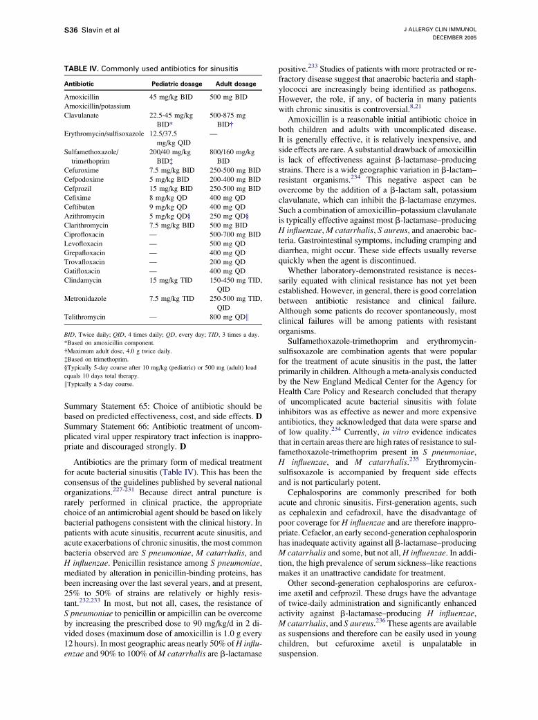
J ALLERGY CLIN IMMUNOL
DECEMBER 2005
S36 Slavin et al
Summary Statement 65: Choice of antibiotic should bebased on predicted effectiveness, cost, and side effects. DSummary Statement 66: Antibiotic treatment of uncom-plicated viral upper respiratory tract infection is inappro-priate and discouraged strongly. D
Antibiotics are the primary form of medical treatmentfor acute bacterial sinusitis (Table IV). This has been theconsensus of the guidelines published by several nationalorganizations.227-231 Because direct antral puncture israrely performed in clinical practice, the appropriatechoice of an antimicrobial agent should be based on likelybacterial pathogens consistent with the clinical history. Inpatients with acute sinusitis, recurrent acute sinusitis, andacute exacerbations of chronic sinusitis, the most commonbacteria observed are S pneumoniae, M catarrhalis, andH influenzae. Penicillin resistance among S pneumoniae,mediated by alteration in penicillin-binding proteins, hasbeen increasing over the last several years, and at present,25% to 50% of strains are relatively or highly resis-tant.232,233 In most, but not all, cases, the resistance ofS pneumoniae to penicillin or ampicillin can be overcomeby increasing the prescribed dose to 90 mg/kg/d in 2 di-vided doses (maximum dose of amoxicillin is 1.0 g every12 hours). In most geographic areas nearly 50% ofH influ-enzae and 90% to 100% ofM catarrhalis are b-lactamase
TABLE IV. Commonly used antibiotics for sinusitis
Antibiotic Pediatric dosage Adult dosage
Amoxicillin 45 mg/kg BID 500 mg BID
Amoxicillin/potassium
Clavulanate 22.5-45 mg/kg
BID*
500-875 mg
BID�Erythromycin/sulfisoxazole 12.5/37.5
mg/kg QID
—
Sulfamethoxazole/
trimethoprim
200/40 mg/kg
BID�800/160 mg/kg
BID
Cefuroxime 7.5 mg/kg BID 250-500 mg BID
Cefpodoxime 5 mg/kg BID 200-400 mg BID
Cefprozil 15 mg/kg BID 250-500 mg BID
Cefixime 8 mg/kg QD 400 mg QD
Ceftibuten 9 mg/kg QD 400 mg QD
Azithromycin 5 mg/kg QD§ 250 mg QD§
Clarithromycin 7.5 mg/kg BID 500 mg BID
Ciprofloxacin — 500-700 mg BID
Levofloxacin — 500 mg QD
Grepafloxacin — 400 mg QD
Trovafloxacin — 200 mg QD
Gatifloxacin — 400 mg QD
Clindamycin 15 mg/kg TID 150-450 mg TID,
QID
Metronidazole 7.5 mg/kg TID 250-500 mg TID,
QID
Telithromycin — 800 mg QDk
BID, Twice daily; QID, 4 times daily; QD, every day; TID, 3 times a day.
*Based on amoxicillin component.
�Maximum adult dose, 4.0 g twice daily.
�Based on trimethoprim.
§Typically 5-day course after 10 mg/kg (pediatric) or 500 mg (adult) load
equals 10 days total therapy.
kTypically a 5-day course.
positive.233 Studies of patients with more protracted or re-fractory disease suggest that anaerobic bacteria and staph-ylococci are increasingly being identified as pathogens.However, the role, if any, of bacteria in many patientswith chronic sinusitis is controversial.8,21
Amoxicillin is a reasonable initial antibiotic choice inboth children and adults with uncomplicated disease.It is generally effective, it is relatively inexpensive, andside effects are rare. A substantial drawback of amoxicillinis lack of effectiveness against b-lactamase–producingstrains. There is a wide geographic variation in b-lactam–resistant organisms.234 This negative aspect can beovercome by the addition of a b-lactam salt, potassiumclavulanate, which can inhibit the b-lactamase enzymes.Such a combination of amoxicillin–potassium clavulanateis typically effective against most b-lactamase–producingH influenzae,M catarrhalis, S aureus, and anaerobic bac-teria. Gastrointestinal symptoms, including cramping anddiarrhea, might occur. These side effects usually reversequickly when the agent is discontinued.
Whether laboratory-demonstrated resistance is neces-sarily equated with clinical resistance has not yet beenestablished. However, in general, there is good correlationbetween antibiotic resistance and clinical failure.Although some patients do recover spontaneously, mostclinical failures will be among patients with resistantorganisms.
Sulfamethoxazole-trimethoprim and erythromycin-sulfisoxazole are combination agents that were popularfor the treatment of acute sinusitis in the past, the latterprimarily in children. Although a meta-analysis conductedby the New England Medical Center for the Agency forHealth Care Policy and Research concluded that therapyof uncomplicated acute bacterial sinusitis with folateinhibitors was as effective as newer and more expensiveantibiotics, they acknowledged that data were sparse andof low quality.234 Currently, in vitro evidence indicatesthat in certain areas there are high rates of resistance to sul-famethoxazole-trimethoprim present in S pneumoniae,H influenzae, and M catarrhalis.235 Erythromycin-sulfisoxazole is accompanied by frequent side effectsand is not particularly potent.
Cephalosporins are commonly prescribed for bothacute and chronic sinusitis. First-generation agents, suchas cephalexin and cefadroxil, have the disadvantage ofpoor coverage for H influenzae and are therefore inappro-priate. Cefaclor, an early second-generation cephalosporinhas inadequate activity against all b-lactamase–producingM catarrhalis and some, but not all, H influenzae. In addi-tion, the high prevalence of serum sickness–like reactionsmakes it an unattractive candidate for treatment.
Other second-generation cephalosporins are cefurox-ime axetil and cefprozil. These drugs have the advantageof twice-daily administration and significantly enhancedactivity against b-lactamase–producing H influenzae,M catarrhalis, and S aureus.236 These agents are availableas suspensions and therefore can be easily used in youngchildren, but cefuroxime axetil is unpalatable insuspension.
J ALLERGY CLIN IMMUNOL
VOLUME 116, NUMBER 6
Slavin et al S37
Cefixime, ceftibuten, cefpodoxime axetil, and cefdinirare third-generation cephalosporins, which can be givenorally once or twice daily. Both cefixime and ceftibutenhave poor activity against S pneumoniae and are espe-cially ineffective against penicillin-resistant strains.Neither of these drugs should be used for acute bacterialsinusitis. Cefpodoxime and cefdinir are suitable agents.237
There is theoretic concern regarding the 2 erythromycinanalogues azithromycin and clarithromycin. Because bothare relatively weak against penicillin-resistant H influen-zae and S pneumoniae, this might lead to increasing resis-tance to macrolides.235
The new ketolide telithromycin provides a targetedspectrum of activity against common respiratory patho-gens, such as S pneumoniae, including macrolide andpenicillin-resistant strains, H influenzae, M catarrhalis,and atypicals.238
In adults ciprofloxacin, levofloxacin, gemifloxacin,moxifloxacin, and grepafloxacin presently have specificindications for the treatment of sinusitis.239 Sparfloxacinmight show enhanced gram-positive coverage but lacksthe specific indication for sinusitis and has a significantrisk for phototoxicity. There has been concern about ad-verse effects on developmental joint formation with allquinolones. In one report involving more than 6000fluoroquinolone-treated children, the incidence of tendonor joint disorders was less than 1% and was comparablewith that of the reference group, children treated withazithromycin.240
In protracted or severe cases of sinusitis, the possibilityof anaerobic pathogens should be considered. Generally,these organisms are sensitive to penicillin, but those thatare resistant, including Prevotella species, respond toamoxicillin-clavulanate. If the clinical course suggeststhat an anaerobe is a likely pathogen, clindamycin ormetronidazole might be considered in combination ther-apy with a broad-spectrum drug. Although clindamycinis generally well tolerated, patients should be alerted tothe possibility of pseudomembranous enterocolitis andtold to contact their physician for any sign of diarrhea orbloody stools.
The appropriate duration of antibiotic therapy forsinusitis is not well defined.228 A 10- to 14-day courseof antibiotic might be adequate for most patients withacute disease. Some experts suggest that patients shouldbe treated until they are free of symptoms plus 7days.228 It has been recommended that if there is no clin-ical improvement in children within 3 days of initiatingantimicrobial therapy, an alternative antibiotic should beconsidered.228
The role of antibiotics in patients with chronic sinusitisis controversial.8,21 These patients might require antibi-otics for acute exacerbations of chronic sinusitis, andthen amoxicillin potassium clavulanate is a good selec-tion. The underlying symptoms, often attributable to ARor NARwill benefit most from therapy directed at the spe-cific problem.
Regrettably, there are no published studies on antibioticprophylaxis in patients with recurrent sinusitis. However,
the prophylactic use of antibiotics in patients with recur-rent sinusitis can be considered in carefully selectedpatients, such as those with immunodeficiency disor-ders.228 Recurrent sinusitis is frequently defined as 3 epi-sodes in 6 months or 4 episodes in 12 months. An initialapproach with antibiotic prophylaxis might include initiat-ing antibiotic therapy at the first sign of a new respiratoryillness. If this fails, the next step would include once-dailyadministration of an antibiotic during the fall and winter,when viral upper respiratory tract infections are morelikely to compromise ostiomeatal clearance. The potentialvalue of this approach is suggested from studies of chil-dren with recurrent otitis media.241,242
There has been interest in administering topical antibi-otics through the inhalation route as a compoundedformulation delivered by means of aerosol or nebulizer.Antibiotics used for this purpose include tobramycin,gentamicin, ciprofloxacin, levofloxacin, and some ceph-alosporins. Several uncontrolled studies have claimedimprovement.243,244 In the absence of adequate random-ized controlled studies demonstrating the clear effective-ness of aerosolized antibiotics for acute or chronicsinusitis, no definite conclusions can be drawn regardingthe effectiveness of this form of therapy.
Because of a lack of convincing evidence, the role ofantifungal agents in chronic sinusitis has not yet beenestablished.
Several agencies have published guidelines concerningthe diagnosis and management of acute bacterial sinusitis,in part to promote the judicious use of antibiotics.227-230
These guidelines, which have been endorsed by the Cen-ters for Disease Control and Prevention, the AmericanCollege of Physicians–American Society of Internal Med-icine, the American Academy of Family Practice, theAmerican Academy of Pediatrics, and the Infectious Dis-eases Society of America, conclude that most cases ofacute sinusitis are caused by uncomplicated viral infec-tions. Antibiotic treatment of uncomplicated viral upperrespiratory tract infections is inappropriate and shouldbe discouraged. In these uncomplicated cases, a 7- to10-day course of watchful waiting for spontaneous resolu-tion of symptoms before prescribing antibiotics is recom-mended. Antibiotics should be considered in thosepatients with severe signs and symptoms of sinusitis, re-gardless of duration of illness. These would include wors-ening symptoms after 3 to 5 days, temperature of greaterthan 39�C, maxillary tooth or facial pain (especiallywhen unilateral), unilateral sinus tenderness, and periorbi-tal swelling.
Antihistamines
Summary Statements
Summary Statement 67: There are no data presently torecommend the use of H1 antihistamines in acute bacte-rial sinusitis. DSummary Statement 68: There might be a role forantihistamines in chronic sinusitis if the underlying riskfactor is AR. D
J ALLERGY CLIN IMMUNOL
DECEMBER 2005
S38 Slavin et al
Both first- and second-generation H1 antihistamineshave a major role in the treatment of AR, allergic conjunc-tivitis, cutaneous allergic disorders, and anaphylaxis. Inaddition, 2 recent studies have shown a modest beneficialeffect of first-generation antihistamines on sneezing andrhinorrhea in the common cold.245,246 However, a rolefor the use of these agents in the treatment of sinusitishas not been demonstrated, and they were not recommen-ded in the recent guidelines for the diagnosis and manage-ment of sinusitis in children.117
a-Adrenergic decongestants
Summary Statements
Summary Statement 69: Topical and oral decongestantsare often used in the therapy of acute or chronic sinusitisbecause they decrease nasal resistance and theoreticallyincrease ostial patency. DSummary Statement 70: Prospective studies are lackingand are needed to assess the value of a-adrenergic agentsin the prevention or treatment of sinusitis. D
There has been relatively little systematic study ofdecongestant therapy in adults with the common cold andno study in either children with the common cold orpatients of any age with sinusitis.247 Decongestants havea modest effect in decreasing nasal airway resistanceand improving symptom scores in adults with a cold.Theoretically, both topical and oral decongestants usedin the therapy of acute or chronic sinusitis might widenthe ostia and reduce turbinate swelling, thus promotingsinus and nasal ventilation.Pharmacology. Decongestants can produce their effectsthrough 2 broad mechanisms. First, direct-acting sympa-thomimetic drugs activate a-adrenergic receptors, result-ing in vasoconstriction.248 Second, indirect-actingsympathomimetic agents function by being taken up intothe prejunctional nerve terminal, where they displace nor-epinephrine from storage vesicles. The displaced norepi-nephrine is then released from the sympathetic nerveterminal to postjunctional a-adrenergic receptors, produc-ing vasoconstriction.Side effects of decongestants. Topical agents used asnasal sprays act rapidly, usually within minutes, andtherapeutic doses have no systemic side effects. Reboundhyperemia or rhinitis medicamentosa, however, is afrequent side effect in patients who use the drugs overan extended period of time.
Oral decongestants cause generalized constriction ofblood vessels, and increased arterial pressure is always ofconcern. Most available oral agents, however, cause bloodpressure increases in healthy persons only at doses thatsignificantly exceed those that are recommended. Otherpossible adverse effects are reflex bradycardia, centralnervous system stimulation and insomnia, urinary reten-tion, mydriasis (with effects on glaucoma), and effectson endocrine and other regulators of metabolic function.Only 2 drugs are commonly used as oral decongestants:pseudoephedrine and phenylephrine. Direct-acting
sympathomimetics, such as phenylephrine and oxymeta-zoline, activate a1- and a2-adrenergic receptors, respec-tively, and both cause vasoconstriction and a decrease innasal congestion.
The US Food and Drug Administration has mandatedthe withdrawal of all over-the-counter products thatcontain phenylpropanolamine because of the risk ofhemorrhagic stroke in women.249,250
Glucocorticosteroids
Summary Statements
Summary Statement 71: The use of systemic corticoste-roid therapy for sinus disease has not been studied sys-tematically in a well-controlled or blinded manner. DSummary Statement 72: A few recent studies suggestthat the addition of intranasal corticosteroids as an ad-junct to antibiotic therapy might be modestly beneficialin the treatment of patients with recurrent acute orchronic sinusitis. C
Researchers and clinicians agree on the potential use-fulness of corticosteroids as potent anti-inflammatoryagents and the fact that sinusitis is an inflammatorydisease. The leap from this logical association to clinicalproof of the effectiveness of corticosteroids in managingsinus disease has been difficult.
The anti-inflammatory activities of corticosteroidsinclude decreased vascular permeability, inhibition ofrelease, and/or formation of mucous secretagogues, in-cluding histamine, leukotrienes, platelet-activating factor,and prostanoids, as well as inhibition of inflammatory cellinfiltration, especially eosinophils.
Topical corticosteroids have a clear effect on inflam-matory cell influx into the nasal mucosa after nasal antigenchallenge.251 Pretreatment of nasal mucosa with inhaledsteroid has been shown to modify both the immediate-and late-phase response after antigen challenge.252
Glucocorticosteroids also inhibit antigen-induced nasalhyperresponsiveness to histamine.253 Numerous clinicaltrials attest to the efficacy of topical corticosteroids incontrolling symptoms of AR.254,255
Although intranasal corticosteroids are unlikely toreach the interior of the paranasal sinuses, their relativesafety, their recognized anti-inflammatory effect, and theirdocumented efficacy in relieving nasal congestion makeintranasal corticosteroids a reasonable adjunctive therapyfor the treatment of sinusitis. Most studies evaluating theeffectiveness of intranasal corticosteroids, even random-ized controlled studies, use study designs that make itdifficult to conclude that the use of intranasal corticoste-roids is effective in the treatment of sinusitis.256-261 It is, inparticular, difficult to separate improvement in manifesta-tions of sinusitis from improvement in nasal congestion,which is a significant component of sinusitis and for whichintranasal corticosteroids have been shown to be effective.The effectiveness of intranasal corticosteroids in the treat-ment of sinusitis will need to be conclusively demon-strated in carefully designed studies in the future.
J ALLERGY CLIN IMMUNOL
VOLUME 116, NUMBER 6
Slavin et al S39
Nasal polyps are a common accompaniment ofchronic sinusitis, and there is good evidence that theyare favorably affected by intranasal and systemiccorticosteroids.262,263
Adjunctive therapies: Saline, mucolytics,and expectorants
Summary Statements
Summary Statement 73: There are several scientific stud-ies that imply but do not directly confirm a role for theseagents in sinusitis. DSummary Statement 74: Use of all these agents as pro-phylaxis for exacerbations of chronic sinusitis is empiricand not supported by clinical data. DSummary Statement 75: These agents are commonlyused and in some instances might be beneficial in somepatients. D
Medical management of sinusitis is based on empiricgoals. Adjunctive agents are frequently prescribed inaddition to antibiotics in the hope of facilitating drainageof retained secretions through the sinus ostia into the nasalcavity. Use is based on clinical experience.264-266
Pharmacologic agents: Guaifenesin. Guaifenesin (glyc-eryl guaicolate; 3-(2 methoxyphonoxy)-1, 2-propanediol)is a water- and alcohol-soluble substance that has beenused as an expectorant to loosen phlegm and bronchialsecretions in the symptomatic management of coughassociated with the upper and lower respiratory tractinfections that occur when these conditions are compli-cated by tenacious mucus, mucus plugs, or both andcongestion. There is clinical evidence that guaifenesin isan effective expectorant in that it increases expectoratedsputum volume over the first 4 to 6 days of a productivecough, decreases sputum viscosity and difficulty inexpectoration, and improves associated symptoms.However, there is currently insufficient evidence to sup-port efficacy of the drug as an adjunct in sinusitis becauseno clinical trials have been reported in sinusitis to dem-onstrate its efficacy.267
Pharmacologic agents: Iodine. Iodine-containing com-pounds, such as potassium iodide or iodinated glycerol,might be expected to have similar effects as guaifenesin.However, they are of limited clinical use, and no studies insinusitis have been reported.Nonpharmacologic adjuncts. Many nonpharmacologicmeasures are advocated for symptomatic relief of acutesinusitis. Because scientific data on efficacy are lacking,physicians might dismiss some of these measures as folkmedicine. For many patients, however, one or more ofthese treatments could provide effective relief of distress-ing symptoms while the infection is resolving. Unfortu-nately, most of these measures are short lived ineffectiveness, and they must be repeated as symptomsrecur.Saline. Saline prevents crusting of secretions in the nasalcavity, especially in the region of the ostiomeatal complex.This facilitates mechanical removal of mucus through
either nose blowing or suction. Theoretically, hypertonicsaline might reduce mucosal edema and enhance muco-ciliary clearance and secondarily improve the patency ofthe sinus ostia.268 In a cross-over study 21 healthy volun-teers underwent nasal irrigation with either buffered nor-mal saline or buffered hypertonic saline, followed byassessment of their mucociliary clearance with the saccha-rin clearance method. The outcome showed hypertonic sa-line nasal irrigation to improve mucociliary transit times,whereas normal saline had no effect. This study shouldbe repeated in patients with acute and also chronicsinusitis.
In a study of 30 patients aged 3 to 16 years with chronicsinusitis,269 subjects were stratified by age and severityand randomized to receive either normal saline or hyper-tonic saline nose drops 3 times daily for 4 weeks.Outcome was evaluated by a comparison of a nasal,cough, and radiology score. The hypertonic saline groupimproved significantly in all scores in comparison withthe normal saline group, in which improvement wasobserved in only the nasal score.
Two other studies have claimed a benefit from hyper-tonic saline nasal washes.270,271 However, the absence of acomparator group in one and possible placebo effect in theother weaken their conclusions. The optimal delivery sys-tem for the hypertonic saline (water pick, ultrasonicRhinoflo nasal spray, or nose drops) has not been clarified.Steam. The traditional method of steam inhalation is toinstruct the patient to do the following:
1. Pour boiling water in a pan or basin on a low table.2. Sit at the table with a towel draped over the head to
make a tent over the pan of water.3. Hold the face a few inches above the water and breathe
through the nose for approximately 10 minutes.
This procedure liquefies and softens crusts whilemoisturizing the dry inflamed mucosa.Astringents. Adding pine oil or mentholated preparations,such as Vicks VapoRub (Proctor & Gamble, Cincinnati,Ohio), oil of eucalyptus, or similar aromatics, might add tothe beneficial effect of the steam treatment. Such additionscould help relieve stuffiness or at least produce a subjec-tive sensation of increased air flow. Again, there are noscientific data to support these therapies, but patients oftenbelieve that they help.Spicy foods. Garlic has an active ingredient (n-allythio-sulphinate) that provides short-lived decongestant effects.Eating foods highly seasoned with garlic has been con-sidered therapeutic. There has been no systemic study ofthe effect of garlic on patients with sinusitis.
IVIG
Summary Statements
Summary Statement 76: Immunodeficiency might be anunderlying risk factor for the development of recurrentacute or chronic sinusitis. BSummary Statement 77: IVIG is approved as a replace-ment therapy for antibody deficiency disorders, including
J ALLERGY CLIN IMMUNOL
DECEMBER 2005
S40 Slavin et al
X-linked agammaglobulinemia, common variable immu-nodeficiency, Wiskott-Aldrich syndrome, and hyper-IgMsyndrome. ASummary Statement 78: Appropriate use of IVIG canprevent complications from chronic sinusitis, includingsubperiosteal and intracranial abscesses, meningitis, sep-sis, and death. B
Immunodeficiency might be an underlying risk factorfor the development of recurrent acute or chronic sinusitis.IVIG is approved as a replacement therapy for antibodydeficiency disorders, including X-linked agammaglobu-linemia, common variable immunodeficiency, Wiskott-Aldrich syndrome, and hyper-IgM syndrome.272 The useof IVIG in other immune disorders (eg, selective antibodydeficiency or IgG subclass deficiencies) remainscontroversial.
The appropriate use of IVIG in patients with immuno-globulin deficiency can prevent complications fromchronic sinusitis, including subperiosteal abscess, intra-cranial abscess, meningitis, sepsis, or even death.
There is considerable controversy regarding gamma-globulin prescribing for therapy of patients with specificantibody deficiency and normal immunoglobulin levels(see ‘‘Practice parameters for the diagnosis and manage-ment of primary immunodeficiency’’). Most experts inthis area consider demonstration of impaired antibodyproduction essential. It must be demonstrated that (1) thepatient has significant and clearly documented infectiousmorbidity (eg, recurrent pneumonias and frequent epi-sodes of documented bacterial sinusitis and not justisolated chronic sinusitis), (2) other disorders (eg, allergyand anatomic defects) have been sought and treatedaggressively if present, and (3) other modes of therapy(antimicrobial, anti-inflammatory, and surgical) are inad-equate or poorly tolerated. If administered to children withmilder antibody deficiencies, gammaglobulin therapyshould be discontinued if there has been an extendedperiod of significant improvement because the suscepti-bility to infection might decrease over time.273 Humoralimmune function should be reassessed but no soonerthan 3 months (preferably 4-6 months) after the last infu-sion. Patients must be followed closely, and therapyshould be discontinued, generally after no more than 3to 6 months, if there is lack of clinical efficacy.
Aspirin desensitization therapy
Summary Statement
Summary Statement 79: Beneficial effects of aspirin de-sensitization on patients with aspirin-exacerbated respira-tory disease (AERD) have been reported. A
The clinical efficacy in patients with AERD of aspirindesensitization followed by daily aspirin treatment hasbeen reported. In one study 65 patients with AERD weretreated with aspirin, 650 mg twice daily, after aspirindesensitization. While the patients were under treatmentwith aspirin, objective clinical criteria demonstrated sig-nificant improvement in their clinical courses, particularly
a reduction in sinusitis. Simultaneous requirement forsystemic corticosteroids decreased significantly.274
In another study275 172 patients with AERD were de-sensitized to and treated with aspirin. By the first 6 monthsof aspirin treatment, there were significant reductions insinus infections and frequency of short courses of predni-sone and improvements in sense of smell and generalassessment of nasal-sinus and asthma symptoms. Of thepatients who completed a year or more of aspirin treat-ment, 87% experienced improvement. Results persistedfor 1 to 5 years.
Treatment of nasal polyposis with daily nasal applica-tion of aspirin-lysine solution has been reported withdecreased swelling, reduction of polyp formation, andreturn of smell.276
Zileuton, a 5-lipoxygenase inhibitor was given as add-on therapy to 40 patients with AERD. This resulted notonly in improvement in asthma but also in return of smell,less rhinorrhea, and a trend toward less stuffiness andhigher nasal inspiratory flow.277
Surgical
Summary Statements
Summary Statement 80: Antral puncture and irrigation isan office procedure that has a place in the managementof acute ethmomaxillary sinusitis refractory to medicaltherapy or in acute sinusitis in an immunosuppressedpatient in whom early identification of pathogenicorganisms is paramount. DSummary Statement 81: Surgical intervention might berequired in acute sinusitis to provide drainage when thereis a significant risk of intracranial complication or in apatient with periorbital or intraorbital abscess or visualcompromise. DSummary Statement 82: Functional endoscopic sinussurgery, in combination with appropriate medical ther-apy, has been shown in uncontrolled studies to havelong-term efficacy in reducing disease-specific symptomsand in improving overall quality of life. C
The surgical approach to sinus disease has undergone asignificant transition, resulting from renewed insights intosinus physiology and the widespread use of nasal endos-copy. In the latter part of the 20th century, several authorsnoted the importance of the middle meatus and of theethmoid sinuses in the cause of frontal and maxillarysinusitis.278 This region was termed the ostiomeatal com-plex in recognition of the importance of this area to diseasein the dependent sinuses.279,280
Our current knowledge continues to indicate thatlimited and localized inflammation within this regionmight cause or exacerbate disease within the dependentsinuses.281 Thus surgical therapy is typically directed to-ward removing mucosal disease and the involved bonewithin the ethmoid sinuses and sinus ostia under endo-scopic visualization (functional endoscopic sinus surgery)rather than stripping inflamed mucosa from the largerdependent sinuses. Indeed, more recently, increased
J ALLERGY CLIN IMMUNOL
VOLUME 116, NUMBER 6
Slavin et al S41
attention has been paid to mucoperiosteal preservation andthe necessity of avoiding bone exposure, even within theregion of the ostiomeatal complex.282
After surgical intervention, most patients with infec-tious sinusitis have a significant improvement in disease-specific symptoms. However, in a significant number ofpatients, endoscopic and radiologic evidence of asymp-tomatic disease might persist after surgical intervention,requiring continued medical therapy or local debride-ment.283 It appears that eventually, with proper ventilationand appropriate systemic and local therapy, residual evi-dence of chronic inflammation slowly resolves in manypatients.284
Evaluation for surgery. In acute sinusitis the necessity forsurgery is usually predicated either by a threatened com-plication or by severe symptoms unresponsive to medicaltherapy. In chronic sinusitis, patient evaluation shouldinclude a careful history and evaluation for environmentaland general host factors that might predispose to sinusitisin addition to evaluation of the local host factors within theostiomeatal complex. It has been demonstrated that pa-tients who continue to smoke after surgery have a signif-icantly worse long-term outcome.285 Thus the advantagesand disadvantages of elective surgical intervention shouldbe carefully considered in patients who continue to smokeor have other ongoing marked environmental exposures.
Surgery is typically required for fungal sinusitis.Fungus balls within the maxillary sinus, allergic fungalsinusitis, and invasive fungal sinusitis generally requiresurgical intervention. Because the radiographic and endo-scopic appearance of unilateral polypoid disease mightfrequently be the result of either fungal disease or tumor(eg, inverted papilloma), biopsy should be considered inthese patients.Surgical approaches. Endoscopic approaches have gen-erally become the surgical standard of care for chronicinfectious sinusitis, especially if there is evidence ofmechanical blockage of the ostiomeatal complex.However, open surgical procedures are still required,depending on the extent and the location of the sinusesinvolved (ie, frontal or sphenoid). Frontal sinus trephineand postoperative irrigation is a valid consideration inpatients with acute or chronic frontal sinusitis. Addition-ally, when endoscopic surgical techniques fail to resolvechronic frontal sinusitis, even with revised surgical inter-vention, the frontal sinus obliteration with fat remains aviable consideration.Operative intervention. Endoscopic sinus surgery can beperformed under local anesthesia.286 However, as the im-portance of carefully removing disease and meticulouslypreserving the mucoperiosteum and normal structureshas increasingly been recognized, surgical cases havetended to become longer. Thus there has been a trend to-ward performing the surgery under general anesthesia.In many cases the surgery can still be performed on an out-patient basis. Patients with significant asthma or other un-derlying medical conditions are usually kept overnight forobservation. The surgical procedure is carried out underendoscopic visualization through the nostril and involves
no external incisions. The extent of the surgical dissectionis dictated by the amount and location of disease identifiedby means of preoperative CT, as well as by the findingsduring the surgical procedure. The standard teaching forthe functional endoscopic approach is that the surgicalprocedure should extend beyond the margins of the ostio-meatal disease. Postoperative pain is typically minimal,and early symptom improvement is generally the rule.The incidence of severe surgical complications is approx-imately 0.5%. However, the meticulous surgical tech-niques used require considerable experience.287,288
Conclusions. The surgical treatment of sinusitis has beensignificantly enhanced by the routine use of nasal endos-copy and by the use of CT imaging. The nasal telescopehas significantly improved our ability to visualize theostiomeatal complex, a critical region in the pathogenesisof chronic sinusitis and a region that is very poorlyvisualized on both anterior rhinoscopy and on standardradiographic films. Although chronic sinusitis is typicallya multifactorial disease with environmental and generalhost factors, localized persistent disease within theostiomeatal complex plays a significant part in continua-tion of the disease process. Functional endoscopic sinussurgery results in significant improvement in the majorityof patients. However, significant improvement oftenrequires a combination of appropriate surgical interven-tion with intensive postoperative local management to theregion and appropriate medical therapy.
Indications for referral
1. Indications for referral to a specialist:d When the condition or its treatment is interferingwith a patient’s performance or causing significantloss of school or work on a chronic or recurrent ba-sis or when the patient’s quality of life is signifi-cantly affected.
d When there are complications of sinusitis, such asotitis, asthma, bronchiectasis, nasal polyps, orbronchitis.
d When there is consideration for an allergic or im-munologic basis for the sinusitis or when immuno-competence needs to be assessed.
d When the condition becomes chronic, persists forseveral months, or recurs 2 to 3 times per year,despite treatment by the primary care physician.
d When there is the need for complex pharmacologyto treat recalcitrant infections caused by underlyingallergies, allergic fungal sinusitis, resistant patho-gens, or aspirin desensitization.
2. What the specialist should provide to the referringphysician:d Clarification of allergic, immunologic, or nonaller-gic causative basis for the patient’s condition.
d Assessment of nasal and sinus outflow tract anat-omy and any contribution these anatomic factorshave in the causation of the sinus problems.
d Identification of specific allergens or other triggersfor the patient’s condition, and education in ways toavoid exposure to these triggers.
J ALLERGY CLIN IMMUNOL
DECEMBER 2005
S42 Slavin et al
d Assistance in developing an effective treatmentplan, including patient education, allergy avoid-ance, pharmacotherapy, anti-infectious therapy,and immunotherapy, if appropriate.
d Provision of specialized services, such as prepara-tion of extracts and provision of immunotherapy.
d Evaluation for associated conditions, such as asthma.3. Indications for surgical intervention:
d When nasal polyps obstruct sinus drainage andpersist despite appropriate medical treatment.
d When there is recurrent or persistent infectious si-nusitis despite adequate trials of medical manage-ment: adequate medical management minimallyinvolves multiple courses of antibiotics chosen tocover the spectrum of pathogens anticipated to becausing the disease.
d For biopsy of the nasal mucosa to rule out granu-lomatous disease, neoplasms, ciliary dyskinesia,or fungal infections.
d When maxillary antral puncture is required.d When anatomic defects exist that obstruct the sinusoutflow tract, particularly including the ostiomeatalcomplex (and adenoidal tissues in children), andare thought to be contributing to recurrent orchronic infectious sinusitis.
d For sinusitis with threatened complications (eg,threat of brain abscess, meningitis, cavernous sinusthrombosis, or Pott’s tumor).
4. What the surgical consultant should provide the refer-ring physician:d An evaluation of the likely pathologic factors in-volved in the disease process and the chances ofimprovement with surgical intervention: this couldinclude puncture and quantitative cultures.
d Determination of whether an adequate trial of med-ical, allergic, or immunologic treatment has beenprovided before recommending surgery.
d Recommendation of a specific procedure or proce-dures for the specific patient and discussion of themerits of alternative approaches.
d Assessment of the need for surgical revision ofabnormal anatomy through rhinolaryngoscopy andCT imaging and the likelihood that such revisionwill reduce the recurrent or chronic sinus disease.
d Assessment of nasal and sinus outflow tract anat-omy through rhinolaryngoscopy and CT imagingand any contribution these anatomic factors havein the causation of the sinus problems.
REFERENCES
1. Anand VK. Epidemiology and economic impact of rhinosinusitis. Ann
Otol Rhinol Laryngol 2004;193(suppl):S3-5. III
2. Gliklich RE, Metson R. The health impact of chronic sinusitis in
patients seeking otolaryngologic care. Otolaryngol Head Neck Surg
1995;113:104-9. IIb
3. Spector SL, Bernstein IL, Li JT, et al. Parameters for the diagnosis and
management of sinusitis. J Allergy Clin Immunol 1998;102(suppl):
S107-44. IV
4. Lanza DC, Kennedy DW. Adult rhinosinusitis defined. Otolaryngol
Head Neck Surg 1997;117:1-7. IV
5. Kaliner MA, Osguthorpe SD, Fireman D, et al. Sinusitis: bench to
bedside. J Allergy Clin Immunol 1997;99(suppl):S829-48. IV
6. Meltzer EO, Hamilos DL, Hadley JA, Lanza DC, Marple BF,
Nicklas RA. Rhinosinusitis: establishing definitions for clinical
research and patient care. J Allergy Clin Immunol 2004;114(suppl):
S156-212. IV
7. Lund VJ, Kennedy DW. Quantification for staging sinusitis. The
Staging and Therapy Group. Ann Otol Rhinol Laryngol 1995;
167(suppl):17-21. IV
8. Wald ER. Chronic sinusitis in children. J Pediatr 1995;127:339-47. IV
9. Stankiewicz JA, Chow JM. A diagnostic dilemma for chronic rhinosi-
nusitis: definition, accuracy and validity. Am J Rhinol 2002;16:
199-202. III
10. Hamilos DL. Chronic sinusitis. J Allergy Clin Immunol 2000;106:
213-27. IV
11. Spaeth J, Krugelstein U, Schlondorff G. The paranasal sinuses in CT-
imaging: development from birth to age 25. Int J Pediatr Otorhinolar-
yngol 1997;39:25-40. IIb
12. Medina J, Hernandez H, Tom LW, Bilaniuk L. Development of the
paranasal sinuses in children. Am J Rhinol 1997;11:203-9. IIb
13. Zeifer B. Pediatric sinonasal imaging: normal anatomy and inflamma-
tory disease. Neuroimaging Clin N Am 2000;10:137-59. IIb
14. Nishioka GJ, Cook PR, McKinsey JP, Rodriguez FJ. Paranasal sinus
computed tomography scan findings in patients with cystic fibrosis.
Otolaryngol Head Neck Surg 1996;114:394-9. IIa
15. Bolger WE. Anatomy of the paranasal sinuses. In: Kennedy DW, Bolger
WE, Zinreich SJ, editors. Diseases of the sinuses: diagnosis and manage-
ment. Hamilton, Ontario, Canada: Decker; 2001. p. 1-11. III
16. Yousem DM, Kennedy DW, Rosenberg S. Ostiomeatal complex risk
factors for sinusitis: CT evaluation. J Otolaryngol 1991;20:419-24. IIa
17. Kennedy DW, Zinreich SJ, Rosenbaum AE, Johns ME. Functional
endoscopic sinus surgery. Theory and diagnostic evaluation. Arch
Otolaryngol 1985;111:576-82. III
18. Aust R, Stierner P, Drettner B. Basic experimental study of ostial
patency and local metabolic environment of the maxillary sinus. Acta
Otolaryngol Suppl 1994;515:7-10. IIa
19. Aust R, Drettner B. Oxygen tension in the human maxillary sinus
during normal and pathological conditions. Acta Otolaryngol 1974;
78:264-9. IIa
20. Aust R, Falck B, Svanholm H. Studies of gas exchange and pressure in
the maxillary sinus in normal and infected humans. Rhinology 1979;17:
245-51. IIa
21. Gwaltney JM Jr. Acute community-acquired sinusitis. Clin Infect Dis
1996;23:1209-25. IV
22. Gordts F, Nasser IA, Clement PAR, Pierard D, Kaufman L. Bacteriol-
ogy of the middle meatus in children. Pediatr Otorhinolaryngol 1999;
48:163-7. IIb
23. Gold SM, Tami TA. Role of middle meatus aspiration culture in the
diagnosis of chronic sinusitis. Laryngoscope 1997;107:1586-9. IIb
24. Gordts F, Halewyck S, Pierard D, von Kaufman L, Clement PA. Micro-
biology of the middle meatus: a comparison between normal adults and
children. J Laryngol Otol 2000;114:184-8. IIa
25. Gwaltney JM Jr, Scheld WM, Sande MA, Sydner A. The microbial
etiology and antimicrobial therapy of adults with acute community-
acquired sinusitis a fifteen year experience at the University of Virginia
and review of other selected studies. J Allergy Clin Immunol 1992;90:
457-62. Ia
26. Sydnor TA, Kapp EJ, Anthony KE, Lococo JM, Kimm SS, Fowler CL.
Open label assessment of levofloxacin for the treatment of acute
bacterial sinusitis in adults. Ann Allergy Asthma Immunol 1998;80:
357-62. III
27. Wald ER, Milmoe GJ, Bowen AD, Ledesma-Medina J, Salmon N,
Bluestone CD. Acute maxillary sinusitis in children. N Engl J Med
1981;304:749-54. III
28. Wald ER, Reilly JS, Casselbrant M, et al. Treatment of acute maxillary
sinusitis in childhood: a comparative study of amoxicillin and cefaclor.
J Pediatr 1984;104:297-302. Ib
29. Wald ER, Byers C, Guerra N, Casselbrant M, Meste D. Subacute sinus-
itis in children. J Pediatr 1989;115:28-32. III
30. Shapiro ED, Milmoe GJ, Wald ER, Rodnan JB, Bowen AD. Bacteriol-
ogy of the maxillary sinuses in patients with cystic fibrosis. J Infect Dis
1982;146:589-93. III

J ALLERGY CLIN IMMUNOL
VOLUME 116, NUMBER 6
Slavin et al S43
31. Friedman R, Ackerman W, Wald E, Casselbrant M, Friday G, Fireman
P. Asthma and bacterial sinusitis in children. J Allergy Clin Immunol
1984;74:185-9. III
32. Goldenhersh MJ, Rachelefsky GS, Dudley J, et al. The bacteriology
of chronic maxillary sinusitis in children with respiratory allergy.
J Allergy Clin Immunol 1990;85:1030-9. III
33. Desrosiers MY, Salas-Prato M. Treatment of chronic rhinosinusitis
refractory to other treatments with topical antibiotic therapy delivered
by means of a large-particle nebulizer: results of a controlled trial.
Otolaryngol Head Neck Surg 2001;125:265-9. Ib
34. Don D, Yellow RF, Casselbrant ML, Bluestone CD. Efficacy of a
stepwise protocol that includes intravenous antibiotic therapy for the
management of chronic sinusitis in children and adolescents. Arch
Otolaryngol Head Neck Surg 2001;127:1093-8. III
35. Slack CL, Dahn KA, Abzug MJ Chan KH. Antibiotic-resistant bacteria
in pediatric chronic sinusitis. Pediatr Infect Dis J 2001;20:247-50. III
36. Brook I, Yocum P, Shah K. Aerobic and anaerobic bacteriology of
concurrent chronic otitis media with effusion and chronic sinusitis in
children. Arch Otolaryngol Head Neck Surg 2000;126:174-6. III
37. Finegold SM, Flynn MJ, Rose FV, et al. Bacteriologic findings associ-
ated with chronic bacterial maxillary sinusitis in adults. Clin Infect Dis
2002;35:428-33. III
38. Jacobs MR, Bajaksouzian S, Zilles A, Lin G, Pankuch GA, Appelbaum
PC. Susceptibilities of Streptococcus pneumoniae and Haemophilus
influenzae to 10 oral antimicrobial agents based on pharmacodynamic
parameters: 1997 US Surveillance study. Antimicrob Agents Chemo-
ther 1999;43:1901-8. III
39. Wald ER, Mason EO Jr, Bradley JS, Barson W. Kaplan SL, the US
Pediatric Multicenter Pneumococcal Surveillance Group. Acute otitis
media caused by Streptococcus pneumoniae in Children’s Hospital
between 1994 and 1997. Pediatr Infect Dis J 2001;20:34-9. III
40. Hozapfel L, Chastang C, Demingeon G, Bohe J, Piralla B, Coupry A.
A randomized study assessing systematic search for maxillary sinusitis
in nasotracheally mechanically ventilated patients. Influence of nosoco-
mial maxillary sinusitis on the occurrence of ventilator-associated
pneumonia. Am J Respir Crit Care Med 1999;159:695-701. Ib
41. Vandenbussche T, DeMoor S, Bachert C, Van Cauwenberge P. Value
of antral puncture in the intensive care patient with fever of unknown
origin. Laryngoscope 2000;110:1702-6. III
42. George DL, Falk PS, Umberto MG, et al. Nosocomial sinusitis in pa-
tients in the medical intensive care unit: a prospective epidemiological
study. Clin Infect Dis 1998;17:463-70. III
43. Bachert C, Gevaert P, Holtappels G. Total and specific IgE in nasal
polyps is related to local eosinophilic inflammation. J Allergy Clin
Immunol 2001;107:607-14. IIb
44. McClay JE, Marple B, Kapadia L, et al. Clinical presentation of allergic
fungal sinusitis in children. Laryngoscope 2002;112:565-9. IV
45. Manning SC, Holman M. Further evidence for allergic pathophysiology
in allergic fungal sinusitis. Laryngoscope 1998;108:1485-96. IV
46. Schubert MS, Goetz DW. Evaluation and treatment of allergic fungal
sinusitis. Demographics and diagnosis. J Allergy Clin Immunol 1998;
102:387-94. IV
47. Klapper SR, Patrinely JR. Orbital involvement in allergic fungal sinus-
itis. Ophthal Plast Reconstr Surg 2001;17:149-51. IV
48. deShazo RD. Fungal sinusitis. Am J Med Sci 1998;316:39-45. IV
49. Rizk SS, Kraus DH, Gerresheim G, Mudan S. Aggressive combination
treatment for invasive fungal sinusitis in immunocompromised patients.
Ear Nose Throat J 2000;79:278-85. IV
50. Washburn RG. Fungal sinusitis. Curr Clin Topics Infect Dis 1998;18:
60-74. IV
51. Shin S, Ponikau JV, Sherris DA, et al. Chronic rhinosinusitis: an
enhanced immune response to ubiquitous airborne fungi. J Allergy
Clin Immunol 2004;114:1369-75. IIA
52. Hickner JM, Bertlett J, Besoler RE, et al. Principles of appropriate
antibiotic use for acute sinusitis in adults: background position paper.
American College of Physicians-American Society of Internal Medicine
Ann Intern Med 2001;134:498-505. IV
53. Gadosnaki AM. Potential interventions for preventing pneumonia
among young children: lack of effect of antibiotic treatment for upper
respiratory infections. Pediatr Infect Dis J 1993;12:115-20. III
54. Lindbaek M, Hjortdahl P. The clinical diagnosis of acute purulent
sinusitis in general practice—a review. Br J Gen Pract 2002;52:491-5. III
55. Lacroix JS, Ricchetti A, Lew D, et al. Symptoms and clinical and radi-
ological signs predicting the presence of pathogenic bacteria in acute
rhinosinusitis. Acta Otolaryngol 2002;122:192-6. Ib
56. Druce HM. Diagnosis of sinusitis in adults: history, physical examina-
tion, nasal cytology, echo, and rhinoscope. J Allergy Clin Immunol
1992;90:436-41. IV
57. Ray DA, Rohren CH. Characteristics of patients with upper respiratory
tract infection presenting to a walk-in clinic. Mayo Clin Proc 2001;76:
169-73. III
58. Alho OP, Ylitalo K, Jokinen K, et al. The common cold in patients with
a history of recurrent sinusitis: increased symptoms and radiologic
sinusitis-like findings. J Fam Pract 2001;50:26-31. III
59. Fendrick AM, Saint S, Brook I, et al. Diagnosis and treatment of upper
respiratory tract infections in the primary care setting. Clin Ther 2001;
23:1683-706. Ia
60. Ioannidis JP, Lau J. Technical report: evidence for the diagnosis and
treatment of acute uncomplicated sinusitis in children: a systematic
overview. Pediatrics 2001;108:1-8. Ia
61. Fireman P. Diagnosis of sinusitis in children: emphasis on the history
and physical examination. J Allergy Clin Immunol 1992;90:433-6. IV
62. Wald ER. Sinusitis in children. N Engl J Med 1992;326:319-24. IV
63. Slavin RG. Nasal polyps and sinusitis in allergy. In: Middleton EJ, et al,
editors. Allergy: principles and practice. St Louis: Mosby; 1993. p.
1455-70. IV
64. Richards W, Roth R, Church J. Underdiagnosis and undertreatment of
chronic sinusitis in children. Clin Pediatr 1991;30:2. IV
65. Cady RK, Schreiber CP. Sinus headache or migraine? Considerations in
making a differential diagnosis. Neurology 2002;58(suppl):S10-4. III
66. Perry BF, Login IS, Kountakis SE. Nonrhinologic headache in a tertiary
rhinology practice. Otolaryngol Head Neck Surg 2004;130:449-52. III
67. Wald ER, Guerra N, Byers C. Upper respiratory tract infections in
young children: duration of and frequency of complications. Pediatrics
1991;87:129-33. IV
68. Settipane GA. Nasal polyps. In: Settipane G3, editor. Rhinitis. 2nd ed.
Providence: Oceanside; 1990. p. 173-83. IV
69. Spector SL, Lotan A, English G, Philpot I. Comparison between trans-
illumination and x-ray in diagnosing paranasal sinus disease. J Allergy
Clin Immunol 1981;67:22-6. IIa
70. Low DE, Desrosiers M, McSherry J, et al. A practical guide for the diag-
nosis and treatment of acute sinusitis. CMAJ 1997;6(suppl):S1-14. IV
71. Williams JW Jr, Simel DL, Roberts L, Samsa G. Clinical evaluation for
sinusitis: making the diagnosis by history and physical examination.
Ann Intern Med 1992;117:705-10. IIa
72. Williams JW Jr, Simel DL. Does this patient have sinusitis? JAMA
1993;270:1242-6. III
73. Lancer JM, Jones AS. Flexible fiberoptic rhinolaryngoscope: results of
338 consecutive examinations. J Laryngol Otol 1985;99:771. III
74. Kennedy DW, Zinreich J, Rosenbaum AE. Functional endoscopic sinus
surgery. Arch Otolaryngol 1985;111:576-82. III
75. Younis RT, Anand VK, Davidson B. The role of computed tomography
and magnetic resonance imaging in patients with sinusitis with compli-
cations. Laryngoscope 2002;112:224-9. III
76. Jones NS. CT of the paranasal sinuses: a review of the correlation with
clinical, surgical and histopathological findings. Clin Otolaryngol 2002;
27:11-7. IIa
77. Mudgil SP, Wise SW, Hopper KD, et al. Correlation between presumed
sinusitis-induced pain and paranasal sinus computed tomographic
findings. Ann Allergy Asthma Immunol 2002;88:223-6. IIa
78. Schwartz RH, Pitkaranta A, Winther B. Computed tomography imaging
of the maxillary and ethmoid sinuses in children with short-duration
purulent rhinorrhea. Otolaryngol Head Neck Surg 2001;124:160-3. IIa
79. Tatli MM, San I, Karaoglanoglu M. Paranasal sinus computed tomo-
graphic findings of children with chronic cough. Int J Pediatr Otolar-
yngol 2001;60:213-7. IIa
80. Zinreich SJ, Kennedy D, Kumar A, et al. MR imaging of the normal
nasal cycle: comparison with sinus pathology. J Comput Assist Tomogr
1988;12:1014-9. III
81. Laine K, Maata T, Voronen H, et al. Diagnosing acute maxillary sinus-
itis in primary care: a comparison of ultrasound, clinical examination
and radiography. Rhinology 1998;36:2-6. III
82. Jannert M, Andreasson L, Honer N-G, et al. Ultrasonic examination of
the paranasal sinuses. Acta Otolaryngol 1982;389(suppl):S1-51. III
J ALLERGY CLIN IMMUNOL
DECEMBER 2005
S44 Slavin et al
83. Shapiro GG, Furukawa CT, Pierson WE, et al. Blinded comparison of
maxillary sinus radiography and ultrasound for diagnosis of sinusitis.
J Allergy Clin Immunol 1986;77:59-64. III
84. Rohr AS, Spector SL, Siegel SC, et al. Correlation between A-mode
ultrasound and radiography in the diagnosis of maxillary sinusitis.
J Allergy Clin Immunol 1986;78:58-61. III
85. Anderson MH, Stafford CT. Comparison of imaging techniques in the
diagnosis of sinusitis [abstract]. Ann Allergy 1991;66:73. III
86. Zinreich SJ. Radiologic diagnosis of the nasal cavity and paranasal
sinuses. In: Druce HM, editor. Sinusitis: pathophysiology and treat-
ment. New York: Marcel Dekker, Inc; 1993. IV
87. Williams JW Jr, Roberts L Jr, Distell B, Simel DL. Diagnosing sinusitis
by x-ray: is a single Waters view adequate. J Gen Intern Med 1992;7:
481-5. III
88. Evans FO Jr, Syndnor JB, Moore WE, et al. Sinusitis of its maxillary
antrum. N Engl J Med 1975;293:735-9. IIa
89. McAllister WH, Lusk R, Muntz HR. Comparison of plain radiographs
and coronal CT scans in infants and children with recurrent sinusitis.
Am J Radiol 1989;153:1259-64. III
90. Lee HS, Majima Y, Sakakura Y, et al. Conventional x-ray versus CT in
diagnosis of chronic sinusitis in children. Nippon Jibinkoka Gakkai
Kaiho 1991;94:1250-6. III
91. Duvoisin B, LandryM, Chapuis L, et al. Low-dose CT and inflammatory
disease of the paranasal sinuses. Neuroradiology 1991;33:403-6. III
92. White PS, Robinson JM, Stewart IA, et al. Computerized tomography
mini-series: and alternative to standard paranasal sinus radiographs.
Aust N Z J Surg 1990;60:25-9. III
93. East CA, Annis JA. Preoperative CT scanning for endoscopic sinus
surgery: a rational approach. Clin Otolaryngol 1992;17:60-6. III
94. Harnsberger HR, Babbel RW, Davis WL. The major obstructive inflam-
matory patterns of the sinonasal region seen on screening sinus
computed tomography. Semin Ultrasound CT MR 1991;12:541-60. III
95. Zinreich SJ. Imaging of chronic sinusitis in adults: x-ray, computed
tomography, and magnetic resonance imaging. J Allergy Clin Immunol
1992;90(suppl):S445-51. IV
96. Conner BL, Phillips K, Roach ES, et al. Nuclear magnetic resonance
(NMR) imaging of paranasal sinuses: frequency of abnormalities.
J Allergy Clin Immunol 1986;77:139. III
97. Rivasi F, Bergamini G. Nasal cytology in allergic processes and other
syndromes caused by hyperreactivity. Diagn Cytopathol 1988;4:
99-105. IV
98. Lans DM, Alfano N, Rocklin R. Nasal eosinophilia in allergic and
nonallergic rhinitis: usefulness of the nasal smear in the diagnosis of
allergic rhinitis. Allergy Proc 1989;10:275-80. IV
99. Davidson AE, Miller SD, Settipane RJ, et al. Delayed nasal mucociliary
clearance in patients with nonallergic rhinitis and nasal eosinophilia.
Allergy Proc 1992;13:81-4. III
100. Wilson NW, Jalowayski AA, Hamburger RN. A comparison of nasal
cytology with sinus x-rays for the diagnosis of sinusitis. Am J Rhinol
1988;2:55-9. IIa
101. Gill FF, Neiburger JB. The role of nasal cytology in the diagnosis of
chronic sinusitis. Am J Rhinol 1989;3:13-5. IIa
102. Osguthorpe JD. Sinus neoplasia. Arch Otolaryngol Head Neck Surg
1994;120:19-25. IV
103. Economou TS, Abemayor E, Ward PH. Juvenile nasopharyngeal angi-
ofibroma: an update of the UCLA experience. Laryngoscope 1988;98:
170-5. IV
104. Blitzer A, Lawson W. Fungal infections of the nose and paranasal
sinuses—part I. Otolaryngol Clin North Am 1993;26:1007-35. III
105. Kopke RD, Jackson RL. Rhinitis. In: Bailey BJ, editor. Head and neck
surgery—otolaryngology. Philadelphia: JB Lippincott; 1993. p. 269-89.
III
106. McDonald TJ. Granulomatous diseases of the nose. In: English GM,
editor. Otolaryngology. 2nd ed. Vol. 2. Philadelphia: JB Lippincott;
1992. III
107. Verra F, Fleury-Feith J, Boucherat M, et al. Do nasal ciliary changes
reflect bronchial changes? Am Rev Respir Dis 1993;147:908-13. IIa
108. Robson AM, Smallman LA, Gregory J, et al. Ciliary ultrastructure in
nasal brushings. Cytopathology 1993;4:149-59. IIa
109. Kaiser L, Morabia A, Stalder H, et al. Role of nasopharyngeal culture in
antibiotic prescription for patients with common cold or acute sinusitis.
Eur J Clin Microbiol Infect Dis 2001;20:445-51. IIa
110. Savolainen S, Jousimies-Somer H, Karjalainen J, et al. Do simple
laboratory tests help in etiologic diagnosis in acute maxillary sinusitis?
Acta Otolaryngol Suppl 1997;529:144-7. III
111. Rao JK, Weinberger EM, Oddone EZ, et al. The role of anti-neutrophil
cytoplasmic antibody (c-ANCA) testing in the diagnosis of Wegener’s
granulomatosis. Ann Intern Med 1995;123:925-32. IV
112. Keicho N, Kitamura K, Takaku F, et al. Serum concentration of soluble
interleukin-2 receptor as a sensitive parameter of disease activity in
sarcoidosis. Chest 1990;98:1125-9. IV
113. Gwaltney JM Jr. Acute community acquired sinusitis. Clin Infect Dis
1996;23:1209-23. IIa
114. Dowell SF, Schwarz B, Phillips WR. Appropriate use of antibiotics for
URIs in children. Otitis media and acute sinusitis. Am Fam Physician
1998;58:1113-8. III
115. Gwaltney JM Jr, Phillips CD, Miller RD, et al. Computed tomographic
study of the common cold. N Engl J Med 1994;330:25-30. IIb
116. Berg O, Carenfelt C, Rystedt G, et al. Occurrence of asymptomatic
sinusitis in common cold and other acute ENT infections. Rhinology
1986;24:223-5. IIb
117. American Academy of Pediatrics. Subcommittee on Management of
Sinusitis and Committee on Quality Improvement. Clinical practice
guideline: management of sinusitis. Pediatrics 2001;108:798-808. IV
118. Sinus and Health Partnership. Antimicrobial treatment guidelines for
acute bacterial rhinosinusitis. Otolaryngol Head Neck Surg 2000;
123(suppl):S1-32. IV
119. Puhakka T, Makela MJ, Alanen A, et al. Sinusitis in the common cold.
J Allergy Clin Immunol 1998;102:403-8. IIb
120. Ramadan HH, Farr RW, Wetmore SJ. Adenovirus and respiratory
syncytial virus in chronic sinusitis using polymerase chain reaction.
Laryngoscope 1997;107:923-5. IIb
121. Pitaranta A, Starck M, Savolainen S, et al. Rhinovirus RNA in the max-
illary sinus epithelium of adult patients with acute sinusitis. Clin Infect
Dis 2001;33:909-11. IIb
122. Broder I, Barlow PP, Hortin RJM. The epidemiology of asthma and hay
fever in a total community, Tecumseh, Michigan. J Allergy Clin Immu-
nol 1974;54:100-10. IIb
123. Edfors-Lubs L. Allergy in 7000 twin pairs. ActaAllergol 1971;26:249. IIb
124. Settipane GA. Allergic rhinitis-update. Otolaryngol Head Neck Surg
1986;94:470. IV
125. Shapiro GG, Virant FS, Furukawa CT, Pierson WE, Bierman CW.
Immunologic defects in patients with refractory sinusitis. Pediatrics
1991;87:311-6. IIb
126. Kogutt MS, Swischuk LE. Diagnosis of sinusitis in infants and young
children. Pediatrics 1973;52:121-4. IIb
127. Rachelefsky GS, Siegel SC, Katz RM, Spector MD, Rohr AS. Chronic
sinusitis in children. J Allergy Clin Immunol 1991;87:219. IIb
128. Savolainen S. Allergy in patients with acute maxillary sinusitis. Allergy
1989;44:1116-22. IIb
129. Van Dishoeck HAE, Franssen MGC. The incidence and correlation of
allergy and chronic sinusitis. Pract Otolaryngol 1957;19:502-6. IIb
130. Van Dishoeck HAE. Allergy and infection of paranasal sinus. Adv
Otolaryngol 1961;10:1-29. IIb
131. Schlerter WW, Man WJ. Sinusitis and allergy. In: Cauwenberg P,
Ekedahl C, editors. Advances in sinusitis—microbiological aspects
and treatment. Belgium: Scientifica Society for Medical Information;
1981. IV
132. Emanuel IA, Shah SB. Chronic rhinosinusitis: allergy and sinus com-
puted tomography relationships. Otolaryngol Head Neck Surg 2000;
123:687-91. IIb
133. Newman LJ, Platts-Mills TAE, Phillips CD, et al. Chronic sinusitis;
relationship of computed tomographic findings to allergy, asthma, and
eosinophilia. JAMA 1994;271:363-8. IIb
134. Ramadan HH, Fornelli R, Ortiz AO, Rodman S. Correlation of allergy
and severity of sinus disease. Am J Rhinol 1999;13:345-7. IIb
135. Berrettini S, Carabelli A, Sellari-Franceschini S, et al. Perennial allergic
rhinitis and chronic sinusitis: correlation with rhinologic risk factors.
Allergy 1999;54:242-8. IIb
136. Iwens P, Clemant PA. Sinusitis in allergic subjects. Rhinology 1994;32:
65-7. IIb
137. Naclerio RM, deTineo ML, Baroody FM. Ragweed allergic rhinitis and
the paranasal sinuses. A computed tomographic study. Arch Otolar-
yngol Head Neck Surg 1997;123:193-6. IIb
J ALLERGY CLIN IMMUNOL
VOLUME 116, NUMBER 6
Slavin et al S45
138. McNally PA, White MV, Kaliner MA. Sinusitis in an allergist’s office:
analysis of 200 consecutive cases. Allergy Asthma Proc 1997;18:
169-76. IIb
139. Karlsson G, Holmberg K. Does allergic rhinitis predispose to sinusitis?
Acta Otolaryngol Suppl 1994;515:26-8. IV
140. Pelikan Z, Pelikan-Filipek M. Role of nasal allergy in chronic maxillary
sinusitis—diagnostic value of nasal challenge with allergen. J Allergy
Clin Immunol 1990;86:484-91. IIb
141. Leipzig J, Martin DS, Eisenbeis JF, Slavin RG. Computed tomographic
study of the paranasal sinuses in allergic rhinitis. J Allergy Clin Immu-
nol 1996;98:1130-3. IIb
142. Adkins TN, Goodgold HM, Hendershott L, Slavin RG. Does inhaled
pollen enter the sinus cavities? Ann Allergy Asthma Immunol 1998;
81:181-4. IIb
143. Georgitis JW, Matthews BL, Stone B. Chronic sinusitis: characteriza-
tion of cellular influx and inflammatory mediators in sinus lavage fluid.
Int Arch Allergy Immunol 1995;106:416-21. IIb
144. Suzuki M, Watanabe T, Suko T, Mogi G. Comparison of sinusitis with
and without allergic rhinitis: characteristics of paranasal sinus effusion
and mucosa. Am J Otolaryngol 1999;20:143-50. IIb
145. Repka-Ramiriz S, Naranch K, Park YJ, Clauw D, Baraniuk JN. Cyto-
kines in nasal lavage fluids from acute sinusitis, allergic rhinitis, and
chronic fatigue syndrome subjects. Allergy Asthma Proc 2002;23:
185-90. IIb
146. Riccio AM, Tosca MA, Cosentino C, et al. Cytokine pattern in allergic
and no-allergic chronic rhinosinusitis in asthmatic children. Clin Exp
Allergy 2002;32:422-6. IIB
147. Kalfa VC, Spector SL, Ganz T, Cole AM. Lysozyme levels in the nasal
secretions of patients with perennial allergic rhinitis and recurrent
sinusitis. Ann Allergy Asthma Immunol 2004;93:288-92. IIB
148. Settipane RA, Lieberman P. Update on nonallergic rhinitis. Ann Allergy
2001;86:494-507. IIb
149. Mullarky MF, Hill JS, Webb DR. Allergic and nonallergic rhinitis: their
characterization with attention to the meaning of nasal eosinophilia.
J Allergy Clin Immunol 1980;65:122-6. IV
150. Togias A. Age relationships and clinical features of nonallergic rhinitis.
J Allergy Clin Immunol 1990;85:182. IV
151. Leynaert B, Bousquet J, Neukirch C, et al. Perennial rhinitis: an inde-
pendent risk factor for asthma in nonatopic subjects. J Allergy Clin
Immunol 1999;104:201-304. IIb
152. Economides A, Kaliner MA. Vasomotor rhinitis: making the diagnosis
and determining therapy. J Respir Dis 1999;20:463-4. IV
153. Settipane RA, Settipane GA. Nonallergic rhinitis. In: Kaliner MA,
editor. Current review of rhinitis. Philadelphia: Current Medicine;
2002. p. 53-66. IV
154. Settipane GA, Klein DE. Nonallergic rhinitis: demography of eosino-
phils in nasal smear, blood total eosinophils counts and IgE levels.
N Engl Reg Allergy Proc 1985;6:363-6. IIb
155. Kaliner MA. Recognizing and treating nonallergic rhinitis. The Female
Patient 2002;27:20-32. IV
156. Dykewicz MS, Fineman S, Skoner DP. Diagnosis and management of
rhinitis: complete guidelines of the Joint Task Force on Practice Param-
eters in Allergy, Asthma and Immunology. American Academy of
Allergy, Asthma and Immunology. American College of Allergy,
Asthma, Immunol. Ann Allergy Asthma Immunol 1998;81:478-518. IV
157. Jacobs RL, Freedman PM, Boswell RN. Nonallergic rhinitis with nasal
eosinophilia (NARES syndrome). J Allergy Clin Immunol 1981;67:
253-62. IIb
158. Barbero GJ. Gastroesophageal reflux and upper airway disease. Otolar-
yngol Clin North Am 1996;29:27-38. IV
159. Phipps CD, Wood WE, Bigson WS, Cochran WJ. Gastroesophageal
reflux contributing to chronic sinus disease in children: a prospective
analysis. Arch Otolaryngol Head Neck Surg 2000;126:831-6. IIa
160. Ulualp SO, Toohill RJ, Hoffmann R, Shaker R. Possible relationship of
gastroesophagopharyngeal acid reflux with pathogenesis of chronic
sinusitis. Am J Rhinol 1999;13:197-202. IIa
161. DiBaise JK, Huerter JV, Quigley EM. Sinusitis and gastroesophageal
reflux disease. Ann Intern Med 1998;129:1078-83. IIa
162. Chee L, Graham SM, Carothers DG, et al. Immune dysfunction in refrac-
tory sinusitis in a tertiary care setting. Laryngoscope 2001;111:233-5. IIa
163. May A, Zielen S, von Ilberg C, Weber A. Immunoglobulin deficiency
and determination of pneumococcal antibody titers in patients with
therapy-refractory recurrent rhinosinusitis. Eur Arch Otorhinolaryngol
1999;256:445-9. III
164. Rosen FS, Cooper M, Wedgwood R. The primary immunodeficiencies.
Part I. N Engl J Med 1984;311:235-42. IV
165. Tahkokallio O, Seppala IJ, Sarvas H, et al. Concentrations of serum im-
munoglobulins and antibodies to pneumococcal capsular polysacchar-
ides in patients with recurrent or chronic sinusitis. Ann Otol Rhinol
Laryngol 2001;110:675-81. IIa
166. Cunningham-Rundles C, Bodian C. Common variable immunodefi-
ciency: clinical and immunological features of 248 patients. Clin Immu-
nol 1999;92:34-48. III
167. Polmar S. The role of the immunologist in sinus disease. J Allergy Clin
Immunol 1992;90:511-4. III
168. Gross S, Blaiss MS, Herrod HG. The role of immunoglobulin sub-
classes and specific antibody determinations in the evaluation of recur-
rent infection in children. J Pediatr 1992;121:516-22. III
169. Edwards E, Razvi S, Cunningham-Rundles C. IgA deficiency: clinical
correlates and responses to pneumococcal vaccine. Clin Immunol
2004;111:93-7. III
170. Shelley CS, Remold OE, Davis AE, et al. Molecular characterization of
sialophorin (CD43), the lymphocyte surface sialoglycoprotein defective
in Wiskott-Aldrich syndrome. Proc Natl Acad Sci U S A 1989;86:
2819-23. III
171. Aucouturier P, Remard-Oury C, Griscelli C, et al. Serum IgG subclass
deficiency in ataxia telangiectasia. Clin Exp Immunol 1987;68:392-6.
III
172. Gorlin RJ, Gelb B, Diaz GA, et al. WHIM syndrome, an autosomal
dominant disorder: clinical, hematological, and molecular studies. Am
J Med Genet 2000;91:368-76. III
173. Chun HJ, Zheng L, Ahmad M, et al. Pleiotropic defects in lymphocyte
activation caused by caspase-8 mutations lead to human immunodefi-
ciency. Nature 2002;419:395-9. III
174. Zurlo JJ, Fuertstein IM, Lebovics R, et al. Sinusitis in HIV infection.
Am J Med 1992;93:157-62. III
175. Sprecht TJ, Rahm SJ, Longworth DI, et al. Frequency of sinusitis in
AIDS patients. In: Proceedings of the IV International AIDS conference;
1988; Stockholm, Sweden. p. 399. IIa
176. Janoff EN, Douglas JM, Gabriel M, et al. Class-specific antibody
response to pneumococcal capsular antibodies in men infected with
human immunodeficiency virus type 1. J Infect Dis 1988;158:
983-90. IIa
177. Riordan JR, Rommens JM, Kerem BS, et al. Identification of the cystic
fibrosis gene: cloning and characterization of complimentary DNA.
Science 1989;245:1066-73. III
178. Coltrera MD, Mathison SM, Goodpaster TA, et al. Abnormal expres-
sion of the cystic fibrosis transmembrane regulator in chronic sinusitis
in cystic fibrosis and non-cystic fibrosis patients. Ann Otol Rhinol
Laryngol 1999;108:576-81. IIa
179. Gysin C, Alothman GA, Papsin BC. Sinonasal disease in cystic fibrosis:
clinical characteristics, diagnosis, and management. Pediatr Pulmonol
2000;30:481-9. III
180. Hall SK, Stableforth DE, Green A. Sweat sodium and chloride concen-
trations—essential criteria for the diagnosis of cystic fibrosis in adults.
Ann Clin Biochem 1990;27:318-20. IIa
181. Ramsey B, Richardson MA. Impact of sinusitis in cystic fibrosis.
J Allergy Clin Immunol 1992;90:547-52. III
182. Hammond KB, Turcios NL, Gibson LE. Clinical evaluation of the mac-
roduct sweat collection system and conductivity analyzer in the diagno-
sis of cystic fibrosis. J Pediatr 1994;255:60. III
183. Boyle MP. The spectrum of CFTR-related disease. Intern Med 2001;40:
522-5. IV
184. Raman V, Clary R, Siegrist KL, et al. Increased prevalence of mutations
in the cystic fibrosis transmembrane conductance regulator in children
with chronic rhinosinusitis. Pediatrics 2002;109:E13. IIa
185. Wiatrak BJ, Myer CM, Cotton RT. Cystic fibrosis presenting with sinus
disease in children. Am J Dis Child 1993;147:258-60. III
186. Welsh MJ, Smith AE. Molecular mechanism of CFTR chloride channel
dysfunction in cystic fibrosis. Cell 1993;73:1251-4. IIa
187. King VV. Upper respiratory disease, sinusitis, and polyposis. Clin Rev
Allergy 1991;9:143. III
188. Cepero R, Smith RJH, Catlin FI, et al. Cystic fibrosis—an otolaryngo-
logic perspective. Otolaryngol Head Neck Surg 1987;97:356-60. IV
J ALLERGY CLIN IMMUNOL
DECEMBER 2005
S46 Slavin et al
189. Crockett DM, McGill TJ, Healy GB, et al. Nasal and paranasal sinus
surgery in children with cystic fibrosis. Ann Otol Rhinol Laryngol
1987;96:367-72. IIa
190. Moss RB, Umetsu DT, Wine JJ, et al. A successful long-term approach
to management of sinusitis in cystic fibrosis. Pediatr Pulmonol 1992;
8(suppl):S301-2. IV
191. Moss RB. Sinusitis and nasal polyposis in cystic fibrosis. Clin Allergy
Immunol 1984;1:247-81. III
192. Lewiston NH, King VV, Umetsu DT, et al. Cystic fibrosis patients who
have undergone heart-lung transplantation benefit from maxillary sinus
antrostomy and repeated sinus lavage. Transplant Proc 1991;23:1207. IIa
193. Umetsu DT, Moss RB, King VV, et al. Sinus disease in patients with
severe cystic fibrosis: relation to pulmonary exacerbation. Lancet
1990;335:1077-8. IIa
194. Kennedy DW, Loury MC. Nasal and sinus pain: current diagnosis and
treatment. Semin Neurol 1988;8:303. IV
195. Miller MA, Greenberger PA, Palmer J, et al. Allergic bronchopulmo-
nary pseudallerscheriosis in a child with cystic fibrosis. Am J Asthma
Allergy Pediatr 1993;6:177-9. III
196. Moss RB, King VV. Management of sinusitis in cystic fibrosis by
endoscopic surgery and serial antimicrobial lavage: reduction in recur-
rence requiring surgery. Arch Otolaryngol Head Neck Surg 1995;121:
566-72. IIa
197. Cowan MJ, Gladwin MT, Shelhamer JH. Disorders of ciliary motility.
Am J Med Sci 2001;321:3-10. III
198. Afzelius BA. Disorders of ciliary motility. Hosp Pract 1986;21:73-80.
III
199. Buchdahl RM, Reiser J, Ingram D, et al. Ciliary abnormalities in respi-
ratory disease. Arch Dis Child 1988;63:238-43. III
200. Afzelius BA, Ewetz L, Palmblad J, et al. Structure and function of
neutrophil leukocytes from patients with the immotile-cilia syndrome.
Acta Med Scand 1980;208:145-54. IIa
201. Carson JL, Collier AM, Hu SCS. Acquired ciliary defects in nasal
epithelium of children with acute viral upper respiratory infections.
N Engl J Med 1985;312:463-8. IIa
202. Kartagener M. Zur Pathologie der Bronkiektasien: Bronchiektasien bei
Situs viscerum inversus. Beitr Klin Tuberk 1933;83:489-501. III
203. Rosman CM, Newhouse MT. Primary ciliary dyskinesia: evaluation and
management. Ped Pulmonol 1988;5:36-50. IIa
204. Stanley P, MacWilliam L, Greenstone M, et al. Efficacy of a saccharin
test for screening to detect abnormal mucociliary clearance. Br J Dis
Chest 1984;78:62-5. IIa
205. Greenstone M, Cole PJ. Ciliary function in health and disease. Br J Dis
Chest 1985;79:9-26. III
206. Rautiinen M, Kiukaanniemi H, Nuutinen J, et al. Ultrastructural
changes in human nasal cilia caused by the common cold and recovery
of ciliated epithelium. Ann Otol Rhinol Laryngol 1992;101:982-7. IIa
207. Asai K, Haruna S, Otori N, et al. Saccharin test of maxillary sinus mu-
cociliary function after endoscopic sinus surgery. Laryngoscope 2000;
110:117-22. IIa
208. Parsons DA, Wald ER. Otitis media and sinusitis—similar diseases.
Otolaryngol Clin North Am 1996;29:11-25. IV
209. Aitken M, Taylor JA. Prevalence of clinical sinusitis in young children
followed up by primary care pediatricians. Arch Pediatr Adolesc Med
1998;152:244-8. III
210. Wald ER, Chiponis D, Ledesma-Medina J. Comparative effectiveness
of amoxicillin and amoxicillin-clavulanate potassium in acute paranasal
sinus infection in children. A double-blind, placebo-controlled trial.
Pediatrics 1986;77:795-800. Ia
211. Grote JJ, Kuijpers W. Middle ear effusion and sinusitis. J Laryngol Otol
1980;94:177-83. IV
212. Hoshaw TC, Nickman NJ. Sinusitis and otitis in children. Arch Otolar-
yngol 1974;100:194-5. III
213. Nickman NJ. Sinusitis, otitis and adenotonsillitis in children: a retro-
spective study. Laryngoscope 1978;138:117-21. III
214. Bresciani M, Paradis L, DesRoches A, et al. Rhinosinusitis in severe
asthma. J Allergy Clin Immunol 2001;107:73-80. III
215. ten Brinke A, Grootendorst DC, Schmidt JT, et al. Chronic sinusitis in
severe asthma is related to spectrum eosinophilia. J Allergy Clin Immu-
nol 2002;109:621-6. IIb
216. Borish L. Sinusitis and asthma: entering the realm of evidence-based
medicine. J Allergy Clin Immunol 2002;109:606-8. IV
217. Kaufman J,Wright GW. The effect of nasal and nasopharyngeal irritation
on airway resistance in man. Am Rev Respir Dis 1969;100:626-30. IIa
218. Ozcan M, Ortaponuk H, Naldoken S, et al. Pulmonary aspiration of
nasal secretions in patients with chronic sinusitis and asthma. Arch
Otolaryngol Head Neck Surg 2003;129:1006-9. IIb
219. Griffin MP, McFadden ER, Ingram RH. Airway cooling in asthmatic
and nonasthmatic subjects during nasal and oral breathing. J Allergy
Clin Immunol 1982;69:354-9. IIb
220. Denburg JA, Sehmi R, Saito H, et al. Systemic aspects of allergic dis-
ease: bone marrow response. J Allergy Clin Immunol 2000;106(suppl):
S242-6. IIb
221. Bucca C, Rolla G, Scappaticci E, et al. Extrathoracic and intrathoracic
airway responsiveness in sinusitis. J Allergy Clin Immunol 1995;95:
52-9. III
222. Rolla G, Cologrand P, Scappaticci E, et al. Damage of the pharyngeal
mucosa and hyperresponsiveness of the airway in sinusitis. J Allergy
Clin Immunol 1997;100:52-7. III
223. Rachelefsky G, Katz RM, Siegel SC. Chronic sinus disease with asso-
ciated reactive airway disease in children. Pediatrics 1984;73:526-9. III
224. Friedman R, Ackerman M, Wald E, et al. Asthma and bacterial sinusitis
in children. J Allergy Clin Immunol 1984;74:185-94. III
225. Palmer JN, Conley DB, Dong RG, et al. Efficacy of endoscopic sinus
surgery in the management of patients with asthma and chronic sinus-
itis. Am J Rhinol 2001;15:49-53. III
226. Ikeda K, Tanno N, Tomura G, et al. Endoscopic sinus surgery improves
pulmonary function in patients with asthma associated with chronic
sinusitis. Ann Otol Rhinol Laryngol 1999;108:355-9. III
227. Sinus and Allergy Health Partnership. Antimicrobial treatment guide-
lines for acute bacterial rhinosinusitis. Otolaryngol Head Neck Surg
2000;123(suppl):S1-32. Ia
228. American Academy of Pediatrics. Subcommittee on the Management of
Sinusitis and Committee on Quality Improvement. Clinical practice
guideline: management of sinusitis. Pediatrics 2001;108:798-808. Ia
229. Hickner JM, Bartlett JG, Besser RE, Gonzales R, Hoffman JR, Sande
MA. Principles of appropriate antibiotic use for acute rhinosinusitis in
adults: background. Ann Intern Med 2001;134:495-7. IV
230. Benninger MS, Sedory-Holzer SE, Lau J. Diagnosis and treatment of
uncomplicated acute bacterial rhinosinusitis: summary of the Agency
for Health Care Policy and Research evidence-based report. Otolar-
yngol Head Neck Surg 2000;122:1-7. Ia
231. Specter SL, Bernstein IL, Li JT, et al. Parameters for the diagnosis and
management of sinusitis. Joint Task Force on Practice Parameters.
J Allergy Clin Immunol 1998;102(suppl):S107-44. IV
232. Wald ER, Mason EO Jr, Bradley JS, Barson WJ, Kaplan SL, the US
Pediatric Multicenter Pneumococcal Surveillance Group. Pediatr Infect
Dis J 2001;20:34-9. III
233. Jacobs MR, Bajaksouzian S, Zilles A, Lin G, Pankuch GA, Applebaum
PC. Susceptibilities of Streptococcus pneumoniae and Haemophilus
influenzae to 10 oral antimicrobial agents based on pharmacodynamic
parameters: 1997 US surveillance study. Antimicrob Agents Chemother
1999;43:1901-8. III
234. De Ferranti SD, Ioannidis JP, Lau J, William V, Barza M. Are amoxi-
cillin and folate inhibitors as effective as other antibiotics for acute
sinusitis? A meta-analysis. BMJ 1998;17:62-7. Ia
235. Hoban DJ, Doern GV, Fluit AC, Roussel-Delallex M, Jones RN.
Worldwide prevalence of antimicrobial resistance in Streptococcus
pneumoniae, Haemophilus influenzae and Moraxella catarrhalis in
the SENTRY Antimicrobial Surveillance Program, 1997-1999. Clin
Infect Dis 2001;32(suppl 2):S81-93. III
236. Camacho AE, Cobo R, Otte J, et al. Clinical comparison of cefuroxime
axetil and amoxicillin/clavulanate in the treatment of patients with acute
bacterial maxillary sinusitis. Am J Med 1992;93:271-6. Ib
237. Gwaltney JM Jr, Savolainen S, Rivas P, et al. Comparative effective-
ness and safety of cefdinir and amoxicillin-clavulanate in treatment of
acute community-acquired bacterial sinusitis. Antimicrob Agents
Chemother 1997;41:1517-20. Ib
238. Clark JP, Langston E. Ketolides: a new class of antibacterial agents for
treatment of community-acquired respiratory tract infections in a
primary case setting. Mayo Clin Proc 2003;78:1113-24. Ib
239. Zhanel GG, Ennis K, Vercaigne L, et al. A crucial review of the
fluoroquinolones: focus on respiratory infections. Drugs 2002;62:
13-59. IV
J ALLERGY CLIN IMMUNOL
VOLUME 116, NUMBER 6
Slavin et al S47
240. Yee CL, Duffy C, Gerbino PG, et al. Tendon or joint disorders in
children after treatment with fluoroquinolones or azithromycin. Pediatr
Infect Dis J 2002;21:525-9. IIb
241. Perrin JM, Charney E, MacWhinney JB Jr, et al. Sulfisoxazole as che-
moprophylaxis for recurrent otitis media: a double-blind crossover
study in pediatric practice. N Engl J Med 1974;291:664. IIa
242. Bluestone CD. Management of otitis media in infants and children:
current role of old and new antimicrobial agents. Pediatr Infect Dis J
1988;7(suppl):S129-36. IV
243. Vaughan WC, Carvalho G. Use of nebulized antibiotics for acute infec-
tions in chronic sinusitis. Otolaryngol Head Neck Surg 2002;127:
558-68. III
244. Scheinberg PA, Otsuji A. Nebulized antibiotics for the treatment of
acute exacerbations of chronic rhinosinustiis. Ear Nose Throat J 2002;
81:648-52. III
245. Gwaltney JM Jr, Druce HM. Efficacy of brompheniramine maleate for
the treatment of rhinovirus colds. Clin Infect Dis 1997;25:1188-94. Ib
246. Turner RB, Sperber SJ, Sorrentino JV, et al. Effectiveness of clemastine
fumarate for treatment of rhinorrhea and sneezing associated with the
common cold. Clin Infect Dis 1997;25:824-30. Ib
247. Taverner D, Bickford L, Draper M. Nasal decongestatnts for the
common cold. Cochrane database of systematic reviews. The Cochrane
Collaboration; Hoboken (NJ): John Wiley and Sons; 2002. Ia
248. Johnson BA, Hricik JG. The pharmacology of a-adrenergic deconges-
tants. Pharmacotherapy 1993;13(suppl):110S-5S. IV
249. Kernan WN, Viscoli CM, Brass LM, et al. Phenylpropanolamine and
the risk of hemorrhagic stroke. N Engl J Med 2000;343:1826-32. IIb
250. Safety issues of phenylpropanolamine (PPA) in over-the-counter drug
products. In: Meeting of the Nonprescription Drugs Advisory Commit-
tee of the Food and Drug Administration Center for Drug Evaluation
and Research; October 19, 2000; Gaithersburg, Md. IV
251. Bascom R, Wachs M, Naclerio RM, et al. Basophil influx occurs after
nasal antigen challenge: effects of topical corticosteroid pretreatment.
J Allergy Clin Immunol 1988;81:580-9. III
252. Pipkorn U, Proud D, Lichtenstein LL, et al. Inhibition of mediator
release in allergic rhinitis by pretreatment with topical glucocorticoste-
roids. N Engl J Med 1987;316:1506-10. Ib
253. Baroody FM, Cruz AA, Lichtenstein LL, et al. Intranasal beclometha-
sone inhibits antigen-induced nasal hyperresponsiveness to histamine.
J Allergy Clin Immunol 1992;90:373-6. IIa
254. Siegel SC. Topical intranasal corticosteroid therapy in rhinitis. J Allergy
Clin Immunol 1988;81:984-91. IV
255. Juniper EF, Guyatt GH, O’Byrne PM, Viveiros M. Aqueous beclome-
thasone dipropionate nasal spray: regular versus ‘‘as required’’ use in
the treatment of seasonal allergic rhinitis. J Allergy Clin Immunol
1990;86:380-6. IIa
256. Cuenant G, Stipori JP, Plante-Longchamp G, Baudoin C, Guerrier Y.
Efficacy of endonasal neomycin-tiocortol pivalate irrigation in the treat-
ment of chronic allergic and bacterial sinusitis. ORL J Otorhinolaryngol
Relat Spec 1986;48:226-32. Ib
257. Qvarnberg Y, Kantola O, Salo J, Toivanen M, Valtonen H, Vuori E. In-
fluence of topical steroid treatment on maxillary sinusitis. Rhinology
1992;30:103-12. Ib
258. Meltzer EO, Orgel HA, Backhaus JW, et al. Intranasal flunisolide spray
as an adjunct to oral antibiotic therapy for sinusitis. J Allergy Clin
Immunol 1993;92:812-23. Ib
259. Barlan TB, Erkan E, Bakir M, Berrak S, Basaran MM. Intranasal bude-
sonide spray as an adjunct to oral antibiotic therapy for acute sinusitis in
children. Ann Allergy Asthma Immunol 1997;78:598-601. Ib
260. Meltzer EO, Charous BL, Busse WW, et al. Added relief in the treat-
ment of acute recurrent sinusitis with adjunctive mometaxone furoate
nasal spray. J Allergy Clin Immunol 2000;106:630-7. Ib
261. Lund V, Black S, Laszlo ZS, Schwerelius C. Budesonide aqueous nasal
spray is effective as monotherapy in stable patients with chronic rhino-
sinusitis. J Allergy Clin Immunol 2002;109:290-3. Ib
262. Ruhno J, Anderson B, Denburg J, et al. A double-blind comparison of
intranasal budesonide with placebo for nasal polyposis. J Allergy Clin
Immunol 1990;946:53. Ib
263. Lildholdt T, Rundcrantz H, Bende M, Larsen K. Glucocorticoid treat-
ment for nasal polyps; the use of topical budesonide powder, intramus-
cular betamethasone and surgical treatment. Arch Otolaryngol Head
Neck Surg 1997;123:595-603. IIb
264. Slavin RG. Nasal polyps and sinusitis. In: Middleton E Jr, Reed CE,
Ellis EF, Adkinson NF Jr, Yunginger JW, Busse WW, editors.
Allergy: principles and practice. 4th ed. St Louis: Mosby; 1993. IV
265. Druce HM, Slavin RG. Sinusitis: a critical need for further study.
J Allergy Clin Immunol 1991;88:675-7. IV
266. Druce HM. Diagnostic and management of chronic sinusitis and its
complications. Immunol Allergy Clin North Am 1987;7:117-32. IV
267. McEvoy GK, editor. AFHS drug information, 1992. Bethesda, Md:
American Society of Hospital Pharmacists; 1992. p. 1600-1. IV
268. Talbot AR, Herr TM, Parsons DS. Mucociliary clearance and buffered
hypertonic saline solution. Laryngoscope 1997;107:500-3. IV
269. Shoseyov D, Bibi H, Shai P, Shoseyov N, Shazberg G, Hurvitz H.
Treatment with hypertonic saline versus normal saline nasal wash of
pediatric chronic sinusitis. J Allergy Clin Immunol 1998;101:602-5. Ib
270. Tomooka LT, Murphy C, Davidson TM. Clinical study and literature
review of nasal irrigation. Laryngoscope 2000;110:1189-93. Ib
271. Heatley DG, McConnell KE, Kille TL, Leverson GE. Nasal irrigation
for the alleviation of sinonasal symptoms. Otolaryngol Head Neck
Surg 2001;125:44-8. Ib
272. Buckley RH, Shiff RI. The use of intravenous immunoglobulin in
immune deficient diseases. N Engl J Med 1991;325:110-7. IIa
273. Herrod HG. Follow-up of pediatric patients with recurrent infection and
mild serologic immune abnormalities. Ann Allergy Asthma Immunol
1997;79:460-4. III
274. Stevenson DD, Hankammer MA, Mathison DA, et al. Aspirin desensi-
tization treatment of aspirin sensitive rhinosinusitis—asthmatic patients:
long term outcomes. J Allergy Clin Immunol 1996;98:751-8. Ia
275. Berges-Gimeno MP, Simon RA, Stevenson DD. Long-term treat-
ment with aspirin desensitization in asthmatic patients with aspirin
exacerbated respiratory disease. J Allergy Clin Immunol 2003;111:
80-6. IA
276. Patriarca G, Bellioni P, Nucera E, et al. Intranasal treatment with lysine
acetyl-salicytate in patients with nasal polyposis. Ann Allergy 1991;67:
558-93. IA
277. Dahlen B, Nizonkowska E, Szczeklik A, et al. Benefits from adding
the 5-lypoxygenase inhibitor Zileuton to conventional therapy in
aspirin-intolerant asthmatics. Am J Respir Crit Care Med 1998;157:
1187-94. Ia
278. Proctor DF. The nose, paranasal sinuses and pharynx. In: Walters W,
editor. Lewis-Walters practice of surgery. Hagerstown: W.F. Prior
Co; 1966. p. 1-37. IV
279. Naumann H. Pathologische Anatomie der Chronischen Rhinitis und
Sinusitis. In: Proceedings VIII International Congress of Oto-rhino-
laryngology. Amsterdam: Excerpta Medica; 1965. p. 80. IV
280. Kennedy DW, Zinreich SJ, Rosenbaum AE, Johns ME. Functional
endoscopic sinus surgery. Theory and diagnostic evaluation. Arch
Otolaryngol 1985;111:576-82. IIb
281. Kennedy DW. functional endoscopic sinus surgery: concepts,
surgical indications and instrumentation. In: Kennedy DW, Bolger
WE, Zinreich SJ, editors. Diseases of the sinuses: diagnosis and
management. Hamilton, Ontario, Canada: Decker; 2001. p. 197-210.
IV
282. Moriyama H, Yanagi K, Otori N, Asai K, Fukami M. Healing process
of sinus mucosa after endoscopic sinus surgery. Am J Rhinol 1996;10:
61-6. IIb
283. Kennedy DW. Prognostic factors, outcomes and staging in ethmoid
sinus surgery. Laryngoscope 1992;102:1-18. IIb
284. Khalid AN, Quraishi SA, Kennedy DW. Long-term quality of life
measures after functional endoscopic sinus surgery. Am J Rhinol
2004;18:131-6. IIb
285. Senior BA, Kennedy DW, Tanabodee J, Kroger H, Hassab M, Lanza D.
Long-term results of functional endoscopic sinus surgery. Laryngo-
scope 1998;108:151-7. IIb
286. Thaler ER, Gottschalk A, Samaranayake R, Lanza DC, Kennedy DW.
Anesthesia in endoscopic sinus surgery. Am J Rhinol 1997;11:409-13.
IIa
287. Kennedy DW, Shaman P, Han W, et al. Complications of ethmoidec-
tomy: a survey of fellows of the American Academy of Otolaryngol-
ogy-Head and Neck Surgery. Otolaryngol Head Neck Surg 1994;111:
589-99. IIb
288. Stankiewicz JA. Complications in endoscopic intranasal ethmoidec-
tomy: an update. Laryngoscope 1989;99:686-90. IIb