the clean microscope - duke...
TRANSCRIPT
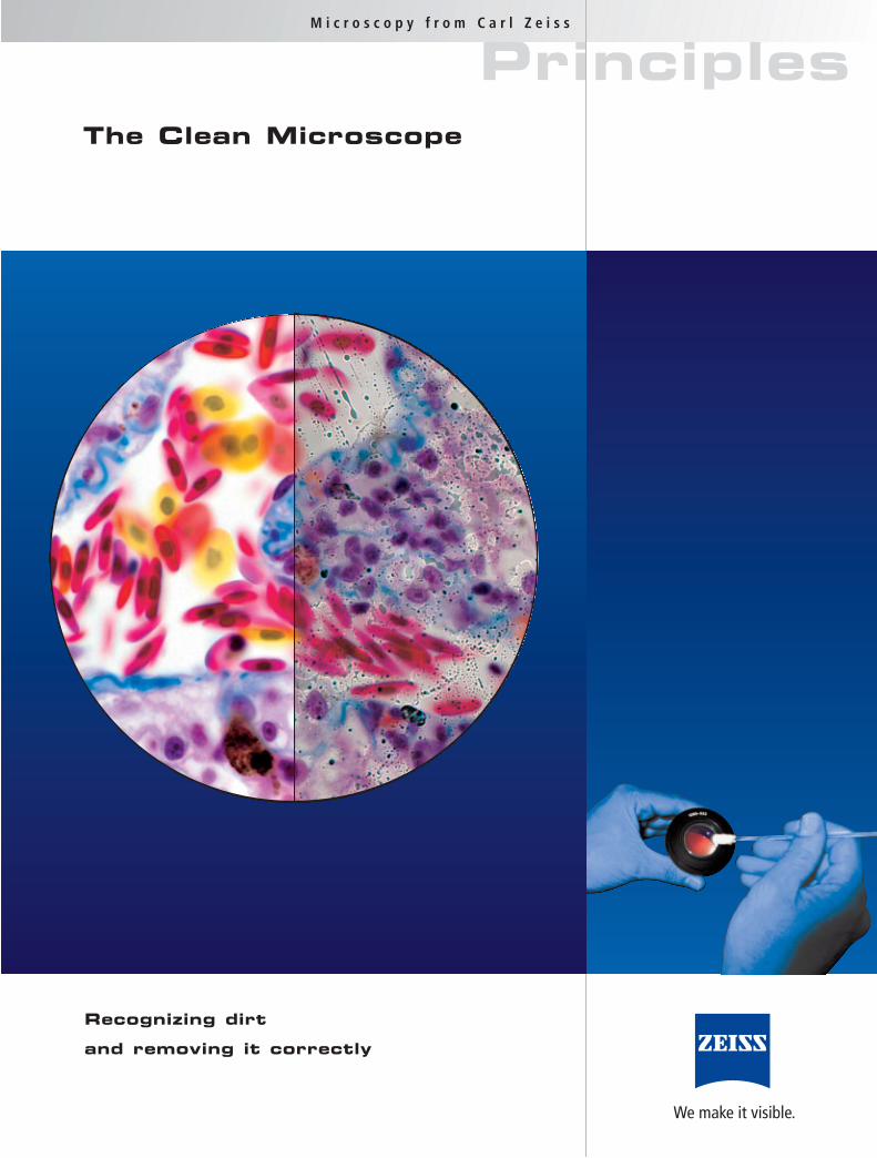
The Clean Microscope
Recognizing dirt
and removing it correctly
PrinciplesM i c r o s c o p y f r o m C a r l Z e i s s
We make it visible.
Clean microscope optics are a prerequisite for suc-
cessful microscopy and perfect images.
Over the years a variety of cleaning procedures
have been recommended. Many users remain un-
sure as to which of these will yield the best results.
The choice of the best cleaning method depends on
the nature of the optical surface and the type of
dirt to be removed.
Please contact the local Carl Zeiss representative
for questions regarding maintenance and service.
Where to find what?
Page
Acetone . . . . . . . . . . . . . . . . . . . . . . . . . 5, 9
Anisole . . . . . . . . . . . . . . . . . . . . . . . . . . . . 4
Blue WINDEX . . . . . . . . . . . . . . . . . . . . . . . 5
Camera adaptor . . . . . . . . . . . . . . . . . . . . . 5
Cameras . . . . . . . . . . . . . . . . . . . . . . . . . . . 5
Chloroform . . . . . . . . . . . . . . . . . . . . . . . . . 5
Cleaning motion . . . . . . . . . . . . . . . . . . . . . 9
Cleaning Solution L . . . . . . . . . . . . . . 6, 9, 12
Coatings . . . . . . . . . . . . . . . . . . . . . . . . . . . 5
Cotton . . . . . . . . . . . . . . . . . . . . . . 6, 7, 8, 12
Cover slips . . . . . . . . . . . . . . . . . . . . . 4, 6, 7
Diethylether . . . . . . . . . . . . . . . . . . . . . . . . 9
Dirt – greasy, loose, oily, water-soluble . . . . . 8
Dirt location . . . . . . . . . . . . . . . . . . . . . . . . 3
Dirt on the camera . . . . . . . . . . . . . . . . . . . 3
Dirty front lens . . . . . . . . . . . . . . . . . . . . 2, 3
Dry objective with correction collar . . . . . . . 4
Dust blower . . . . . . . . . . . . . . . . . . . . . . 6, 8
Dust . . . . . . . . . . . . . . . . . . . . . . . . . . . . . 10
Ethanol . . . . . . . . . . . . . . . . . . . . . . . . . . . . 9
Fluorescence filter sets . . . . . . . . . . . . . . . . . 5
Fungus . . . . . . . . . . . . . . . . . . . . . . . . . . . 10
IMMERSOL . . . . . . . . . . . . . . . . . . . . . . . 2, 4
JENA-Microscopes 250 CF . . . . . . . . . . . . . 11
Kleenex . . . . . . . . . . . . . . . . . . . . . . . . . . . 6
LD-Dry objective . . . . . . . . . . . . . . . . . . . . . 2
MIKROVAL-Microscopes . . . . . . . . . . . . . . . 11
Optical surfaces – concave or convex . . . . 5, 6
Optical surfaces – flat . . . . . . . . . . . . . . . 5, 6
Painted surface . . . . . . . . . . . . . . . . . . . . . 10
Petroleum ether . . . . . . . . . . . . . . . . . 6, 9, 12
Polyester swab . . . . . . . . . . . . . . . . . . . 6, 7, 8
Polystyrene stick . . . . . . . . . . . . . . . . . . . . 11
SIDOLIN . . . . . . . . . . . . . . . . . . . . . . . . . . . 5
Solvents . . . . . . . . . . . . . . . . . . . . . . . . . 6, 8
SPARKLE . . . . . . . . . . . . . . . . . . . . . . . . . . . 5
Spherical aberration . . . . . . . . . . . . . . . . . . 4
STANDARD-Microscopes . . . . . . . . . . . . . . . 11
Swabs . . . . . . . . . . . . . . . . . . . . . . 6, 7, 8,12
WHATMAN paper . . . . . . . . . . . . . . . . 6, 7, 12
Xylene . . . . . . . . . . . . . . . . . . . . . . . . . . . . 9
The Effect of Dirt on the Image
2
The closer dirt is to the object or to the camera
sensor, the greater its effect on the visual or
photographed image. The critical areas are the
following:
1. The external surface of the front lens of the
objective
2. The surface of the camera sensor and its
protective glass cover
3. Both surfaces of the cover slip
4. The surface of the microscope slide
5. The surface of the camera adapter optics
6. Surface of the upper lens of the condenser
7. The outer and inner surfaces of the eyelens
of the eyepiece as well as the upper surface
of the graticule
8. The outer surface of the protective glass cover-
ing the opening through which light exits
9. Other glass surfaces in the light path e.g. bulbs
of halogen- or high pressure lights, fluores-
cence filters and beam splitters, collector lenses,
contrast and heat filters.
Some optical surfaces are more sensitive to dirt
than others. The front lens of the objective is par-
ticularly critical and is therefore discussed in
greater detail below:
For any dry objective, the smaller the free working
distance and the smaller the surface area of the
concave front lens, the greater the danger of soil-
ing the front lens with embedding media, immer-
sion liquids or dust.
Examples of such objectives are the EC Plan-Neo-
fluar 40x/0.75, EC Plan-Neofluar 63x/0.95 Korr,
Achroplan 63x/0.80, 63x/0.95 o.D., Fluar 20x/
0.75, Planapo 20x/0.80, Planapo 40x/0.95 Korr, all
dry objectives of the type Epiplan and EC Epiplan-
Neofluar as well as EC Epiplan-Apo-Objectives with
magnifications of 20x, 50x and 100x.
When working with inverted microscopes, the
front lens of every objective will be more exposed
to dust than that in an upright microscope; all LD
dry objectives with magnifications of 32x, 40x and
63x need to be regularly checked.
The front lens of an immersion objective should be
cleaned to remove residue both after use and addi-
tionally, before applying fresh immersion liquid.
The mixing of different immersion media, as well
as different lots of the same medium e.g. the
immersion oil IMMERSOL F™, can result in blurred
images.
The cameras are always to be handled with the
utmost care and protected from dirt using all avail-
able methods.
Before every critical use, check the front lens
of the objective for dirt.
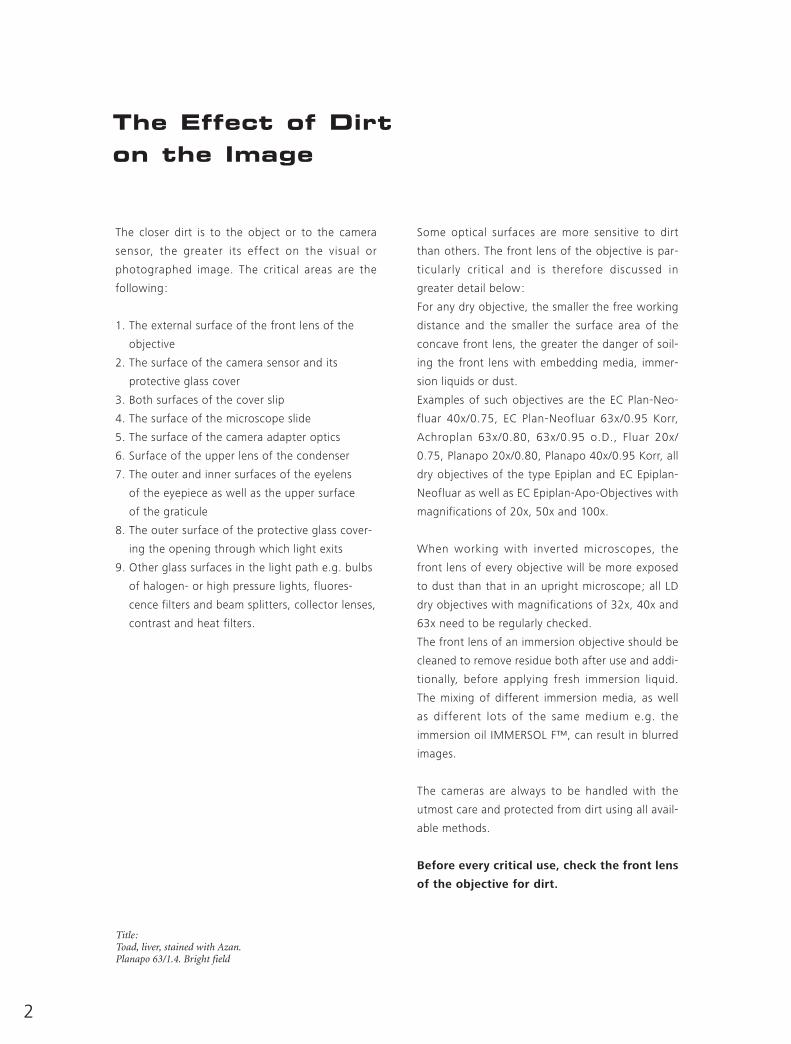
Title: Toad, liver, stained with Azan.Planapo 63/1.4. Bright field
3
How to Recognize Dirt
Familiarity with the best possible results obtainable
with a specific technique and procedure permits a
user to recognize the consequences of dirty optics.
The ability to compare the expected image and the
image actually obtained with respect to optimal
sharpness, contrast and the absence of contamina-
tion-dependent visual artefacts, allows the user to
immediately recognize when the microscope is
dirty or not.
If the image sharpness or contrast is not
optimal, then there is a high probability that
the microscope optics are not clean.
In order to determine the location of the dirt,
please proceed as follows:
Carefully rotate objectives and cameras a small
amount within their thread.
Check the slide and cover slip by moving the spec-
imen while focusing initially on the upper and then
the lower surfaces.
Check the condenser while moving it up and down
and if applicable, by swiveling the front lens slightly.
The affected optical surface is identified when a
suspected optical component is moved and the dirt
follows this movement. The single exception to this
rule is the camera: dirt located within the camera
will not move when the camera is moved!
A macroscopic check for larger dust particles and
scratches on optical surfaces can be carried out
using either a magnifying glass (with a magnifica-
tion of 3 –6x) or an eyepiece held the wrong way
round.
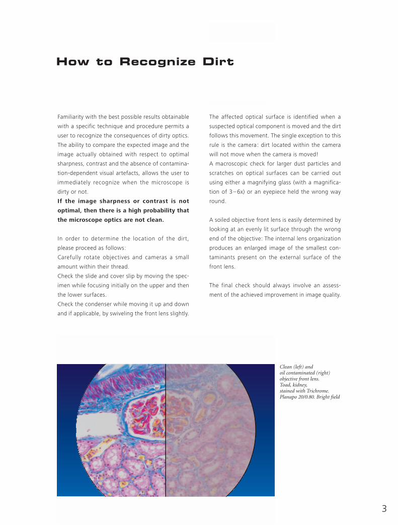
A soiled objective front lens is easily determined by
looking at an evenly lit surface through the wrong
end of the objective: The internal lens organization
produces an enlarged image of the smallest con-
taminants present on the external surface of the
front lens.
The final check should always involve an assess-
ment of the achieved improvement in image quality.
Clean (left) and oil contaminated (right) objective front lens.Toad, kidney,stained with Trichrome.Planapo 20/0.80. Bright field
4
Different Types of Dirt
It is crucial to differentiate between dust (e.g. glass
dust from slides, flakes of skin from the micro-
scopist, fluff from clothing, pollen from spring and
summer flowering) and other dirt (e.g. liquid or
dried-out embedding or immersion media, culture
solutions, residue from improper cleaning at-
tempts, fingerprints and grease).
Dust particles can either rest loosely or can be
more or less stuck to optical surfaces. Other dirt is
either water-soluble or may only be completely
removed using organic solvents.
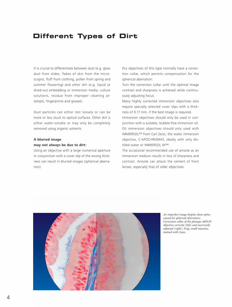
A blurred image
may not always be due to dirt:
Using an objective with a large numerical aperture
in conjunction with a cover slip of the wrong thick-
ness can result in blurred images (spherical aberra-
tion).
Dry objectives of this type normally have a correc-
tion collar, which permits compensation for the
spherical aberration:
Turn the correction collar until the optimal image
contrast and sharpness is achieved while continu-
ously adjusting focus.
Many highly corrected immersion objectives also
require specially selected cover slips with a thick-
ness of 0.17 mm, if the best image is required.
Immersion objectives should only be used in con-
junction with a suitable, bubble-free immersion oil.
Oil immersion objectives should only used with
IMMERSOL™ from Carl Zeiss; the water immersion
objective, C-APOCHROMAT, ideally with only dis-
tilled water or IMMERSOL W™.
The occasional recommended use of anisole as an
immersion medium results in loss of sharpness and
contrast. Anisole can attack the cement of front
lenses, especially that of older objectives.
An imperfect image despite clean optics caused by spherical aberration: Correction collar of the planapo 40/0.95 objective correctly (left) and incorrectly adjusted (right). Frog, small intestine,stained with Azan.
5
Different Optical Surfaces
Concave or convex surfaces (e.g. front lens of dry
objectives and condensers, the eye-lens of some
eyepieces) should be distinguished from planar
parallel or flat surfaces (e.g. the front lens of most
of the immersion objectives and condensers, fil-
ters, the protective glass covering camera sensors
or the opening through which light exits). Concave
or convex surfaces are cleaned using either the cot-
ton or the new polyester swabs as described on
page 6.
Easily accessible flat surfaces may be similarly
cleaned or with soft disposable cellulose wipes.
Microscope optics can be composed either of opti-
cal glass, quartz or polymers. The upper surface of
almost all will be coated to minimize stray light.
Some anti-reflex coatings may be wipeable (e.g.
the eyelens of eyepieces) or not, due to their soft-
ness. Generally, anti-reflex coatings are composed
of magnesium fluoride and should only be cleaned
with agents free from ammonia and acid. Some-
times household glass cleaning agents, which con-
tain dilute ammonia (e.g. SIDOLIN, SPARKLE, Blue
WINDEX), are recommended but they should not
be used routinely.
Some optical components are surrounded by black
anti-reflex surfaces, which are sensitive to organic
solvents. The plastic and rubber parts of the eye-
piece will likewise be attacked by organic solvents
(e.g. acetone, chloroform).
In older microscopes, lenses are cemented using an
alcohol soluble cement such as Canada balsam.
These days the lens cement is generally a poly-
acrylic synthetic resin, which does not have this
problem.
The internal workings of the microscope – the opti-
cal surfaces, components of the fluorescent filter
sets, cameras and camera adapters should never be
cleaned by the user, but by experienced customer
representatives from the original manufacturer.
The user should only clean: the external surface of
the objective front lens, the condenser front lens,
the eyepiece eyelens, glass color- and conversion
filters, and the external surface of the protective
glass covering the opening through which light
exits.
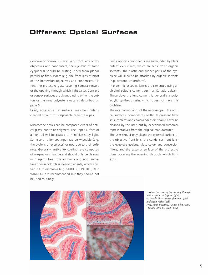
Dust on the cover of the opening throughwhich light exits (upper right),extremely dirty camera (bottom right)and clean optics (left).Frog, small intestine, stained with Azan.Planapo 10/0.45. Bright field.
For the easy cleaning of flat surfaces (e.g. the
removal of immersion media from cover slips or the
front lenses of immersion objectives), soft tissue
(e.g. Kleenex) soaked in diluted washing-up liquid
is suitable.
Care: The smooth lens paper (so-called Joseph
paper), which is often readily available in research
laboratories, is not for cleaning but is intended
only for the dust-free storage and protection of
optical components. For cleaning purposes this
lens paper is too harsh; it also doesn’t absorb the
dirt effectively or quickly enough. WHATMAN Lens
Cleaning Tissue 105 represents the single excep-
tion.
For the cleaning of all other optical surfaces either
freshly made cotton swabs or the new polyester
swabs, ITW Texwipe Clean Tips®, are used.
6
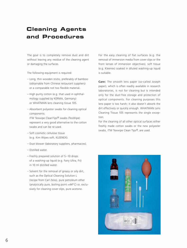
Cleaning Agents and Procedures
The goal is to completely remove dust and dirt
without leaving any residue of the cleaning agent
or damaging the surfaces.
The following equipment is required:
– Long, thin wooden sticks, preferably of bamboo
(obtainable from Chinese restaurant suppliers)
or a comparable not too flexible material.
– High purity cotton (e.g. that used in ophthal-
mology supplied by KERMA, Germany)
or WHATMAN lens cleaning tissue 105.
– Absorbent polyester swabs for cleaning optical
components.
ITW Texwipe CleanTips® swabs (TexWipe)
represent a very good alternative to the cotton
swabs and can be re-used.
– Soft cosmetic cellulose tissue
(e.g. Kim Wipes soft, KLEENEX).
– Dust blower (laboratory suppliers, pharmacies).
– Distilled water.
– Freshly prepared solution of 5–10 drops
of a washing-up liquid (e.g. Fairy Ultra, Fit)
in 10 ml distilled water.
– Solvent for the removal of greasy or oily dirt,
such as the Optical Cleaning Solution L
(recipe from Carl Zeiss), pure petroleum ether
(analytically pure, boiling point <44º C) or, exclu-
sively for cleaning cover slips, pure acetone.

Preparation of cotton swabs
■ Wash hands (powdered, latex gloves are not suit-
able).
■ Dip the stick into the cleaning solution (aqueous-
or organic solvent). The cotton fibers attach better
to the stick as a result.
■ Dab the stick onto the wad of cotton and loosen
some fibers. Do not compact the cotton otherwise
the fibers will not separate easily.
■ Turn the stick so that an even, elliptical cotton bud
forms at the tip.
■ Remove the cotton tip after every wipe and replace
it with a fresh cotton bud.
■ The stick can be used for a long period of time. Use
separate sticks for water-based solutions and
organic solvents.
■ To protect the cotton tip from dirt, the stick should
be stored in a polythene bag. It should not be
handled as perspiration and grease, from the fin-
gers of the users, will significantly affect its ability
to clean.
If the use of WHATMAN Lens Cleaning Tissue 105
is preferred, fold the sheet around the stick so that
a sharp point is generated. The point should not be
handled. Use the tissue only once and then replace
it.
The polyester swabs, ITW Texwipe CleanTips®, can
be used until they no longer clean well.
7
1 3
2 4
8
Cleaning Procedure
1. Blow all loose dust particles away with a dust
blower.
2. Remove all water-soluble dirt with distilled
water. If this is unsuccessful repeat using a solu-
tion of diluted washing-up liquid. Remove any
remaining residue with a dry cotton swab, but
breathe on the surface first to generate a film of
moisture. In so doing, be careful not spray
droplets of saliva.
3. To remove oily dirt, use a solution of dilute
washing-up liquid initially. If this does not pro-
duce a satisfactory result, repeat the cleaning
using a solvent (Optical Cleaning Solution L,
petroleum ether).
4. Greasy dirt must always be removed using a
solvent.
5. After cleaning check the surface (see the section
“How to recognize dirt”).
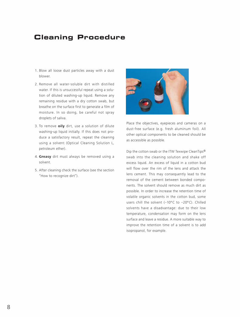
Place the objectives, eyepieces and cameras on a
dust-free surface (e.g. fresh aluminum foil). All
other optical components to be cleaned should be
as accessible as possible.
Dip the cotton swab or the ITW Texwipe CleanTips®
swab into the cleaning solution and shake off
excess liquid. An excess of liquid in a cotton bud
will flow over the rim of the lens and attack the
lens cement. This may consequently lead to the
removal of the cement between bonded compo-
nents. The solvent should remove as much dirt as
possible. In order to increase the retention time of
volatile organic solvents in the cotton bud, some
users chill the solvent (–10ºC to –20ºC). Chilled
solvents have a disadvantage: due to their low
temperature, condensation may form on the lens
surface and leave a residue. A more suitable way to
improve the retention time of a solvent is to add
isopropanol, for example.
9
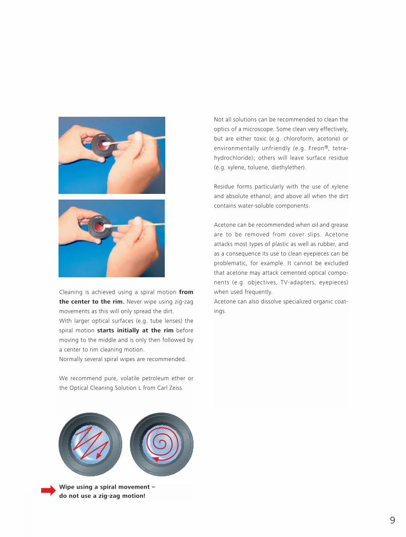
Cleaning is achieved using a spiral motion from
the center to the rim. Never wipe using zig-zag
movements as this will only spread the dirt.
With larger optical surfaces (e.g. tube lenses) the
spiral motion starts initially at the rim before
moving to the middle and is only then followed by
a center to rim cleaning motion.
Normally several spiral wipes are recommended.
We recommend pure, volatile petroleum ether or
the Optical Cleaning Solution L from Carl Zeiss.
Not all solutions can be recommended to clean the
optics of a microscope. Some clean very effectively,
but are either toxic (e.g. chloroform, acetone) or
environmentally unfriendly (e.g. Freon®, tetra-
hydrochloride) ; others will leave surface residue
(e.g. xylene, toluene, diethylether).
Residue forms particularly with the use of xylene
and absolute ethanol, and above all when the dirt
contains water-soluble components.
Acetone can be recommended when oil and grease
are to be removed from cover slips. Acetone
attacks most types of plastic as well as rubber, and
as a consequence its use to clean eyepieces can be
problematic, for example. It cannot be excluded
that acetone may attack cemented optical compo-
nents (e.g. objectives, TV-adapters, eyepieces)
when used frequently.
Acetone can also dissolve specialized organic coat-
ings.
Wipe using a spiral movement –
do not use a zig-zag motion!

10
How CanContaminationbe Avoided?
Cleaning ExternalMicroscopeComponents
The opening of the binocular tubes should always
be protected either with the eyepieces or with dust
covers. If no dust covers from the manufacturers
are available, aluminum foil is a suitable substitute.
The best fundamental method to prevent dust
accumulation is to first cover the microscope
with two additional plastic bags and then the dust
cover supplied by the manufacturer.
In tropical regions this measure is not recommended
as it can frequently lead to a build up of fungus.
Fungal contamination can best be minimized by
reducing the humidity of the room either by air
conditioning or by installing an infrared lamp
above the microscope (at a minimum distance of
150 cm or 5 Feet). Fungal contamination is almost
impossible to remove.
The microscope should never be located in a posi-
tion where it could be affected by acidic or alka-
line vapors, such as in or near a wet chemical
photographic laboratory.
The painted surfaces of microscopes from the AXIO
range are powder coated and extremely durable.
They can be cleaned well with a very lightly mois-
tened microfiber cloth. Loose dust and other dirt
can be removed using a brush of soft hair used
exclusively for this purpose.
A thin and clean preparationof the fresh water protozoan,Dimorpha mutans.Planapo 63/1.4 Phase contrast.
Together with the cleanliness of the micro-
scope optics, perfect sample preparation is
the deciding factor for optimal results:
e.g. the thickness of the histological section,
staining intensity, the refractive index and disper-
sion of the embedding media and immersion
fluids, and when performing high resolution
microscopy – the distance of a living cell from the
cover slip.
11
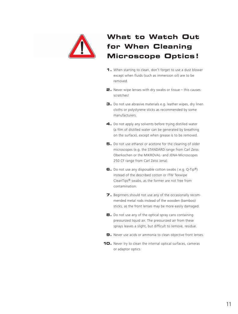
What to Watch Outfor When CleaningMicroscope Optics!
1. When starting to clean, don’t forget to use a dust blower
except when fluids (such as immersion oil) are to be
removed.
2. Never wipe lenses with dry swabs or tissue – this causes
scratches!
3. Do not use abrasive materials e.g. leather wipes, dry linen
cloths or polystyrene sticks as recommended by some
manufacturers.
4. Do not apply any solvents before trying distilled water
(a film of distilled water can be generated by breathing
on the surface), except when grease is to be removed.
5. Do not use ethanol or acetone for the cleaning of older
microscopes (e.g. the STANDARD range from Carl Zeiss
Oberkochen or the MIKROVAL- and JENA-Microscopes
250 CF range from Carl Zeiss Jena).
6. Do not use any disposable cotton swabs ( e.g. Q-Tip®)
instead of the described cotton or ITW Texwipe
CleanTips® swabs, as the former are not free from
contamination.
7. Beginners should not use any of the occasionally recom-
mended metal rods instead of the wooden (bamboo)
sticks, as the front lenses may be more easily damaged.
8. Do not use any of the optical spray cans containing
pressurized liquid air. The pressurized air from these
sprays leaves a slight, but difficult to remove, residue.
9. Never use acids or ammonia to clean objective front lenses.
10. Never try to clean the internal optical surfaces, cameras
or adaptor optics.
Suppliers and Recipes
KERMA cotton N 1. DAB 6.
The cotton used in ophthalmology is 100 % pure
cotton (DIN 61 640-A, Ph. Eur., DAB). It is absolutely
pure, highly absorbent and soft. The fibers can be
blown away from optical surfaces.
www.sbh-hainichen.de/kerma/prod/spez2.htm
WHATMAN Lens Cleaning Tissue 105
Lens paper in folders 10 cm x 15 cm,
25 blocks each with 25 sheets, Order Nr. 2105 841.
The only lens tissue recommended by Carl Zeiss. It
is chemically pure, silicon free and contains
absolutely no additives.
This product is also supplied by other companies
e.g. KODAK
www.whatman.plc.uk
Absorbent polyester swabs for cleaning
optical components ITW Texwipe CleanTips®
(Alpha®-, Clean Foam®- or Absorbond® series.)
Obtainable in different sizes and absorption
abilities e.g. from the company Basan under
TEXWIPE TX743B.
www.texwipe.com www.basan.com
Recipe for Optical Cleaning Solution L
from Carl Zeiss
Recipe 85 % petroleum ether, 15 % isopropanol.
(The solution is not sold by Carl Zeiss.)
The petroleum ether (also known as benzine or rub-
bing alcohol) should be analytically pure and have
the lowest possible boiling point (≤ 44ºC). Heavier
benzene fractions are not suitable as they leave an
insoluble residue on the optical surface.
Acetone that is recommended exclusively for the
occasional cleaning of oil contaminated cover slips
should also be analytically pure.
Carl Zeiss Post Box 4041Light Microscopy 37030 Göttingen
Phone: 0551-5060 660Fax: 0551-5060 464E-Mail: [email protected]/courses
Safety adviceWhen working with chemicals, solvents and
other possible hazards, please be sure to follow
the current, country-specific, safety regula-
tions.
1246-0009 e/04.05
Prin
ted
on e
nviro
nmen
tally
frie
ndly
pap
er
blea
ched
with
out
the
use
of c
hlor
ine.