the chest xray and electrocardiogram - uclaheart.ucla.edu/workfiles/adult_congenital/xrayand...
TRANSCRIPT
The Chest Xray and
Electrocardiogram
Roentgen/Einthoven
The State of Their Art
Ahmanson/UCLA Adult Congenital Heart Disease Center
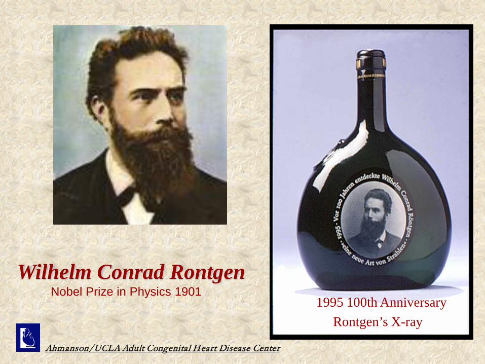
1995 100th Anniversary Rontgen’s X-ray
Wilhelm Conrad Rontgen Nobel Prize in Physics 1901
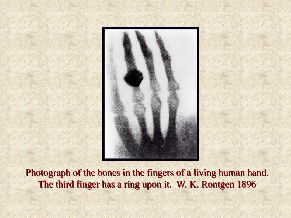
Photograph of the bones in the fingers of a living human hand. The third finger has a ring upon it. W. K. Rontgen 1896
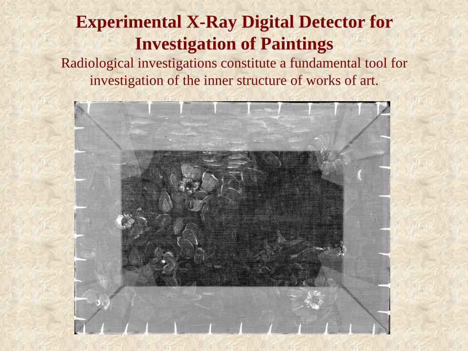
Experimental X-Ray Digital Detector for Investigation of Paintings
Radiological investigations constitute a fundamental tool for investigation of the inner structure of works of art.
Chest X-ray in Congenital Heart Disease
•Age and sex •Right/left orientation •Positions and malpositions -- above and below the diaphragm, thoracic and abdominal situs •The bones •Extrapulmonary soft tissue densities •Intrapulmonary soft tissue densities – vascular and parenchymal •The great arteries and great veins •The atria •The ventricles or ventricle
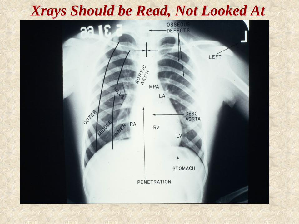
Xrays Should be Read, Not Looked At
Positions and Malpositions
Above and below the diaphragm. Thoracic and abdominal situs.
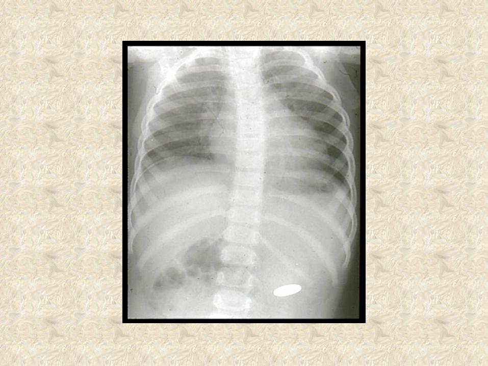
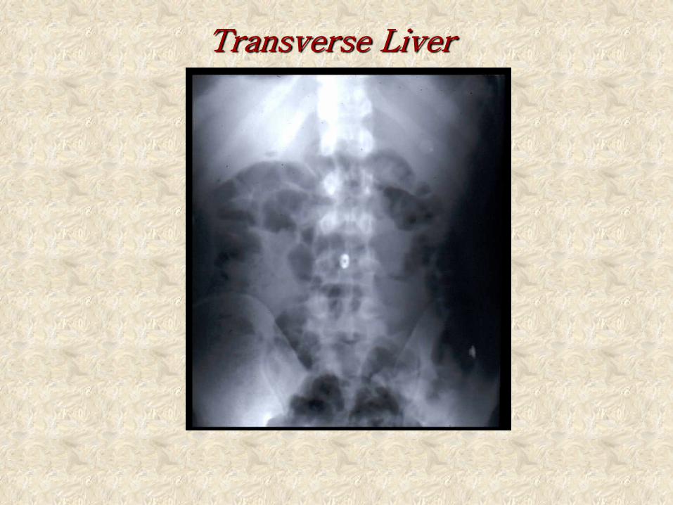
Transverse Liver
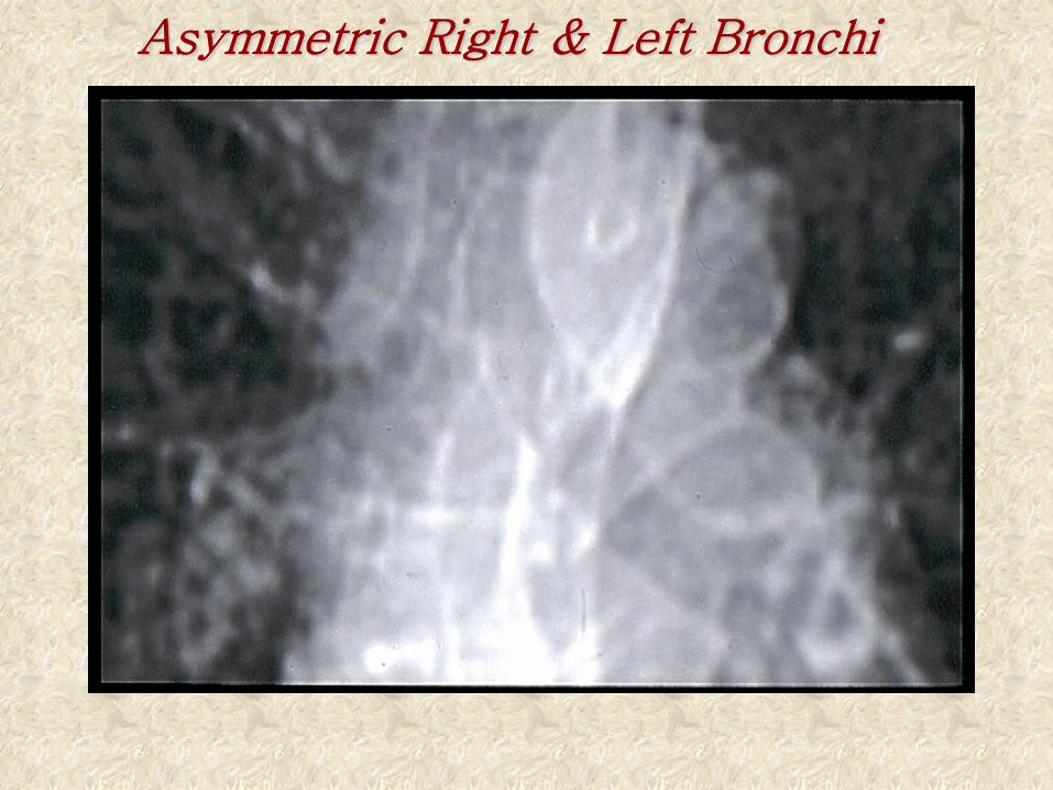
Asymmetric Right & Left Bronchi

Symmetric Right Bronchi. Bilateral Trilobed Lungs.
Right Isomerism.
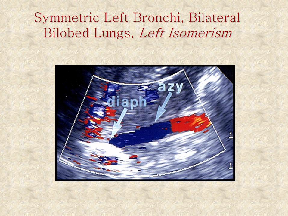
Symmetric Left Bronchi, Bilateral Bilobed Lungs, Left Isomerism
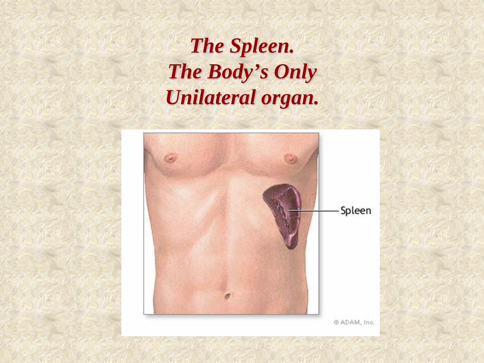
The Spleen. The Body’s Only Unilateral organ.
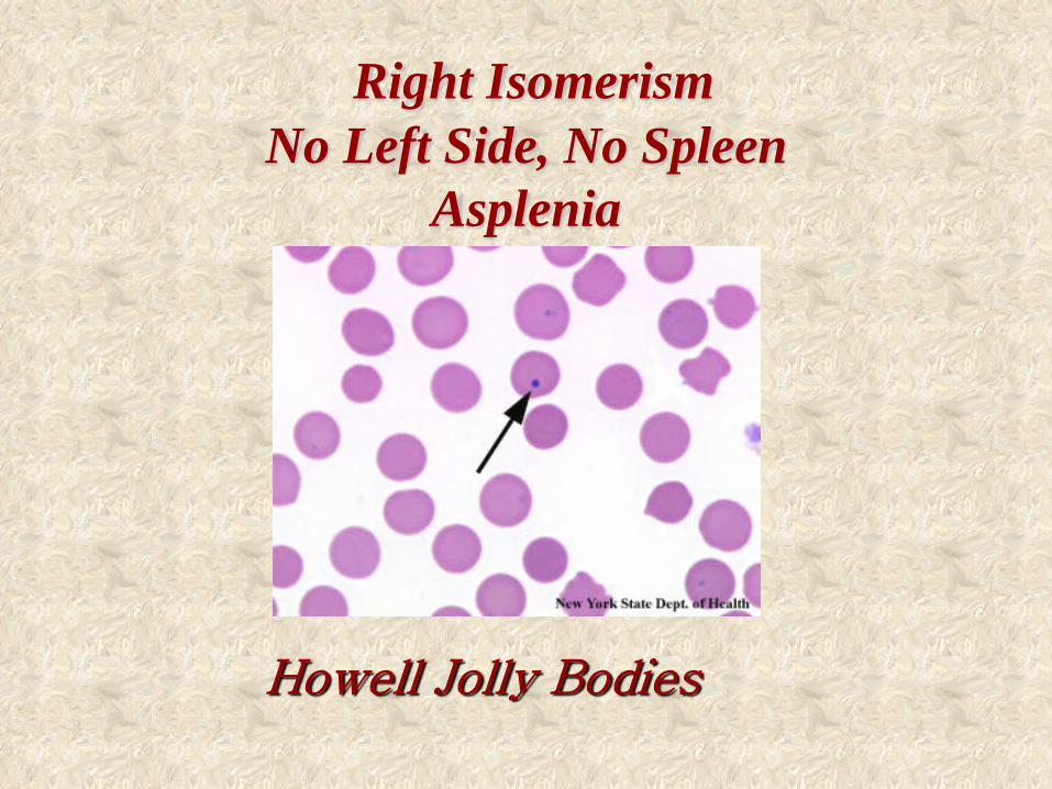
Right Isomerism
No Left Side, No Spleen Asplenia
Howell Jolly Bodies
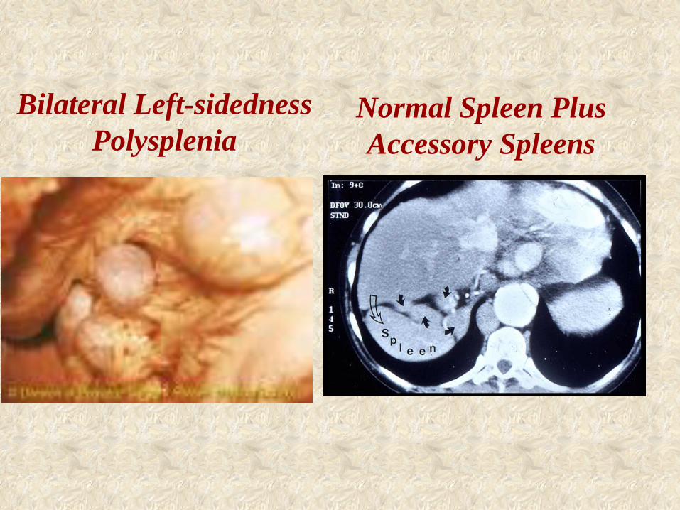
Bilateral Left-sidedness
Polysplenia
Normal Spleen Plus Accessory Spleens

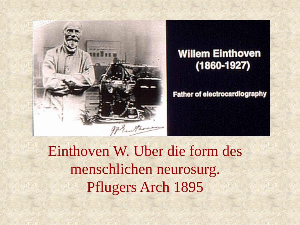
Einthoven W. Uber die form des menschlichen neurosurg.
Pflugers Arch 1895
The Electrocardiogram
Many brilliant minds have contributed to the development of electrocardiography as a clinical science. The early history (1900-1945) was dominated by Professor Willem Einthoven in the Netherlands, Sir Thomas Lewis in England and Dr. Frank N. Wilson in the United States. These three pioneers laid the foundation for modern electrocardiography.
Charles Fisch, The ECG Centennial
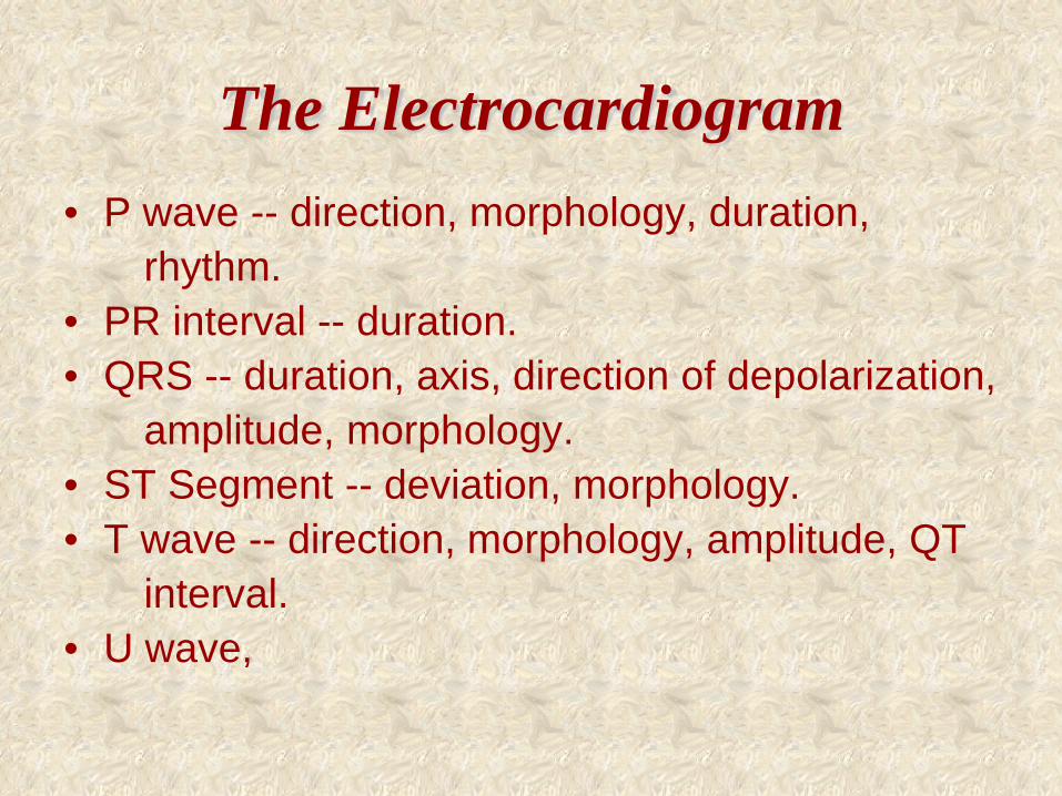
The Electrocardiogram • P wave -- direction, morphology, duration, rhythm. • PR interval -- duration. • QRS -- duration, axis, direction of depolarization, amplitude, morphology. • ST Segment -- deviation, morphology. • T wave -- direction, morphology, amplitude, QT interval. • U wave,
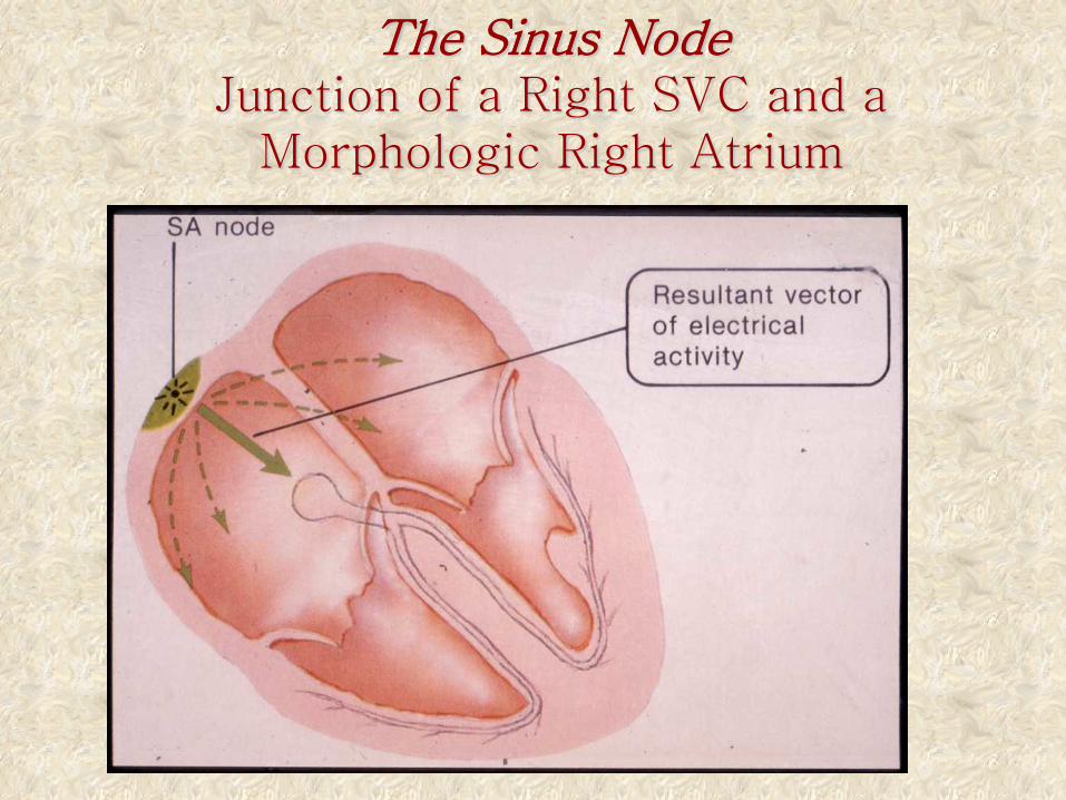
The Sinus Node Junction of a Right SVC and a
Morphologic Right Atrium
Einthoven W. Uber die form des menschlichen neurosurg.
Pflugers Arch 1895
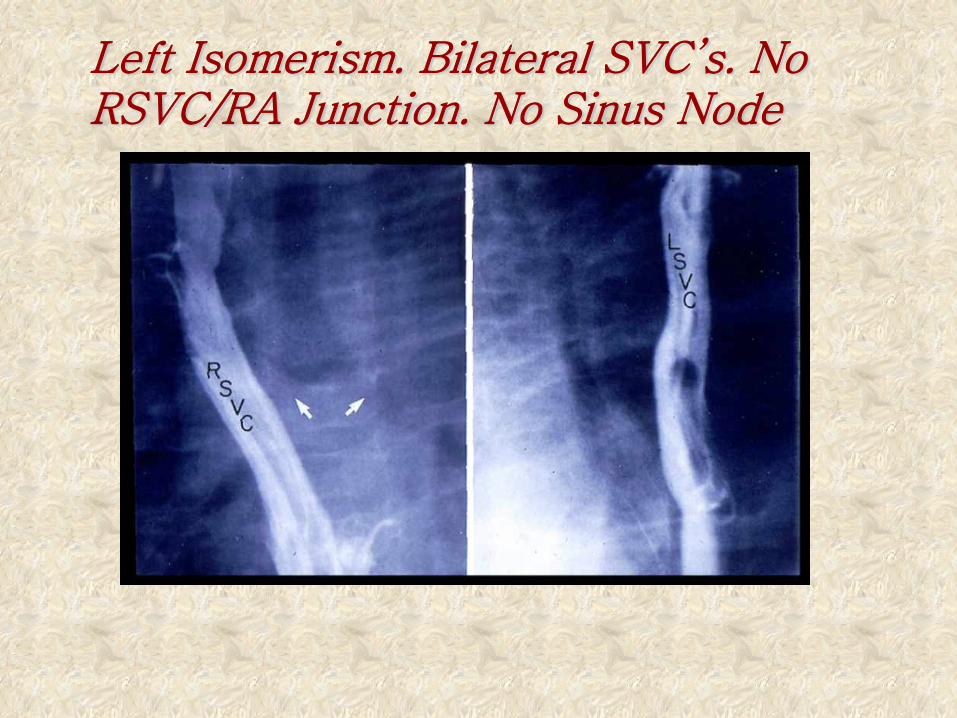
Left Isomerism. Bilateral SVC’s. No RSVC/RA Junction. No Sinus Node
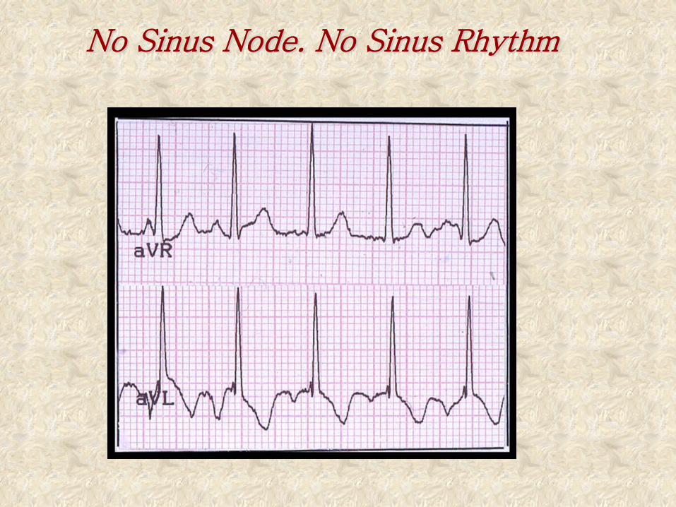
No Sinus Node. No Sinus Rhythm
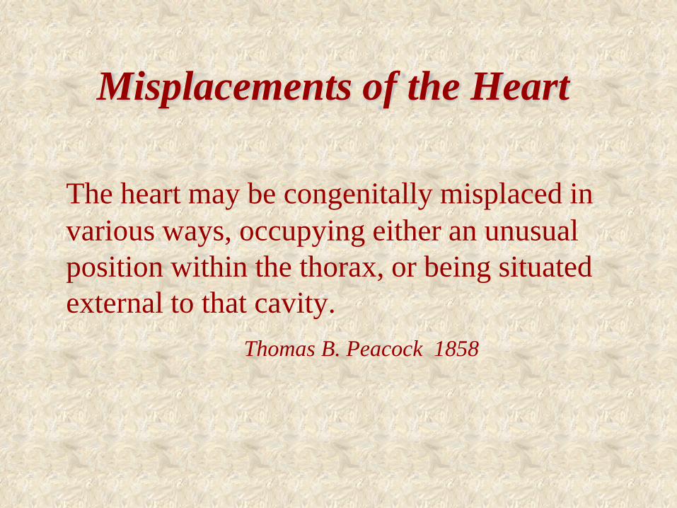
Misplacements of the Heart The heart may be congenitally misplaced in
various ways, occupying either an unusual position within the thorax, or being situated external to that cavity.
Thomas B. Peacock 1858

The Egyptians believed that the heart was the seat of personal and moral integrity. If the heart were not in its right
place, the individual would be beside himself.
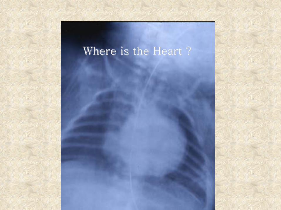
Where is the Heart ?
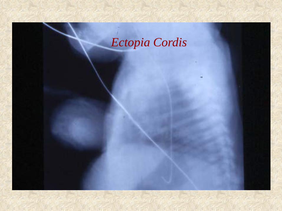
Ectopia Cordis
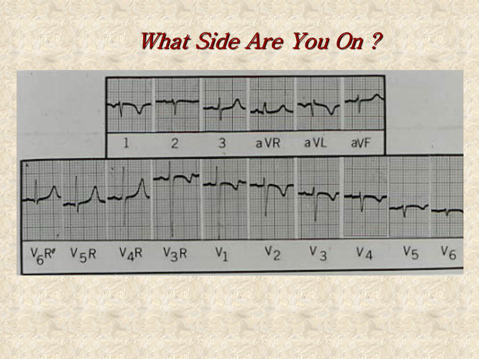
What Side Are You On ?
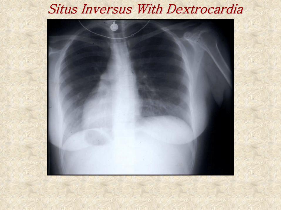
Situs Inversus With Dextrocardia
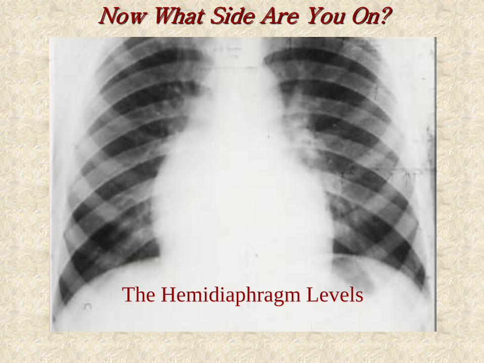
Now What Side Are You On?
The Hemidiaphragm Levels
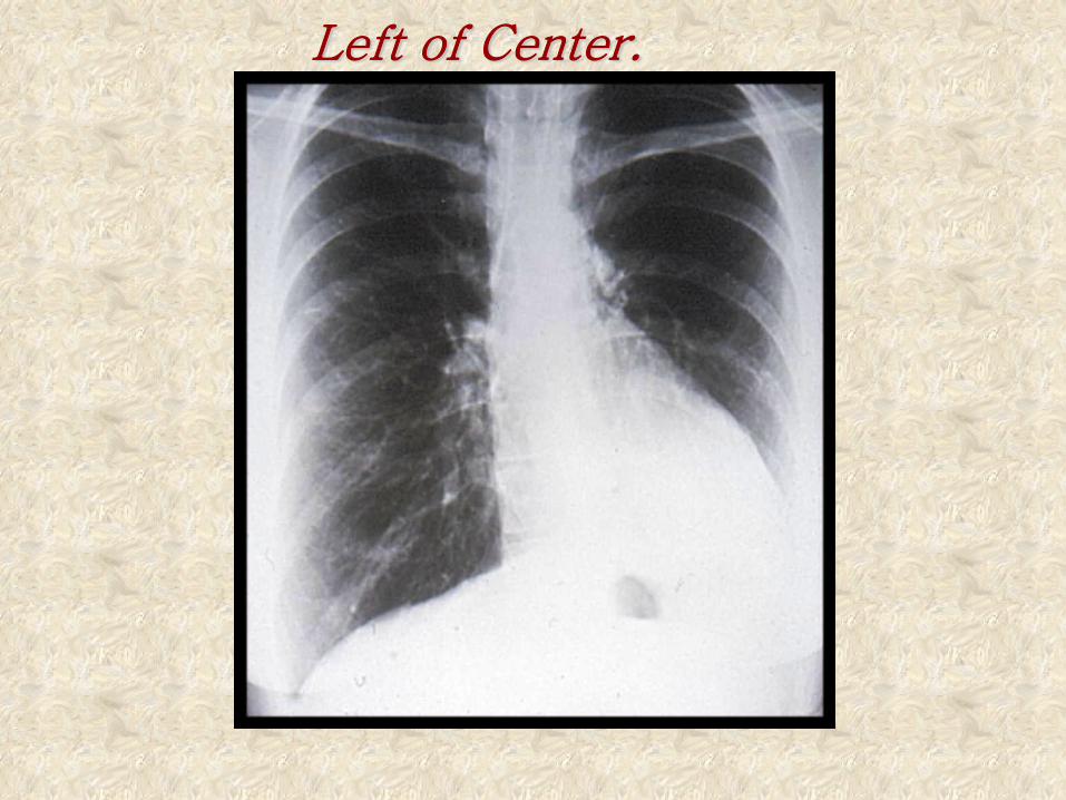
Left of Center.
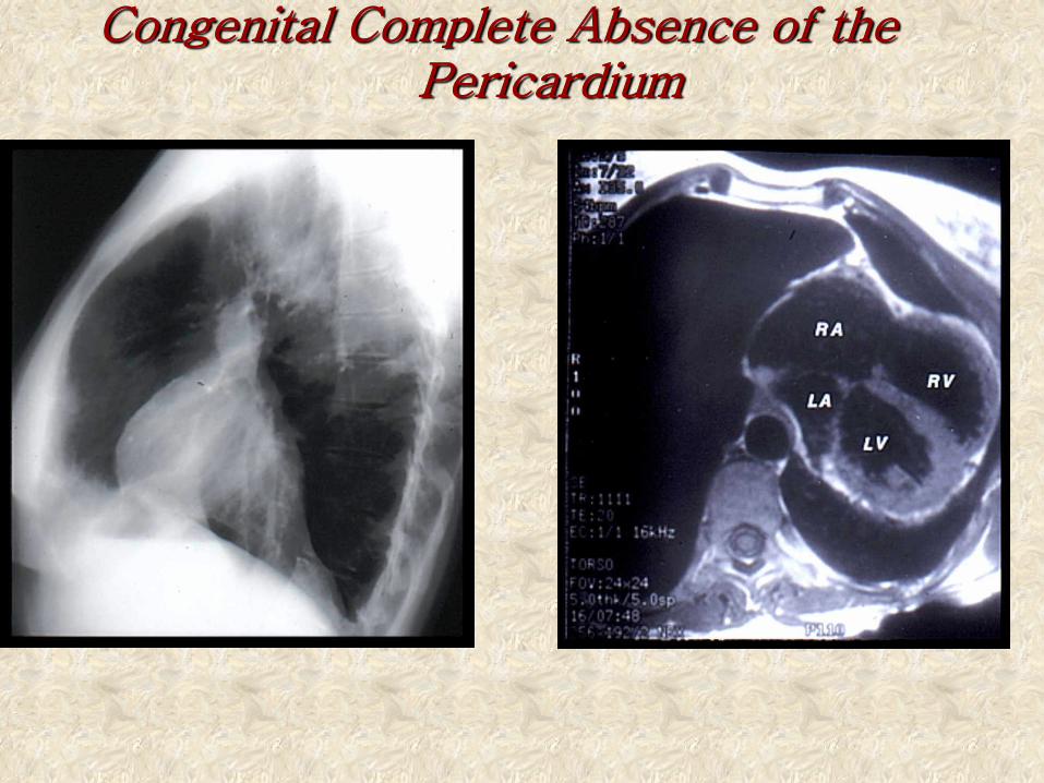
Congenital Complete Absence of the Pericardium
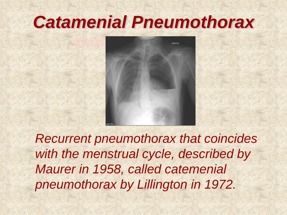
Catamenial Pneumothorax
Recurrent pneumothorax that coincides with the menstrual cycle, described by Maurer in 1958, called catemenial pneumothorax by Lillington in 1972.
Pleural Endometriosis
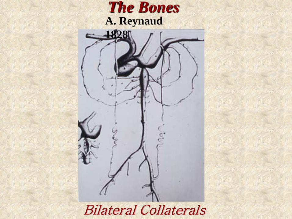
A. Reynaud 1828
Bilateral Collaterals
The Bones
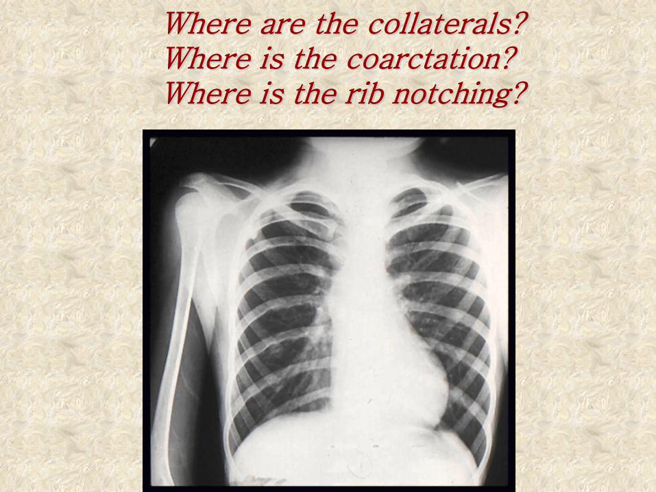
Where are the collaterals? Where is the coarctation? Where is the rib notching?
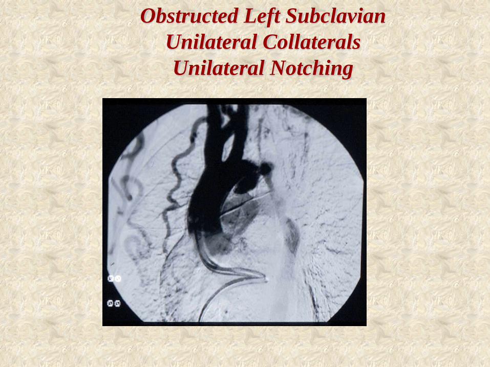
Obstructed Left Subclavian Unilateral Collaterals Unilateral Notching
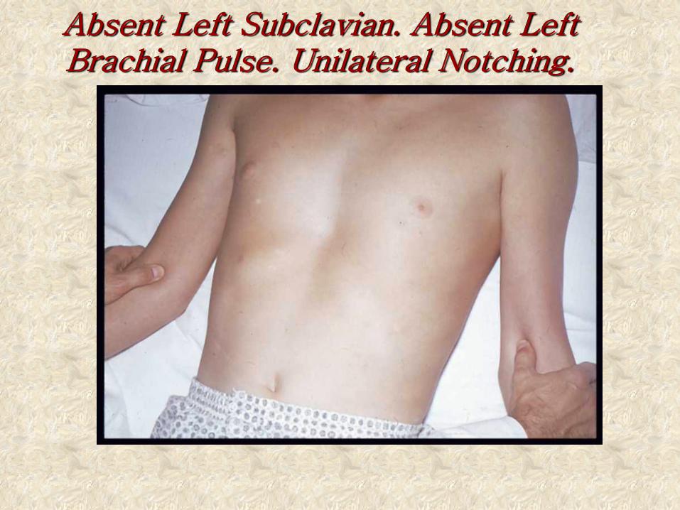
Absent Left Subclavian. Absent Left Brachial Pulse. Unilateral Notching.
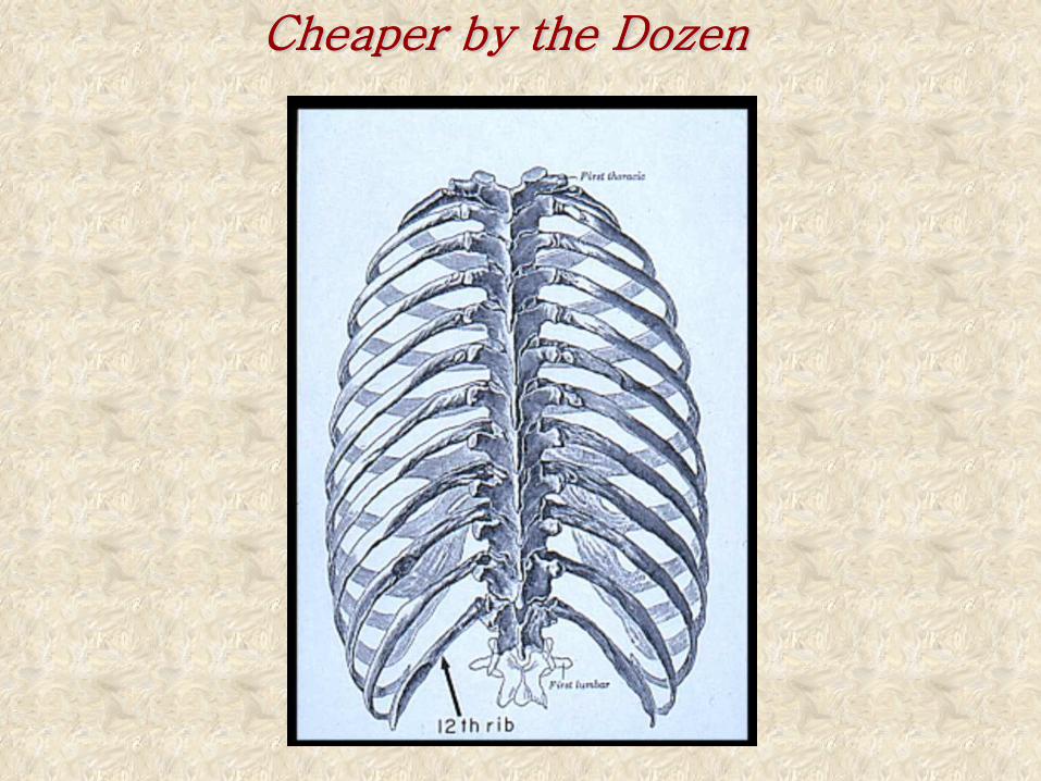
Cheaper by the Dozen
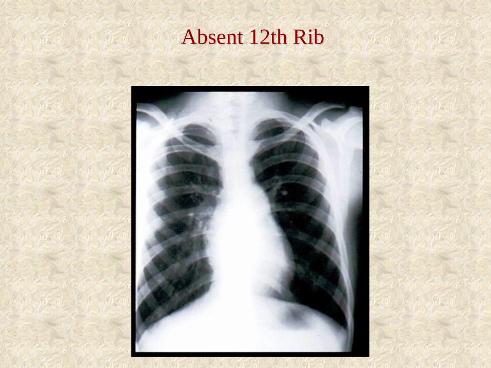
Absent 12th Rib
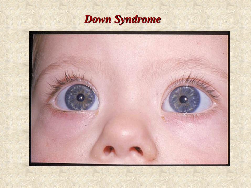
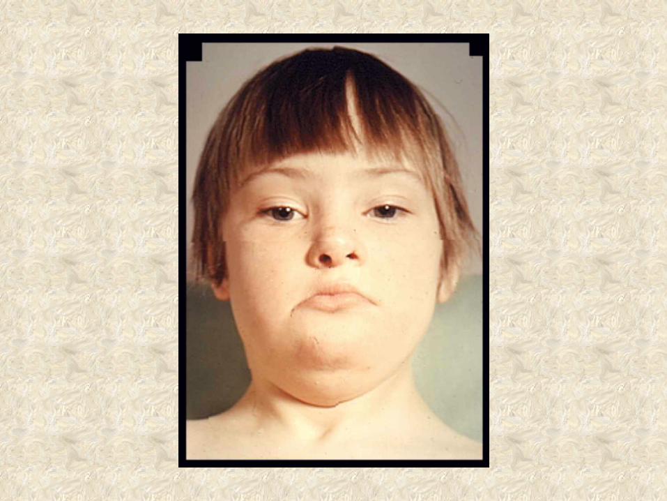
Down Syndrome

Intrapulmonary Soft Tissue Densities:
Vascular/parenchymal
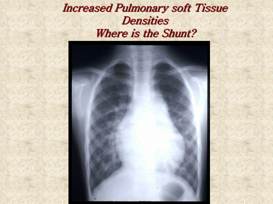
Increased Pulmonary soft Tissue Densities
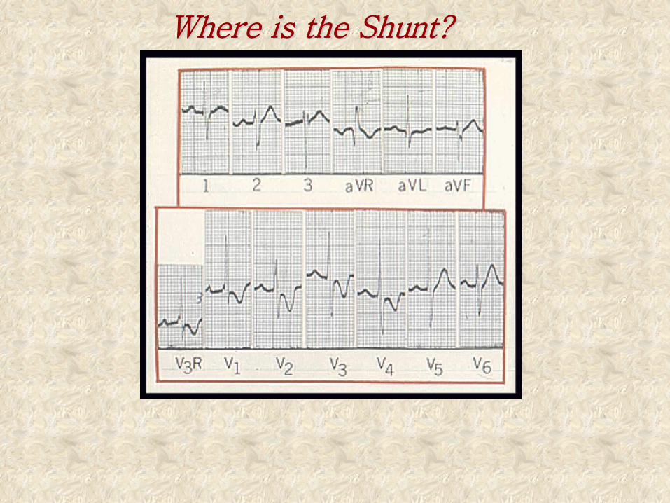
Where is the Shunt?
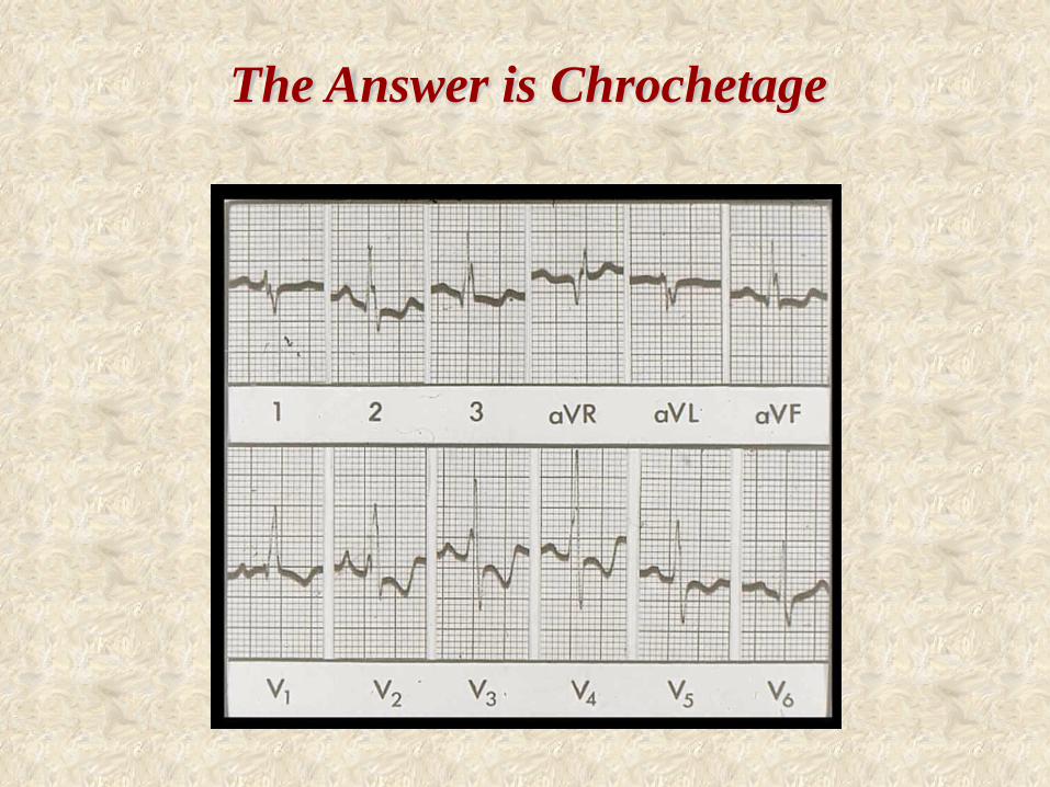
The Answer is Chrochetage

Sinus arrhythmia in children with atrial septal defect: An analysis of heart rate variability before and after surgical repair
Finley JP, et al. Br Heart J 1989
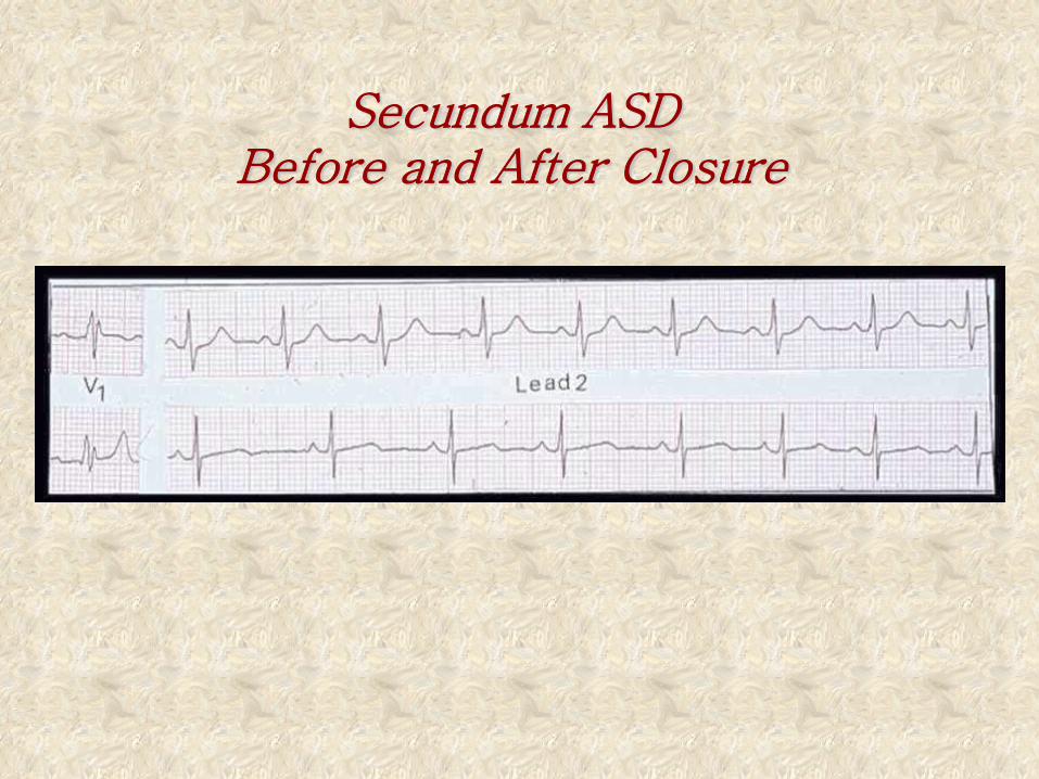
Secundum ASD Before and After Closure
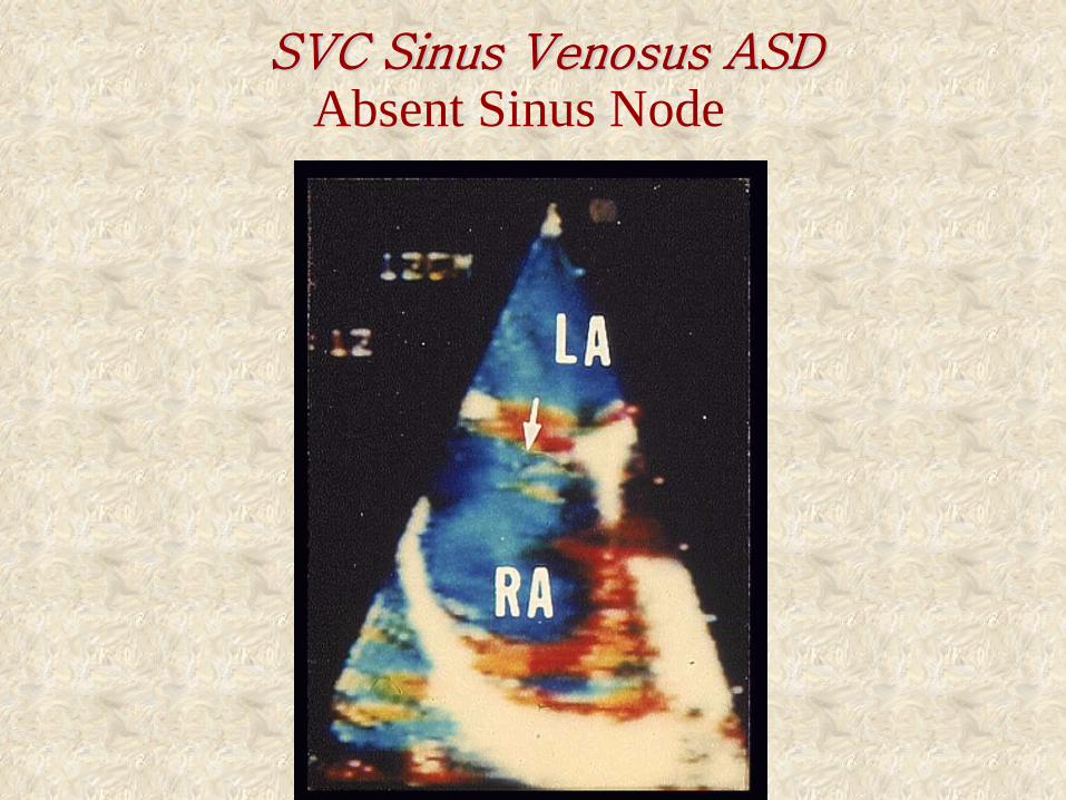
SVC Sinus Venosus ASD Absent Sinus Node
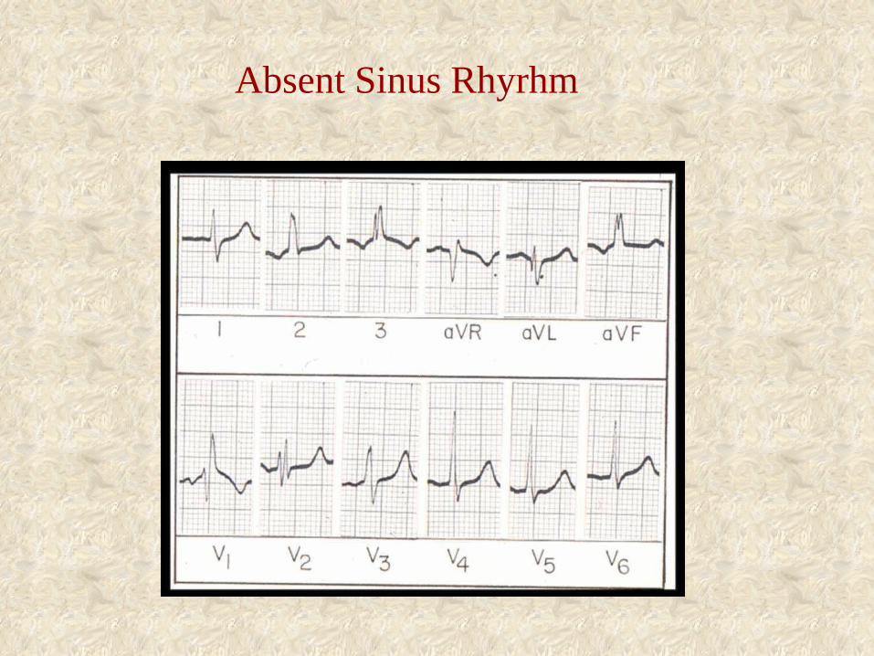
Absent Sinus Rhyrhm
Where is the Shunt?
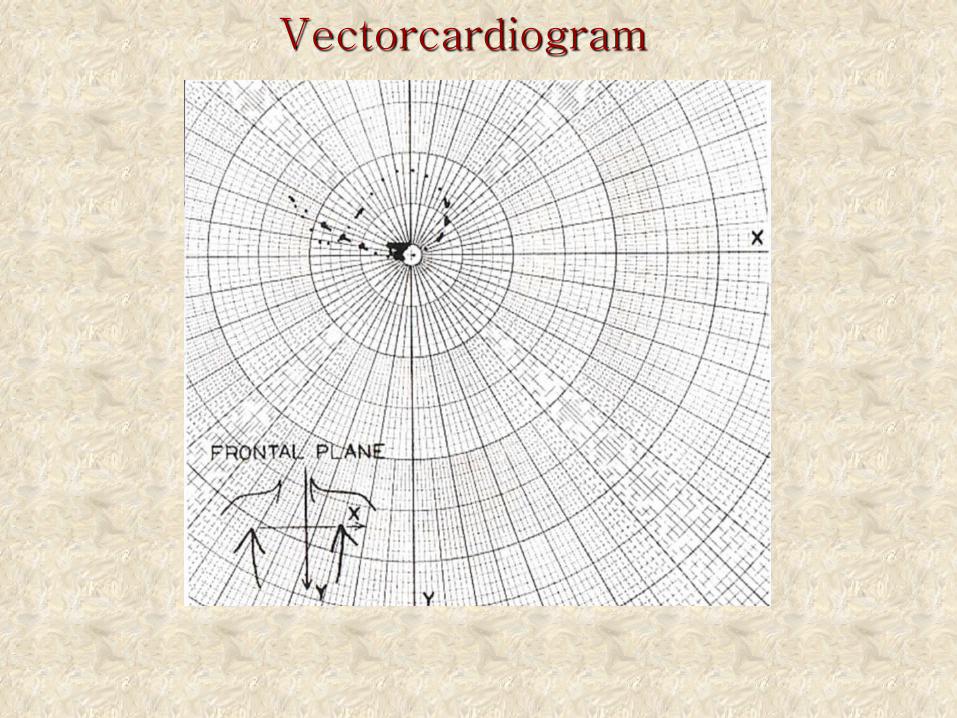
Vectorcardiogram
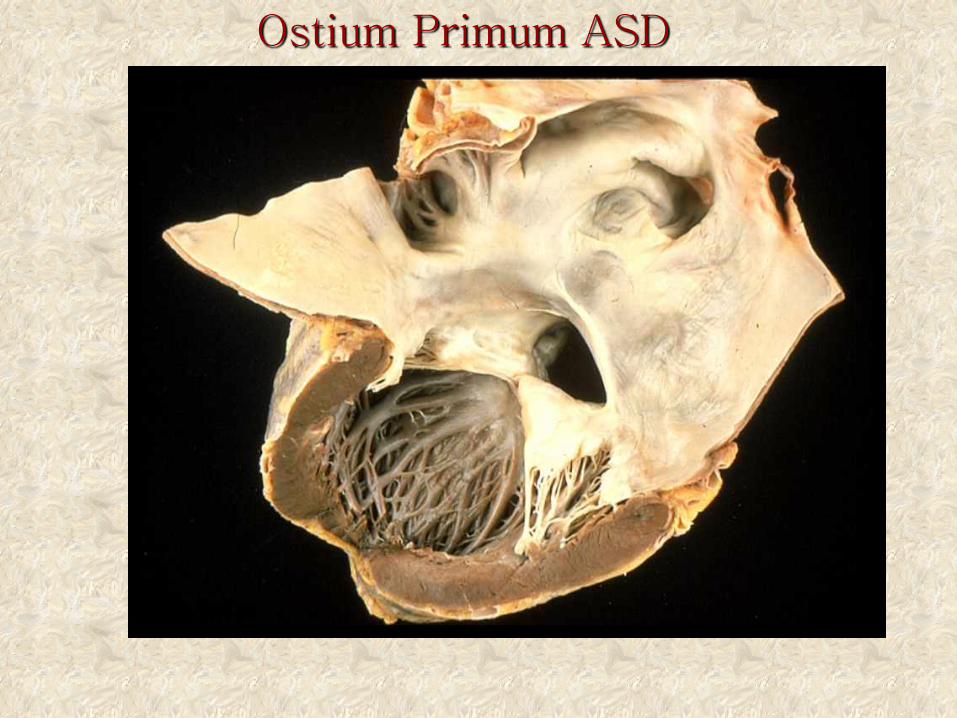
Ostium Primum ASD
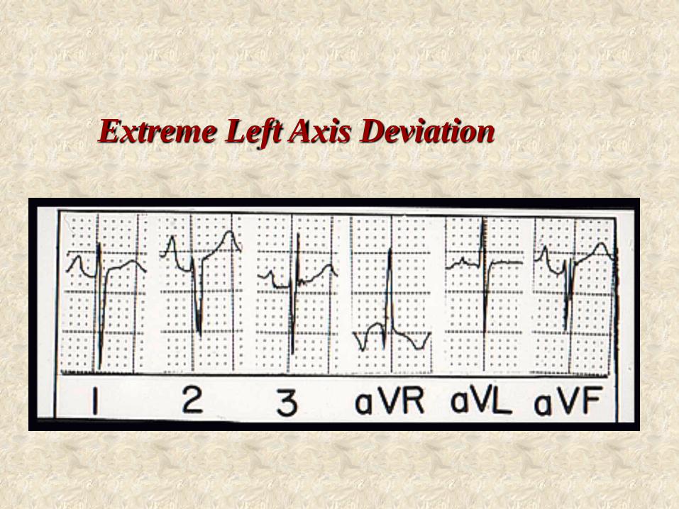
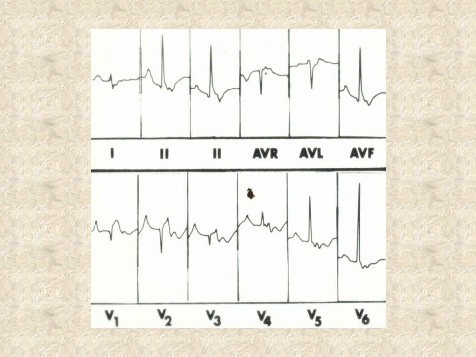
Extreme Left Axis Deviation
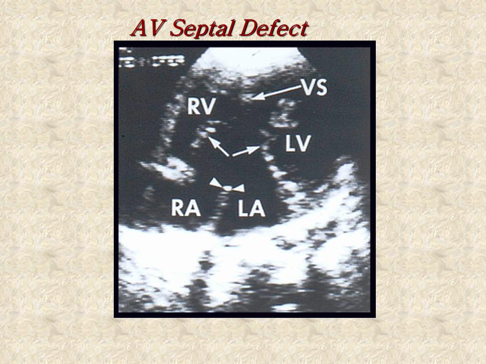
AV Septal Defect

The Atria
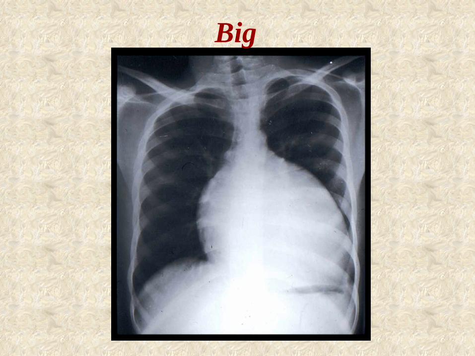
Big
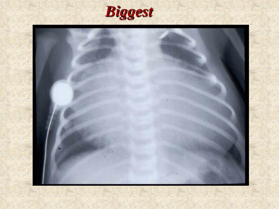
Biggest
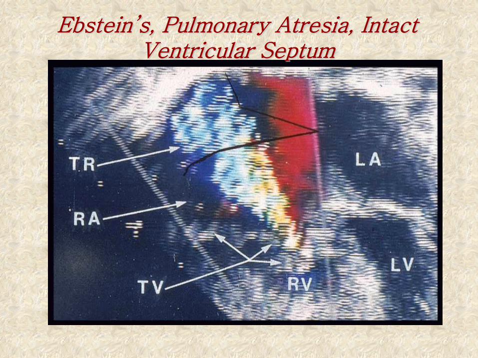
Ebstein’s, Pulmonary Atresia, Intact Ventricular Septum
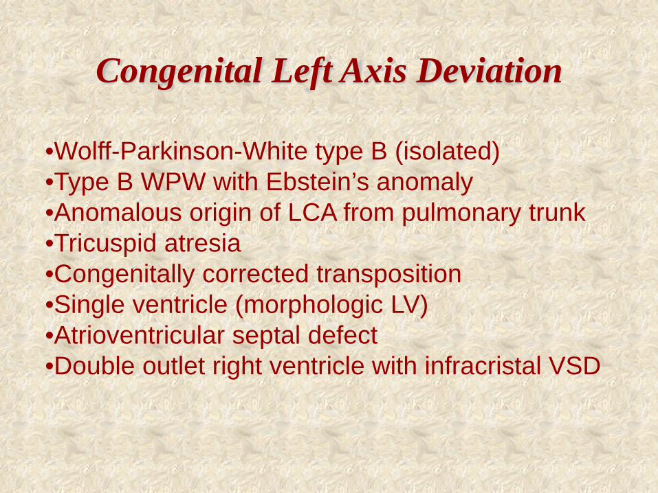
Congenital Left Axis Deviation
•Wolff-Parkinson-White type B (isolated) •Type B WPW with Ebstein’s anomaly •Anomalous origin of LCA from pulmonary trunk •Tricuspid atresia •Congenitally corrected transposition •Single ventricle (morphologic LV) •Atrioventricular septal defect •Double outlet right ventricle with infracristal VSD
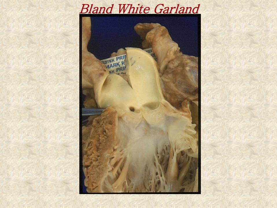
Bland White Garland
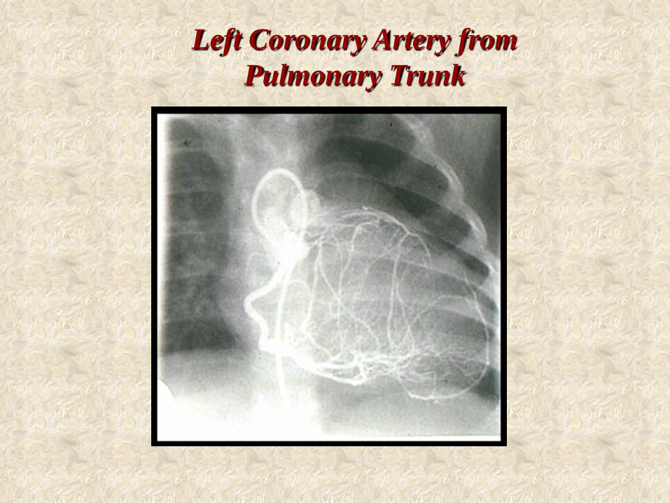
Left Coronary Artery from Pulmonary Trunk
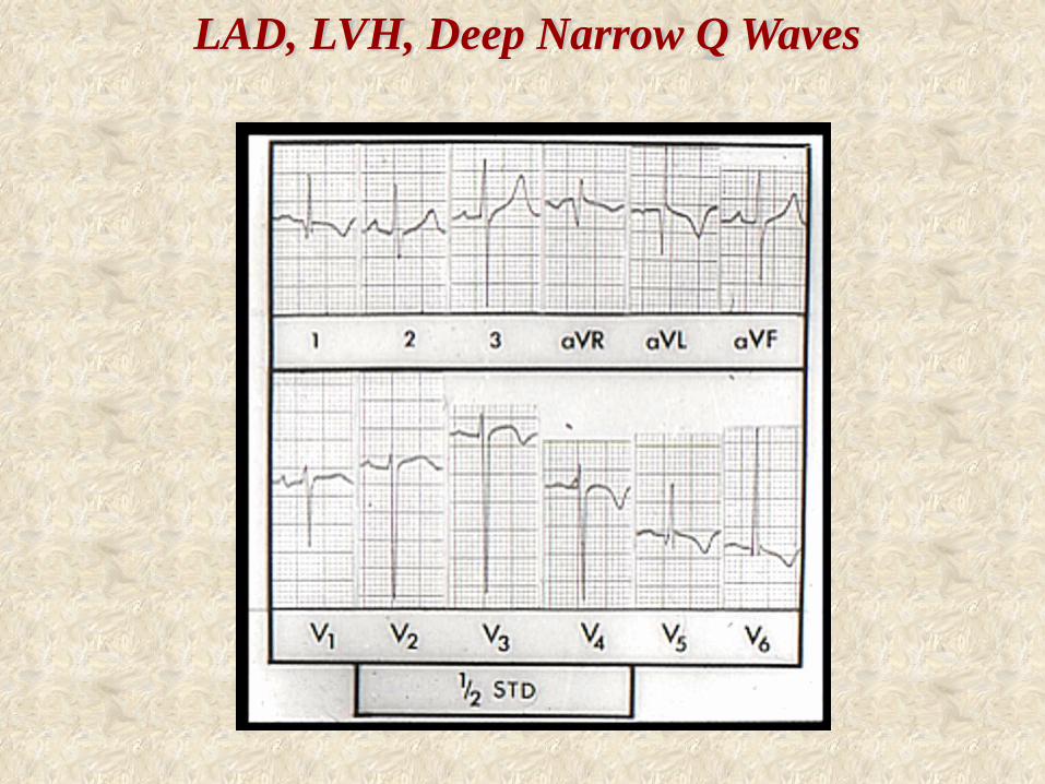
LAD, LVH, Deep Narrow Q Waves
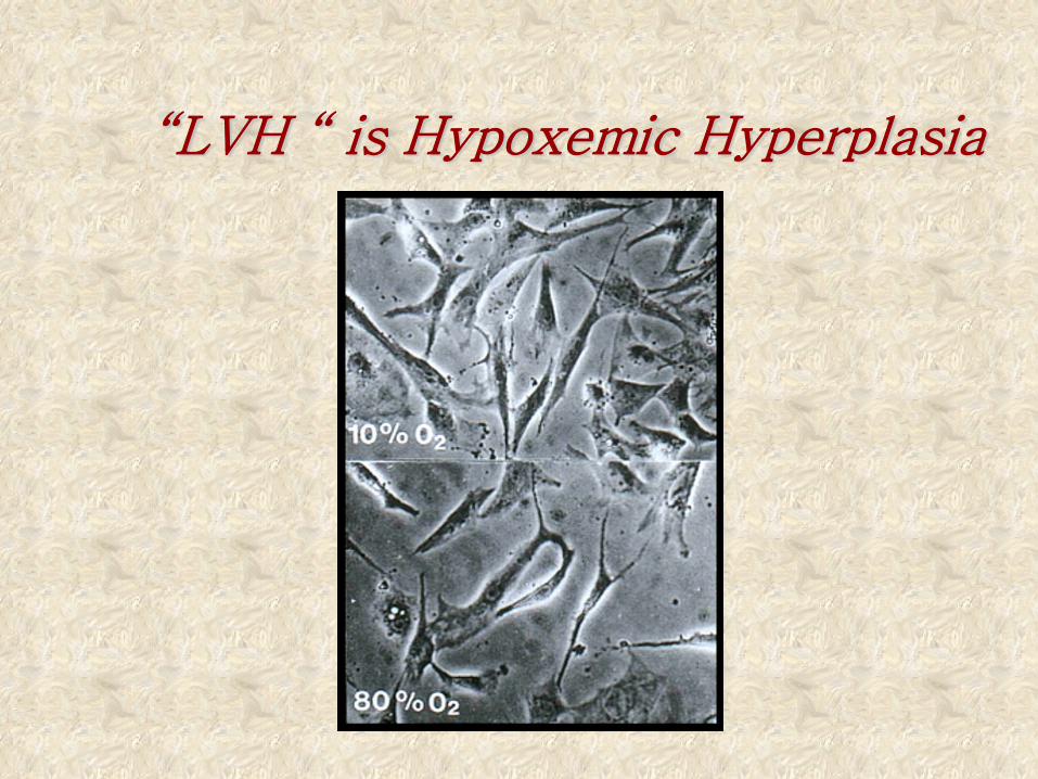
“LVH “ is Hypoxemic Hyperplasia
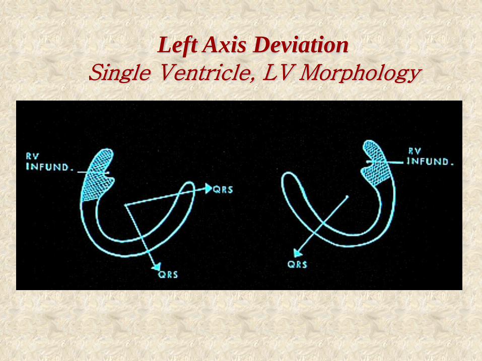
Left Axis Deviation Single Ventricle, LV Morphology
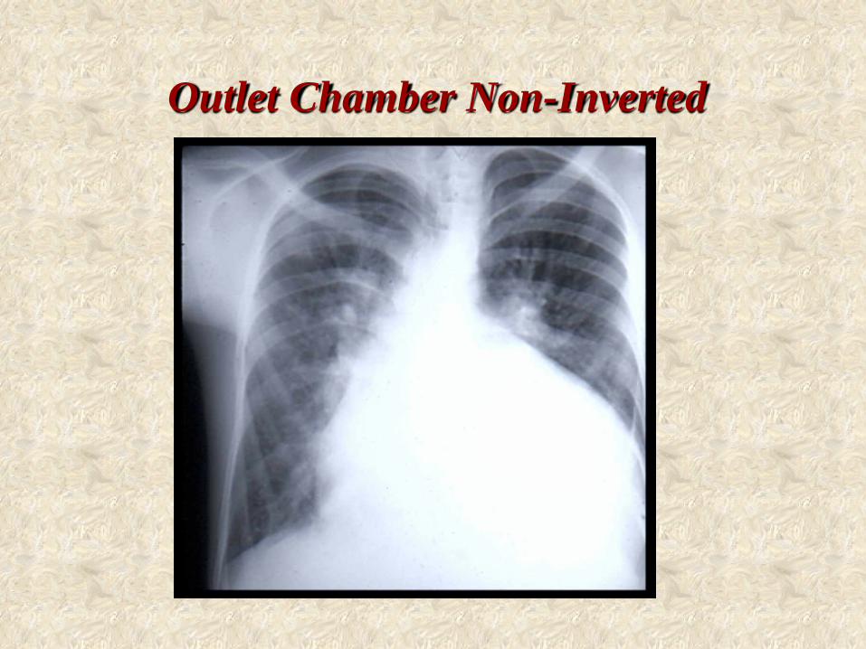
Outlet Chamber Non-Inverted
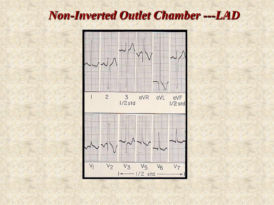
Non-Inverted Outlet Chamber ---LAD
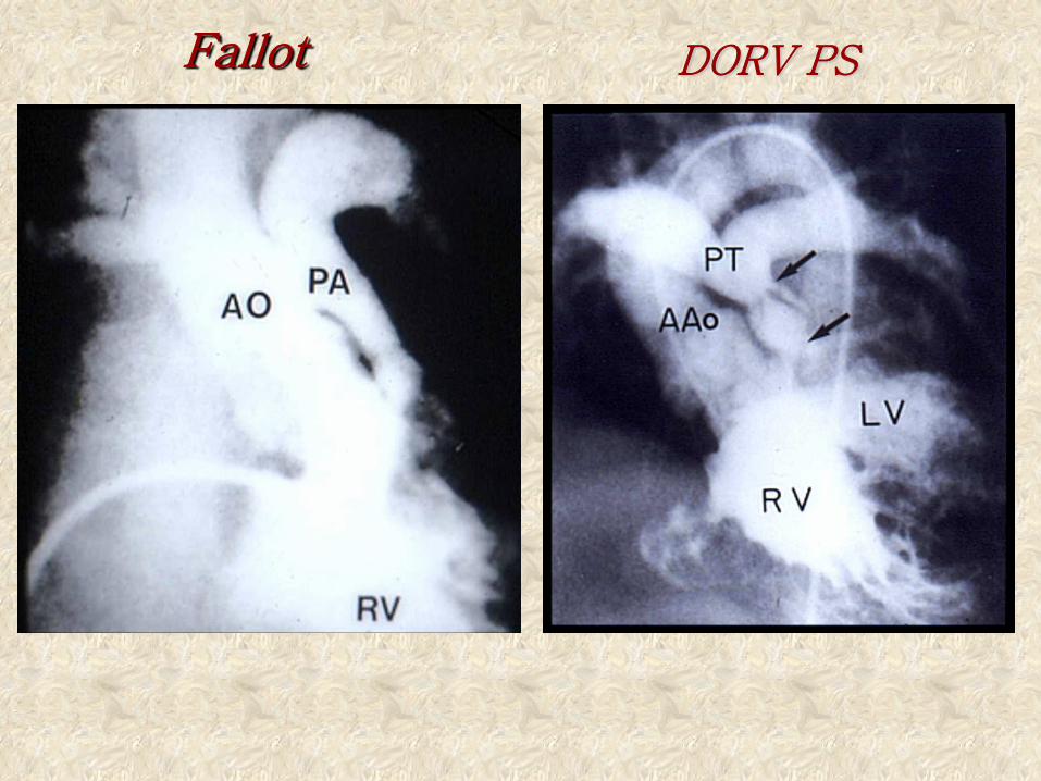
Fallot DORV PS
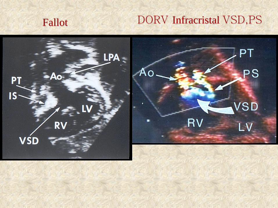
DORV Infracristal VSD,PS Fallot
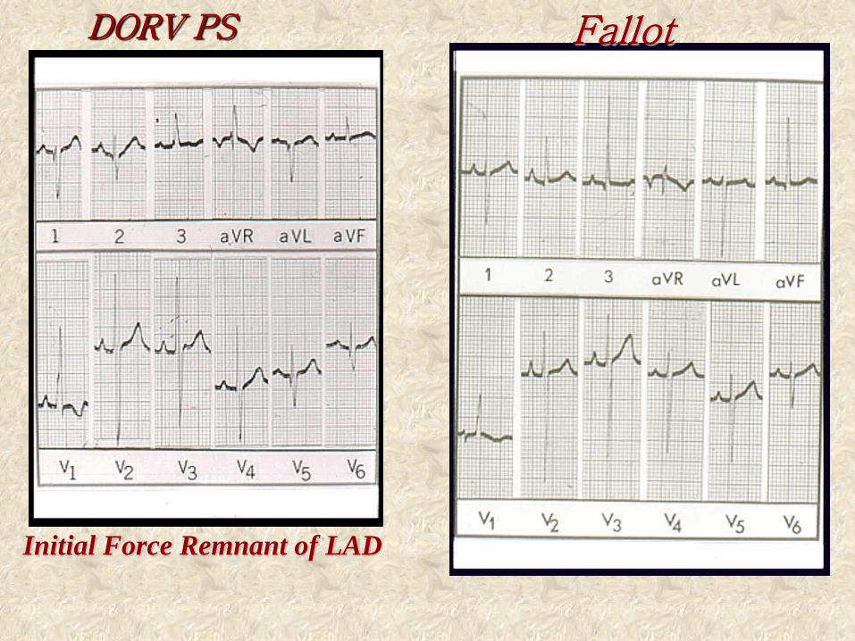
DORV PS Fallot
Initial Force Remnant of LAD

Congenital Deafness with Cardiac Arrhythmias:
The Jervell and Lange-Nielsen Syndrome

A dog’s Life.

Coupled Rhythms

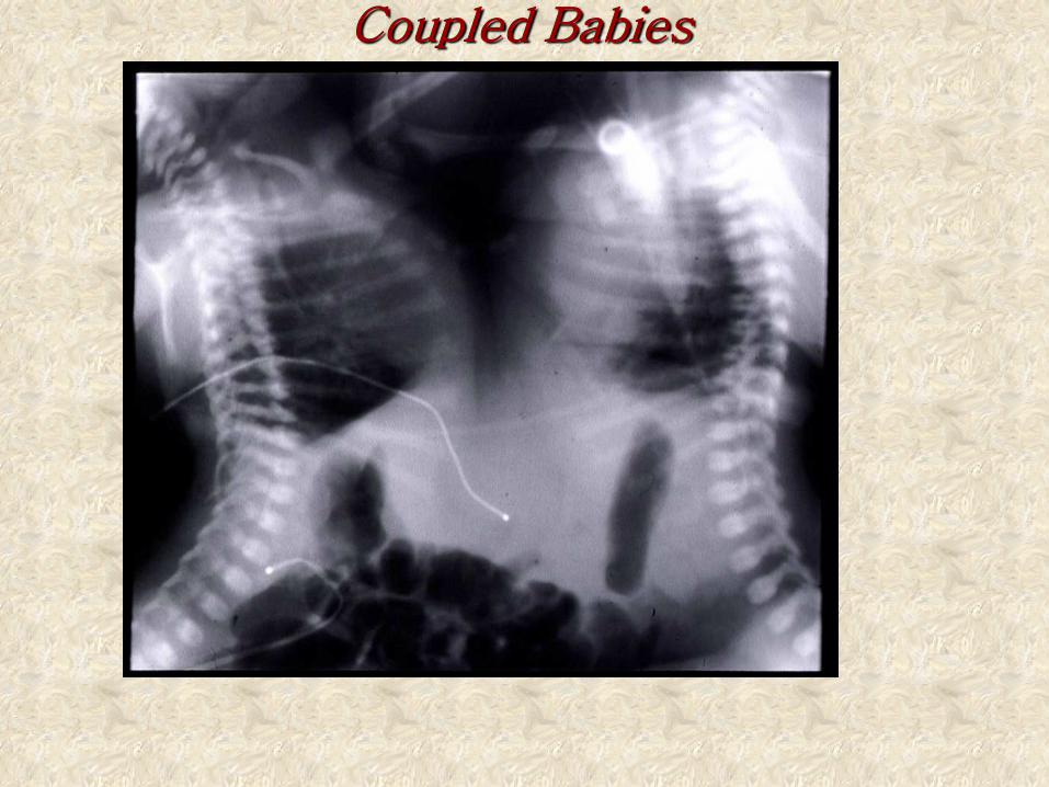
Coupled Babies
A Duet
Played by two hearts beating as one.
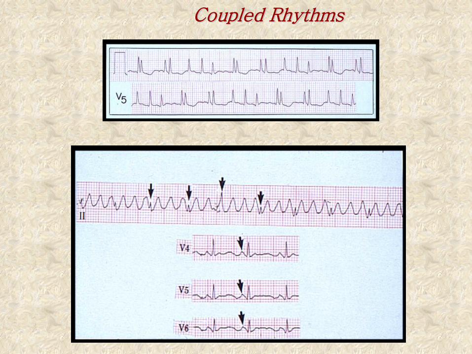
Coupled Rhythms
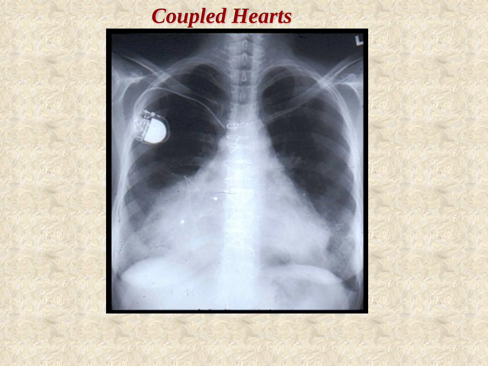
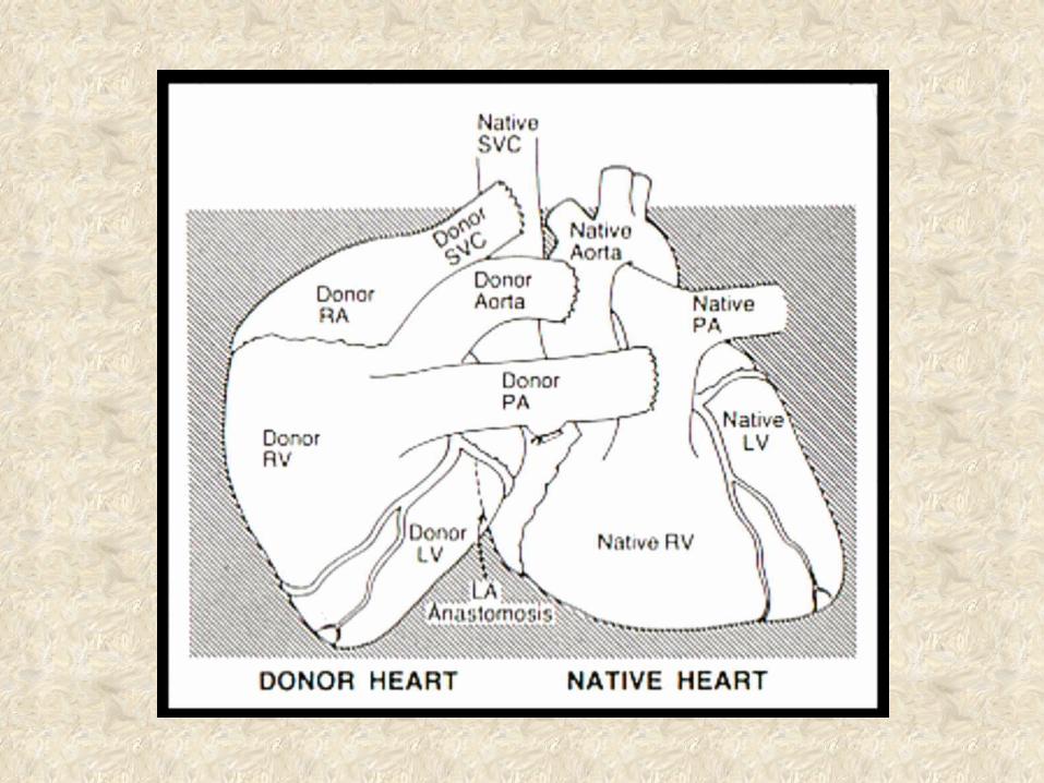
Coupled Hearts
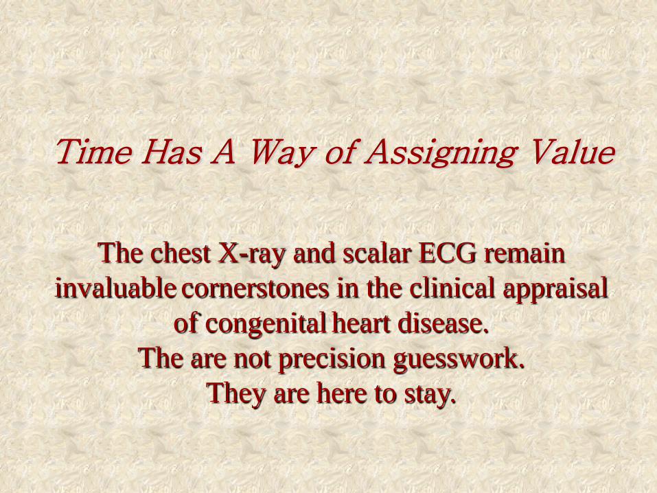
Time Has A Way of Assigning Value
The chest X-ray and scalar ECG remain invaluable cornerstones in the clinical appraisal
of congenital heart disease. The are not precision guesswork.
They are here to stay.
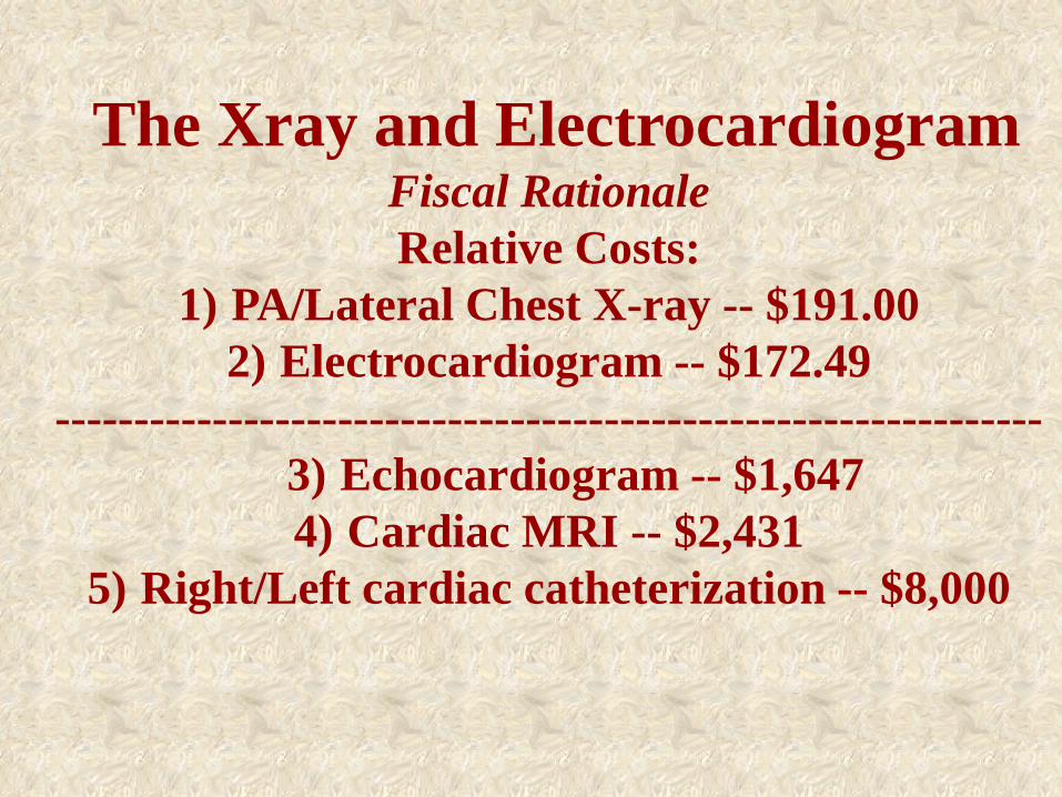
The Xray and Electrocardiogram
Fiscal Rationale Relative Costs:
1) PA/Lateral Chest X-ray -- $191.00 2) Electrocardiogram -- $172.49
---------------------------------------------------------------3) Echocardiogram -- $1,647 4) Cardiac MRI -- $2,431
5) Right/Left cardiac catheterization -- $8,000

Thank You
.


I shall focus on two aspects of this topic:
1) Unusual or atypical arrhythmias.
2) The signal averaged electrocardiogram

Einthoven W. Uber die form des menschlichen neurosurg.
Pflugers Arch 1895

The Electrocardiogram
Many brilliant minds have contributed to the development of electrocardiography as a clinical science. The early history (1900-1945) was dominated by Professor Willem Einthoven in the Netherlands, Sir Thomas Lewis in England and Dr. Frank N. Wilson in the United States. These three pioneers laid the foundation for modern electrocardiography.
Charles Fisch, The ECG Centennial

Sinus arrhythmia in children with atrial septal defect: An analysis of heart rate variability before and after surgical repair
Finley JP, et al. Br Heart J 1989

Sinus arrhythmia in children with atrial septal defect: An analysis of heart rate variability before and after surgical repair
Finley JP, et al. Br Heart J 1989

Secundum ASD Before and After Closure
Congenital Deafness with Cardiac Arrhythmias:
The Jervell and Lange-Nielsen Syndrome

A dog’s Life.
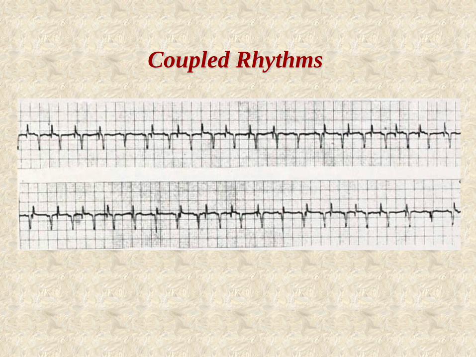
Coupled Rhythms
Coupled Babies
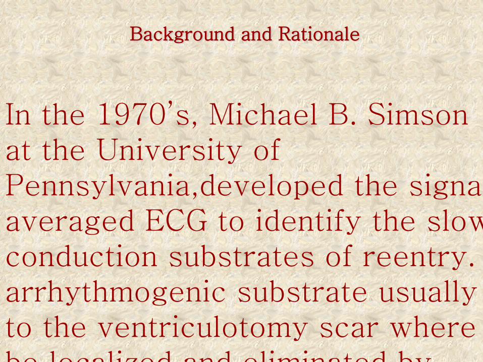
In the 1970’s, Michael B. Simson at the University of Pennsylvania,developed the signa averaged ECG to identify the slow conduction substrates of reentry. arrhythmogenic substrate usually to the ventriculotomy scar where be localized and eliminated by
Background and Rationale
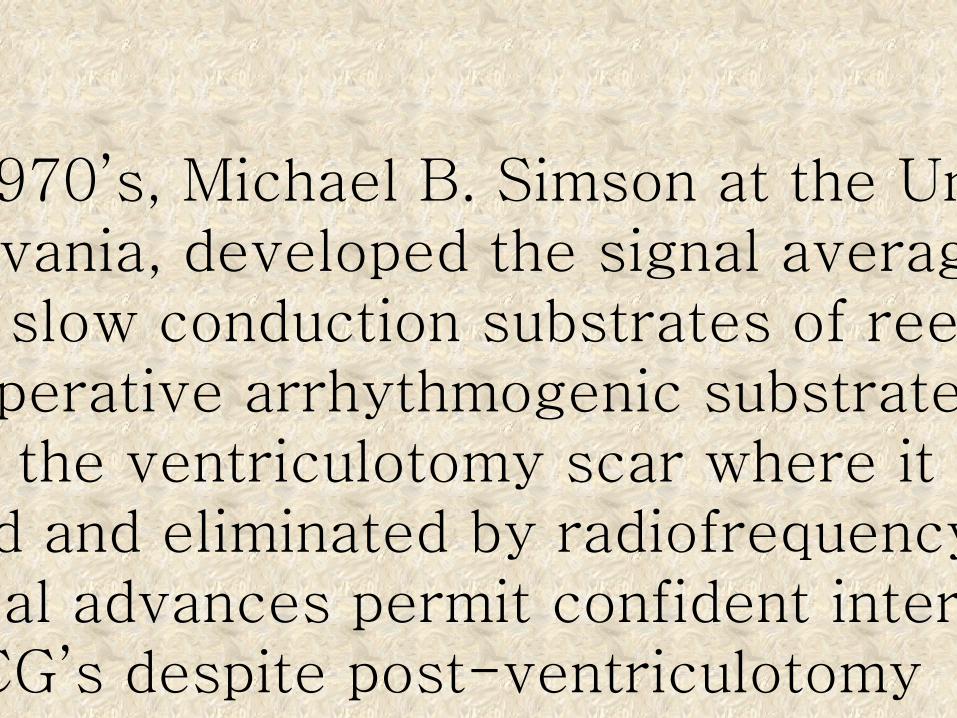
970’s, Michael B. Simson at the Un vania, developed the signal averag slow conduction substrates of ree
perative arrhythmogenic substrate the ventriculotomy scar where it
d and eliminated by radiofrequency al advances permit confident inter
CG’s despite post-ventriculotomy
The Signal Averaged Elecrocardiogram for Detection of Post-ventriculotomy Late
Potentials of Reentrant Monomorphic Ventricular Tachycardia
A Step in the Right Direction ?
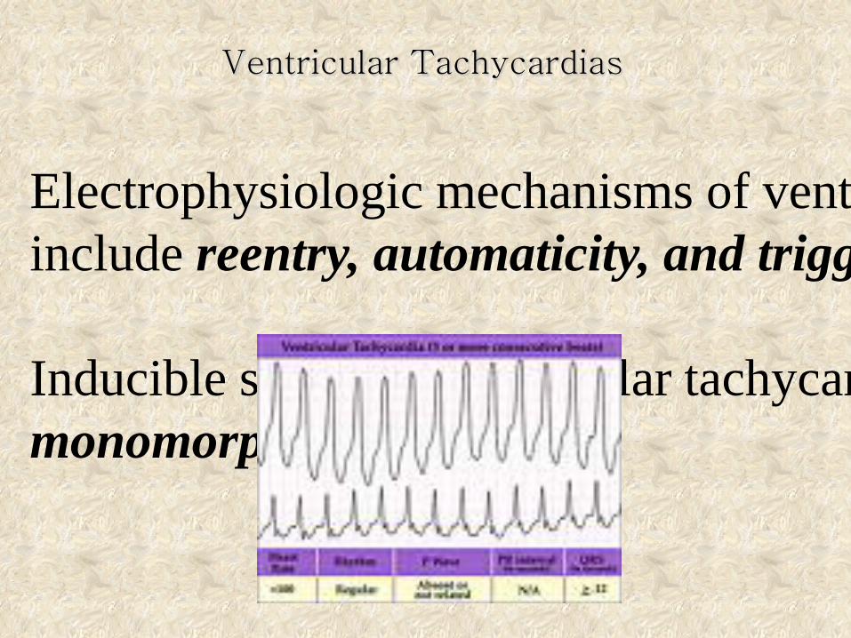
Electrophysiologic mechanisms of vent include reentry, automaticity, and trigg Inducible sustained ventricular tachycar monomorphic.
Ventricular Tachycardias
Basis for the Judgment & Recommendations in this Report
A prospective study that extended from J included 242 consecutive patients in who after---and often before and after---right repair of CHD.
Perloff JK, Middlekauf HR, Stevenson WG, et al. Usefulness of Post-ventriculotomy Signal Averaged Elecrocardiograms in Congenital Heart Diseae. Am J Cardiol 2006;98:1646-1651

Coupled Babies
A positive SAECG is defined as a fil duration >145 msec plus root mean squa of the terminal 40 msec of the filtered Q microvolts, and/or low amplitude signal terminal filtered QRS > 50 msec. A positive SAECG indicates the presenc
Definition & Implications
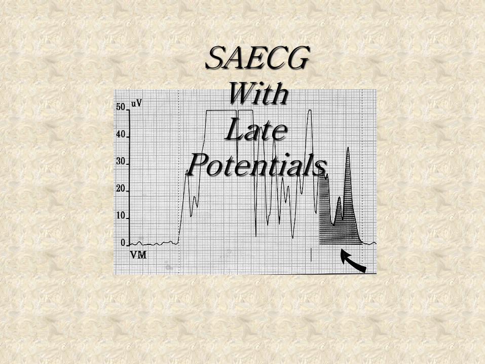
SAECG With Late
Potentials

The Trigger A slow conduction arrhythmogenic reentrant substrate remains dormant unless activated (triggered). Accordingly, the overt expression of reentrant MVT requires a susceptible substrate and an effective trigger. Severe pulmonary regurgitation is such a trigger.
These factors include scalar QRS durati increase in QRS duration ≥ 30 msec over pulmonary regurgitation, depressed right function, ventricular ectopic beats induce ≥3 consecutive monomorphic ventricular ≥age at ventriculotomy, and a decade or Importantly, patients with QRS duratio SAECG’s, and patients with QRS duratio negative SAECG’s, so the QRS duration
Established Risk Factors for MVT
Intracardiac Electrophysiology
When sustained MVT is inducible in SAECG’s, the commonest site of the slo substrate is along the ventriculotomy sc
Substrates can be localized by mappi by radiofrequency ablation.
The Best Results