the cd3 conformational change in the γδ t-cell receptor is not
TRANSCRIPT

Cell Reports, Volume 7
Supplemental Information
The CD3 Conformational Change in the T Cell
Receptor Is Not Triggered by Antigens, but Can
Be Enforced to Enhance Tumor Killing
Elaine P. Dopfer, Frederike A. Hartl, Hans-Heinrich Oberg, Gabrielle M. Siegers,
Sascha Yousefi, Sylvia Kock, Gina J. Fiala, Beatriz Garcillán, Andrew
Sandstrom, Balbino Alarcón, Jose R. Regueiro, Dieter Kabelitz, Erin J. Adams,
Susana Minguet, Daniela Wesch, Paul Fisch, and Wolfgang W.A. Schamel

1
The CD3 conformational change in the γδ T-cell receptor is not triggered by antigens, but can be enforced to enhance tumor killing
Supplemental Figures
Figure S1, related to Figure 1. CD3 CC induction in murine γδTCRs.
(A) The murine γδ T-cell hybridoma F30L31 was stimulated for 5 min at 37°C with 5 µg/ml of the anti-
TCRγδ mAb GL3. After lysis, one part of the lysates was incubated with SH3-beads and another part
with protein A- and protein G-beads. Lysates and bead-purifed proteins were analyzed by anti-ζ WB.
This experiment shows that the murine γδTCR can undergo the CD3 CC upon anti-TCR γδ mAb
stimulation.
(B) G8 γδ T cells were stimulated with 1, 5, 10 and 20 µg/ml T22t or 5 µg/ml anti-TCRγδ mAb (GL3) for 5
min, and the SH3-PD assay was performed. This titration of the T22 tetramer suggested that tetramer
binding to the G8 γδTCR significantly reduced the CD3 CC at different ligand concentrations, as
compared to the basal level.
(C) G8 γδ hybridoma cells were incubated with 5 µg/ml T22 tetramers and T1.4 αβ hybridoma cells with
5 µg/ml H2-Kd-pepABA tetramers. Both tetramers were prepared from the same stock of PE-labelled
streptavidin, thus the PE fluorescence measured by flow cytometry can be compared. Unstained cells
served as a negative control. This experiment shows that binding of the G8 γδ hybridoma cells to T22
tetramers is comparable to that of T1.4 αβ hybridoma cells binding to pMHC tetramers.
(D) The G8 γδ hybridoma was kept unstimulated or stimulated for 1 hr at 37°C with LPS-activated
splenic B cells from a CD3ε-deficient mouse or with 2C11. The SH3-PD was performed as before. The
significance between unstimulated and APC-stimulated cells was determined by the Student’s t-test; ns:
non significant. This experiment shows that stimulation of the G8 γδTCR by its natural ligand T22 did not
induce the CD3 CC. We detected a small reduction in the basal CD3 CC suggesting again that T22 had
a small impact on the γδTCR conformation.

2
Figure S2, related to Figure 2. Expression of chimeric γδ-αβTCRs.
(A) Schematic of the murine wt and chimeric TCRs used in figure 2. The domains of the G8 γδTCR are
depicted in beige, including the T22-binding CDR3δ loop derived from the G8 γδTCR; the αβTCR
domains are in blue. The amino acid sequences are shown below each TCR in the same colour code as
in the pictures. The CDR3δVCαβTCR was published before (Adams et al., 2008).
58α-β- T cells were lentivirally infected with vectors encoding for the wt or chimeric TCRs. A flow
cytometric analysis using the anti-CD3 mAb 2C11 demonstrated that all receptors except the VγδCαβ
TCR were expressed on the cell surface. Thus, the 58.VγδCαβ cells were excluded from any further
analysis.
(B) The lentivirally infected 58α-β- T cells were stained with 5 µg/ml PE-labelled T22 tetramers or H2-Kd-
pepABA (pMHC) tetramers and analysed by flow cytometry (red lines). Unstained cells served as a
negative control (black line). This Experiment shows that the wt and chimeric TCRs bind to their
respective tetrameric antigens.
(C) The experiment as in (B) was repeated 13 times. The mean fluorescence intensity (MFI) normalized
to the unstained cells ± standard eroor of the mean (SEM) is shown. The higher binding capacity of the
58.VγδCγδ and 58.CDR3δVCαβ cells compared to the 58.VαβCαβ and 58.VαβCγδ cells, could be due to
the higher affinity of the T22-G8 γδTCR interaction.

3
Figure S3, related to Figure 3. Induction of the CD3 CC at the human γδTCR. (A) Jurkat (αβTCR-expressing) and Jk.Vγ9Vδ2 (γδTCR-expressing) cells were left untreated (-) or
stimulated for 5 min at 37°C with 5 µg/ml of the anti-CD3 mAbs UCHT1 and OKT3 or pervanadate (PV).
After lysis the SH3-PD assay was performed (n=3).
(B) Jk.Vγ9Vδ2 cells were left untreated or stimulated for 5 min at 37°C with 5 µg/ml of anti-TCRγδ mAbs.
After lysis the SH3-PD was performed (n=2).
(C) Jk.Vγ9Vδ2 cells were stimulated in triplicate as in main figure 3E. The ratio of SH3-bound TCR to
total TCR (ζ) was calculated using the Odyssey infrared imager. The mean ± standard deviation (SD) is
shown. Significance between unstimulated and cells stimulated with Daudi+ZOL was determined by the
Student’s t-test; n.s. = not significant.
These experiments demonstrate that UCHT1 and anti-TCRγδ mAbs, but neither OKT3 nor ZOL-pulsed
Daudi cells, trigger the CD3 CC in a γδTCR independent of the cellular environment.

4
Figure S4, related to Figure 4. Induction of the CD3 CC in γδTCRs promotes proximal signalling. (A) Jurkat cells were loaded with Indo-1 and stimulated with 5 µg/ml of the anti-CD3 mAbs UCHT1 and
OKT3. The Indo-1 ratio was integrated over 6 min and measured by flow cytometry, showing that both
mAbs induce Ca2+-influx in αβ T cells. This is in line with the capacity of both mAbs to induce the CD3
CC in the αβTCR.
(B) Human freshly isolated, untouched purified γδ T cells (γδPBMCs) were stimulated with 5 µg/ml
UCHT1 and OKT3 for 1, 3, 5 and 15 min. Phospho-Akt (solid line, y-axis on left) and phospho-Erk
(dashed line, y-axis on right) were measured by a multiplexed-bead assay performed on cell lysates.
(C) The Vγ9Vδ2 T-cell clone was stimulated in triplicates for 4 hr with 5 µg/ml soluble UCHT1 or OKT3.
Subsequently, cells were stained with anti-TCRγδ (5A6E9) mAb for flow cytometry, Significances
between UCHT1- and OKT3-stimulated cells were determined by the Student’s t-test; ***: p<0.001. This
experiment shows that UCHT1 induces stronger γδTCR downmodulation than OKT3.
(D) Jk.Vγ9Vδ2 cells were left untreated or degylcosylated with N-acetyl neuraminidase (NA) for 1 hr at
37°C (left). Subsequently, cells were stimulated with 5 µg/ml UCHT1 or OKT3 for 5 min. Jk.Vγ9Vδ2 T
cells were stimulated with 5 µg/ml UCHT1 or OKT3 in the presence of an anti-IgG antibody (2.5 µg/ml,
right). After cell lysis the SH3-PD was performed. Thus, a γδTCR expressed in an αβ T cell undergoes
the CD3 CC upon OKT3 stimulation when deglycosylated. In addition, multimerization of OKT3
promotes CD3 CC induction.
(E) The Vγ9Vδ2 γδ T clone (left panel) or human freshly isolated, untouched purified γδ T cells
(γδPBMCs, right panel) were loaded with Indo-1 and stimulated with 5 µg/ml UCHT1 or OKT3 with or
without 2.5 µg/ml anti-IgG and Ca2+ influx was measured as in (A). This result demonstrates that
supplementing OKT3 stimulation with the CD3 CC by multimerizing with an anti-IgG antibody leads to
Ca2+-influx.
The experiments shown in all panels were done at least three times (n=3).

5
Figure S5, related to Figure 6. TNFα production by a γδ and αβ clone stimulated with UCHT1 and
OKT3. (A) The murine γδ T-cell line F30L31 exogenously expressing CD3εwt, CD3εK76T or CD3εC80G was
stained with an APC-labelled anti-CD3 mAb 145-2C11 and analysed by flow cytometry. The histogram
shows that all three lines express similar levels of γδTCR.
(B) The human Vγ9Vδ2 γδ T-cell clone was left untreated (-) or stimulated with UCHT1 or OKT3 using 5
µg/ml or 1 µg/ml for 20 hours as indicated. The cellular supernatants were used to measure TNFα by a
multiplexed bead assay.
(C) The human αβ T-cell clone was stimulated as in (A). TNFα in the cellular supernatant was measured
as in (A). Together, these data show that the difference in the activity of UCHT1 and OKT3 in γδ T cells
(Figure 5D) is not detected in αβ T cells. Significances between UCHT1- and OKT3-stimulated cells in
(B) and (C) were determined by the Student’s t-test; ***: p<0.001; *: p<0.05; ns: non significant.
(D) A primary γδ T-cell culture was labelled with 1 µM Cell Trace Violet and left unstimulated or
stimulated with 5 µg/ml soluble UCHT1 or OKT3 as indicated. After 4 days proliferation was determined
by flow cytometry. Quantification of the mean fluorescence intensity (MFI) of triplicates is given (n=3).
Mean ± SD of triplicates is shown. Significances between unstimulated and UCHT-stimulated cells as
well as between UCHT1- and OKT3-stimulated cells were determined by the Student’s t-test; ns: not
significant; **: p<0.01; ***: p<0.001.
6
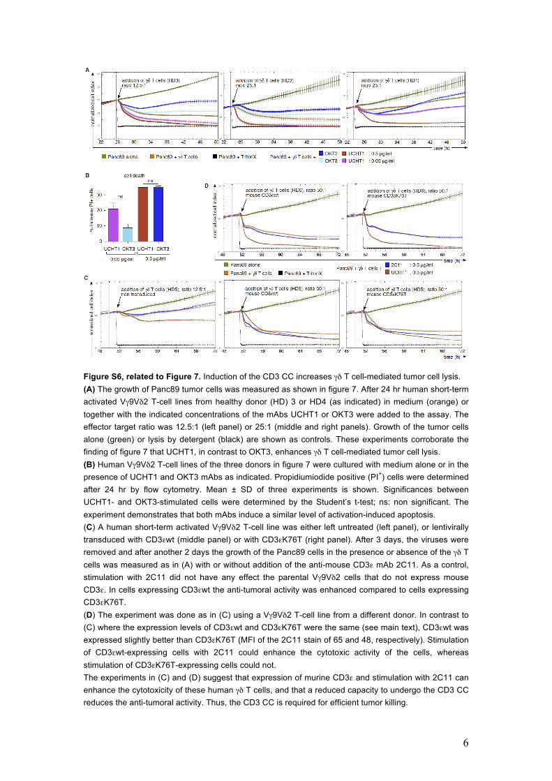
Figure S6, related to Figure 7. Induction of the CD3 CC increases γδ T cell-mediated tumor cell lysis. (A) The growth of Panc89 tumor cells was measured as shown in figure 7. After 24 hr human short-term activated Vγ9Vδ2 T-cell lines from healthy donor (HD) 3 or HD4 (as indicated) in medium (orange) or together with the indicated concentrations of the mAbs UCHT1 or OKT3 were added to the assay. The effector target ratio was 12.5:1 (left panel) or 25:1 (middle and right panels). Growth of the tumor cells alone (green) or lysis by detergent (black) are shown as controls. These experiments corroborate the finding of figure 7 that UCHT1, in contrast to OKT3, enhances γδ T cell-mediated tumor cell lysis. (B) Human Vγ9Vδ2 T-cell lines of the three donors in figure 7 were cultured with medium alone or in the presence of UCHT1 and OKT3 mAbs as indicated. Propidiumiodide positive (PI+) cells were determined after 24 hr by flow cytometry. Mean ± SD of three experiments is shown. Significances between UCHT1- and OKT3-stimulated cells were determined by the Student’s t-test; ns: non significant. The experiment demonstrates that both mAbs induce a similar level of activation-induced apoptosis. (C) A human short-term activated Vγ9Vδ2 T-cell line was either left untreated (left panel), or lentivirally transduced with CD3εwt (middle panel) or with CD3εK76T (right panel). After 3 days, the viruses were removed and after another 2 days the growth of the Panc89 cells in the presence or absence of the γδ T cells was measured as in (A) with or without addition of the anti-mouse CD3ε mAb 2C11. As a control, stimulation with 2C11 did not have any effect the parental Vγ9Vδ2 cells that do not express mouse CD3ε. In cells expressing CD3εwt the anti-tumoral activity was enhanced compared to cells expressing CD3εK76T. (D) The experiment was done as in (C) using a Vγ9Vδ2 T-cell line from a different donor. In contrast to (C) where the expression levels of CD3εwt and CD3εK76T were the same (see main text), CD3εwt was expressed slightly better than CD3εK76T (MFI of the 2C11 stain of 65 and 48, respectively). Stimulation of CD3εwt-expressing cells with 2C11 could enhance the cytotoxic activity of the cells, whereas stimulation of CD3εK76T-expressing cells could not. The experiments in (C) and (D) suggest that expression of murine CD3ε and stimulation with 2C11 can enhance the cytotoxicity of these human γδ T cells, and that a reduced capacity to undergo the CD3 CC reduces the anti-tumoral activity. Thus, the CD3 CC is required for efficient tumor killing.

7
Figure S7, related to Figures 4, 6 and 7. Influence of the CD3 CC on γδTCR signaling. (A) Activation of the αβTCR requires CD3 CC induction (red CD3 tails) by antibody/antigen (orange).
Without induction of the CD3 CC by artificial ligands or in a mutated TCR the αβTCR is not activated
(blue CD3 tails). In contrast, several activation events mediated by the three γδTCRs studied here did
not require the CD3 CC and the γδ T-cell antigens tested (yellow) did not induce this change. However,
artificial induction of the CD3 CC did enhance tumor killing by γδ T cells. (B) The mAb UCHT1 triggers
the CD3 CC in the human Vγ9Vδ2 TCR, whereas OKT3 does not. Stimulation of the γδTCR with
UCHT1 results in strong proximal signaling and TCR downmodulation, upregulation of the activation
markers CD25 and CD69, weak cytokine secretion and massive tumor killing. In contrast, stimulation
with OKT3 hardly induces proximal signaling and TCR downmodulation, upregulates CD25, CD69 and
cytokines, promotes proliferation but inhibits tumor killing by the Vγ9Vδ2 T cells. These findings were
confirmed using mutant CD3ε chains that cannot undergo the CD3 CC.

8
Supplemental Experimental Procedures
Cells and mice. The human Vγ9Vδ2 T-cell clone (γδPF55) was generated and maintained as described (Fisch et al., 1990; Fisch et al., 1997). JK.Vγ9Vδ2 T cells were generated from TCRαβ-/- Jurkat T cells transfected with TCR Vγ9 and TCR Vδ2 (Alibaud et al., 2001). The murine γδ T-cell hybridomas G8 and F30L31 were obtained from Y.H. Chien, Stanford, USA, and K. Eichmann, Freiburg, Germany, respectively, and the murine αβ T-cell hybridoma 2B4 from J. Ashwell, Bethesda. The pancreatic ductal adenocarcinoma cell line Panc89 was provided by H. Kalthoff, Section of Molecular Oncology, UKSK, Campus Kiel, Germany. All cells were maintained in RPMI 1640 medium supplemented with 10% fetal bovine serum. TCRβ-/-Vγ1.1Vδ6 transgenic mice have been previously described (Siegers et al., 2007). The CD3ε knock out mouse was described before (DeJarnette et al., 1998). Mice were killed between 6 and 12 weeks of age and lymphocytes were isolated from the indicated tissues according to standard protocols. Human γδ peripheral blood mononuclear cells (γδPBMCs) were isolated from a healthy donor using a Ficoll-Hypaque gradient followed by negative isolation using the human TCRγ/δ+ untouched T-Cell Isolation Kit human (Miltenyi Biotech) to enrich the γδ T cells. Mouse experiments were approved by the relevant institutional review boards (code number: T-02/31). Informed consent was obtained from all human blood donors, and the research was approved by the relevant institutional review boards (code number: D 405/10). Short-time cultured human γδ T-cell lines were used in the tumor cell killing assays. To this end, PBMCs from healthy donors were cultured in RPMI 1640 supplemented with 10 % fetal calf serum, and then stimulated with 5 µmol/l of aminobisphosphonate zoledronic acid (Novartis) and 50 U/ml rIL-2 (Novartis). Additionally, rIL-2 was added every two days over a culture period of 20 days. The purity of expanded γδ T cells was analyzed by flow cytometry and was > 97% Vγ9Vδ2 T cells. Antibodies, antigens and reagents. The rabbit anti-ζ antiserum 448 was described (San Jose et al., 1998). UCHT1 (anti-hCD3ε) was provided by P. Beverly, London, U.K. The following reagents were purchased: OKT3 hybridome (anti-hCD3ε, American Type Culture Collection), GL3-PE (anti-mTCRγδ, eBiosciences), 5A6.E9-PE (anti-hTCRγδ, Invitrogen), 145-2C11 (anti-mCD3ε, eBioscience), anti-hCD69-APC (Invitrogen), anti-hCD25-PE, goat anti-rabbit IgG-HRPO (both, Pierce), and anti-κ and anti-IgG (Southern Biotech). Zoledronate was supplied as hydrated disodium salt by Novartis Pharma. BrHPP was kindly provided by Innate Pharma. T22 monomers were expressed and purified as previously described (Adams et al., 2008). H2-Kd-pepABA (pMHC) monomers were obtained from TCMetrix. PE-coupled streptavidin (Life Technologies) was used to generate the T22 and pMHC tetramers.

9
Cloning and expression of the chimeric TCRs. The CDR3δVCαβTCR has been published (Adams et al., 2008). Here we inserted before the start codon of CDR3δ172α or 172β with a fusion-PCR a XhoI restriction site and a mouse TCRδ leader peptide sequence. A stop codon was inserted at the original place followed by a XbaI restriction site. The resulting PCR products were cloned into pLXV-IRES-ZsGreen1 (Clontech Lab). TCRα-β- cells were transduced with virus containing CDR3δ172α or 172β plasmids and plated to obtain single clones. Transduced cells were tested for TCR expression by anti-CD3 (2C11) flow cytometry. The CMV promoter of the lentiviral vector pLVX-IRES-ZsGreen1 (Clontech Lab) was exchanged by the SFFV promoter from the pHRSIN CSGW plasmid. Furthermore, the ZsGreen1 cDNA was exchanged by either the cDNA of the puromycin N-acetyl-transferase (from pIRESpuro3, Clontech Lab) or of the hygromycin B phosphotransferase (pLHCX, Clontech Lab). Thus, three lentiviral backbones were obtained: pLVX-S IZ, pLVX-S IP and pLVX-S IH (pLVX with the SFFV promoter and either IRES-ZsGreen1, IRES-puromycin resistance or IRES-hygromycin B resistance, respectively). For the VαβCαβ TCR, the sequences encoding the TCRα and TCRβ chains of the T1 αβTCR were amplified from cDNA of the T1.4 hybridoma cells. The TCRα sequence was cloned into pLVX-S IP and the TCRβ sequence into pLVX-S IH. The cDNAs of the VγδCγδ TCR (G8 TCRγ and G8 TCRδ sequences) were cloned into the same vectors. The chimeric VαβCγδ and VγδCαβ TCRs were generated by joining the T1 TCRα and G8 TCRδ sequences, as well as the T1 TCRβ and G8 TCRγ sequences at the V-C borders as defined by the respective exons (see also Figure S2A). For each chimeric TCR, one sequence was cloned into pLVX-S IP and the other one into pLVX-S IH. All constructs were verified by sequencing. 58α-β- cells were transduced with lentivirus supernatants encoding for both chains and co-transduced cells were selected with puromycin (PAA) and hygromycin B (Invitrogen). Transduced cells were tested for TCR expression by anti-CD3 (2C11) flow cytometry. Cloning and expression of the mutant CD3ε molecules. Murine CD3ε wild-type as well as the CD3εK76T mutant sequences were amplified from pHRSIN CSGW plasmids and cloned into pLVX-S IZ. The CD3εC80G mutant sequence was derived from the wt CD3ε sequence by site-directed mutagenesis. All constructs were verified by sequencing. F30L31 and human γδ T cells were transduced with lentivirus encoding for the wt or mutant CD3ε chains. Proliferation assay. γδ T-cell cultures derived from healthy donor PBMC were generated as described (Siegers et al., 2012). On day 9, γδ T cells were labeled with 1 µM Cell Trace Violet (CTV, Invitrogen, Burlington, Canada) and resuspended at 106 cells/ml in complete medium: RPMI 1640 with 10% fetal bovine serum, 1x MEM NEAA, 10 mM HEPES, 1 mM

10
sodium pyruvate (all Gibco/Life Technol) containing 10 ng/ml recombinant IL-2 (rIL-2, Proleukin, Novartis Pharmaceuticals, Canada) and 10 ng/ml IL-4 (Life Technol). Cells were untreated, or stimulated with 5 µg/ml anti-human CD3 mAb UCHT1 or OKT3 (Biolegend) and plated at 200 µl/well in a 96-well round bottom plate (BD Biosciences). Apoptosis assay. 250.000 human freshly isolated, purifed γδ T cells were cultured with medium alone or with the indicated stimuli for 24 hr. Propidium iodide positive (PI+) cells were determined in all cultures after 24 hr by flow cytometry using a FACSCalibur. Results are presented as enhancement of OKT3- or UCHT-1 mAb mediated cell death beyond the medium control.
Supplemental References
Adams, E.J., Strop, P., Shin, S., Chien, Y.H., and Garcia, K.C. (2008). An autonomous CDR3δ is sufficient for recognition of the nonclassical MHC class I molecules T10 and T22 by γδ T cells. Nat Immunol 9, 777-784. Alibaud, L., Arnaud, J., Llobera, R., and Rubin, B. (2001). On the role of CD3δ chains in TCRγδ/CD3 complexes during assembly and membrane expression. Scand J Immunol 54, 155-162. DeJarnette, J.B., Sommers, C.L., Huang, K., Woodside, K.J., Emmons, R., Katz, K., Shores, E.W., and Love, P.E. (1998). Specific requirement for CD3epsilon in T cell development. Proc Natl Acad Sci U S A 95, 14909-14914. Fisch, P., Malkovsky, M., Kovats, S., Sturm, E., Braakman, E., Klein, B.S., Voss, S.D., Morrissey, L.W., DeMars, R., Welch, W.J., et al. (1990). Recognition by human Vγ9/Vδ2 T cells of a GroEL homolog on Daudi Burkitt's lymphoma cells. Science 250, 1269-1273. Fisch, P., Meuer, E., Pende, D., Rothenfusser, S., Viale, O., Kock, S., Ferrone, S., Fradelizi, D., Klein, G., Moretta, L., et al. (1997). Control of B cell lymphoma recognition via natural killer inhibitory receptors implies a role for human Vγ9/Vδ2 T cells in tumor immunity. Eur J Immunol 27, 3368-3379. San Jose, E., Sahuquillo, A.G., Bragado, R., and Alarcon, B. (1998). Assembly of the TCR/CD3 complex: CD3 epsilon/delta and CD3 epsilon/gamma dimers associate indistinctly with both TCR alpha and TCR beta chains. Evidence for a double TCR heterodimer model. Eur J Immunol 28, 12-21. Siegers, G.M., Ribot, E.J., Keating, A., and Foster, P.J. (2012). Extensive expansion of primary human gamma delta T cells generates cytotoxic effector memory cells that can be labeled with Feraheme for cellular MRI. Cancer Immunol Immunother 62, 571-583. Siegers, G.M., Swamy, M., Fernandez-Malave, E., Minguet, S., Rathmann, S., Guardo, A.C., Perez-Flores, V., Regueiro, J.R., Alarcon, B., Fisch, P., et al. (2007). Different composition of the human and the mouse γδ T cell receptor explains different phenotypes of CD3γ and CD3δ immunodeficiencies. J Exp Med 204, 2537-2544.