the capillary network of normal and emphysematous human lungs
TRANSCRIPT
Thorax (1963), 18, 201
The capillary network of normal and emphysematoushuman lungs studied by injections of Indian ink
J. ALEXANDER REID1 AND BRIAN E. HEARD
From the Postgraduate Medical School ofLondon, Du Cane Road, London, W. 12
In the past few years there has been renewedinterest in the pathology of pulmonary emphy-sema, and the need has arisen to demonstratethe patterns of pulmonary capillaries more clearly.The present paper describes an injection methoddeveloped for such purposes using a mixture ofblack waterproof ink and gelatin. Some prelimi-nary findings in normal and emphysematous lungsare described.
METHOD
The lung was removed from the chest with care toavoid perforating the pleura. Where there were denseadhesions, extrapleural dissection was carried out.Fresh material was used wherever possible. Bloodclots were massaged from the large pulmonary vessels,and a selected cannula of suitable size, made bymounting a grooved rubber bung broad and distallyon glass tubing, was tied into the pulmonary artery.Two litres of normal saline was run through thearteries slowly to wash out some of the blood. Mucuswas sucked from the bronchi with a narrow glasscannula attached to a filter pump, and the lungs weredistended once with air via the bronchi and floated onwarm water.
Early trials with dilute mixtures of Raybar cream(50% barium sulphate) and gelatin failed to fill thecapillary bed. Since radio-opacity was not desired,barium sulphate was replaced by ink as follows:
Pelican black waterproof drawing ink 400 ml.Distilled water 100 ml.Gelatin 45 g.Thymol 0 5 g.
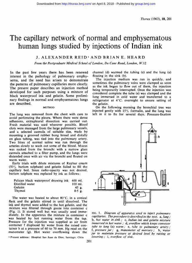
The water was heated to about 80°C. in a conicalflask and the gelatin stirred in until dissolved. Theink and thymol were added to the hot gelatin, and themixture was filtered through gauze into container c(Fig. 1). It stored well but was usually used imme-diately. In the apparatus the mixture in container cwas heated by hot running water from the tap.Pressure for the injection was supplied as air fromcontainer f displaced by water from the raised con-tainer h at a pressure of 60 to 70 mm. Hg read on themanometer (g). Hot water overflowing down the1 Present address: Hospital San Juan de Dios, Santiago, Chile
channel (d) warmed the tubing (e) and the lung (a)floating in the sink (b).The injection medium was run in quickly, and
sometimes the pulmonary veins were clamped as soonas the ink began to flow out of them, the injectionbeing temporarily interrupted. Once the injection wasconsidered complete the tube (e) was clamped and thelung immersed in cold water and transferred to arefrigerator at 4°C. overnight to ensure setting ofthe gelatin.On the following morning the bronchial tree was
injected gently with 15% formalin, and the lung wasleft in it to fix for several days. Pressure-fixation
9
FIG. 1. Diagram of apparatus used to inject pulmonarycapillaries. Theprocedure is described in the text. a, lung;b, hot water in sink; c, Indian ink and gelatin mixtureimmersed in hot water; d, overflow which keeps conveyortube to lung (a) warm; e, tube to pulmonary artery;f, pressure jar; g, manometer of mercury; h, waterjar to maintain pressure at desired level by raising orlowering; i, overflow of sink.
201
f
group.bmj.com on April 8, 2018 - Published by http://thorax.bmj.com/Downloaded from

J. Alexander Reid and Brian E. Heard
(Heard, 1958; 1960) was used in early cases to fix theair spaces evenly for emphysema. but it was foundthat raising the intra-alveolar pressure to 25-30 cm.of water in the absence of a similar pressure in thepulmonary arteries squeezed some of the injectionmedium from the capillary bed. It should be notedthat this is not a contra-indication to the use ofpressure-fixation in studying other aspects of emphy-sema. Antero-posterior slices were cut from the lateralsurface of the lung, using a ham knife supported onrails, and some of these were impregnated with bariumsulphate (Heard. 1958; 1960).Our original plan was to study injected pulmonary
capillaries in barium sulphate-treated slices under thedissecting microscope (Figs. 2. 3. and 4). The densityof the capillaries in the alveolar walls was so great.however, that it was found necessary to cut frozensections, 250 to 500 ,. thick, of selected blocks oftissue to study fine detail. Both the above procedureswere followed in each case. Frozen sections were cutin Gurr's gum syrup and mounted in Canada balsam.(D.P.X. mountant was abandoned because it shrankseverely on drying.) Some sections were stained lightlywith neutral red to advantage. Ordinary 5 u, sectionswere often prepared also.
Blocks were taken routinely from a point 4 cm.from the apex and 8 cm. from the base of the lungat right angles to large pulmonary arteries. Otherblocks were selected from emphysematous areasdetected in the slices after barium sulphate impreg-nation.The injection material sometimes escaped into and
filled alveoli, rendering areas of lung unsuitable forexamination. Rarely, however, was the whole lung soaffected.
FINDINGS
Over the last year we have injected 16 humanlungs, some normal and others showing varyingdegrees of emphysema. Information has beenobtained on the normal capillary bed, and somepreliminary findings in moderate emphysema aredescribed.The cut surface of a normal lung after ink
injection is shown in Fig. 2. Close inspection ofthe photograph reveals that the dark grey appear-ance is due to an enormous number of closely-packed capillaries, especially in alveoli. The capil-laries of the non-respiratory bronchioles are lessdense and separated by more pale tissue. In theacinus partly illustrated in Figs. 3 and 4 the laterorders of respiratory bronchioles are darker thanthe earlier orders because they carry more alveoli.
Ordinary 5-7 ,u thick paraffin sections showclosely-packed capillaries in the alveolar walls(Fig. 5) but do not give a clear view of their dis-tribution. A great improvement in visualizationof the fine vessels was achieved by the use ofthicker sections (Fig. 6). The photograph does notconvey the full stereoscopic effect of the prepar-ation, for the vessels appear here as silhouettes.This method of sectioning was used for most ofthe detail of the present study.A remarkable system of intercommunicating
vessels was seen in the normal lung, especiallyaround bronchi. The injection material ran intopulmonary and bronchial arteries and veins. Since
FIG. 2. Appearance ofcut surfaceoflung after injection of capillariesand barium sulphate impregnation.The centr-e of the secondary lobule
_ t... is in the middle of the photographand the periphery is demarcatedby the whitened septum along thelower margin. The vessels are tooclosely set to be studied in detailin this way (x 26).
""a --.1 1.11w
202
group.bmj.com on April 8, 2018 - Published by http://thorax.bmj.com/Downloaded from

FIG. 3. Surface view of the point where two terminal bronchioles originate. In the mucosa the injected network isnot so fine (arrow) as in the alveoli (see Fig. 4). This photograph overlaps with Fig. 4. TB= Terminal bronchiole;A= bronchiole repeated in Fig. 4 (x 38).
FIG. 4. The upper bronchiole A in Fig. 3 is on the right, and it divides into a pair of respiratory bronchioles. Note howdark the alveoli of these bronchioles are internally (arrow), due to the dense network of injected capillaries (x 38).
group.bmj.com on April 8, 2018 - Published by http://thorax.bmj.com/Downloaded from
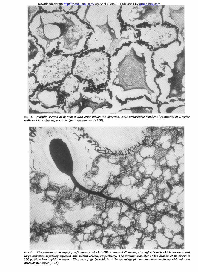
FIG. 5. Paraffin section of normal alveoli after Indian ink injection. Note remarkable number ofcapillaries in alveolarwalls and how they appear to bulge in the lumina (x 100).
FIG. 6. The pulmonary artery (top left corner), which is 600 Ht internal diameter, gives off a branch which has small andlarge branches supplying adjacent and distant alveoli, respectively. The internal diameter of the branch at its origin is100 H. Note how rapidly it tapers. Plexu.,es of the bronchiole at the top of the picture communicate freel.y with adjacentalveolar networks (x 33).
group.bmj.com on April 8, 2018 - Published by http://thorax.bmj.com/Downloaded from

The capillary network of normal and emphysematous human lungs
all vessels were black they were identified by theirsize and shape and their position in the secondarylobule. Normal pulmonary arteries entered thecentres of the secondary lobules after accompany-ing bronchi and bronchioles through the lung.Venules drained the peripheries of the lobulesto veins lying in the septa. These anatomical pointsallowed identification of many, but not all, vessels.Pulmonary arteries were normally similar indiameter to the bronchi they accompanied whenthey were medium-sized, but near the lobule theywere smaller in comparison.The pulmonary artery in the upper left corner
of Fig. 6 is about 600 IL (0-6 mm.) internally andit accompanies a bronchus (top centre). The 100 ,ubranch arising from its lower right-hand regiongives off an enormous number of side branchesas soon as it leaves the parent vessel. The largestbranches, which are arterioles up to 30 ,u ininternal diameter, in turn throw off many sidebranches of a similar pattern to supply more distalalveoli. The small side arterioles, 15 1i or lessinternally, supply the immediately adjacentalveolar capillary network. The main branchtapers rapidly. The large side arterioles run intissue at the corners of adjacent alveolar walls,sending out frequent radial branches to supply thealveolar capillary network.The very small side branches joined the capillary
network immediately as in Fig. 7. In that photo-graph the large 75 ,u parent pulmonary arteriole,crossing the field obliquely, gives off a small 15 ,uarteriolar branch (A). After a short course throughthe wall of the parent vessel the branch (A) dividesinto three or more smaller arterioles which branchas they join the capillary network.The alveolar capillaries intercommunicated to
form a network of polygonal spaces with aboutsix lengths of capillary around each space hole.The capillaries are 5 ,u across in Fig. 7, but otherauthors have found them wider (see Discussion).The spaces between them vary greatly in size,being, on an average, 20 u across but often downto 5 u in this preparation. Occasionally a largerround or oval space was seen in normal lungand presumed to be a pore of Cohn. Holes weremore common and larger in emphysema (seebelow).
In the walls of respiratory bronchioles the capil-laries had a larger mesh, average 15 x 40 A (centreof Fig. 8), than those of alveoli (edge of Fig. 8),and the oval shape of the meshes gave themdirectional axes. They were thus easily dis-tinguished from the smaller, rounder alveolarmeshes. The capillary and bronchiolar types of
network merged almost imperceptibly, for thecapillaries themselves were similar in calibre.
Occasionally in a normal area of lung the net-work was interrupted by a round or oval holewhich we presumed to be a pore of Cohn. Therewas usually a circular capillary around the edgeof the hole which communicated freely with capil-laries all round and was sometimes slightly largerthan these. The holes were sometimes more fre-quent and larger in emphysematous areas (Fig. 9)where they could measure over 100 ,u in diameter.More observations will be carried out to establishby our method the upper limit of size of normalpores of Cohn. The presence of numerous holes inemphysematous areas supported the descriptionof fenestration in emphysema known over 100years ago (e.g., Rainey, 1848; Waters, 1862). Thepossibility that many of the holes arose as tearswas borne out by the occasional finding of minute,blind-ended, short capillaries on the inner aspectof the surrounding ring capillary.One pattern of capillaries in a patch of emphy-
sema from a left upper lobe is illustrated in Fig.10. The large vessel in the right upper corner isa pulmonary venule and it collects blood from anabnormal capillary bed. The original alveolarcapillaries are scarce (arrow), and most of thefinest remaining network is bronchiolar in typewith a wide mesh and directional axes. Theslightly thicker branches coursing through thelung are small arterioles and venules. Thesenormally lie at the junctions of adjacent alveolarwalls and are somewhat obscured by the capillarynetworks (Figs. 6, 12, and 13). In Fig. 10, perhapsthey are slightly thicker than their normal counter-parts. In some places the bronchiolar capillariesare also becoming thinned out. Many of theemphysematous holes are ringed by a marginalcapillary. While there is a drastic reduction in thealveolar capillary bed which could cut downgaseous exchange, the remaining vessels stillappear capable of carrying a good flow of bloodfrom artery to vein. Incidentally, this is a localizedpatch of diffuse emphysema from a left upperlobe. An adjacent secondary lobule was normal.Where emphysema was more severe than in
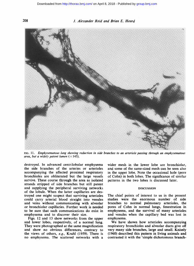
the last example and all that remained of theparenchyma was strands crossing large spaces, theinjection showed that in the core of each strandwas a large artery or arteriole, usually patent(Fig. 11). In the residual tissue around the arterywere a few smaller arterioles or a trace of abronchiolar or capillary network. An occasionalweb of tissue showed more of a network persist-ing but the original respiratory pattern was almost
205
group.bmj.com on April 8, 2018 - Published by http://thorax.bmj.com/Downloaded from

FIG. 7. Higher magnification of a pulmonary arteriole and branches. The large arteriole crossing the field obliquely is75 ju in diameter, internally. In the centre ofthe photograph a branch 15 i in diameter (A) passes down to join a capillarynetwork via three short vessels of intermediate size (x 200).
FIG. 8. The walls of respiratory bronchioles in the centre show a capillary network with a wider mesh than alveoli (seethis in top right corner). Th. oval shape of the meshes gives them direction in contrast to alveolar capillaries (top rightcorner) which are more rounded. BC=Bron?chiolar capillaries; AC=alveolar capillaries (x 134).
.w
'A
group.bmj.com on April 8, 2018 - Published by http://thorax.bmj.com/Downloaded from

FIG. 9. Emphysematous lung showing holes (arrows) inceapillary network, each bordered byaceontinuousceapillary (xl175).
FIG. 10. Emphysematous lung showing scanty alveolar networks (arrow), moderately wellpreserved bronchiolar networks,and persisting arterioles and venules. The large vessel (top right corner) is a venule (x 50).
group.bmj.com on April 8, 2018 - Published by http://thorax.bmj.com/Downloaded from
J. Alexander Reid and Brian E. Heard
-W
I
FFIG. 11. Emphysematous lung showing reduction in side branches to an arteriole passing through an emphysematousarea, but a widely patent lumen (x 145).
destroyed. In advanced centrilobular emphysemathe side branches of the arteries or arteriolesaccompanying the affected proximal respiratorybronchioles are obliterated but the large vesselssurvive. These course through the area as isolatedstrands stripped of side branches but still patentand supplying the peripheral surviving networksof the lobule. When the latter capillaries are des-troyed one might suspect that surviving arteriolescould carry arterial blood straight into venulesand veins without communicating with alveolaror bronchiolar capillaries. Further work is neededto be sure that such communications do exist inemphysema and to discover their size.
Figs. 12 and 13 show networks from the upperand lower lobes, respectively, of a normal lung.They were photographed at the same magnificationand show no obvious differences, contrary tothe views of others, e.g., Krahl (1959). There isno emphysema. The scattered networks with a
wider mesh in the lower lobe are bronchiolar,and some of the same-sized mesh can be seen alsoin the upper lobe. Note the occasional hole (poreof Cohn) in both lobes. The significance of similarpatterns in the two lobes is discussed later.
DISCUSSION
The chief points of interest to us in the presentstudies were the enormous number of sidebranches to normal pulmonary arterioles, thepores of Cohn in normal lungs, fenestration inemphysema, and the survival of many arteriolesand venules when the capillary bed was lost inemphysema.We have shown how arterioles accompanying
respiratory bronchioles and alveolar ducts give offvery many side branches, large and small. Knisely(1960) described this pattern in living animals andcontrasted it with the 'simple dichotomous branch-
208
group.bmj.com on April 8, 2018 - Published by http://thorax.bmj.com/Downloaded from

FIG. 12. Normal upper lobe showing similar pattern to normal lower lobe in Fig. 13. Both from same lung (x 80).
F1l shows-imi ptr walFIG. 13. Normal lower lobe showing similar pattern to normal upper love in Fig. 12 (x 80).
group.bmj.com on April 8, 2018 - Published by http://thorax.bmj.com/Downloaded from

J. Alexander Reid and Brian E. Heard
ing of systemic arterioles supplying capillary beds'.Krahl (1959) injected Indian ink into the rightventricle of the beating heart of a mouse and alsofound this pattern. It appears to be typical ofmammals. Incidentally, judging by Krahl's photo-graphs (his Fig. 10), the alveolar capillary networkin the mouse appears slightly simpler thanin man, having smaller collections of vessels andfewer meshes. His intercapillary distances cannotbe compared with those in our preparations, how-ever, because our air spaces are more fully dis-tended and the walls more spread out.
Miller (1947) stated that the bronchial arteriescould be followed as far as the first order ofrespiratory bronchioles where alveoli appeared,and there they joined an arterial plexus in thefibrous layer of the bronchiole. He found thatsmall branches penetrated the muscle coat andformed a network of capillaries, the long axis ofwhich corresponded to the long axis of thebronchiole. Venous radicals arose from that andformed another plexus with an irregular rectangu-lar mesh of thin vessels still on the inner side ofthe muscle coat. This drained through shortbranches in the muscle coat to form another plexuson the outside, composed of larger vessels, andfrom this arose one of the sources of origin ofthe pulmonary vein.The significance of the many side branches run-
ning out from pulmonary arteries and arteriolesis that if the alveolar walls supplied by the sidebranches atrophy or are destroyed, a good bloodflow may still pass through that region in theparent vessels. In centrilobular emphysema, forexample, loss of respiratory tissue from the centresof the lobules strips the arteries and arterioles ofthe central zone of their branches to alveolarcapillary networks, but that does not preventblood from flowing in the larger vessels and tothe alveoli in the peripheral paraseptal zone.Dunnill (1961) has made the interesting suggestionthat pulmonary hypertension may be caused byair pressure in the lesion compressing the survivingvessels.When a secondary lobule shows more extensive
loss of alveolar capillary networks, as in diffuse(panacinar) emphysema, surviving arterioles andvenules may still convey blood through that lobulealthough their side branches have vanished. Theprocess may be similar to what we have seen incentrilobular emphysema where pruning of theside branches does not affect the patency of theparent vessels. Preliminary findings suggest thatthe ends of arterioles and venules in emphy-sematous lung are connected by broadened capil-
laries, but further studies on this are in progressat the moment. Spain (1959) studied capillaries inemphysematous lungs that were hyperaemic orcongested so that the vessels were more easilyseen. There was diminution in the number ofcapillaries in many parts of the emphysematouswalls, and their distribution was irregular. Rela-tively more pre-capillary arterioles and smallarteries were seen.The persistence of arterioles and bronchiolar
networks when alveolar networks are lost wouldbe compatible with the growing amount of clinicalevidence that patients with advanced pulmonaryemphysema do not necessarily have hypertrophyof the right ventricle. Mounsey, Ritzmann,Selverstone, Briscoe, and McLemore (1952) showedthat pulmonary hypertension in emphysema maybe reversible, being high during attacks of acutebronchial infection but falling to normal betweenattacks.A striking feature of our preparations was the
way in which vessels intercommunicated on sucha large scale in normal lungs. This was especiallynotable around the bronchi and bronchioleswhere bronchial-type networks and adjacentalveolar networks communicated freely. Weibel(1959) has carried out injection studies in thehuman lung and suggests that anastomoses nor-mally occur between arteries and veins, pulmonaryor bronchial, in any combination. Liebow (1962,personal communication) has observed wide com-munications between arteries and between veinsbut not between arteries and veins in normal lungs.
Staub (1961) has described a method for exam-ining the pulmonary capillaries of animals in astate comparable to that in life. He opens thechest of an anaesthetized cat and freezes a lungvery rapidly by inundating it with up to threelitres of liquid propane cooled to -180°C. withliquid nitrogen. This procedure stops the capillaryblood flow almost instantly in the outermost partof the lung, and his findings are of great physio-logical interest. For example, he has viewed vesselsdown to 50 u in diameter and noted that whena cat is ventilated with 100% oxygen, the bloodin the frozen pulmonary arterioles (even quitelarge ones) is wholly or partially oxygenated(bright red) whereas in a cat breathing room airthe pulmonary arteriolar blood is dark red. Thatobservation raises the possibility that the arterioleswe observe surviving in an emphysematous areaof lung may still be capable of carrying out adegree of gaseous exchange.
Staub claims that the pulmonary capillaries donot bulge into the lumina of air spaces in his
210
group.bmj.com on April 8, 2018 - Published by http://thorax.bmj.com/Downloaded from

The capillary network of normal and emphysematous human lungs
preparations. We find this surprising since in someof our preparations of deep alveolar networksthe capillaries were only about 3 to 5 u indiameter and still bulged into the air spaces (seeFig. 5). The calibre of the human pulmonarycapillaries under normal living conditions is notknown. Knisely (1960) described some valuableobservations made on the surfaces and edges ofliving animal lungs using quartz-rod trans-illumination and a dissecting microscope. He gavefigures of 12 to 14 u for the diameter of pul-monary capillaries of living animals, a little widerthan systemic capillaries. Intercapillary distancesare commonly claimed to be sometimes less thanthe width of the capillaries themselves. In ourpreparations the alveolar intercapillary distanceswere very variable, averaging 20 u but as low as5 ,u, and greater where capillaries were interruptedby the very rounded holes of the pores of Cohn.Von Hayek (1953) put the diameters of pulmonarycapillaries (? human) at 10 to 12 ,u, as judged byfixation in situ by perfusion at approximately thepulmonary blood pressure. He found between fourand 12 capillary meshes from pre-capillary topost-capillary with a consequent variation in thelength of the capillary course of the blood (onlyabout 60 ,u through four capillary meshes butabout 250 ,u through 12 meshes). The diametersof capillaries in our preparations varied accordingto the quantity of injection material retained, andthat was affected by the viscosity and temperatureof the injection material, the unusual pressureseach side of the capillary wall, and the fact thatthe vessel walls were dead and toneless.
Orsos (1936) used a silver impregnation techniquefor pulmonary capillaries. He found emphy-sematous changes in the lungs in the first yearof life, namely enlargement and an increase ofpores, stretching of the inter-alveolar partitions,etc. In the adult he found, especially in the upperparts of the lungs, widening of alveoli and a pro-gressive decrease in density of the capillaries.Many capillaries were obliterated and otherswidened so that in places the regular polygonalcapillary network was replaced by a more sparsewide-meshed capillary system of wide lumina.Along with this was an increase in elastic andcollagen. He demonstrated these differences be-tween the lobes with two photographs, one fromeach lobe, taken at the same magnification. Krahl(1959) states that the capillary networks varywidely in their density, depending upon theirlocation in the lung. We have included Figs. 12and 13 to show that in the absence of emphysemathe two lobes had the same pattern of capillaries.
Since emphysema is common in the upper partsof the upper lobes in the general population,comparison of the vasculature of the lobes mustbe undertaken only after great care has been takento exclude emphysema (Heard, 1958; 1960). Res-piratory bronchioles more than 0-7 mm. acrossin the adult lung (excluding attached alveoli)indicate emphysema, and isolated strands of tissuepassing through air spaces indicate emphysemawith destruction.Most pulmonary vascular studies in emphysema
have concerned the larger vessels and anastomosesbetween bronchial and pulmonary circulations.Liebow (1959) summarized the changes in largervessels, viz., medial hyperplasia of the musculararteries, sometimes extreme, and atheromatouschanges in the larger arteries with associatedthrombosis. The venous side of the pulmonarycirculation is greatly expanded in emphysema(Liebow, 1953); the bronchial veins double indiameter and may allow blood to travel in eitherdirection between the atria. Cudkowicz andArmstrong (1953) described obliterative changesin the bronchial arteries in emphysema. Marchand,Gilroy, and Wilson (1950) described widespreadmacroscopic bronchopulmonary arterial anas-tomoses in emphysema which must have beendue to dilatation of existing anastomoses. McLean(1958) put forward a theory that sclerosis of thesmall arteries (adjoining terminal and respiratorybronchioles) in emphysema resulted from theorganization of thrombi, this thrombosis beingprecipitated by the cause of the inflammation ofthe adjoining air passages.
SUMMARY
The capillaries of human lungs at necropsy havebeen studied by an injection procedure using amixture of gelatin and black waterproof ink.The capillary networks were similar in upper
and lower lobes in the absence of emphysema.Alveolar capillary networks had small and
rounded meshes. Bronchiolar capillary networkshad larger oval meshes.
Occasional pores of Cohn were seen with a ring-like capillary.
In emphysema 'fenestrae' were seen, with sur-rounding ring-like capillaries.The remarkably numerous side branches to
arterioles are illustrated. It is pointed out thatwhen these side branches are lost in emphysemaa good blood flow may still be possible throughsurviving arterioles. That would be compatiblewith the clinical observations of others that the
211
group.bmj.com on April 8, 2018 - Published by http://thorax.bmj.com/Downloaded from

J. Alexander Reid and Brian E. Heard
pulmonary artery pressure in emphysema may benormal between attacks of infection.
We wish to thank Professor C. V. Harrison for hisadvice and Mr. W. Brackenbury for the photographs.One of us (J. A. R.) was in receipt of a grant from theBritish Council.
REFERENCES
Cudkowicz, L., and Armstrong, J. B. (1953). The bronchial arteriesin pulmonary emphysema. Thorax, 8, 46.
Dunnill, M. S. (1961). An assessment of the anatomical factor in corpulmonale in emphysema. J. clin. Path., 14, 246.
Heard, B. E. (1958). A pathological study of emphysema of thelungs with chronic bronchitis. Thorax, 13, 136.(1960). Pathology of pulmonary emphysema * methods of
study. Amer. Rev. resp. Dis., 82, 792.von Hayek, H. (1953). Die menschliche Lunge. Springer, Berlin. Trans.
(1960) Krahl, V. E., Hafner Publishing Co., New York.Knisely, W. H. (1960). In vivo architecture of blood vessels supplying
and draining alveoli. Amter. Rev. resp. Dis., 81, 735.Krahl, V. E. (1959). Microscopic anatomy of the lungs. Ibid., 80,
No. 1, Pt. 2, p. 24.
Liebow, A. A. (1953). The bronchopulmonary venous collateralcirculation with special reference to emphysema. Amer. J. Path.,29, 251.
- (1959). Pulmonary emphysema with special reference to vascularchanges. Amer. Rev. resp. Dis., 80, No. 1, Pt. 2, p. 67.
McLean, K. H. (1958). The significance of pulmonary vascularchanges in emphysema. Aust. Ann. Med., 7, 69.
Marchand, P., Gilroy, J. C., and Wilson, V. H. (1950). An anatomicalstudy of the bronchial vascular system and its variations indisease. Thorax, 5, 207.
Miller, W. S. (1947). The Lung, 2nd ed., p. 74. Thomas, Springfield,Illinois.
Mounsey, J. P. D., Ritzmann, L. W., Selverstone, N. J., Briscoe,W. A., and McLemore, G. A. (1952). Circulatory changes insevere pulmonary emphysema. Brit. Heart J., 14, 153.
Orsos, F. (1936). Die Ger0stsysteme der Lunge und deren physio-logische und pathologische Bedeutung. Beitr. klin. Tuberk., 87,568.
Rainey, G. (1848). On the minute anatomy of the emphysematouslung. Med.-C'iir. Trans. (Lond.), 31. 297.
Spain, D. M. (1959). In Pulmonary Circulation. An ItnterntationalSymposium, 1958. Ed. Adams, W. R., and Veith. I., p. 99. Gruneand Stratton, New York and London.
Staub, N. C. (1961). Microcirculation of the lung utilizing very rapidfreezing. Angiology, 12, 469.
Waters, A. T. H. (1862). Researches on the Nature, Pathology andTreatment of Emphysema of the Lungs, and its Relations withother Diseases of the Chest. Churchill, London.
Weibel, E. (1959). Die Blutgefassanastomosen in der menschlichenLunge. Z. Zellforsch., 50, 653.
212
group.bmj.com on April 8, 2018 - Published by http://thorax.bmj.com/Downloaded from

Injections of Indian InkHuman Lungs Studied byNormal and Emphysematous The Capillary Network of
J. Alexander Reid and Brian E. Heard
doi: 10.1136/thx.18.3.2011963 18: 201-212 Thorax
http://thorax.bmj.com/content/18/3/201.citationUpdated information and services can be found at:
These include:
serviceEmail alerting
the online article. article. Sign up in the box at the top right corner of Receive free email alerts when new articles cite this
Notes
http://group.bmj.com/group/rights-licensing/permissionsTo request permissions go to:
http://journals.bmj.com/cgi/reprintformTo order reprints go to:
http://group.bmj.com/subscribe/To subscribe to BMJ go to:
group.bmj.com on April 8, 2018 - Published by http://thorax.bmj.com/Downloaded from