the burden of late onset sepsis: infection prevention … prevention and antibiotic stewardship...
TRANSCRIPT

The Burden of Late Onset Sepsis: Infection Prevention and Antibiotic
Stewardship
Sandra Sundquist Beauman, MSN, RNC-NIC
CNS Consulting, owner
Manager, Clinical Trials Operations
Department of Pediatrics
University of New Mexico, Albuquerque

Objectives
} Discuss risk factors for and reported rates of late onset sepsis
} Identify sources for various bundle recommendations including CLABSI, VAP and others such as feeding tube related infections
} Discuss incidence and prevention of multi-drug resistant organisms in the NICU
} Identify recommended measures to improve antibiotic stewardship and expected outcomes for a successful program

Timing of Onset
u Early onset sepsis (EOS)
u Less than 3 days (term)
u Less than 7 days (preterm)
u Late onset sepsis (LOS)
u Onset at more than 3 or 7 days of age up to 30 days
u Late, late onset sepsis
u Onset beyond 30 days of age
Shah et al, 2014; Bizzarro, Rashkind, Baltimore, Gallagher, 2005

Incidence -
} Culture-proven sepsis overall◦ 0.77 to 1 per 1000 live births
� (Weston, Pondo, Lewis et al, 2011)
} Late-onset sepsis
◦ O.61% to 14.2% world-wide (Dong, Ying, Speer, 2014)
◦ 400-500 gms 65.5%◦ 501-750 gms 51.2%◦ 751-1000 gms 32.5% (Boghassian et al, 2013)
◦ 1000-1499 gms 10.2%◦ 1500-2500 gms 2.2%◦ > 2500 gms 1.6% (Vergnano et al, 2011)

Late Onset Sepsis-Risk Factors
} Flora in NICU/hospital environment
} Invasive monitoring and techniques◦ Central line (CLABSI)◦ Intubation (VAP)◦ Surgery (SSI)
} Extended length of stay (NICU)
o Feeding intolerance/necrotizing enterocolitis
Camacho-Gonzales, Spearman, Stoll, 2013Shah & Padbury, 2014

Etiology of LOS
u Coagulase-negative Staph
u Coagulase-negative Staph
u Coagulase-negative Staph
u Staphylococcus aureus
u Candida albicans
u Escherichia coli
u Klebsiella pneumoniae
u Enterococci
u Pseudomonas aeruginosa
u Group B streptococci
Shah & Padbury, 2014

Incidence of MDRO
u 30 – 70 % of humans worldwide are carriers
u 0.6 – 8.4% of NICU patients are carriers or infected
u Previous colonization = increased risk of infection
u Screening variesu Upon admission
u Upon admission for transports
u Upon admission and weekly
u Only during outbreaks
Newman, K. 2016, Advances in Neonatal Care
Management of MDRO
u Infection control practices
u Handwashing
u Isolation/cohorting
u Decolonization
u Reported 33.8% NICUs decolonize MRSA+ patients
u Nasal mupirocin
u 2% chlorhexidine baths/cloths
u Nasal polysporin

Infection Prevention

Infection Prevention
u Handwashing, handwashing, handwashing!
u Use of alcohol-based hand disinfectants
u Clean environment/work area
u Universal gloving?

Universal Gloving vs Good Hand Hygiene
u Intervention: “During RSV season, all health care workers in the pediatric units must wear a new pair of gloves every time they see a patient. They must remove the gloves and perform hand hygiene before leaving the patient’s room.”
u Extended to year round
u No compliance data offered
u Results showed statistically significant decrease in BSI and CLABSI in NICU
Yin, et al, 2013, Pediatrics

Universal Gloving vs Hand Hygiene
u RCT of handwashing only or handwashing + clean gloving showed statistically significant decrease in infection
u 32% (19 of 60) in gloving group vs 45% (27 of 60) in handwashing only group
u 64% fewer CLABSI in gloving group
Kaufman et al, 2014, JAMA

Universal Gowning/Gloving for MDRO
u Part of the BUGG study – adult ICUs
u During the study, universal gowning/gloving for all patients
u After the study, return to usual care
u Gowning/gloving for MDRO patients
u Clothes were cultured at beginning and near end of shift
u 25% of HCW practicing usual care had contaminated clothing at the end of the shift.
u This was reduced by 70% with universal gowning and gloving
Williams, McGraw, Schneck, et al, 2015

Universal Gloving vs Good Hand Hygiene
u Hands cultured quarterly after completion of hand hygiene. Infection surveillance for BSI and CLABSI over three year period
u Results: Species isolated from clean hands and neonatal BSI were significantly different
u Consideration of universal gloving
u Many potential positive and negative impacts to be considered
Larson et al, 2005

CLABSI
Consistently reported as the leading type of device-associated infection in the NICU and consistently among the highest CLABSI source throughout the hospital (Hocevar et al, 2014)
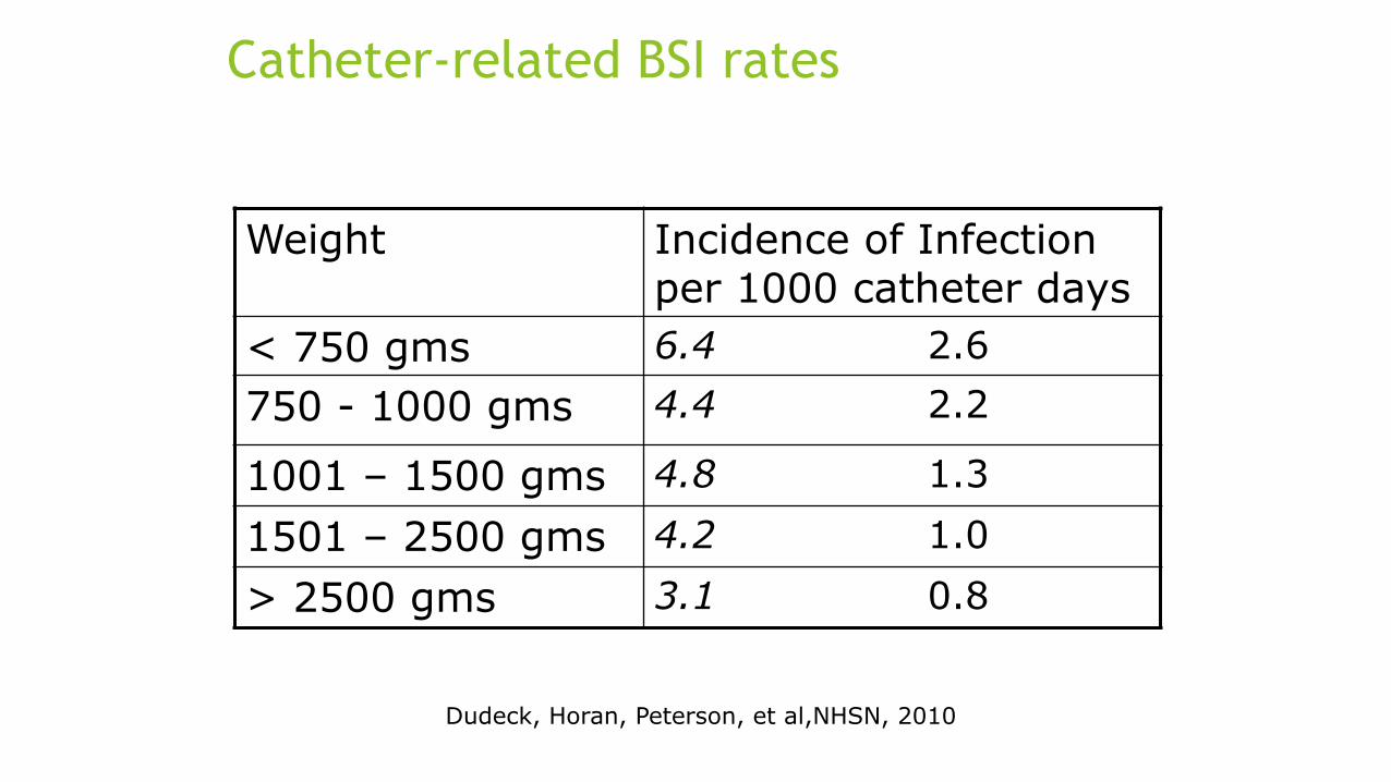
Catheter-related BSI rates
Weight Incidence of Infection per 1000 catheter days
< 750 gms 6.4 2.6750 - 1000 gms 4.4 2.2
1001 – 1500 gms 4.8 1.3
1501 – 2500 gms 4.2 1.0
> 2500 gms 3.1 0.8
Dudeck, Horan, Peterson, et al,NHSN, 2010

CLABSI bundles
u Insertion
u Hand hygiene before insertion
u All necessary supplies available at bedside before insertion
u Maximum sterile barrier precautions
u Face mask worn within 3 feet of sterile field
u Perform skin antisepsis with povidone-iodine or CHG – allow to dry
u Procedure stopped if anyone notes sterility compromised

Maintenanceu Daily assessment of catheter need
u Dressing integrity and site cleanliness assessed (daily at minimum)
u Sterile/2-person dressing change
u Use “closed” systems for infusion, blood draws & medication administration
u Meticulous care when changing IV tubing (q72hrs à 96 hrs) (CDC, 2011)
u Clean vs sterile?
u Scrub-the-hub prior to entry
u Clean gloves for all VAD entries & hand hygiene utilized before & after glove use

Maintenance
u Frequency/method of tubing changes
u Needleless connector changes
(Sandora, Graham, Conway, Dodson, Potter-Bynoe, Margossian, 2014; Pettit & Sharpe survey)

Scrub the Hub
u Hub colonization demonstrated highest risk for infection in NICU & much higher than exit site colonization or other factors. Mahlieu et al 2001, J Hospital Infection, 48.
u CHG/70% IPA superior to single use of either agent
u CHG benefits
u Residual activity
u Effectiveness in presence of blood
u Addition of alcohol increases kill rate & drying time
u Marschall et al, 2008 AJIC,36; Pratt et al, 2007, J Hospital Infection, 655; Safer Healthcare NOW!Quebec Campaign 2008; Milstone et al, 2008,Clinical Infect Dis, 46.

Biofilm on Hubs
u Biofilm can be found in as little as 3.8 days
u Colonization increased dramatically by 3.8 days
u Biofilms tenaciously attach to external and internal surfaces of needleless connector & are not easily removed
u Adequate disinfection will require disinfectants capable of biofilm eradication as well as sufficient mechanical friction
Ryder et al. Microscopic evaluation of microbial colonization on needleless connectors. 2009, APIC Poster Presentation
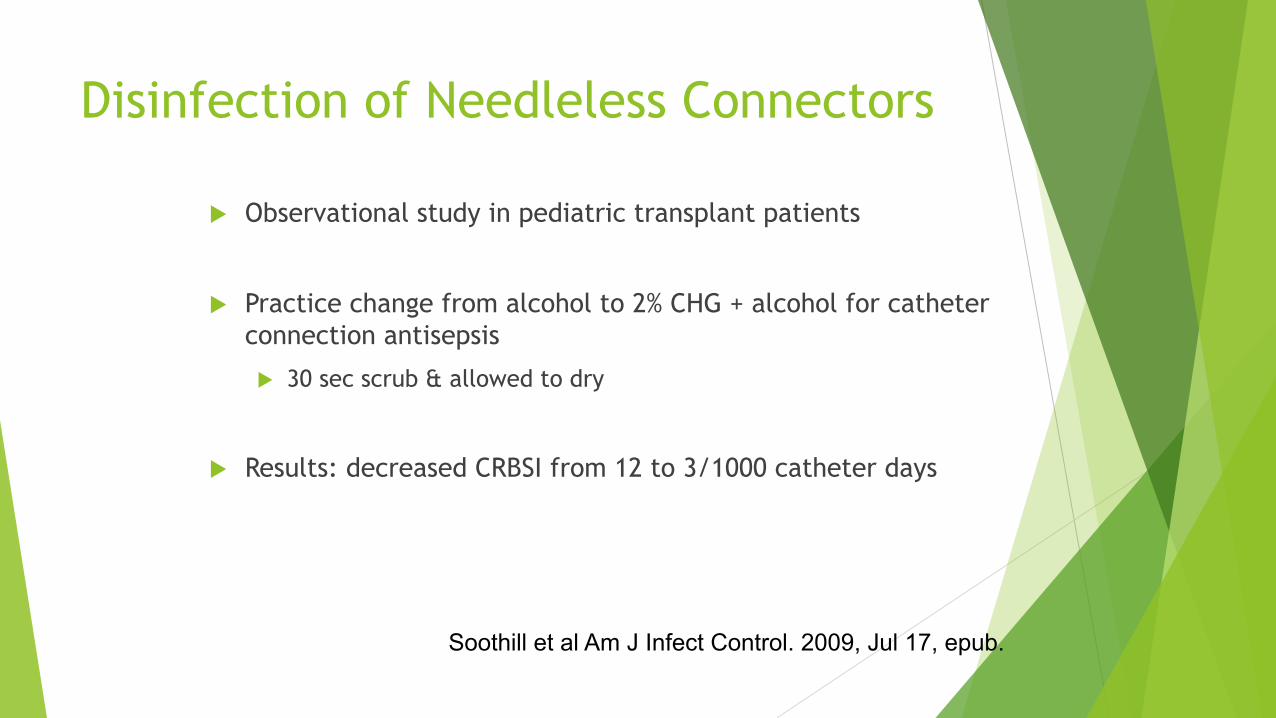
Disinfection of Needleless Connectors
u Observational study in pediatric transplant patients
u Practice change from alcohol to 2% CHG + alcohol for catheter connection antisepsis
u 30 sec scrub & allowed to dry
u Results: decreased CRBSI from 12 to 3/1000 catheter days
Soothill et al Am J Infect Control. 2009, Jul 17, epub.

Needleless Connectors – Are they all created equal?
u Positive flush: CLC 2000, Posi-flow, Ultrasite
u Negative flush: Clave, Interlink, Clearlink, Q-syte
u Neutral flush: MicroCLAVE and others
u Split septum
u Mechanical valve
Hadaway & Richardson, 2010. Needleless Connectors: A primer on terminology.Journal of Infusion Nursing. 33(1):1-10

Fluid Pathway

Disinfection of Needleless Connectors
u Various types of needleless access devices inoculated with organisms, cleansed with 70% alcohol or with 3.15% CHG + 70% alcohol
u Flushed with NS or hyperal and cultured
u No organisms grew after cleansing with either solution for 15 seconds with twisting motion
u All models of needleless access devices tested
Kaler & Chinn, 2007

Use of povidone-iodine vs chlorhexidine gluconatefor skin prep
u CHG recommended by CDC (2000)
u Risk of skin irritation/breakdown
u Commercially available CHG in U.S. has base of 70% or 2% alcohol
u FDA required labeling to change January 1, 2012
u Use with caution in premature infants and infants under 2 months
u Common criteria
u > 28 wks GA OR > 1 wk old
u Not usually used on peri-umbilical area
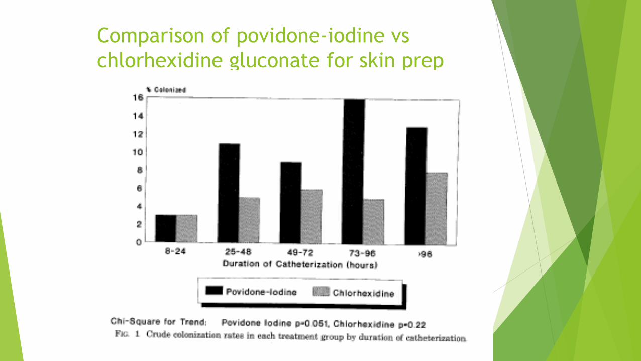
Comparison of povidone-iodine vs chlorhexidine gluconate for skin prep
Garland et al, 1995

Site Selection & Infection
u 396 infants with PICCs
u 370 upper extremity
u 107 lower body extremity
u Results
u Lower extremity catheters in place longer, time to complications longer and decrease in cholestasis
u CRBSI
u 7.1/1000 upper extremity catheters
u 4.8/1000 lower extremity catheters
u Infants with lower GA and birthweight had significantly higher CRBSI in upper extremity lines
Tsai et al, 2009

Complications Associated with 2 Different Types of Catheters in VLBW Infants
u Total of 808 catheters inserted
u 410 femoral placement
u 398 non femoral placement
u Femoral placement was an independent risk factor for catheter-related sepsis
Tsai, 2011, Infection Control and Hospital Epidemiology

CLABSI rates vs BSI rates
u Over 5 year period
u CLABSI = 18.7% of noncontaminant BSI during study period (Leekha et al, 2013)
u With implementation of CLABSI prevention, non-CLABSI BSI also fell
u CLABSI portion of BSI = ~33%
u California Collaborative 2013
u After almost 8 years of CLABSI bundle implementation

Where Can We Effectively Impact BSI Prevention?
u Skin
u Good skin care
u Efforts to prevent skin breakdown, treat effectively
u Gastrointestinal
u Prevent introduction of bacteria into GI tract leading to bacterial translocation
u Management of gastric tube similar to IV tubing

GI Tract Pathophysiology
u Several studies show contamination and bacterial growth of feedings tubes
u Enteral tube hub as a reservoir for transmissible enteric bacteria. MatlowAm J Infect Control. 2006
u Enteral tubes quickly become covered with biofilms of potentially pathogenic organisms. Hurrel BMC Infectious Diseases 2009, 9:146

Feeding Tube Colonization
Electron microscopy of enteral feeding tube inner wall from neonate fed breast milk and ready to feed formula. Bar indicates 4 µm size marker
Hurrell, E., Kucerova, E., Loughlin, M., et al. (2009). Neonatal enteral feeding tubes as loci for colonisation by members of the Enterobacteriaceae. BMC infectious diseases, 9(1), 146.

Complications of Bacterial Contamination of Enteral Feeding in Neonates
u 50 infants, tubes cultured after 7 days of indwelling gravity feeds
u 71/125 tubes heavily “contaminated” with 3 different types of bacteria
u Feeding intolerance occurred in 24/32 wks with “contaminated feeds” vs 0/44 wks with non-contaminated tubes
Mehall, 2002, J Pediatric Surgery

Colonization of neonatal enteral feeding tubes
u 129 feeding tubes from 2 NICUs
u 44% receiving BM + fortifier, the rest formula
u 76% were colonized, many with species associated with “outbreaks,” e.g. Enterobacteriaceae, S Marcescens, Klebsiellapneumoniae
u No difference in isolation rates by feeds, but biofilm isolation was less in BM fed infants
u Colonization related to duration of insertion (up to 48hr)
u Increased colony counts related to postnatal age
Hurrell BMC Infectious Diseases 2009, 9:146

Colonization of extension tube & connector
u Investigated colonization of extension tube vs feeding tube
u Similar organisms found with most colonization at > 48 hrs, none at < 12 hrs
u Same organisms found in infant stool
Gomez et al, 2016

Ventilator Associated Pneumonia

Ventilator-Associated Events
u Pneumonia
u Acute respiratory distress syndrome
u Pneumothorax
u Pulmonary embolism
u Atelectasis
u Pulmonary edema
Klompas, Branson, Eichenwals, Greene, Howel et al, 2014

Adult Definition
u VAC (ventilator-associated conditions)
u Increased ventilator/FiO2 requirements (4 days)
u IVAC (infectious VAC)
u Above + fever, +/- WBC count, antibiotics x > 4 days
u Possible VAP
u Above + positive gram stain of secretions
u Probably VAP
u Above + growth of pathogenic organism beyond specified thresholds or positive test for respiratory viruses
Klompas, Branson, Eichenwals, Greene, Howel et al, 2014

Recommendations for VAE prevention
u NHSN rates vary from 0.2 – 1.8/1000 vent days (Klompas et al, 2014)
u May be as high as 37.2/1000 vent days (Weber, 2016)

Recommendations for VAE prevention
Recommendation Intervention
Basic Practices• May lower VAP rates,• Minimal risk of harm, • Benefits likely outweigh potential risks
•Avoid intubation
•Extubate as soon as possible
•Evaluate for extubation daily
•Manage without sedation when possible
•Avoid unplanned extubation
•Provide regular oral care with sterile water
•Minimize breaks in ventilator circuit
•Change circuit only if visibly soiled or malfunctioning
Klompas, Branson, Eichenwals, Greene, Howel et al, 2014

Recommendations for VAE prevention
Recommendation Intervention
Special approaches• Unknown impact on VAP rates but risk of harm likely minimal
• Lateral recumbent positioning
• Elevated HOB
• Closed/in-line suctioning systems
Klompas, Branson, Eichenwals, Greene, Howel et al, 2014

Recommendations for VAE prevention
Recommendation Intervention
Generally not recommended•Unknown impact on VAP rates, inadequate data on risks•May be harmful
•Not recommended because appropriate products not available for use in this population
•Regular oral care with antiseptics
•Histamine 2 receptor antagonists•Prophylactic broad-spectrum antibiotics•Daily spontaneous breathing trials•Daily sedative interruptions
•Prophylactic probiotics or synbiotics•ETT with subglottic secretion drainage ports•Silver-coated endotracheal tubes
Klompas, Branson, Eichenwals, Greene, Howel et al, 2014

Ventilator Associated Pneumonia
} Extubate early/avoid intubation
} Eliminate contamination while suctioning
} Maintain separate suction cannisters
} Change ventilator circuit only when visibly soiled or malfunctioning
} Suction only as needed, avoid using normal saline
} Resuscitation bag outside patient bed, cleanse connector with alcohol wipe Van Der Zwet, Parlevliet, Savelkoul, 2000
} Oral care with HM or sterile H2O q3-4h
} Avoid abdominal distension
} Elevate HOB 15-30 degreesCeballos et al, 2013; Smulders et al, 2013, Weber, 2016
Colostrum as Oral Care
u No clinical trials specific to VAP
u Safety & feasibility evaluated (Rodriguez, et al, 2010)
u Decrease in surgical NEC rates and death, not statistically significant (Seigel, et al, 2013)

Buccal administration of human colostrum: Impact on the oral microbiota of premature infants
u 12 VLBW infants randomized to receive oral colostrum (own mother) or standard care
u Buccal administration influenced oral colonization with differences persisting at least 48 hrs after intervention
u Planococcaceae in colostrum group
u Moraxellaceae & Staphylococcaceae in standard care group
Sohn, Kalanetra, Mills, Underwood, 2016

Oropharyngeal Colostrum Administration in Extremely Premature Infants
u Administered oral colostrum to 48 infants < 28 wks
u Measured urinary levels of secretory IgA, lactoferrin and other immune agents
u Levels significantly higher in colostrum group
u Observed lower incidence of clinical sepsis in colostrum group
Lee, Kim, Jung et al, 2015

Preventing Surgical Site Infections

Reported incidence of SSI
u 4.3 Surgical site infections/100 cases
u 3.1/100 clean interventions
u 4.4/100 clean-contaminated interventions
u 19/100 dirty interventions
Segal, Kang, et al, 2014

Surgical Site Infection
u Highest risk of infection (adult)u Orthopedic
u Cardiac (open heart)
u Gastrointestinal
u Highest risk of infection (neonatal)u Thoracic (27%)
u Abdominal (50%)
u Gastroschesis reported at highest incidence
(Segal et al, 2014)

Risk factors for SSI
u Increased risk related to
u Use of parenteral nutrition
u Previous/extended use of antibiotics
u Multiple surgical procedures
u Timing of surgery (later = increased risk)
Romanelli et al, 2014; Segal et al, 2014

Risk factors for SSI
u Decreased risk related to:
u Non-invasive ventilation
u No difference r/t
u Gestational age,
u Birth weight,
u Use of perioperative antibiotics,
u Wound class
Romanelli et al, 2014; Segal et al, 2014

Surgical Site Infection
u Antibiotic Prophylaxis
u Appropriate choice, timing and duration of antibiotics unknown
u Skin Preparation
u Peri-operative hypothermia prevention
u Peri-operative euglycemia maintenance

Antibiotic Prophylaxis in Neonates
u Based on wound type
u Clean = None
u Clean-contaminated = Single pre-operative
u Contaminated = 24 hours
u Dirty or infected = > 24 hours
u Inconsistent definition of wound type by pediatric surgeons reported(Vu, Nobuhara, Lee, Farmer, 2009)

Reducing Surgical Site Infections (SSI) at a Pediatric Academic Medical Center
u All class I and II surgeries included. Special measures for VP shunt placement and spinal fusionsu Antibiotic administration within 1 hru Skin prep
u Room and body temperature goals
u Oxygenation at least 95%
u Glycemic control – blood glucose < 200 mg% during surgery and 24 hrs postoperatively
u Leadership involvement and results reported regularly
u 64% reduction of SSI seenu 1.5 per 100 procedures to 0.54 per 100 procedures
u Improvement continued for 2 years
Ryckman et al JT Commission Qual Safety 2009

Antibiotic Stewardship

Antibiotic Stewardship
u Prolonged/prophylactic antibiotic exposure increases incidence of resistant bacteria
u MRSA, VRE, C Diff
u Colonizes infant with pathogenic bacteria thus increasing rate of infection
u More than 10 days antibiotic exposure = 3 fold NEC incidence u Each day associated with about 20% increased NEC risk
Alexander, 2011

Effects of Inappropriate Antibiotic Administration
u Inadequate antibiotic treatment results in growth of multi-drug resistant Gram-negative bacilli
u Specifically exposure to third generation cephalosporin and carbapenem
Tsai et al, 2014

Effects of Inappropriate Antibiotic Administration
u Prolonged initial empirical antibiotic therapy
u 2-fold higher incidence of late onset sepsis, NEC or death
u 3-fold higher incidence of late onset sepsis onlyu Kuppala et al, 2011
u About a 4% increase in odds of NEC or death with each additional day of initial antibiotic treatment
u Almost 7% risk of NEC alone
u About 16% increase in mortality (Cotton et al, 2009)

Results of SCOUT study

Examples of antibiotic stewardship programs
u Create an antibiotic stewardship team
u neonatologist, ID physician, neonatal or ID–trained pharmacist, infection preventionists, bioinformatician, neonatal nurse
u Determine current practices (accurately)

Examples of antibiotic stewardship programs
u Set goals/metrics
u Avoid redundant antibiotic use
u Limit broad spectrum antibiotic use
u Reduce duration of antibiotic exposure
u Avoid inadequate therapy
Cantey & Patel, 2014

Antibiotic Stewardship
u Limit antibiotic use
u Blood culture negative useu Limit to 48 hours
u Surgery prophylaxis not beyond 48 hrs
u Awareness of third generation cephalosporin and vancomycin use
u Be aware of unit/hospital specific antibiogram
Committee of the Fetus and Newborn, Pediatrics, 2012

Leadership
u Determine/define process measures
u Set goals
u Audit performance measures and/or outcomes
u Standardize care processes
u Build in reminders
Leadership
u Accountability and engagement at all levels
u Involve local champions
u Utilize peer networks
u Provide feedback/celebrate successes
u Believe that zero is possible!!

Evaluate/Celebrate Success
u Post compliance rates
u Inform staff of outcomes

Thank you!