the aryl hydrocarbon receptor: differential contribution ... · the aryl hydrocarbon receptor:...
TRANSCRIPT
The Aryl Hydrocarbon Receptor: DifferentialContribution to T Helper 17 and T Cytotoxic 17 CellDevelopmentMark D. Hayes1*, Vitalijs Ovcinnikovs1, Andrew G. Smith2, Ian Kimber1, Rebecca J. Dearman1
1 Faculty of Life Sciences, The University of Manchester, Manchester, United Kingdom, 2 MRC Toxicology Unit, The University of Leicester, Leicester, United Kingdom
Abstract
The aryl hydrocarbon receptor (AhR) has been shown to be required for optimal Thelper (Th) 17 cell activation. Th17 cellsprovide immunity against extracellular pathogens and are implicated in autoimmune diseases. Herein, the role of the AhR incytokine production by Th17, and by the analogous population of T cytotoxic (Tc)17 cells, has been examined. Lymph nodeTc (CD8+) and Th (CD4+) cells were isolated by negative selection from naive AhR+/2 and AhR2/2 mice and polarised to Tc1/Th1 or Tc17/Th17 phenotypes with appropriate cytokines. Cell differentiation was assessed as a function of mRNA andprotein (ELISA and flow cytometry) expression for interferon (IFN)-c and for key Th17 cytokines. In AhR+/2 mice, Th17 cellsdisplayed an exclusive IL-17 profile, which was markedly inhibited by a selective AhR antagonist to levels observed in AhRknockout mice. Addition of the natural AhR agonist 6-formylindolo[3,2-b]carbazole (FICZ) markedly enhanced Th17 cellactivity in the heterozygotes. In contrast, Tc17 cells polarised into 3 distinct subsets: producing either IL-17 or IFN-c alone, orboth cytokines. Blocking AhR was also detrimental to Tc17 development, with reduced responses recorded in AhR2/2 miceand antagonist-mediated reduction of IL-17 expression in the heterozygotes. However, Tc17 cells were largely refractory toexogenous FICZ, presumably because Tc17 cells express baseline AhR mRNA, but unlike Th17 cells, there is no marked up-regulation during polarisation. Thus, Th17 cell development is more dependent upon AhR activation than is Tc17 celldevelopment, suggesting that endogenous AhR ligands play a much greater role in driving Th17 cell responses.
Citation: Hayes MD, Ovcinnikovs V, Smith AG, Kimber I, Dearman RJ (2014) The Aryl Hydrocarbon Receptor: Differential Contribution to T Helper 17 and TCytotoxic 17 Cell Development. PLoS ONE 9(9): e106955. doi:10.1371/journal.pone.0106955
Editor: Jose Carlos Alves-Filho, University of Sao Paulo, Brazil
Received February 5, 2014; Accepted August 11, 2014; Published September 9, 2014
Copyright: � 2014 Hayes et al. This is an open-access article distributed under the terms of the Creative Commons Attribution License, which permitsunrestricted use, distribution, and reproduction in any medium, provided the original author and source are credited.
Funding: This work was funded by the medical research council grant number MR/J500410/1. The funders had no role in study design, data collection andanalysis, decision to publish, or preparation of the manuscript.
Competing Interests: The authors have declared that no competing interests exist.
* Email: [email protected]
Introduction
An important property of adaptive immune function is the
orchestration of polarised immune responses by the generation of
discrete populations of CD4+ T helper (Th) and CD8+ cytotoxic T
(Tc) lymphocytes [1,2]. A more recent addition to known subsets
of CD4+ Th cells are Th17 cells that have a characteristic cytokine
secretion profile that includes IL-17A, IL-17F, IL-21 and IL-22.
These cells play important roles in protective immunity and have
been implicated in the pathogenesis of certain autoimmune
diseases including multiple sclerosis, psoriasis, rheumatoid arthritis
and Crohn’s disease [3–5]. The effector functions of Th17 cells are
cytokine-mediated. Proinflammatory IL-17A acts as a potent
neutrophil recruiter, as well as stimulating other neutrophil-
attracting and activating cytokines and chemokines, and is an
important component of resistance to pathogenic microorganisms
[6]. IL-22 targets non-hematopoietic cells (keratinocytes, hepato-
cytes and colonic epithelial cells) inducing proliferative and anti-
apoptotic pathways and the production of anti-microbial mole-
cules that aid tissue repair [7]. In autoimmunity, Th17 cytokines
are thought to enhance inflammation by paracrine induction of
other proinflammatory factors, such as tumour necrosis factor-aand IL-6 [8,9].
In addition to the various CD4+ Th cell subsets, CD8+ Tc cells
analogous with Th1 and Th2 cells have been described both in
vitro and in vivo, and have been categorised as Tc1 and Tc2 cells,
respectively [10]. The most recent addition to the family are CD8+
cells secreting IL-17, and classified as Tc17 [11]. It has been
claimed previously that Tc17 cells are an in vitro artefact due to
their lack of cytotoxic activity, associated with the absence of
perforin and Granzyme B[12]. However, more recent studies have
provided evidence for the existence of Tc17 cells in both mouse
and humans [13–15]. Although Tc17 cells express cytokine
profiles similar to their CD4+ counterparts, their roles in protective
immunity and autoimmune disease have yet to be established. An
interesting characteristic of both Th17 and Tc17 cells is their
plasticity. The switch from Th17 to Th1 phenotype has been
shown in vivo using Th17 reporter mice and a range of
inflammatory and autoimmune conditions. For example, the
majority of Th1 cells that had infiltrated spinal tissue during the
development of experimental autoimmune encephalomyelitis had
at some time previously expressed IL-17A, thus demonstrating
that they had derived from Th17 cells [16]. Tc17 cells have also
been shown to display plasticity. Tc17 cells generated ex vivo were
found to switch off IL-17 production when transferred into mice,
and interestingly, this coincided with the acquisition of cytotoxic
ability, even in the absence of interferon (IFN)-c production [17].
The conditions for Th17 development have been investigated
thoroughly and although there are similar requirements for Tc17
PLOS ONE | www.plosone.org 1 September 2014 | Volume 9 | Issue 9 | e106955
development, there may also be some differences. Th17 and Tc17
polarisation have both been shown to require transforming growth
factor (TGF)-b and IL-6, and to be enhanced further by IL-1b, IL-
21 and IL-23 [18,19]. In addition, it has been shown that
activation of the aryl hydrocarbon receptor (AhR) is required for
optimal Th17 polarisation. The AhR was first described as a
receptor for ligands that are environmental toxicants, such as
2,3,7,8-tetrachlorodibenzo-p-dioxin (TCDD) or dioxin [20]. This
receptor is a cytoplasmic transcription factor that following
ligation translocates to the nucleus where it binds to the AhR
nuclear translocator forming a heterodimer that can activate
various AhR responsive genes [20,21]. AhR ligands fall into two
categories: synthetic and natural. Although initial characterisation
of AhR focused primarily on TCDD and other synthetic
halogenated hydrocarbons, more recently ligation by natural
ligands and the role of AhR in immune function has attracted
increasing interest. Natural ligands include plant-derived materi-
als, such as flavonoids and by-products of dietary indoles, such as
6-formylindolo[3,2-b]carbazole (FICZ) that is a photoproduct of
tryptophan [22]. Although the expression of AhR is ubiquitous in
vertebrate cells [23], so far there are only two known conventional
T cell populations that actively up-regulate this receptor when
activated: regulatory T cells and Th17 cells [24]. However, to date
there have been no investigations reported of the involvement of
the AhR in Tc17 development.
The similarity of Tc17 cells to their CD4+ counterparts with
respect to their requirements for polarisation and cytokine
expression profiles has led us to hypothesise that activation of
the AhR may impact on Tc17 phenotype. We have therefore
investigated the contribution of the AhR to Tc17 polarisation invitro. In addition, comparisons have been made between Th17
and Tc17 development in T cells obtained from AhR knockout
mice. Successful polarisation has been measured as a function of
frequency of IL-17A-expressing cells by flow cytometry, and at the
levels of mRNA and secreted protein for IL-17A and IL-22.
Materials and Methods
AnimalsAhR2/2 and AhR+/2 mice on a C57BL/6J background,
originally obtained from the Jackson Laboratory via B. Stockinger
(National Institute for Medical Research, London, UK), were bred
in the specific pathogen free facility at the University of
Manchester. The AhR transgenic mice were originally derived
from 129X1 x 129S1 via R1 (+Kitl-SlJ) ES cell line and donated to
the Jackson Laboratory by Dr. Christopher Bradfield (University
of Wisconsin Medical School). Mice were bred from male
homozygous (AhR2/2) and female heterozygous (AhR+/2)
breeding pairs.
Ethics statementAll animal procedures were approved by the UK Home Office
and carried out in compliance with the Animals (Scientific
Procedures) Act 1986 under a Home Office granted project
licence. Mice were provided with environmental stimuli (bedding
and nesting materials); with food (Beekay Rat and Mouse Diet
No1 pellets; B&K Universal, Hull, UK) and water being available
ad libitum. The ambient temperature was maintained at 2162uCand relative humidity was 55610% with a 12 h light/dark cycle.
Mice were sacrificed by exposure to CO2 gas in rising
concentration followed by dislocation of the neck in concordance
with schedule 1 (Animals [Scientific Procedures] Act 1986).
Genotyping of AhR miceGenotyping was performed with two sets of PCR primers
(Sigma Aldrich, Gillingham, UK). The first amplified a region of
AhR exon 2 present in the wild type but not the knockout mice,
that is, forward TTCAGAGTAAAGCCCATCCC and reverse
ATCAAAGAAGCTCTTGGCCC. The second set amplified a
region of the neomycin gene only present in the knockout animals,
that is, forward TGGGTGGAGAGGCTATTC and reverse
ATGGTGAGATGACAGGAGATC (http://blast.ncbi.nlm.nih.
gov/). Mice were genotyped prior to experimentation using
DNA derived from ear clip tissue.
Th1/Tc1 and Th1/Tc17 polarisationCD4+ and CD8+ T cells were isolated from skin draining lymph
nodes (LN: axillary, inguinal and auricular) by magnetic separa-
tion (Stemcell Technologies, Manchester, UK). The culture
medium for both Th1/Tc1 and Th17/Tc17 polarisation was
IMDM (Sigma), supplemented with 25 mM HEPES, 400 mg/ml
streptomycin/penicillin, 292 mg/ml L-Glutamine, 5 mM 2-mer-
captoethanol and 5% heat-inactivated fetal calf serum (v/v) (all
from Life Technologies, Paisley, UK). A total of 2.56105 T cells/
well were differentiated in wells coated with 1 mg/ml anti-CD3
plus 10 mg/ml anti-CD28 (eBioscience, Hatfield, UK) with a
cytokine cocktail of 50 ng/ml human IL-6, 1 ng/ml human TGF-
b, and 10 ng/ml IL-1b for Th17/Tc17 polarisation and 3 ng/ml
IL-12 for Th1/Tc1 polarisation (all cytokines from R & D systems,
Abingdon, UK). The AhR antagonist CH-223191 (Calbiochem,
Nottingham, UK) or AhR agonist FICZ (Enzo Life sciences,
Exeter, UK) were added at 3 mM and 300 nM respectively. Both
were formulated in dimethyl sulfoxide (DMSO) at the start of
culture with control wells received appropriate amounts of
DMSO.
RT-PCRTotal RNA was purified from enriched CD4+ or CD8+ cells
using TRIzol and Purelink RNA mini kit (Life Technologies). The
mRNA expression levels of mouse IL-17A, IL-17F, IFN-c, T-bet,
RORC, AhR, AhRR, Cyp1A1 genes were determined by RT-
PCR using Taqman primers (Life Technologies) on a RT-PCR
machine (StepOne plus; Life Technologies). Expression was
normalised using freshly isolated unpolarised cells (control) and
to hypoxanthine–guanine phosphoribosyl transferase (HPRT),
with the DD cyclic threshold (Ct) method used to calculate relative
fold change.
Flow cytometrySingle cell suspensions of freshly isolated CD8+ enriched cells
were characterised for memory/effector phenotype by flow
cytometry. Cells were treated with 5 mg/ml anti-CD16/32
blocking antibody prior to staining with specific antibodies;
5 mg/ml fluorescein isothiocyanate (FITC) labelled anti-CD8
(eBioscience), 2 mg/ml allophycocyanin (APC) labelled anti-127
(eBioscience), 2 mg/ml phycoerythin (PE) labelled anti-CD62L
(Becton Dickinson, Oxford, UK), or with appropriate isotype
controls. Cells were defined as follows: naıve, single positive
CD62L; central memory, double positive; effector memory, single
positive CD127; effector, double negative [25]. In addition, the
frequency of cytokine expressing cells was characterised by flow
cytometry. Single cell suspensions of in vitro polarised T cells were
treated with phorbol 12,13-dibutyrate (PdBu; 500 ng/ml), iono-
mycin (500 ng/ml) and 10 mg/ml brefeldin A for 4 h (Sigma).
Following restimulation cells were fixed and permeabilised using
2% paraformaldehyde and saponin (0.1% w/v) (Sigma). Blocking
AhR Influence on Tc17 Polarisation
PLOS ONE | www.plosone.org 2 September 2014 | Volume 9 | Issue 9 | e106955
antibodies were applied prior to staining for specific antibodies;
10 mg/ml anti-CD16/32, 10 mg/ml hamster IgG and 10 mg/ml
rat IgG (BD, Oxford, UK). Cells were then stained with 2.5 mg/ml
PE labelled anti-IL-17A and 10 mg/ml APC labelled anti-IFN-cand appropriate isotype controls (Biolegend, San Diego, CA,
USA). In each case, acquisition of samples (25000 cells) was
performed on a flow cytometer (FACScalibur; Becton Dickinson)
and data were analysed using FlowJo v7.5 software (Tree Star,
Ashland, OR, USA). For memory effector phenotype, cells were
initially gated on forward and side scatter and non-viable cells
excluded using 7-aminoactinomycin D (7-AAD; 1 mg/ml). Cells
that stained positive for CD8, which constituted 84–96% of the
total enriched population, were analysed for memory/effector
marker expression. Cytokine expressing cells were initially gated
on forward scatter and side scatter. In both cases, gates were
drawn based upon the position of cells stained with isotype
controls, with .99% of the double isotype control stained cells
residing in the lower left hand quadrant (Th1/Tc1 gating strategy
supplied as Figure S1).
Cytokine determinations using ELISAThe cytokines IL-17A, IL-22 and IFN-c were analysed in
supernatants using DuoSet ELISA kits (R&D systems) following
the manufacturers’ instructions. Optical density at 450 nm was
measured using an automated reader (Multiskan, Thermo
scientific, Basingstoke, UK). The lower limit of accurate detection
was approximately 25–50 pg/ml. Standard errors were less than
10% in most experiments.
Statistical analysesStatistical significance of differences between groups was
determined using one-way ANOVA with a Tukey post test using
Prism 5 software (GraphPad Software, La Jolla, CA, USA).
Significant differences are illustrated by * p,0.05, ** p,0.01 and
*** p,0.001.
Results
In vitro polarised Th17 and Tc17 cells display differentcytokine profiles
Naıve CD4+ or CD8+ cells from both AhR+/2 and AhR2/2
mice were polarised under conditions shown previously to
generate Th17 cells (medium known to contain AhR agonists;
IMDM) [24,26]. Following 5 days in culture the extent of
successful polarisation was analysed as a function of intracellular
cytokine expression by flow cytometry, cytokine mRNA levels by
RT-PCR and secreted cytokine by ELISA. The contribution of
the AhR was assessed by either blocking the AhR with a selective
antagonist (CH-223191) or by addition of excess natural agonist
(FICZ). In the control vehicle treated group, Th17 cells generated
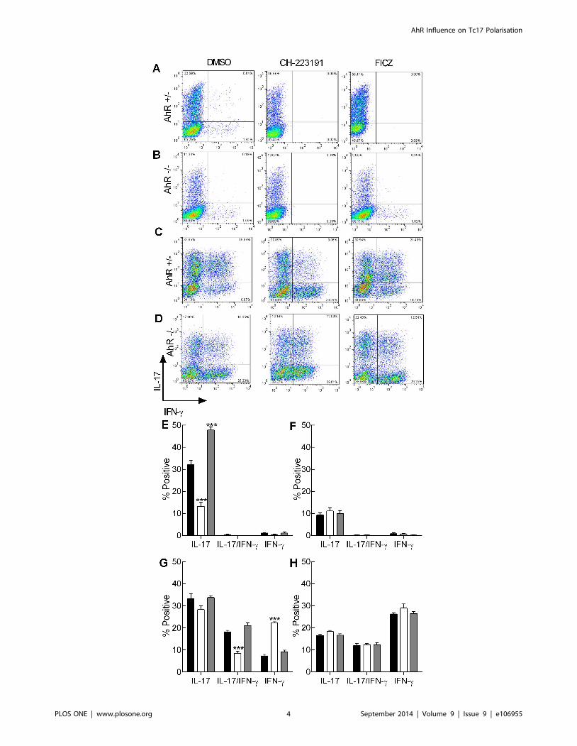
from CD4+ precursors from AhR+/2 mice were similar to those
published previously, with approximately 30% of cells expressing
intracellular IL-17 (Figure 1A, E). This frequency was significantly
reduced (by ,50%) in the presence of the AhR antagonist.
Conversely this population was markedly enhanced by the
addition of exogenous FICZ (Figure 1A, E). When Th17 cells
were generated from CD4+ precursors from AhR2/2 mice, the
maximum yield of IL-17+ cells varied between ,10–14%, and was
unaffected by the addition of the agonist or antagonist (Figure 1B,
F). Analysis of IFN-c production revealed that independent of
treatment or genotype, ,2% of cells produced the Th1 cytokine.
Further, no cells expressed both cytokines. However, CD8+ cells
polarised under identical conditions displayed significantly differ-
ent phenotypes. CD8+ cells cultured under Th17 conditions
displayed 3 distinct cytokine profiles; those that expressed only IL-
17 or IFN-c or those that expressed both cytokines. In cells from
AhR+/2 mice, the frequency of the Tc17 population was similar to
the relative proportion of Th17 cells (Figure 1C, G). However
unlike Th17 cells, there was no impact of AhR agonist or
antagonist (,30% of cells were IL17+ irrespective of treatment).
The population that was most susceptible to the addition of the
AhR ligands was the IL-17/IFN-c fraction, where a ,50%
reduction was observed following addition of the antagonist but
the agonist was without effect (Figure 1C, G). Interestingly, the
reduction in the double positive population coincided with a
reciprocal increase in the IFN-c single positive population. CD8+
cells from AhR2/2 mice polarised into 3 distinct populations
similar to those found in the heterozygotes. However, the
frequency of the single positive IL-17 cells remained around
20% under all conditions, and the double positive IL-17/IFN-cand single positive IFN-c populations remained approximately
10% and 25% respectively (Figure 1D, H). Thus, for the CD8+
dual expressing cells and the single positive IFN-c populations, cell
frequencies recorded for AhR+/2 derived cells in the presence of
antagonist were equivalent to those recorded for the AhR
knockout mice. In contrast, frequencies of single IL-17 expressing
AhR+/2 cells were somewhat higher than in the AhR2/2 mice,
suggesting that this population is less sensitive to the antagonist, at
least using this end point.
It has been suggested that the influence of AhR on CD8 cells is
dependent upon the maturation status of the cell [27]. Therefore
in order to rule out potential skewing of responses due to
differential numbers of effector versus naıve cells in the starting
CD8 cell population, the frequency of naive, central memory,
effector memory and effector cells was characterised by flow
cytometry (Table 1). In both strains of mice, fewer than 2% of cells
were of the effector memory or effector cell phenotype; the
majority of cells were central memory cells (,60%) and the
remainder (,40%) displayed a naıve phenotype, regardless of the
AhR genotype. These starting cell populations were also assessed
for cytokine expression by flow cytometry: consistent with the lack
of CD8 effector cells, there was no detectable IL-17 or IFN-cexpression by these initial populations of cells (data not shown).
The cells were also cultured under Tc0 conditions (anti-CD3 and
anti-CD28 but in the absence of polarising cytokines) for 5 days
and the frequency of cytokine expressing cells enumerated by flow
cytometry. Under these conditions, ,15% IFN-c only expressing
cells and ,1% double positive cells were recorded for cells isolated
from either AhR+/2 or AhR2/2 mice (Table 1), and there was no
impact of either antagonist or FICZ on these frequencies (data not
shown). Taken together, these data demonstrate that not only is
there is no detectable difference between the two mouse strains
with respect to the distribution of naive/memory/effector cells in
the starting population but also that it is unlikely that
precommitted CD8 effector cells are contributing to the observed
phenotype.
In parallel with measuring the frequency of cytokine expressing
cells, in vitro polarised cells were analysed for IL-17, IL-22 and
IFN-c expression at the level of mRNA by RT-PCR and cytokine
secretion by ELISA. The pattern of IL-17 mRNA and protein
secretion paralleled closely the results seen for intracellular
cytokine staining, with expression by AhR+/2 Th17 cells
significantly reduced by the antagonist and enhanced by the
addition of FICZ (Figure 2A, B). Furthermore, the AhR+/2 Th17
cells were the only population that expressed significant amounts
of IL-22 and then only in response to exogenous FICZ. In
contrast, production of both IL-17 and IL-22 by AhR2/2 Th17
cells was reduced to levels to those recorded for AhR+/2 Th17
AhR Influence on Tc17 Polarisation
PLOS ONE | www.plosone.org 3 September 2014 | Volume 9 | Issue 9 | e106955
AhR Influence on Tc17 Polarisation
PLOS ONE | www.plosone.org 4 September 2014 | Volume 9 | Issue 9 | e106955
cells following treatment with the antagonist (Figure 2); with these
relatively low levels unaffected by addition of either ligand. In
addition, neither heterozygous nor knockout Th17 cells expressed
significant IFN-c (Figure 2E, F). The levels of IL-17 expressed by
Tc17 cells from AhR+/2 mice were markedly lower than those
produced by Th17 cells from the same mice. Tc17 IL-17
production at the level of both message and secreted cytokine
was inhibited by the presence of the antagonist whereas addition of
exogenous FICZ was without effect (Figure 2A, B). As noted for
the AhR2/2 Th17 cells, Tc17 cells derived from the same mice
expressed relatively low levels of IL-17, again comparable with the
amounts produced when the AhR+/2 Tc17 cells were cultured
with the antagonist. This suggests that the lack of effect of the
antagonist on the frequency of single positive IL-17 CD8 cells
detected by flow cytometry is not functionally relevant. For the
majority of Tc17 cultures, IL-22 production was low or
undetectable, with the exception of AhR+/2 Tc17 cells cultured
in the presence of exogenous FICZ. However, this was still
dramatically lower than the amount expressed by Th17 cells from
the same mice cultured under the same conditions. Although the
Th17 cell populations did not express IFN-c, all CD8+ cells
cultured under Tc17 conditions produced this cytokine. Treating
the AhR+/2 CD8+ cells with the antagonist enhanced IFN-cexpression, whereas FICZ was without effect (Figure 2E, F).
Vigorous IFN-c expression was recorded for AhR2/2 CD8+ cells
which was largely unaffected by AhR ligands.
Kinetics of polarisation reveals differences between Th17and Tc17 development
To characterise potential differences between Th17 and Tc17
development, the kinetics of expression of a panel of genes was
examined. Th1 and Tc1 cells were also analysed, the former being
known to develop independently of AhR [24]. The expected Th1
and Tc1 patterns of cytokine expression and lack of impact of AhR
ligands were confirmed in day 5 polarised cells isolated from AhR
heterozygous and knockout mice (Figures S2–S4). Subsequently,
the kinetics of IL-17, IL-22 and IFN-c expression in Th1/Th17
and Tc1/Tc17 cells derived from AhR heterozygous mice was
examined. As expected, expression of IFN-c was highest in Th1
and Tc1 populations, with no impact of the AhR antagonist or
FICZ treatment. IFN-c levels were lower in the Tc17 populations
and reduced further still in the Th17 group (Figure 3A, B).
Conversely, analysis of IL-17 expression levels revealed that the
highest levels were achieved for Th17 cells, then Tc17 cells, with
FICZ treatment increasing IL-17 expression only for Th17 cells,
and the antagonist decreasing cytokine expression in each case
(Figure 3C, D). The pattern of IL-22 expression was very similar
for Th17 and Tc17 cells, with very profound inhibition by the
antagonist recorded (Figure 3E, F). Interestingly, whereas Tc17
cells expressed relatively high levels of IFN-c, levels of IL-17A, and
to some extent IL-22, were markedly down-regulated in Tc1 cells,
to levels considerably lower than those recorded in the naıve
precursor CD8+ cell population.
Finally, genes associated with AhR activation (AhRR; the AhR
repressor and Cyp1A1) and expression levels of the AhR itself
were analysed. Similar to previous reports [26], the AhR was up-
regulated under Th17 polarising conditions, whereas Th1 or Tc1
stimulation resulted in active down-regulation (Figure 4E, F).
Importantly, there was no up-regulation of AhR under Tc17
polarising conditions, with expression maintained at baseline levels
(Figure 4F). Expression of AhRR, which is stimulated in response
to AhR activators forming a negative feedback loop [28], was
remarkably similar between CD4+ and CD8+ cell polarisations.
The highest levels of this gene were recorded under Th17/Tc17
conditions in the presence of FICZ. While AhRR expression in all
cells, regardless of polarising conditions, was down-regulated by
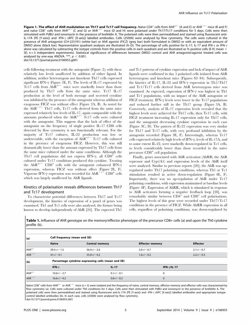
Figure 1. The effect of AhR modulation on Th17 and Tc17 cell frequency. Naıve CD4+ cells from AhR+/2 (A and E) or AhR2/2 mice (B and F)and naıve CD8+ cells from AhR+/2 (C and G) or AhR2/2 mice (D and H) were polarised under Th17/Tc17 conditions for 5 days. Cells were thenstimulated with PdBU and ionomycin in the presence of brefeldin A. The polarised cells were then permeabilised and stained using fluorescent anti-IL-17A (PE [Y-axis]) and -IFN-c (APC [X-axis]) labelled antibodies. Cells (25000) were analysed by flow cytometry. The cells were cultured in thepresence of AhR antagonist (CH-223191) (white bar) or AhR agonist (FICZ) (grey bar) both formulated in DMSO or with an equivalent amount ofDMSO alone (black bar). Representative quadrant analyses are illustrated (A–D). The percentage of cells positive for IL-17, IL-17 and IFN-c or IFN-calone was calculated by subtracting the isotype controls from the positive cells in each quadrant and are illustrated as % positive cells (E-H; mean 6SE; n = 3 independent experiments). Statistical significance of differences between DMSO control and AhR antagonist/agonist treated cells wasanalysed by one-way ANOVA. ***, p , 0.001.doi:10.1371/journal.pone.0106955.g001
Table 1. Influence of AhR genotype on the memory/effector phenotype of the precursor CD8+ cells (a) and upon the Th0 cytokineprofile (b).
(a) Cell frequency (mean and SE)
Naıve Central memory Effector memory Effector
AhR+/2 39.5+/21.5 56.4+/22.6 2.0+/20.7 2.1+/20.7
AhR2/2 41+/24.1 55.0+/24.2 1.6+/20.2 2.2+/20.3
(b) Percentage cytokine expressing cells (mean and SE)
IFN-c IL-17 IFN-c/IL-17
AhR+/2 10.6+/22.7 0.1+/20.1 0
AhR2/2 16.6+/24.2 0.6+/20.2 0
Naıve CD8+ cells from AhR+/2 or AhR2/2 mice (n = 3) were isolated and the frequency of naıve, central memory, effector memory and effector cells was characterised byflow cytometry (a). Cells were cultured under Th0 conditions for 5 days. Cells were then stimulated with PdBU and ionomycin in the presence of brefeldin A. Thepolarised cells were then permeabilised and stained using fluorescent anti-IL-17A (PE [Y-axis]) and -IFN-c (APC [X-axis]) labelled antibodies and appropriate isotypecontrol labelled antibodies (b). In each case, cells (25000) were analysed by flow cytometry.doi:10.1371/journal.pone.0106955.t001
AhR Influence on Tc17 Polarisation
PLOS ONE | www.plosone.org 5 September 2014 | Volume 9 | Issue 9 | e106955
the antagonist; the most marked effects were recorded for Th17/
Tc17 cells (Figure 4G, H). As Cyp1A1 has long been used as a
biomarker of AhR activation and also forms part of the negative
feedback loop by metabolising AhR ligands [29], the kinetics of its
expression was also investigated. The hierarchy of the responses
was striking similar to those recorded for AhRR. Despite being
independent of AhR for their polarisation (Figures S2–S4), the
Th1 and Tc1 cells also up-regulated Cyp1A1 in response to
exogenous FICZ, but to a lesser extent than Th17/Tc17 cells
(Figure 4I, J).
Discussion
In the investigations described here, dual strategies have been
employed to characterise the role played by AhR in the regulation
of Tc17 cell development. Both approaches (AhR agonism/
antagonism and AhR null mice) have revealed differences in the
development of Th17 and Tc17 cells, despite the fact the same
cytokine cocktail (IL-1b, IL-6 and TGF-b) is able to drive the
induction of both cell types. In line with previous studies [24,26] it
has been shown here that Th17 (but not Th1) cell development
requires AhR. Thus, Th17 responses were significantly compro-
mised in AhR2/2 mice, and were inhibited by AhR antagonist.
Conversely, Th17 responses were enhanced markedly by FICZ in
AhR+/2 mice. We have shown here that AhR is also required for
the development of Tc17 cells. Development of Tc17 cells was
reduced in AhR2/2 mice, and inhibited by the antagonist in
AhR+/2 mice. However, in contrast to Th17 cells, Tc17 cells were
largely refractory to FICZ, particularly at the level of IL-17
secretion. The implication is that baseline levels of expression of
AhR are necessary and sufficient to sustain maximal Tc17
polarisation. As expected, Tc1 development was completely
independent of AhR, indeed, during the polarisation of these
cells, AhR (and to a lesser extent RORC) was rapidly and
substantially down-regulated compared with baseline levels in
naıve CD8+ precursor cells.
Although the results described here on the role of AhR in Th17
polarisation are largely consistent with those reported previously,
there are some intriguing differences. Thus, Veldhoen et al [26]
demonstrated that availability of natural AhR ligands (namely,
FICZ) was sufficient in IMDM media (as a result of UV light
exposure from natural sunlight or laboratory lighting [26,30,31])
to stimulate IL-22 expression by Th17 cells. In contrast, in our
hands, supplementation of IMDM media with additional FICZ
was required for optimal IL-22 expression. The inference being
that there exists inter-laboratory variation in storage conditions
may impact on the AhR ligand content of media. It could be
argued that such differences might be attributable to the use in our
studies of heterozygous AhR+/2 control mice on a C57BL/6J
background, rather than wild type C57BL/6 mice as used by
Veldhoen et al [26]. Consistent with this is the fact that loss of a
single AhR allele can have effects on regulation of blood pressure
and heme metabolism [32,33]. Furthermore, reduced IL-17+ Th
cell numbers have been reported in AhR+/2 mice compared with
wild type controls [34], although it should be noted that baseline
levels were very low in that series of experiments (,10%). In order
to address this question directly, comparisons were made of IL-17
polarisation in cells derived from AhR2/2, AhR+/2 and AhR+/+
mice (Figure S5). These experiments revealed that all measures of
IL-17 expression (IL-17+ cell frequency, IL-17 transcript expres-
sion and protein production) were equivalent between AhR+/+ and
AhR+/2 littermates, for both baseline and FICZ inducible
responses. In the current series of experiments, therefore, the loss
of one AhR allele does not impact on Th17 cell development, and
thus AhR+/2 littermates are appropriate controls for the AhR null
mice.
The primary focus of these investigations was, however, to
characterise the role of AhR in Tc17 development. One marked
difference between Tc17 cells compared with Th17 cells was the
plasticity of the cytokine producing phenotype and the impact of
blocking of the AhR on this phenotype. Whereas in vitro polarised
Th17 cells produced only IL-17, Tc17 cells were found to produce
IL-17 and/or IFN-c. Furthermore, Tc17 (but not Th17)
development in vitro was associated with initial up-regulation of
the type 1 master regulator, T-bet. This is consistent with previous
reports of the existence of IL-17/IFN-c double positive Tc17 cells
in mice, albeit with considerably lower frequencies than those
reported here [12]. Strikingly, the IL-17 single positive population
of Tc17 cells was unresponsive to either stimulation or antagonism
of AhR, whereas the IL-17/IFN-c double positive cells were
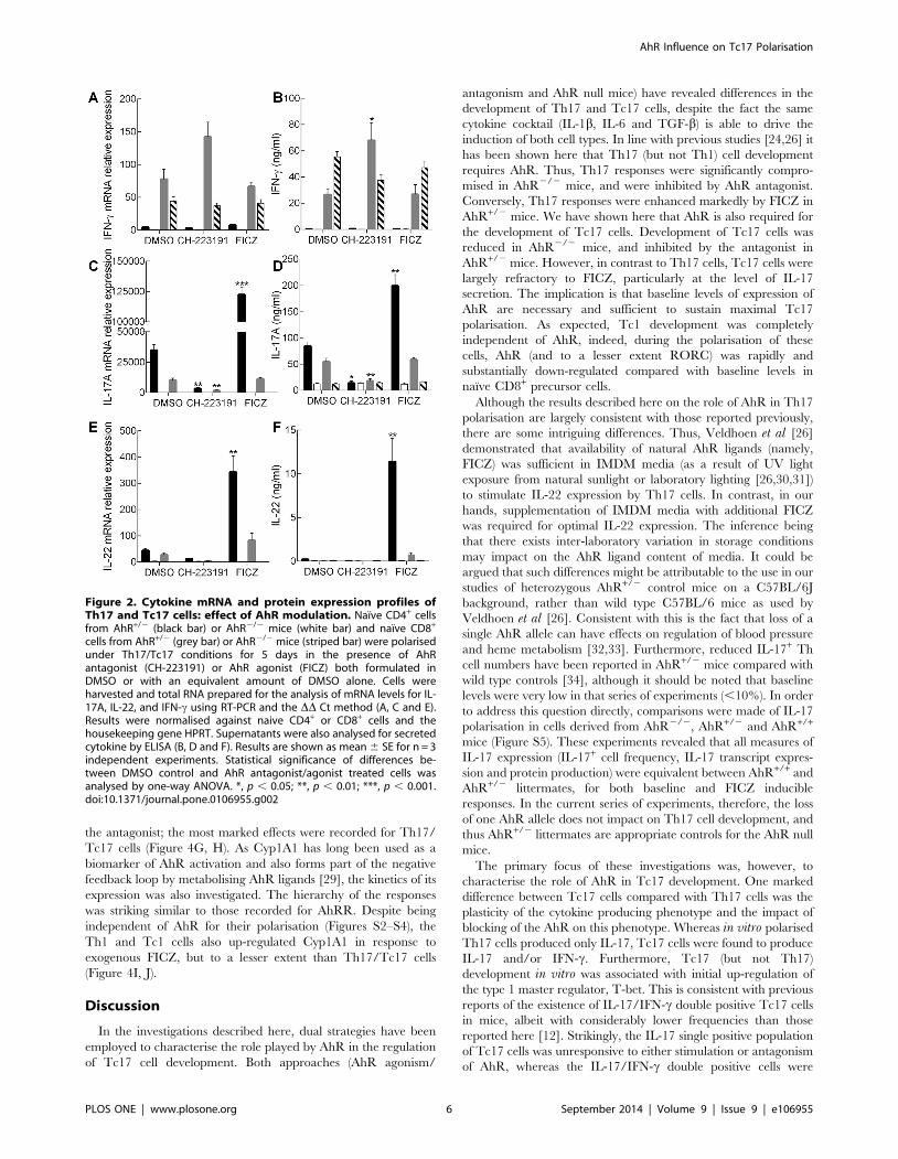
Figure 2. Cytokine mRNA and protein expression profiles ofTh17 and Tc17 cells: effect of AhR modulation. Naıve CD4+ cellsfrom AhR+/2 (black bar) or AhR2/2 mice (white bar) and naıve CD8+
cells from AhR+/2 (grey bar) or AhR2/2 mice (striped bar) were polarisedunder Th17/Tc17 conditions for 5 days in the presence of AhRantagonist (CH-223191) or AhR agonist (FICZ) both formulated inDMSO or with an equivalent amount of DMSO alone. Cells wereharvested and total RNA prepared for the analysis of mRNA levels for IL-17A, IL-22, and IFN-c using RT-PCR and the DD Ct method (A, C and E).Results were normalised against naive CD4+ or CD8+ cells and thehousekeeping gene HPRT. Supernatants were also analysed for secretedcytokine by ELISA (B, D and F). Results are shown as mean 6 SE for n = 3independent experiments. Statistical significance of differences be-tween DMSO control and AhR antagonist/agonist treated cells wasanalysed by one-way ANOVA. *, p , 0.05; **, p , 0.01; ***, p , 0.001.doi:10.1371/journal.pone.0106955.g002
AhR Influence on Tc17 Polarisation
PLOS ONE | www.plosone.org 6 September 2014 | Volume 9 | Issue 9 | e106955
susceptible to both, converting to exclusive IFN-c expression when
the AhR was blocked. This functional plasticity driven by AhR
activation may provide for a fine tuning mechanism in vivo,
permitting effector activity to be tailored according to local need in
response to microenvironmental cues. Plasticity of IL-17 express-
ing cells (primarily Th17 cells) has been demonstrated previously
in vivo in mouse models of autoimmune disease, particularly in
chronic inflammatory conditions, or during pathogenic challenge
[16,17,35]. Th17 conversion to IL-17/IFN-c double positive cells
and ultimately IFN-c producing Th1 cells (designated exTh17) has
been shown to be dependent upon IL-23 [16]. Whereas IL-12,
rather than IL-23, has been shown to permit the conversion of
mouse IL-17-producing Tc17 cells to IL-17/IFN-c-double pro-
ducing Tc17/IFN-c cells [12,36]. Furthermore, cells that copro-
duce IL-17A and Tc1 cytokines (IL-2, IFN-c and TNF-a) in
response to Salmonella Typhi have been identified in humans,
indicating that the distinction between Tc17 and Tc1 responses is
not as clearly differentiated as suggested previously and that such
cells may be important in protection [37]. We demonstrate here
that Tc17 cells can develop into IL-17/IFN-c expressing cells in
the absence of exogenous IL-12, and that the conversion to
exclusive IFN-c producers can be influenced by AhR modulation,
providing a further potential level of regulation for such cells invivo.
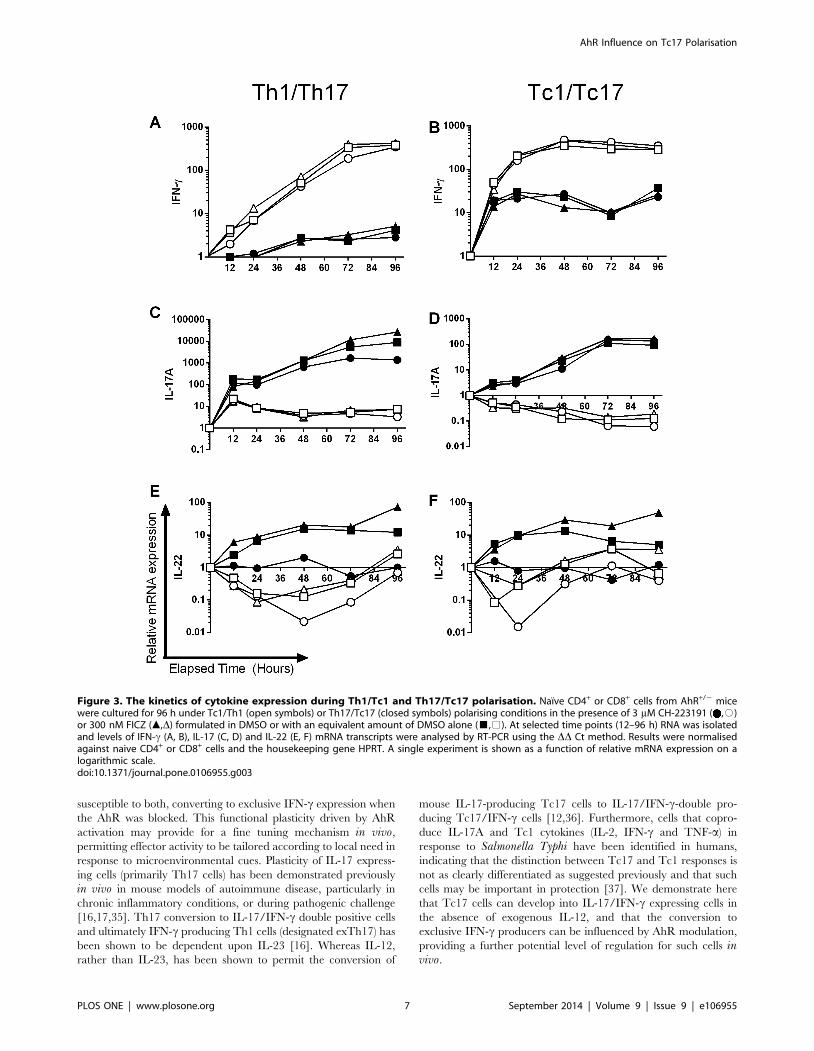
Figure 3. The kinetics of cytokine expression during Th1/Tc1 and Th17/Tc17 polarisation. Naıve CD4+ or CD8+ cells from AhR+/2 micewere cultured for 96 h under Tc1/Th1 (open symbols) or Th17/Tc17 (closed symbols) polarising conditions in the presence of 3 mM CH-223191 ( ,#)or 300 nM FICZ (m,D) formulated in DMSO or with an equivalent amount of DMSO alone (&,%). At selected time points (12–96 h) RNA was isolatedand levels of IFN-c (A, B), IL-17 (C, D) and IL-22 (E, F) mRNA transcripts were analysed by RT-PCR using the DD Ct method. Results were normalisedagainst naive CD4+ or CD8+ cells and the housekeeping gene HPRT. A single experiment is shown as a function of relative mRNA expression on alogarithmic scale.doi:10.1371/journal.pone.0106955.g003
AhR Influence on Tc17 Polarisation
PLOS ONE | www.plosone.org 7 September 2014 | Volume 9 | Issue 9 | e106955
AhR Influence on Tc17 Polarisation
PLOS ONE | www.plosone.org 8 September 2014 | Volume 9 | Issue 9 | e106955
There were a number of other differences between Th17 and
Tc17 cells, including the fact that Tc17 polarisation, unlike Th17
activation, was not associated with increased expression of AhR,
and was relatively refractory to exogenous FICZ. The Tc17 cells
were sensitive to AhR stimulation as Cyp1A1, a biomarker of AhR
activation, was noticeably up-regulated. We speculated that
differential regulation of AhR in CD4+ and CD8+ cells might be
driven through variable expression of AhRR, which is involved in
a negative feedback loop [28]. However, apart from a slightly
more rapid induction of the repressor in Tc17 cells, maximal levels
and persistence of stimulation were very similar between the two
cell types. Regardless of the potential mechanism, the results
demonstrate that Th17 cells are up-regulated more effectively
through the AhR and are more responsive to the natural ligand
FICZ than are their Tc17 counterparts. This mechanism in CD4+
cells may be necessary to ensure the robust differentiation of Th17
cells. Unlike the skewing of Th1 and Th2 cells, where their
respective major cytokines (IFN-c and IL-4) act as self-amplifying
immunomodulators, it is IL-21, not the major cytokine IL-17, that
acts in autocrine fashion on differentiation of Th17 cells. Further
amplification through AhR therefore provides a supplementary
mechanism through which vigorous Th17 responses are main-
tained.
Given the increased interest in the role of the AhR in
orchestrating Th17 cell responses, it is not surprising that it is
being considered currently as a therapeutic target in a range of
Th17 autoimmune diseases [38,39]. However, in mouse models of
psoriasis blocking of the AhR unexpectedly resulted in exacerba-
tion of the disease [40]. In the light of the data presented herein, it
is possible that this finding might be related to Tc17 responses, as
blocking the AhR may result in the acquisition of an exTc17
phenotype, with these cells having lytic ability[17], which could
cause disease progression. Interestingly, there are a number of
other examples of autoimmune disorders that were initially
attributed to Th17 cell pathogenesis that may in fact be elicited,
at least partly, by Tc17 cells. These include experimental (mouse)
models of autoimmune uveoretinitis, autoimmune encephalomy-
elitis and hepatic fibrosis and clinical studies in which Tc17 cells
have been identified in peripheral blood or lesions, including
rheumatoid arthritis and multiple sclerosis [15,41–43].
In conclusion, Tc17 cells have been shown to exhibit identical
cytokine requirements for polarisation to their Th17 counterparts.
Additionally, AhR plays a role in Tc17 development. However,
although baseline AhR expression is required for optimal Tc17
development, these cells do not up-regulate the receptor during
development, and do not respond vigorously to high levels of
endogenous ligand. Thus, maximal Th17 cell responses are more
dependent upon AhR activation than is Tc17 cell development,
suggesting that endogenous AhR ligands play a much greater role
in driving Th17 cell responses. Furthermore, Tc17 cells display
greater plasticity than do Th17 cells, and may be able, therefore,
to respond to different microenvironments by switching between
Tc1 and Tc17 phenotypes.
Supporting Information
Figure S1 Gating strategy for in vitro polarised Th1/Tc1 cells.
Naive CD4+ (A) or CD8+ (B) (1.25 6105/ml) cells from AhR
heterozygote controls were cultured for 5 days under Th1/Tc1
(IL-12) polarising conditions. After 5 days in culture cells were
stimulated with PdBU and ionomycin in the presence of brefeldin
A. The polarised cells were then permeabilised and labelled using
fluorescent anti-IL-17A (PE) and -IFN-c (APC) labelled antibodies
in combination with appropriate isotype controls. Cells (25000)
were analysed by flow cytometry. Results from a representative
experiment are displayed as gated analyses. An identical gating
strategy was employed for the AhR-/- mice (data not shown).
(TIFF)
Figure S2 The effect of AhR modulation on Th1 and Tc1 cell
frequency. Naıve CD4+ cells from AhR+/- (A) or AhR-/- mice (B)
and naive CD8+ cells from AhR+/- (C) or AhR-/- mice (D) were
polarised under Th1/Tc1 conditions for 5 days. The cells were
cultured in the presence of AhR antagonist (CH-223191) (white
bar) or AhR agonist (FICZ) (grey bar) both formulated in DMSO
or with an equivalent amount of DMSO alone (black bar). Cells
were then stimulated with PdBU and ionomycin in the presence of
brefeldin A, followed by permeabilisation and staining with
fluorescent-labelled anti-IL-17A (PE [Y-axis]) and -IFN-c (APC
[X-axis]) labelled antibodies. Cells (25000) were analysed by flow
cytometry. The percentage of cells positive for IL-17, IL-17 and
IFN-c or IFN-c alone was calculated by subtracting the isotype
controls from the stained cells in each quadrant. Representative
quadrant analyses are shown (A-D) and percentage positive cells
(E) are displayed as mean 6 SE for n = 3 independent
experiments. The statistical significance of differences between
DMSO control and AhR antagonist/agonist treated cells and of
differences between AhR+/- and AhR-/- mice was analysed by one-
way ANOVA. No significant differences were recorded.
(TIF)
Figure S3 Cytokine mRNA and protein expression profiles of
Th1 cells : effect of AhR modulation. Naıve CD4+ cells from
AhR+/2 (black bar) or AhR2/2 mice (white bar) were polarised
under Th1 conditions for 5 days. The cells were cultured in the
presence of AhR antagonist (CH-223191) or AhR agonist (FICZ)
both formulated in DMSO or with an equivalent amount of
DMSO alone. Total RNA was isolated and levels of mRNA
transcripts for IFN-c, IL-17A and IL-22 were analysed using RT-
PCR and the DD Ct method (A, C and E). Results were
normalised against naive CD4+ cells and the housekeeping gene
HPRT. Supernatants were also analysed for secreted cytokine by
ELISA (B, D and F). Results are shown as mean 6 SE for n = 3
independent experiments. The statistical significance of differences
between DMSO control and AhR antagonist/agonist was
analysed by one-way ANOVA. **, p,0.01.
(TIF)
Figure S4 Cytokine mRNA and protein expression profiles of
Tc1 cells: effect of AhR modulation. Naıve CD8+ cells from
AhR+/2 (black bar) or AhR2/2 mice (white bar) were polarised
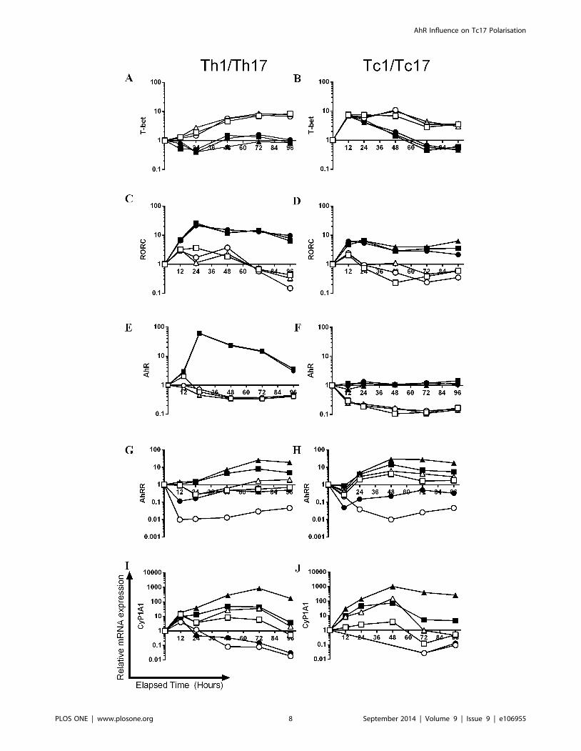
Figure 4. The kinetics of expression of master transcription factors and AhR associated genes during Th1/Tc1 and Th17/Tc17polarisation. Naıve CD4+ or CD8+ cells from AhR+/2 mice were cultured for 96 h under Tc1/Th1 (open symbols) or Th17/Tc17 (closed symbols)polarising conditions in the presence of presence of 3 mM CH-223191 ( ,#) or 300 nM FICZ (m,D) formulated in DMSO or with an equivalent amountof DMSO alone (&,%). At selected time points (12–96 h) total RNA was isolated and levels of mRNA transcripts for RORC (A, B), T-bet (C, D), AhR (E, F),AhRR (G, H) and Cyp1A1 (I, J) were analysed by RT-PCR using the DD Ct method. Results were normalised against naive CD4+ or CD8+ cells and thehousekeeping gene HPRT. A single experiment is shown as a function of relative mRNA expression on a logarithmic scale. If expression levels weretoo low to achieve a meaningful CT value for a given sample, due to the logarithmic scale, these data were not illustrated graphically (12–48 hCH223191 data in J).doi:10.1371/journal.pone.0106955.g004
AhR Influence on Tc17 Polarisation
PLOS ONE | www.plosone.org 9 September 2014 | Volume 9 | Issue 9 | e106955
under Th1/Tc1 conditions for 5 days. The cells were cultured in
the presence of AhR antagonist (CH-223191) or AhR agonist
(FICZ) both formulated in DMSO or with an equivalent amount
of DMSO alone. Total RNA was isolated and levels of mRNA
transcripts for IFN-c, IL-17A and IL-22 were analysed using RT-
PCR and the DD Ct method (A, C and E). Results were
normalised against naive CD8+ cells and the housekeeping gene
HPRT. Supernatants were also analysed for secreted cytokine by
ELISA (B, D and F). The statistical significance of differences
between DMSO control and AhR antagonist/agonist treated cells
was analysed by one-way ANOVA. No significant differences were
recorded.
(TIF)
Figure S5 Th17 polarisation and impact of exogenous FICZ :
role of AhR phenotype. Naive CD4+ cells from wild type, AhR+/2
and AhR2/2 mice were cultured for 5 days under Th17 (IL-6,
TGF-b and IL-1b) polarising conditions in the presence of the
AhR agonist FICZ (&) (300 nM) or DMSO vehicle alone ( ).
Cells were stimulated with PdBU and ionomycin in the presence of
brefeldin A. The polarised cells were then permeabilised and
labelled using fluorescent anti-IL-17A (PE) labelled antibodies.
Cells (25000) were analysed by flow cytometry and are shown as
percentage IL-17A positive for each condition (A). Changes in IL-
17A mRNA levels were analysed using RT-PCR and the DDCt
method, normalised against naive CD4+ cells and the housekeep-
ing gene HPRT (B). Concentrations of IL-17A were analysed by
ELISA in supernatants prepared following 5 days culture of the
CD4+ cells (C). Results are displayed as individual animals (n = 3).
The statistical significance of differences between DMSO control
and AhR agonist treated cells were analysed by one-way ANOVA.
**, p,0.01, ***, p,0.001.
(TIF)
Acknowledgments
We would like to thank Professor Brigitta Stockinger for supply of the
AhR2/2 and heterozygote mice and also for her help and advice regarding
methods for in vitro polarisation of IL-17 secreting cells. We would also like
to thank Kieran Mellody for technical assistance.
Author Contributions
Conceived and designed the experiments: MH VO AS IK RD. Performed
the experiments: MH VO RD. Analyzed the data: MH VO AS IK RD.
Contributed reagents/materials/analysis tools: MH VO AS IK RD. Wrote
the paper: MH VO AS IK RD.
References
1. Cerwenka A, Carter LL, Reome JB, Swain SL, Dutton RW (1998) In vivo
persistence of CD8 polarized T cell subsets producing type 1 or type 2 cytokines.
J Immunol 161: 97–105.
2. Doherty PC, Topham DJ, Tripp RA, Cardin RD, Brooks JW, et al. (1997)
Effector CD4+ and CD8+ T-cell mechanisms in the control of respiratory virus
infections. Immunol Rev 159: 105–17.
3. Waite JC, Skokos D (2012) Th17 response and inflammatory autoimmune
diseases. Int J Inflam 2012: 819467.
4. Di Cesare A, Di Meglio P, Nestle FO (2009) The IL-23/Th17 axis in the
immunopathogenesis of psoriasis. J Invest Dermatol 129: 1339–50.
5. Brand S (2009) Crohn’s disease: Th1, Th17 or both? The change of a paradigm:
new immunological and genetic insights implicate Th17 cells in the pathogenesis
of Crohn’s disease. Gut 58: 1152–67.
6. Hirota K, Ahlfors H, Duarte JH, Stockinger B (2012) Regulation and function of
innate and adaptive interleukin-17-producing cells. EMBO Reports 13: 113–20.
7. Sonnenberg GF, Fouser LA, Artis D (2011) Border patrol: regulation of
immunity, inflammation and tissue homeostasis at barrier surfaces by IL-22. Nat
Immunol 12: 383–90.
8. Hwang SY, Kim JY, Kim KW, Park MK, Moon Y, et al. (2004) IL-17 induces
production of IL-6 and IL-8 in rheumatoid arthritis synovial fibroblasts via NF-
kappaB- and PI3-kinase/Akt-dependent pathways. Arthritis Res Ther 6: R120–
8.
9. Beklen A, Ainola M, Hukkanen M, Gurgan C, Sorsa T, et al. (2007) MMPs, IL-
1, and TNF are regulated by IL-17 in periodontitis. J Dent Res 86: 347–51.
10. Kourilsky P, Truffa-Bachi P (2001) Cytokine fields and the polarization of the
immune response. Trends Immunol 22: 502–9.
11. Hamada H, Garcia-Hernandez MeL, Reome JB, Misra SK, Strutt TM, et al.
(2009) Tc17, a unique subset of CD8 T cells that can protect against lethal
influenza challenge. J Immunol 182: 3469–81.
12. Yen HR, Harris TJ, Wada S, Grosso JF, Getnet D, et al. (2009) Tc17 CD8 T
cells: functional plasticity and subset diversity. J Immunol 183: 7161–8.
13. Hu Y, Ma DX, Shan NN, Zhu YY, Liu XG, et al. (2011) Increased number of
Tc17 and correlation with Th17 cells in patients with immune thrombocyto-
penia. PLoS One 6: e26522.
14. Hamada H, Bassity E, Flies A, Strutt TM, Garcia-Hernandez MeL, et al. (2013)
Multiple redundant effector mechanisms of CD8+ T cells protect against
influenza infection. J Immunol 190: 296–306.
15. Eysteinsdottir JH, Sigurgeirsson B, Olafsson JH, Friðriksson T, Agnarsson BA, et
al. (2013) The role of Th17/Tc17 peripheral blood T cells in psoriasis and their
positive therapeutic response. Scand J Immunol 78(6): 529–37.
16. Hirota K, Duarte JH, Veldhoen M, Hornsby E, Li Y, et al. (2011) Fate mapping
of IL-17-producing T cells in inflammatory responses. Nat Immunol 12: 255–
U95.
17. Yeh N, Glosson NL, Wang N, Guindon L, McKinley C, et al. (2010) Tc17 cells
are capable of mediating immunity to vaccinia virus by acquisition of a cytotoxic
phenotype. J Immunol 185: 2089–98.
18. Bettelli E, Korn T, Kuchroo VK (2007) Th17: the third member of the effector
T cell trilogy. Curr Opin Immunol 19: 652–7.
19. McGeachy MJ, Cua DJ (2008) Th17 cell differentiation: the long and winding
road. Immunity 28: 445–53.
20. Ko HP, Okino ST, Ma Q, Whitlock JP (1996) Dioxin-induced CYP1A1transcription in vivo: the aromatic hydrocarbon receptor mediates transactiva-
tion, enhancer-promoter communication, and changes in chromatin structure.
Mol Cell Biol 16: 430–6.
21. Ho PP, Steinman L (2008) The aryl hydrocarbon receptor: a regulator of Th17
and Treg cell development in disease. Cell Res 18: 605–8.
22. Denison MS, Nagy SR (2003) Activation of the aryl hydrocarbon receptor by
structurally diverse exogenous and endogenous chemicals. Annu Rev PharmacolToxicol 43: 309–34.
23. Harper PA, Riddick DS, Okey AB (2006) Regulating the regulator: factors thatcontrol levels and activity of the aryl hydrocarbon receptor. Biochem Pharmacol
72: 267–79.
24. Veldhoen M, Hirota K, Westendorf AM, Buer J, Dumoutier L, et al. (2008) The
aryl hydrocarbon receptor links TH17-cell-mediated autoimmunity to environ-mental toxins. Nature 453: 106–9.
25. Bachmann MF, Wolint P, Schwarz K, Jager P, Oxenius A (2005) Functionalproperties and lineage relationship of CD8+ T cell subsets identified by
expression of IL-7 receptor alpha and CD62L. J Immunol. 175: 4686–96.
26. Veldhoen M, Hirota K, Christensen J, O’Garra A, Stockinger B (2009) Natural
agonists for aryl hydrocarbon receptor in culture medium are essential foroptimal differentiation of Th17 T cells. J Exp Med 206: 43–9.
27. Kerkvliet NI, Shepherd DM, Baecher-Steppan L (2002) T lymphocytes aredirect, aryl hydrocarbon receptor (AhR)-dependent targets of 2,3,7,8-tetrachlo-
rodibenzo-p-dioxin (TCDD): AhR expression in both CD4+ and CD8+ T cells is
necessary for full suppression of a cytotoxic T lymphocyte response by TCDD.Toxicol Appl Pharmacol 185(2): 146–152.
28. Evans BR, Karchner SI, Franks DG, Hahn ME (2005) Duplicate arylhydrocarbon receptor repressor genes (ahrr1 and ahrr2) in the zebrafish Danio
rerio: structure, function, evolution, and AHR-dependent regulation in vivo.Arch Biochem Biophys 441: 151–67.
29. Hu W, Sorrentino C, Denison MS, Kolaja K, Fielden MR (2007) Induction ofcyp1a1 is a nonspecific biomarker of aryl hydrocarbon receptor activation:
results of large scale screening of pharmaceuticals and toxicants in vivo and in
vitro. Mol Pharmacol 71: 1475–86.
30. Oberg M, Bergander L, Hakansson H, Rannug U, Rannug A (2005)Identification of the tryptophan photoproduct 6-formylindolo[3,2-b]carbazole,
in cell culture medium, as a factor that controls the background aryl
hydrocarbon receptor activity. Toxicol Sci 85: 935–43.
31. Rannug U, Rannug A, Sjoberg U, Li H, Westerholm R, et al. (1995) Structure
elucidation of two tryptophan-derived, high affinity Ah receptor ligands. ChemBiol 2: 841–5.
32. Zhang N, Agbor LN, Scott JA, Zalobowski T, Elased KM, et al. (2010) Anactivated renin-angiotensin system maintains normal blood pressure in aryl
hydrocarbon receptor heterozygous mice but not in null mice. BiochemPharmacol 80: 197–204.
33. Davies R, Clothier B, Robinson SW, Edwards RE, Greaves P, et al. (2008)Essential role of the AH receptor in the dysfunction of heme metabolism induced
by 2,3,7,8-tetrachlorodibenzo-p-dioxin. Chem Res Toxicol 21: 330–40.
34. Kimura A, Naka T, Nohara K, Fujii-Kuriyama Y, Kishimoto T (2008) Aryl
hydrocarbon receptor regulates Stat1 activation and participates in thedevelopment of Th17 cells. Proc Natl Acad Sci U S A 105: 9721–6.
AhR Influence on Tc17 Polarisation
PLOS ONE | www.plosone.org 10 September 2014 | Volume 9 | Issue 9 | e106955
35. Bending D, De la Pena H, Veldhoen M, Phillips JM, Uyttenhove C, et al. (2009)
Highly purified Th17 cells from BDC2.5NOD mice convert into Th1-like cellsin NOD/SCID recipient mice. J Clin Invest 119: 565–72.
36. Satoh T, Tajima M, Wakita D, Kitamura H, Nishimura T (2012) The
development of IL-17/IFN-c-double producing CTLs from Tc17 cells is drivenby epigenetic suppression of Socs3 gene promoter. Eur J Immunol 42: 2329–42.
37. McArthur MA, Sztein MB (2012) Heterogeneity of multifunctional IL-17Aproducing S. Typhi-specific CD8+ T cells in volunteers following Ty21a typhoid
immunization. PLoS One 7: e38408.
38. Quintana FJ, Basso AS, Iglesias AH, Korn T, Farez MF, et al. (2008) Control ofT(reg) and T(H)17 cell differentiation by the aryl hydrocarbon receptor. Nature
453: 65–71.39. Zhang L, Ma J, Takeuchi M, Usui Y, Hattori T, et al. (2010) Suppression of
experimental autoimmune uveoretinitis by inducing differentiation of regulatory
T cells via activation of aryl hydrocarbon receptor. Invest Ophthalmol Vis Sci
51: 2109–17.40. Di Meglio P, Duarte JH, Muller H, Hirota K, Nestle FO, et al. (2012) The Aryl
hydrocarbon receptor modulates psoriasis-like skin inflammation. J Invest
Dermatol 132: S14–S35.41. Huber M, Heink S, Grothe H, Guralnik A, Reinhard K, et al. (2009) A Th17-
like developmental process leads to CD8(+) Tc17 cells with reduced cytotoxicactivity. Eur J Immunol 39: 1716–25.
42. Huber M, Heink S, Pagenstecher A, Reinhard K, Ritter J, et al. (2013) IL-17A
secretion by CD8+ T cells supports Th17-mediated autoimmune encephalo-myelitis. J Clin Invest 123: 247–60.
43. Henriques A, Gomes V, Duarte C, Pedreiro S, Carvalheiro T, et al. (2013)Distribution and functional plasticity of peripheral blood Th(c)17 and Th(c)1 in
rheumatoid arthritis. Rheumatol Int 33: 2093–9.
AhR Influence on Tc17 Polarisation
PLOS ONE | www.plosone.org 11 September 2014 | Volume 9 | Issue 9 | e106955