the application of medical infrared thermography...
TRANSCRIPT
14
The Application of Medical Infrared Thermography in Sports Medicine
Carolin Hildebrandt1, Karlheinz Zeilberger2, Edward Francis John Ring3 and Christian Raschner1
1University of Innsbruck, Department of Sport Science, Innsbruck, 2Medical Practices for Internal and Sports Medicine, Munich,
3Medical Imaging Research Group, Faculty of Advanced Technology, University of Glamorgan,
1Austria 2Germany
3UK
1. Introduction
Medical Infrared Thermography (MIT) is a non-radiating and contact-free technology to monitor physiological functions related to skin temperature control. The efficiency, safety and low cost of MIT make it a useful auxiliary tool for detecting and locating thermal abnormalities characterized by increases or decreases in skin surface temperature. It has been successfully utilized in the field of veterinary medicine to detect locomotion injuries in racehorses and to monitor their health status. However, research on human athletes with modern infrared sensor technology is more rare. Athletes are exposed to physical stress in training and during competition season. Overuse reactions and so-called “minor traumas” are very frequent; therefore, early detection is critical to avoid injuries. Research suggests that the most beneficial application of MIT is the screening of individuals for overuse injuries. In the following chapters, the use of MIT in clinical practice is presented with special focus on sports injuries and exercise-induced physiological functions. Case studies illustrate the clinical applicability.
2. MIT – Quo Vadis?
2.1 History and development The association between changes in temperature and disease is almost as old as medicine itself. Hippocrates stated, “should one part of the body be hotter or colder than the rest, then disease is present in that part”. The first application of thermal imaging was in the early 19th century and did not have any commercial purpose. Following the 2nd World War, infrared imaging systems were used to monitor changes in skin temperature in relation to certain diseases (Ring, 2007). Poor quality imaging systems and a lack of methodological standards in the past has limited quality, resulting in non-acceptance of the technique (Elliot & Head, 1999). Technological advances in infrared cameras within the last few years have promoted MIT as a powerful measurement tool. A new generation of high-resolution cameras,
www.intechopen.com
An International Perspective on Topics in Sports Medicine and Sports Injury
258
appropriate software and standardized protocols have been developed for medical imaging, resulting in improved diagnostic capability and reliability (Plassmann et al., 2006; Diakides & Bronzino, 2008). In 1987, the American Medical Association recognized MIT as a feasible diagnostic tool. The following worldwide Thermographic organizations promote the proper application of medical thermal imaging. International Academy of Clinical Thermology International Thermographic Society American Academy of Medical Infrared Imaging European Association of Thermology Northern Norwegian Centre for Medical Thermography German Society of Thermography and Regulation Medicine
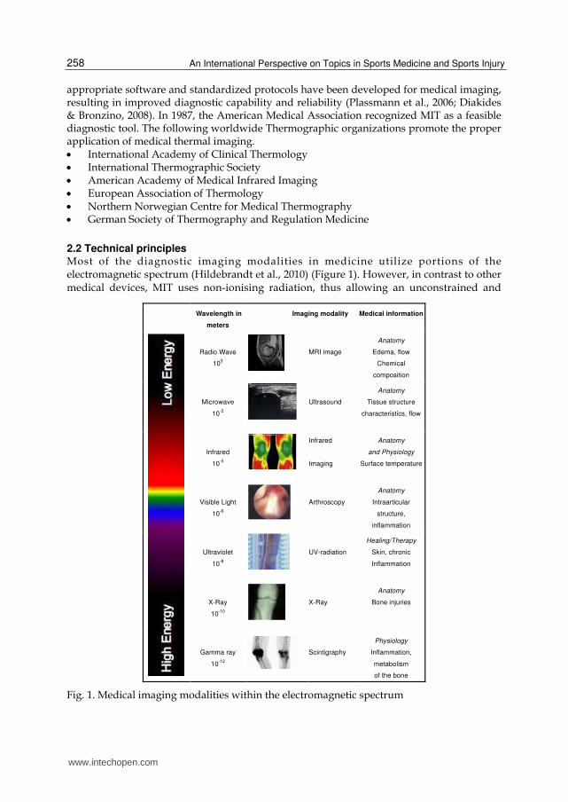
2.2 Technical principles Most of the diagnostic imaging modalities in medicine utilize portions of the electromagnetic spectrum (Hildebrandt et al., 2010) (Figure 1). However, in contrast to other medical devices, MIT uses non-ionising radiation, thus allowing an unconstrained and
Wavelength in
meters
Imaging modality Medical information
Radio Wave
103
MRI image
Anatomy
Edema, flow
Chemical
composition
Microwave
10-2
Ultrasound
Anatomy
Tissue structure
characteristics, flow
Infrared
10-5
Infrared
Imaging
Anatomy
and Physiology
Surface temperature
Visible Light
10-6
Arthroscopy
Anatomy
Intraarticular
structure,
inflammation
Ultraviolet
10-8
UV-radiation
Healing/Therapy
Skin, chronic
Inflammation
X-Ray
10-10
X-Ray
Anatomy
Bone injuries
Gamma ray
10-12
Scintigraphy
Physiology
Inflammation,
metabolism
of the bone
Fig. 1. Medical imaging modalities within the electromagnetic spectrum
www.intechopen.com
The Application of Medical Infrared Thermography in Sports Medicine
259
harmless application in patients. Using infrared radiation, infrared cameras generate
thermal images based on the amount of heat dissipated at the surface. Roughly 80% of the
emitted infrared radiation of human skin is in the wavelength range of 8-15µm (Steketee,
1973). The technology operates in the long-wave infrared region and is a sophisticated way
of receiving electromagnetic radiation and converting it into electrical signals. These signals
are finally displayed and matched to colors on the screen for calculations. Modern focal
plane array detectors ensure a stable image with high thermal resolution. Sensitivity and
resolution are important parameters for medical devices (Plassmann et al., 2006). High-
resolution cameras with focal plane arrays of 320×240 pixels, a thermal sensitivity less than
50mK and a spatial resolution of 25-50μm ensure useful thermal and spatial details (Ring &
Ammer, 2000). The resulting information can be used to provide instant feedback on the
patient or athlete. Unlike other medical imaging modalities, MIT is not related to
morphology. However, to study cutaneous circulation, the non-contact method of MIT was
compared with other medical imaging modalities. Merla et al. (2007) calculated blood flow
by using MIT and laser Doppler imaging (LDI) and showed that cutaneous blood perfusion
values obtained from MIT correlate with those obtained by means of LDI and have the
advantage of a better time resolution.
2.3 Biological principles Human skin, with an emissivity (an object’s ability to emit radiation) of 0.98, is almost equal
to a black body radiator (Steketee, 1973). The physics of heat radiation and the physiology of
thermoregulation in the human body make the reliable and valid interpretation of thermal
images difficult. Skin temperature regulation is a complex system that depends on blood-
flow rate, local structures of subcutaneous tissues and the activity of the sympathetic nervous
system (Kellog & Pergola, 2000). However, there is evidence that the sympathetic nervous
system is the primary regulator of blood circulation in the skin and is, therefore, the primary
regulator of thermal emission (Charkoudian, 2003). Vasoconstriction and vasodilation of the
blood vessels function to regulate blood flow in the skin. Thermoreceptors in the skin, also
known as Ruffini corpuscles, recognize the ambient temperature. An increased temperature
results in vasodilation, leading to increased blood flow to the skin, whereas vasoconstriction
occurs by a decrease in temperature and results in reduced blood flow to the skin (Wallin,
1990). These physiological processes combine with heat transfer and thermoregulation in
convection, conduction, radiation and sweat evaporation. Heat transfer by radiation is of great
value in medicine (Blatteis, 1998). To date, the mechanism of thermoregulatory adaption to
exercise is complex and not entirely understood.
3. MIT – What is its place in medicine?
3.1 Human medicine MIT is used in a variety of medical applications in the fields of neurology, oncology,
orthopedics, and dermatology (Diakides & Bronzino, 2007). The technique has gained
widespread use in breast cancer research (Arora et al., 2008; Ng, 2009; Kontos et al., 2011).
Tumors are characterized by increased angiogenesis and, therefore, increased metabolic
activity, leading to higher temperature gradients compared to surrounding tissue. In
addition, MIT is well accepted in surgery. In aortic-coronary bypass surgery, it is possible to
monitor the restart of blood flow through the coronary blood vessels (Wild et al., 2003). In
www.intechopen.com
An International Perspective on Topics in Sports Medicine and Sports Injury
260
plastic surgery, an infrared camera can evaluate the reperfusion of perforator flaps (de
Weerd, 2006). For all medical areas, it should be noted that MIT, as an outcome measure,
provides a visual map of the skin temperature distribution but cannot quantify absolute
temperature values. In addition, MIT alone should not be used as a diagnostic tool; clinical
examinations must be included for interpreting thermograms. Several global medical
institutions are concerned about scientific work, and the practical application of MIT in
medicine has lead to an increased number of publications in peer-reviewed journals. Figure
2 illustrates medical applications including relevant and recent studies.
Fig. 2. Recent medical applications of MIT
3.2 Sports medicine MIT has been successfully utilized in the field of veterinary medicine to detect locomotion injuries in racehorses and to monitor their health status (Turner, 2000; Eddy et al., 2001). By using an infrared camera, Turner et al. (2000) examined tendonitis in race horses and detected hot spots before clinical evidence of swelling and lameness. However, research on human athletes is more rare. Sports medicine must provide high-quality care for athletes, and a modern approach for identifying risk factors and injury prevention should be of primary importance (Bruckner & Khan, 2006). Athletes are exposed to great physical stress in training and during competition. Overuse reactions are frequent; therefore, their early detection is important. Furthermore, early detection and localization of inflammation is a critical step in determining the appropriate treatment. Inflammation will usually cause a localized increase in skin temperature, thereby disturbing the “normal” symmetry. Nerve damage or disturbances to the autonomic nervous system may also cause a change and may
impingement
www.intechopen.com
The Application of Medical Infrared Thermography in Sports Medicine
261
lead to a localized cooling of the affected area. Because this is a remote sensing technique, it is possible to monitor body surface temperature during and after movement and thereby detect changes in skin temperature caused by the exercise or therapy (Ring & Ammer, 1998, Hardaker et al., 2007). Within the field of sports medicine, long-time sport specific changes in physiology and therefore thermoregulatory processes, as well as changes in anatomy such as muscle structures, needs to be considered.
3.3 Standardization methods Modern state-of-the-art technology has made MIT a reliable measurement tool (Jiang et al.,
2005). When used as an outcome measure it must satisfy the basic criteria of measurement.
The quality of thermal imaging depends on the technical equipment and the experience of
the examiner (Plassmann et al., 2006, Ring & Ammer, 2000). Proper care must be taken with
standardization of the imaging procedure to avoid misinterpretation of the thermograms.
Thermography societies provide protocols including examination recommendations and
technical guidelines. The following aspects are considered:
Control of Examination Room Conditions
Patient Preparation
Number of Studies and Views
Equipment
Patient Identification
Thermogram Analysis
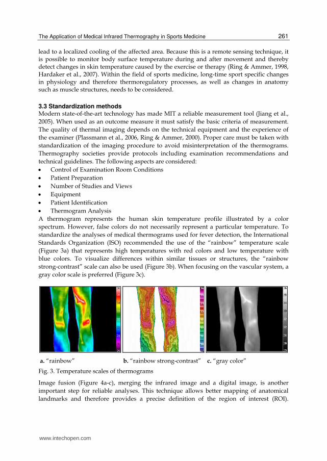
A thermogram represents the human skin temperature profile illustrated by a color
spectrum. However, false colors do not necessarily represent a particular temperature. To
standardize the analyses of medical thermograms used for fever detection, the International
Standards Organization (ISO) recommended the use of the “rainbow” temperature scale
(Figure 3a) that represents high temperatures with red colors and low temperature with
blue colors. To visualize differences within similar tissues or structures, the “rainbow
strong-contrast” scale can also be used (Figure 3b). When focusing on the vascular system, a
gray color scale is preferred (Figure 3c).
a. “rainbow” b. “rainbow strong-contrast” c. “gray color”
Fig. 3. Temperature scales of thermograms
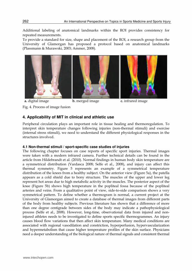
Image fusion (Figure 4a-c), merging the infrared image and a digital image, is another
important step for reliable analyses. This technique allows better mapping of anatomical
landmarks and therefore provides a precise definition of the region of interest (ROI).
www.intechopen.com
An International Perspective on Topics in Sports Medicine and Sports Injury
262
Additional labeling of anatomical landmarks within the ROI provides consistency for
repeated measurements.
To provide a standard for size, shape and placement of the ROI, a research group from the University of Glamorgan has proposed a protocol based on anatomical landmarks (Plassmann & Murawski, 2003; Ammer, 2008).
a. digital image b. merged image c. infrared image
Fig. 4. Process of image fusion
4. Applicability of MIT in clinical and athletic use
Peripheral circulation plays an important role in tissue healing and thermoregulation. To interpret skin temperature changes following injuries (non-thermal stimuli) and exercise (internal stress stimuli), we need to understand the different physiological responses in the structures involved.
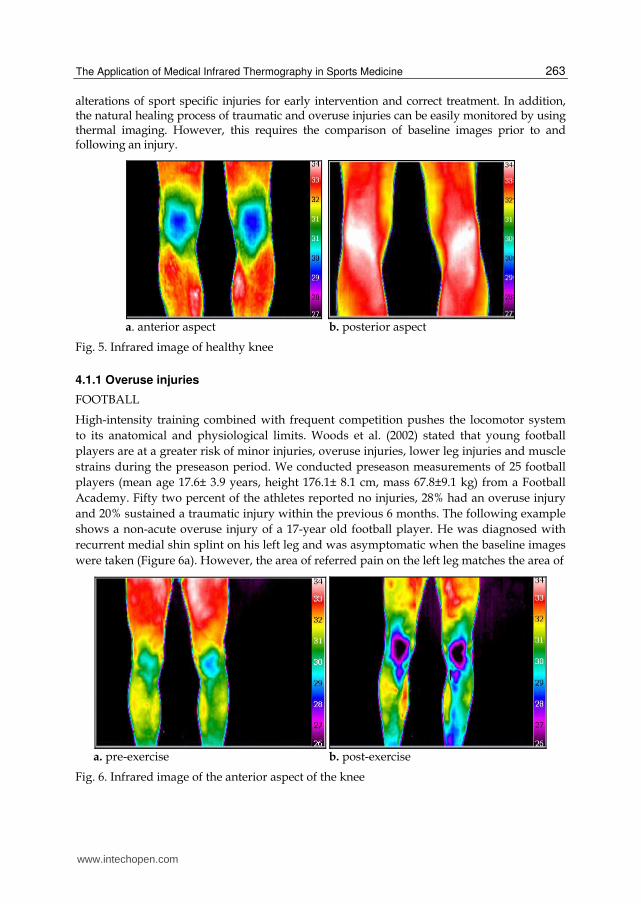
4.1 Non-thermal stimuli / sport-specific case studies of injuries The following chapter focuses on case reports of specific sport injuries. Thermal images were taken with a modern infrared camera. Further technical details can be found in the article from Hildebrandt et al. (2010). Normal findings in human body skin temperature are a symmetrical distribution (Vardasca 2008; Selfe et al., 2008), and injury can affect this thermal symmetry. Figure 5 represents an example of a symmetrical temperature distribution of the knees from a healthy subject. On the anterior view (Figure 5a), the patella appears as a cold shield due to bony structure. The muscles of the upper and lower leg represent hot areas due to high metabolic activity in the muscles. The posterior aspect of the knee (Figure 5b) shows high temperature in the popliteal fossa because of the popliteal arteries and veins. From a qualitative point of view, side-to-side comparison shows a very symmetrical pattern. To define whether a thermogram is normal, a current project at the University of Glamorgan aimed to create a database of thermal images from different parts of the body from healthy subjects. Previous literature has shown that a difference of more than one degree centigrade between sides of the body may indicate a pathophysiological process (Selfe et al., 2008). However, long-time, observational data from injured and non-injured athletes needs to be investigated to define sports specific thermogrammes. An injury causes blood flow variations that then affect skin temperature. Many medical conditions are associated with regional vasodilation and constriction, hyperperfusion, hypervascularization and hypermetabolism that cause higher temperature profiles of the skin surface. Physicians need a deeper understanding of the biological nature of thermal signals and consistent thermal
www.intechopen.com
The Application of Medical Infrared Thermography in Sports Medicine
263
alterations of sport specific injuries for early intervention and correct treatment. In addition, the natural healing process of traumatic and overuse injuries can be easily monitored by using thermal imaging. However, this requires the comparison of baseline images prior to and following an injury.
a. anterior aspect b. posterior aspect
Fig. 5. Infrared image of healthy knee
4.1.1 Overuse injuries
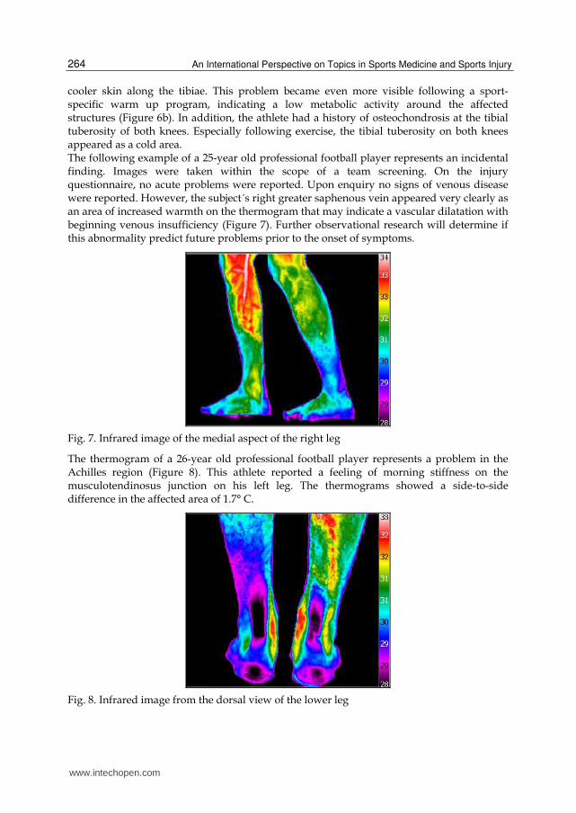
FOOTBALL
High-intensity training combined with frequent competition pushes the locomotor system
to its anatomical and physiological limits. Woods et al. (2002) stated that young football
players are at a greater risk of minor injuries, overuse injuries, lower leg injuries and muscle
strains during the preseason period. We conducted preseason measurements of 25 football
players (mean age 17.6± 3.9 years, height 176.1± 8.1 cm, mass 67.8±9.1 kg) from a Football
Academy. Fifty two percent of the athletes reported no injuries, 28% had an overuse injury
and 20% sustained a traumatic injury within the previous 6 months. The following example
shows a non-acute overuse injury of a 17-year old football player. He was diagnosed with
recurrent medial shin splint on his left leg and was asymptomatic when the baseline images
were taken (Figure 6a). However, the area of referred pain on the left leg matches the area of
a. pre-exercise b. post-exercise
Fig. 6. Infrared image of the anterior aspect of the knee
www.intechopen.com
An International Perspective on Topics in Sports Medicine and Sports Injury
264
cooler skin along the tibiae. This problem became even more visible following a sport-specific warm up program, indicating a low metabolic activity around the affected structures (Figure 6b). In addition, the athlete had a history of osteochondrosis at the tibial tuberosity of both knees. Especially following exercise, the tibial tuberosity on both knees appeared as a cold area. The following example of a 25-year old professional football player represents an incidental finding. Images were taken within the scope of a team screening. On the injury questionnaire, no acute problems were reported. Upon enquiry no signs of venous disease were reported. However, the subject´s right greater saphenous vein appeared very clearly as an area of increased warmth on the thermogram that may indicate a vascular dilatation with beginning venous insufficiency (Figure 7). Further observational research will determine if this abnormality predict future problems prior to the onset of symptoms.
Fig. 7. Infrared image of the medial aspect of the right leg
The thermogram of a 26-year old professional football player represents a problem in the Achilles region (Figure 8). This athlete reported a feeling of morning stiffness on the musculotendinosus junction on his left leg. The thermograms showed a side-to-side difference in the affected area of 1.7° C.
Fig. 8. Infrared image from the dorsal view of the lower leg
www.intechopen.com
The Application of Medical Infrared Thermography in Sports Medicine
265
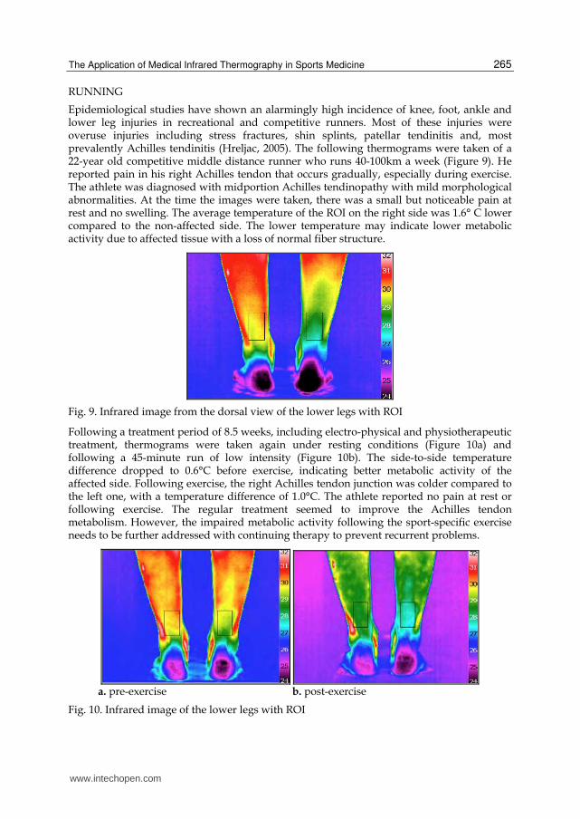
RUNNING
Epidemiological studies have shown an alarmingly high incidence of knee, foot, ankle and lower leg injuries in recreational and competitive runners. Most of these injuries were overuse injuries including stress fractures, shin splints, patellar tendinitis and, most prevalently Achilles tendinitis (Hreljac, 2005). The following thermograms were taken of a 22-year old competitive middle distance runner who runs 40-100km a week (Figure 9). He reported pain in his right Achilles tendon that occurs gradually, especially during exercise. The athlete was diagnosed with midportion Achilles tendinopathy with mild morphological abnormalities. At the time the images were taken, there was a small but noticeable pain at rest and no swelling. The average temperature of the ROI on the right side was 1.6° C lower compared to the non-affected side. The lower temperature may indicate lower metabolic activity due to affected tissue with a loss of normal fiber structure.
Fig. 9. Infrared image from the dorsal view of the lower legs with ROI
Following a treatment period of 8.5 weeks, including electro-physical and physiotherapeutic treatment, thermograms were taken again under resting conditions (Figure 10a) and following a 45-minute run of low intensity (Figure 10b). The side-to-side temperature difference dropped to 0.6°C before exercise, indicating better metabolic activity of the affected side. Following exercise, the right Achilles tendon junction was colder compared to the left one, with a temperature difference of 1.0°C. The athlete reported no pain at rest or following exercise. The regular treatment seemed to improve the Achilles tendon metabolism. However, the impaired metabolic activity following the sport-specific exercise needs to be further addressed with continuing therapy to prevent recurrent problems.
a. pre-exercise b. post-exercise
Fig. 10. Infrared image of the lower legs with ROI
www.intechopen.com

An International Perspective on Topics in Sports Medicine and Sports Injury
266
SWIMMING
A study, by Sein and co-workers in 2008, investigated shoulder pain in elite swimmers and found that 91% of the swimmers reported shoulder pain; moreover, 84% of the athletes demonstrated a positive impingement sign. The following thermal image was taken of a 27-year-old elite female swimmer under resting conditions (Figure 11). Following a high- volume swimming program, she reported pain and stiffness in both shoulders. With her right arm, she had difficulty reaching behind her back. The clinical examination confirmed overloading of the supraspinatus tendon and general stiffness of the shoulder muscles on both sides. The thermal image shows a hot area above the right deltoid muscle and a hot spot on both shoulders in the region of the humeral head, near the insertion of the supraspinatus muscle. Based on healthy baseline thermal images, MIT should be used to further monitor pathophysiological thermal changes during high-volume swim training prior to the onset of symptoms.
a. right shoulder b. left shoulder
Fig. 11. Infrared image from the lateral view of the shoulder
YOUTH SPORTS
A common problem, predominantly in young, male athletes is the occurrence of enthesopathy of the ligamentum patellae (Gholve et al., 2007). This insertion tendinitis, caused by repetitive mechanical strain of the patella tendon, is characterized by pain, swelling and tenderness above the tibial tuberosity (Brukner & Khan, 2006). Thermal images clearly show a hyperthermic area above the tibial tuberosity (Figure 12). Long term evaluation of affected athletes from alpine skiing (n= 7), football (n=3), running (n=2) and tennis (n=1), who showed acute symptoms in one leg, revealed a side-to-side temperature difference of 1.1°C (± 0.71 °C). The technique provides a quick screening tool and should be used as a first-line detection tool prior to ultrasound or conventional X-rays.
Fig. 12. Infrared images from athletes with enthesopathy of the ligamentum patellae
affected
knee
affected
knee
affected
knee
www.intechopen.com
The Application of Medical Infrared Thermography in Sports Medicine
267
4.1.2 Traumatic injuries Traumatic injuries usually involve a long, costly rehabilitation period, and they are challenging for the athlete. An injured athlete is under pressure to return to competition as soon as possible. High-quality treatment can reduce the duration and negative impact of the rehabilitation period. It is well known that richly vascularized areas heal faster compared to poorly vascularized areas (Singer et al., 1999). MIT may give information about the state of vascularization and the on-going healing process to ensure the most effective treatment and provide recovery information to decrease the likelihood of re-injury by returning to the sport too quickly.
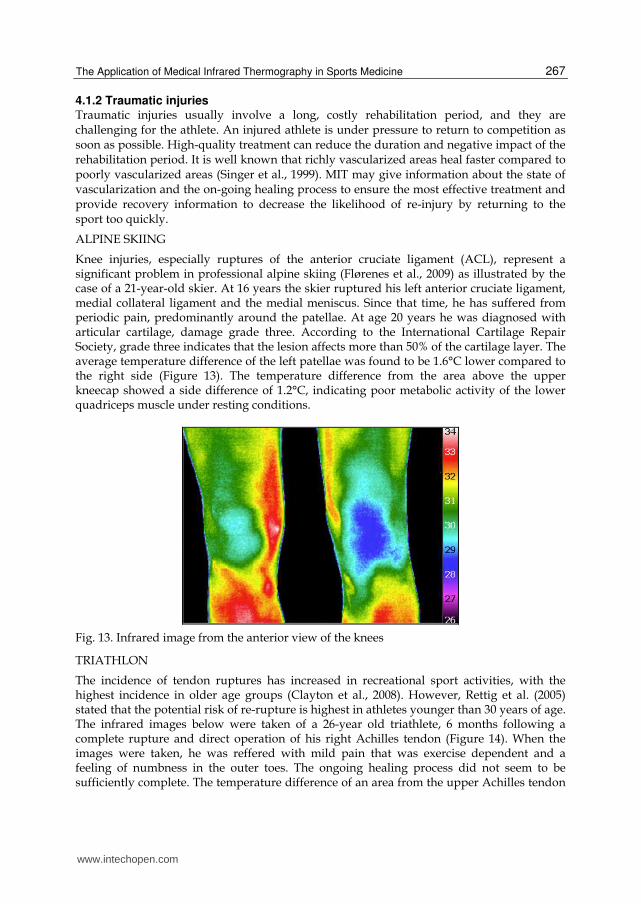
ALPINE SKIING
Knee injuries, especially ruptures of the anterior cruciate ligament (ACL), represent a significant problem in professional alpine skiing (Flørenes et al., 2009) as illustrated by the case of a 21-year-old skier. At 16 years the skier ruptured his left anterior cruciate ligament, medial collateral ligament and the medial meniscus. Since that time, he has suffered from periodic pain, predominantly around the patellae. At age 20 years he was diagnosed with articular cartilage, damage grade three. According to the International Cartilage Repair Society, grade three indicates that the lesion affects more than 50% of the cartilage layer. The average temperature difference of the left patellae was found to be 1.6°C lower compared to the right side (Figure 13). The temperature difference from the area above the upper kneecap showed a side difference of 1.2°C, indicating poor metabolic activity of the lower quadriceps muscle under resting conditions.
Fig. 13. Infrared image from the anterior view of the knees
TRIATHLON
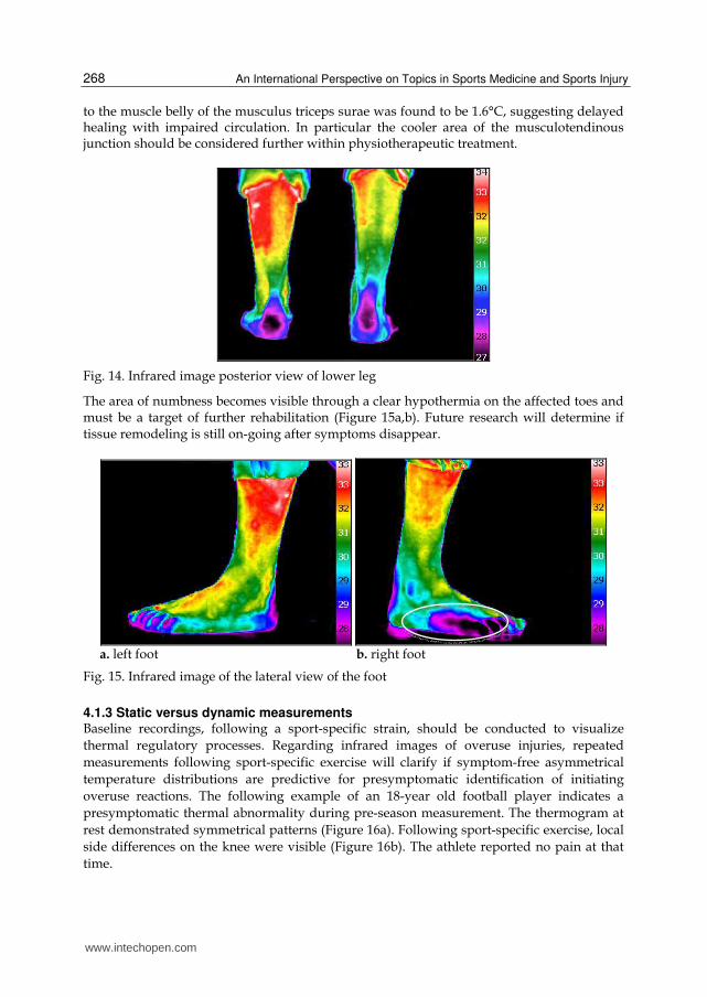
The incidence of tendon ruptures has increased in recreational sport activities, with the highest incidence in older age groups (Clayton et al., 2008). However, Rettig et al. (2005) stated that the potential risk of re-rupture is highest in athletes younger than 30 years of age. The infrared images below were taken of a 26-year old triathlete, 6 months following a complete rupture and direct operation of his right Achilles tendon (Figure 14). When the images were taken, he was reffered with mild pain that was exercise dependent and a feeling of numbness in the outer toes. The ongoing healing process did not seem to be sufficiently complete. The temperature difference of an area from the upper Achilles tendon
www.intechopen.com
An International Perspective on Topics in Sports Medicine and Sports Injury
268
to the muscle belly of the musculus triceps surae was found to be 1.6°C, suggesting delayed healing with impaired circulation. In particular the cooler area of the musculotendinous junction should be considered further within physiotherapeutic treatment.
Fig. 14. Infrared image posterior view of lower leg
The area of numbness becomes visible through a clear hypothermia on the affected toes and must be a target of further rehabilitation (Figure 15a,b). Future research will determine if tissue remodeling is still on-going after symptoms disappear.
a. left foot b. right foot
Fig. 15. Infrared image of the lateral view of the foot
4.1.3 Static versus dynamic measurements Baseline recordings, following a sport-specific strain, should be conducted to visualize
thermal regulatory processes. Regarding infrared images of overuse injuries, repeated
measurements following sport-specific exercise will clarify if symptom-free asymmetrical
temperature distributions are predictive for presymptomatic identification of initiating
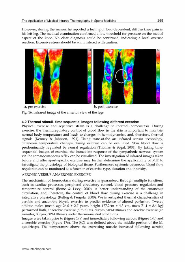
overuse reactions. The following example of an 18-year old football player indicates a
presymptomatic thermal abnormality during pre-season measurement. The thermogram at
rest demonstrated symmetrical patterns (Figure 16a). Following sport-specific exercise, local
side differences on the knee were visible (Figure 16b). The athlete reported no pain at that
time.
www.intechopen.com
The Application of Medical Infrared Thermography in Sports Medicine
269
However, during the season, he reported a feeling of load-dependent, diffuse knee pain in his left leg. The medical examination confirmed a low threshold for pressure on the medial aspect of the knee. No clear diagnosis could be confirmed, indicating a local overuse reaction. Excessive stress should be administered with caution.
a. pre-exercise b. post-exercise
Fig. 16. Infrared image of the anterior view of the legs
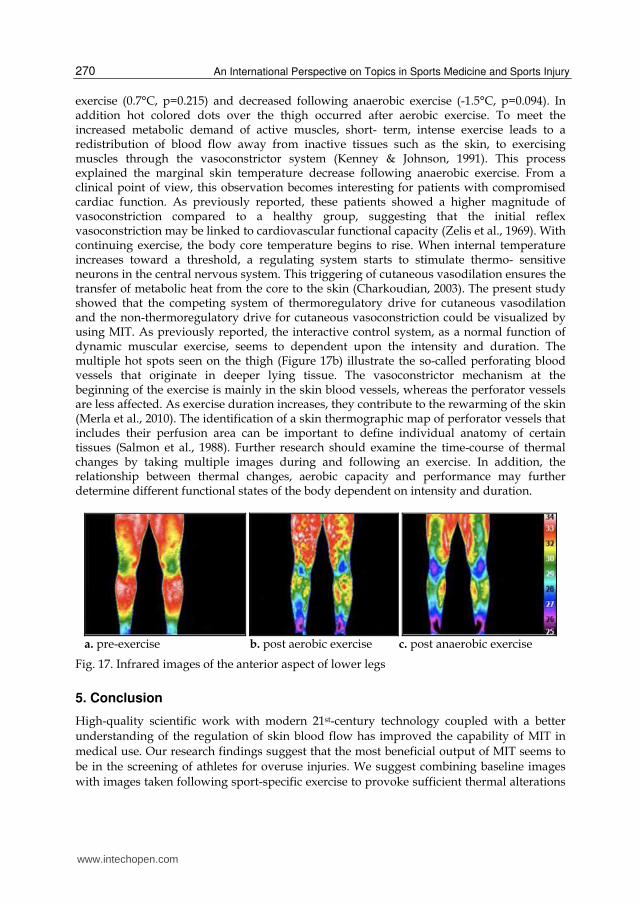
4.2 Thermal stimuli- time sequential images following different exercise Physical exercise and repetitive strain is a challenge to thermal homeostasis. During exercise, the thermoregulatory control of blood flow in the skin is important to maintain normal body temperature and leads to changes in hemodynamics, and, therefore, thermal signals (Kenney & Johnson, 1991). Using state-of-the art infrared sensor technology, cutaneous temperature changes during exercise can be evaluated. Skin blood flow is predominantly regulated by neural regulation (Thomas & Segal, 2004). By taking time- sequential images of exercise, the immediate response of the sympathetic nervous system via the somatocutaneous reflex can be visualized. The investigation of infrared images taken before and after sport-specific exercise may further determine the applicability of MIT to investigate the physiology of biological tissue. Furthermore systemic cutaneous blood flow regulation can be monitored as a function of exercise type, duration and intensity.
AEROBIC VERSUS ANAEROBIC EXERCISE
The mechanism of homeostasis during exercise is guaranteed through multiple functions, such as cardiac processes, peripheral circulatory control, blood pressure regulation and temperature control (Berne & Levy, 2000). A better understanding of the cutaneous circulation, and, therefore, the control of blood flow during exercise is a challenge in integrative physiology (Kellog & Pérgola, 2000). We investigated thermal characteristics of aerobic and anaerobic bicycle exercise to predict evidence of altered perfusion. Twelve athletic males (mean age 26.0 ± 2.7 years, height 177.2cm ± 4.3 cm, mass 71.1 ± 8.4 kg) performed both, anaerobic exercise (5 minutes, 80rpm, 90%HRmax) and aerobic exercise (45 minutes, 80rpm, 60%HRmax) under thermo-neutral conditions. Images were taken prior to (Figure 17a) and immediately following aerobic (Figure 17b) and anaerobic exercise (Figure 17c). The ROI was defined above the middle portion of the M. quadriceps. The temperature above the exercising muscle increased following aerobic
www.intechopen.com
An International Perspective on Topics in Sports Medicine and Sports Injury
270
exercise (0.7°C, p=0.215) and decreased following anaerobic exercise (-1.5°C, p=0.094). In addition hot colored dots over the thigh occurred after aerobic exercise. To meet the increased metabolic demand of active muscles, short- term, intense exercise leads to a redistribution of blood flow away from inactive tissues such as the skin, to exercising muscles through the vasoconstrictor system (Kenney & Johnson, 1991). This process explained the marginal skin temperature decrease following anaerobic exercise. From a clinical point of view, this observation becomes interesting for patients with compromised cardiac function. As previously reported, these patients showed a higher magnitude of vasoconstriction compared to a healthy group, suggesting that the initial reflex vasoconstriction may be linked to cardiovascular functional capacity (Zelis et al., 1969). With continuing exercise, the body core temperature begins to rise. When internal temperature increases toward a threshold, a regulating system starts to stimulate thermo- sensitive neurons in the central nervous system. This triggering of cutaneous vasodilation ensures the transfer of metabolic heat from the core to the skin (Charkoudian, 2003). The present study showed that the competing system of thermoregulatory drive for cutaneous vasodilation and the non-thermoregulatory drive for cutaneous vasoconstriction could be visualized by using MIT. As previously reported, the interactive control system, as a normal function of dynamic muscular exercise, seems to dependent upon the intensity and duration. The multiple hot spots seen on the thigh (Figure 17b) illustrate the so-called perforating blood vessels that originate in deeper lying tissue. The vasoconstrictor mechanism at the beginning of the exercise is mainly in the skin blood vessels, whereas the perforator vessels are less affected. As exercise duration increases, they contribute to the rewarming of the skin (Merla et al., 2010). The identification of a skin thermographic map of perforator vessels that includes their perfusion area can be important to define individual anatomy of certain tissues (Salmon et al., 1988). Further research should examine the time-course of thermal changes by taking multiple images during and following an exercise. In addition, the relationship between thermal changes, aerobic capacity and performance may further determine different functional states of the body dependent on intensity and duration.
a. pre-exercise b. post aerobic exercise c. post anaerobic exercise
Fig. 17. Infrared images of the anterior aspect of lower legs
5. Conclusion
High-quality scientific work with modern 21st-century technology coupled with a better
understanding of the regulation of skin blood flow has improved the capability of MIT in
medical use. Our research findings suggest that the most beneficial output of MIT seems to
be in the screening of athletes for overuse injuries. We suggest combining baseline images
with images taken following sport-specific exercise to provoke sufficient thermal alterations
www.intechopen.com
The Application of Medical Infrared Thermography in Sports Medicine
271
in the tissues. A main challenge is to combine the anatomical and physiological information
demonstrated by the thermal pattern of the skin. The biological nature of thermal signals
and consistent thermal alterations of different sport-specific injuries should be further
addressed. Thermal screening of injured and non-injured athletes is the first step to create a
sport-specific database with individual thermograms. Repeated follow-up measurements
during the sport season will further clarify the link between asymmetrical temperature
distributions, pathophysiological changes on the skin surface and the extent of injury. The
long-term aim is to create a knowledge-based database of thermograms of overuse and
traumatic injuries. However, it should be considered that within a certain time span,
different pathologies could alter their patterns of temperature. A deeper understanding of
the different time courses of injuries is important to clarify the benefit of MIT in injury
management and to define whether a thermogram is “normal” or not. In terms of
quantification of side-to side differences within a defined ROI, it is important to use the
medical analysis function of image fusion. The main advantage of MIT is its safety,
however, the disadvantage of MIT results from its physical limitations. The non-radiating,
two-dimensional technique provides information about surface structures. A conclusion of
processes in deeper tissues needs to be further investigated by combining different medical
imaging modalities. In addition, it must be clearly stated that the aim of MIT use in sports
medicine is not to be a substitute for clinical examination, but to enhance and support it. It
can be concluded that MIT is a reliable, low-cost detection tool that should be applied for
pre-scanning athletes.
6. References
Ammer, K. (2006). Diagnosis of Raynaud´s phenomen by thermography. Skin Research and Technology, Vol.2, No.4, pp. 182-185, ISSN 0909-752X
Ammer, K. (2008). The Glamorgan Protocol for recording and evaluation of thermal images of the human body. Thermology International, Vol.18, No.4, pp. 125-144, ISSN1560-604X
Ammer, K. (2008). The sensitivity of infrared imaging for diagnosing Raynaud´s phenomenon or Thoracic Outlet Syndrome is dependent on the method of temperature extraction from thermal images. Thermology International, Vol.18, No. 3, pp.81-88, ISSN1560-604X
Arora, N.; Martins, D.; Ruggerio, D.; Tousimis, E.; Swistel, A.J.; Osborne, M.P.& Simmons, R.M. (2008). Effectiveness of a noninvasive digital infrared thermal imaging system in the detection of breast cancer. The American Journal of Surgery, Vol.196, No.4, pp. 523-526, ISSN 0002- 9610
Berne, R.M. & Levy, M.N. (2000). Principles of Physiology. Third Edition, Mosby, ISBN 84-8174-550-2, St. Louis
Bharara, M. (2006). Thermography and Thermometry in the Assessment of Diabetic Neuropathic Foot: A Case for furthering the role of thermal techniques. The International Journal of Lower Extremity Wounds, Vol.5, No.4, pp. 250-260, ISSN 1534-7346
Blatteis, C.M. (1998). Physiology and pathophysiology of temperature regulation. First edition, World scientific printers, ISBN 981-02-3172-5, Singapore
www.intechopen.com

An International Perspective on Topics in Sports Medicine and Sports Injury
272
Bruckner, P. & Khan, K. (2006). Fundamental principals, In: Clinical Sports Medicine. Third edition. Bruckner, P; Khan, K pp. 3-7, McGraw-Hill Medical Publishing Division, ISBN 0070278997, Canada
Bruehl, S.; Lubenow, T.; Nath, H. & Ivankovich, O. (1996). Validation of Thermography in the Diagnosis of Reflex Sympathetic Dystrophy. Clinical Journal of Pain, Vol.12, No.4, pp. 316-325, ISSN 07498047
Charkoudian, N. (2003). Skin blood flow in adult human thermoregulation: how it works, when it does not and why. Mayo Clinic Proceedings, Vol.78, No.5, pp. 603-612, ISSN 0025-6196
Clayton, R.A.E. & Court-Brown, C.M. (2008). The epidemiology of musculoskeletal tendinous and ligamentous injuries. Injury, Vol.39, No.12, pp.1338-1344, ISSN 0020-1383
Cochrane, D.J.; Sanard, S.R.; Firth, E.C. & Rittweger, J. (2010). Comparing Muscle Temperature during static and dynamic Squatting with and without Whole- Body Vibration. Clinical Physiology and Functional Imaging, Vol.30, No.4, pp. 223-229, ISSN 1475-0961
Denoble, A.E.; Hall, N.; Pieper, C.F. & Kraus, V.P. (2010). Patellar skin surface temperature by thermography reflects knee osteoarthritis. Clinical medicine insights. Arthritis and musculoskeletal disorders, Vol.3, No.1, pp. 69-75, ISSN 11795441
Diakides, N.A. & Bronzino J.D. (2008). Medical Infrared Imaging, First Edition, CRC Press, ISBN 0849390272, Broken
de Weerd, L.; Mercer J. & Setså, L. (2006). Intraoperative Dynamic Infrared Thermography and Free-Flap Surgery. Annals of Plastic Surgery, Vol.57, No.3, pp. 279-284, ISSN 01487043
Eddy, A.L.; Van Hoogmoed, L.M. & Snyder, J.R. (2001). The role of Thermography in the Management of Equine Lameness. The Veterinary Journal, Vol.162, No.3, pp. 172-181, ISSN 1090-0233
Elliot, R.L. & Head, J.F. (1999). Medical infrared imaging in the twenty-first century. Thermology International, Vol.9., No.4, pp. 111, ISSN 1560-604X
Flørenes, T.W.; Bere, T.; Nordsletten, L. Heir, S. & Bahr, R. (2009). Injuries among male and female world cup alpine skiers. British Journal of Sports Medicine, Vol.43, No.13, pp. 973-978, ISSN 0306-3674
Gholve, P.; Scher, D.; Khakharia, S.; Widmann, R. & Green, D. (2007). Osgood Schlatter syndrome. Current Opinion in Pediatrics, Vol.19, No.1, pp. 44-50, ISSN 1531-698X
Hardaker, N.J.; Moss, A.D.; Richards, J.; Jarvis, S.; McEwan, C. & Selfe, J. (2007). The relationship between skin surface temperature measured via non-contact thermal imaging and intra-muscular temperature of the rectus femoris muscle. Thermology International, Vol.17, No.2; pp. 45-50, ISSN 1560-604X
Hildebrandt, C.; Ammer, K. & Raschner, C. (2010). An Overview of Recent Application of Medical Infrared Thermography in Sports Medicine in Austria. Sensors, Vol.10, No.5, pp. 4700-4715, ISSN 1424-8220
Hreljac, A. (2005). Etiology, prevention and early intervention of overuse injuries in runners: a biomechanical perspective. Physical medicine and rehabilitation clinics of North America, Vol.16, No.3, pp. 651-667, ISSN 1047-9651
www.intechopen.com
The Application of Medical Infrared Thermography in Sports Medicine
273
Jiang, L.J.; Ng, E.Y.K.; Yeo, A.C.B.; Wu, S.; Pan, F.; Yau, W.Y.; Chen, J.H. & Yang, Y. (2005). A perspective on medical infrared imaging. Journal of Medical Engineering and Technology, Vol.29, No.6, pp. 257-267, ISSN 0309-1902
Kellog D. L. & Pérgola P. (2000). Skin Response to exercise and training. In: Exercise and Sports Science, Garrett, W.E.; Kirkendall, D.T. published by Lippincott Williams &Wilkins, pp. 239-250, ISBN 0-683-03421-9, Philadelphia
Kenney W.L. & Johnson J.M. (1992). Control of skin blood flow during exercise. Medicine and Science in Sports and Exercise, Vol. 24, No.3, pp. 303-312, ISSN 1530-0315
Kontos, M.; Wilson, R. & Fentiman, I. (2011). Digital infrared thermal imaging (DITI) of breast lesions: sensitivity and specificity of detection of primary breast cancers. Clinical Radiology, Vol. 66, No.6, pp. 536-539, ISSN 0033-8419
Merla, A.; DiRomualdo, S.; DiDonato, L.; Proietti, M.; Salsano, F. & Romani, G.L. (2007). Combined thermal and laser Doppler imaging in the assessment of cutaneous tissue perfusion. Conference Proceedings of the IEEE Engineering Medicine and Biology Society, pp. 2630-2633, ISSN 1557-170X
Merla, A; Mattei, P.A.; di Donato, L. & Romani, G.L. (2010). Thermal Imaging of Cutaneous Temperature Modifications in Runners During Graded Exercise. Annals of Biomedical Engineering, Vol.38, No.1, pp.158-163, ISSN 1573-9686
Ng, E.Y.K. (2009). A review of thermography as promising non-invasive detection modality for breast tumor. International Journal of Thermal Science, Vol.48, No.5, pp. 849-859, ISSN 1290-0729
Niehof, S.P.; Huygen, F.; van der Weerd, R.; Westra, M. & Zijlstra, F.J. (2006). Thermography imaging during static and controlled thermoregulation in complex regional pain syndrome type 1. Biomedical Engineering OnLine, Vol.5, No.30, pp. 1-13, ISSN 1475-925X
Park, J.Y.; Hyun, J.K. & Seo, J.B. (2007). The effectiveness of digital infrared thermographic imaging in patients with shoulder impingement syndrome. Journal of Shoulder and Elbow Surgery, Vol.16, No.5, pp. 548-554, ISSN 1058-2746
Plassmann,P. & Murawski, P. (2003). CTHERM for standardized thermography, Proceedings of Abstracts the 9th congress of Thermology, ISBN N/A, Poland
Plassman, P.; Ring, E.F.J. & Jones, C.D. (2006). Quality assurance of thermal imaging systems in medicine. Thermology International, Vol.16, No.1, pp.10-15, ISSN 1560-604X
Rettig, A.C.; Liotta, F.J.; Klootwyk, T.E.; Porter, D.A. & Mieling, P. (2005). Potential Risk of Rerupture in Primary Achilles Tendon Repair in Athletes Younger Than 30 Years of Age. American Journal of Sports Medicine, Vol.33, No.1, pp.119-123, ISSN 0363-5465
Ring, E.F.J. & Ammer, K. (1998). Thermal imaging in sports medicine. Sport and Medicine Today, Vol.1, No.2, pp.108-109, ISSN N/A
Ring, E.F.J. & Ammer, K. (2000). The technique of infrared imaging in medicine. Thermology international, Vol.10, No1, pp. 7-14, ISSN 1560-604X
Ring E.F.J. (2007). The Historical development of temperature measurement in medicine. Infrared Physics and Technology, Vol.49, No.3, pp. 297- 301, ISSN 1350-4495
Romano, C.L.; Logoluso, N.; Dellóro, F.; Elia, A. & Drago, L. (2011) Telethermographic findings after uncomplicated and septic total knee replacement. Knee, Epub ahead of print, ISSN 0968-0160
Salmon, M.; Taylor, G.I. & Tempest, M.N. (1988). Arteries of the skin, Churchill Livingstone, ISBN 0443036055, London
www.intechopen.com

An International Perspective on Topics in Sports Medicine and Sports Injury
274
Sein, M.L.; Walton, J.; Linklater, J.; Appleyard, R.; Kirkbride, B.; Kuah, D. & Murrell G.A.C. (2010). Shoulder pain in elite swimmers: primarily due to swim-volume-induced supraspinatus tendinopathy. British Journal of Sports Medicine, Vol.44, No.2, pp.105-113, ISSN 0306-3674
Selfe, J.; Whitaker, J. & Hardaker, N. (2008). A narrative literature review identifying the minimum clinically important difference for skin temperature asymmetry at the knee. Thermology International, Vol.18, No.2, 41-44, ISSN 1560-604X
Singer, A.J. & Clark, R.A.F. (1999). Cutaneous wound healing. The New England Journal of Medicine, Vol. 341, No.10, pp. 738-746, ISSN 0028-4793
Steketee, J. (1973). Spectral emissivity of skin and pericardium. Physics in Medicine and Biology. Vol. 18, No. 5, pp. 686-694, ISSN 0031-9155
Thomas, G.D. & Segal, S.S. (2004). Neural control of muscle flow during exercise. Journal of Applied Physiology, Vol.97, No.2, pp. 731-738, ISSN 8750-7587
Turner, T.A. (2000). Diagnostic thermography. Veterinary Clinics of North America-Equine Practice, Vol.17, No.1, pp. 95-113, ISSN 0749-0739
Vardasca, R. (2008). Symmetry of temperature distribution in the upper and lower extremities. Thermology International, Vol.18, No.4, pp. 154-155, ISSN 1560-604X
Wallin, B.G. (1990). Neural control of human skin blood flow. Journal of the autonomic nervous system, Vol. 30, No.S1, pp.185-190, ISSN 1529-8027
Wild, W.; Schütte, S.R.; Pau, H.W.; Kramp, B. & Just, T. (2003). Infrared thermography as a non invasive application for medical diagnostic. Proceedings XVII IMEKO World Congress, June 22-27, 2003, ISBN 0-7803-8493-8, Dubrovnik Croatia
Woods, C.; Hawkins, R.; Hulse, M. & Hodson A. (2002). The Football Association Medical Research Programme: an audit of injuries in professional football—analysis of preseason injuries. British Journal of Sports Medicine, Vol.36, No.6, pp. 436–441, ISSN 0306-3674
Zaprodina, N.; Ming, Z. & Hänninen, O.P. (2006). Plantar infrared thermography measurements and low back pain intensity. Journal of Manipulative Physiological Therapeutics, Vol.29, No.3, pp.219-223, ISSN 0161-4754
Zelis, R.; Mason, D.T. & Braunwald, D. (1969). Partition of blood flow to the cutaneous and muscular beds of the forearm at rest and during leg exercise in normal subjects and in patients with heart failure. Circulation Research, Vol.24, No.6, pp.799-806, ISSN 0009-7330
www.intechopen.com
An International Perspective on Topics in Sports Medicine andSports InjuryEdited by Dr. Kenneth R. Zaslav
ISBN 978-953-51-0005-8Hard cover, 534 pagesPublisher InTechPublished online 17, February, 2012Published in print edition February, 2012
InTech EuropeUniversity Campus STeP Ri Slavka Krautzeka 83/A 51000 Rijeka, Croatia Phone: +385 (51) 770 447 Fax: +385 (51) 686 166www.intechopen.com
InTech ChinaUnit 405, Office Block, Hotel Equatorial Shanghai No.65, Yan An Road (West), Shanghai, 200040, China
Phone: +86-21-62489820 Fax: +86-21-62489821
For the past two decades, Sports Medicine has been a burgeoning science in the USA and Western Europe.Great strides have been made in understanding the basic physiology of exercise, energy consumption and themechanisms of sports injury. Additionally, through advances in minimally invasive surgical treatment andphysical rehabilitation, athletes have been returning to sports quicker and at higher levels after injury. Thisbook contains new information from basic scientists on the physiology of exercise and sports performance,updates on medical diseases treated in athletes and excellent summaries of treatment options for commonsports-related injuries to the skeletal system.
How to referenceIn order to correctly reference this scholarly work, feel free to copy and paste the following:
Carolin Hildebrandt, Karlheinz Zeilberger, Edward Francis John Ring and Christian Raschner (2012). TheApplication of Medical Infrared Thermography in Sports Medicine, An International Perspective on Topics inSports Medicine and Sports Injury, Dr. Kenneth R. Zaslav (Ed.), ISBN: 978-953-51-0005-8, InTech, Availablefrom: http://www.intechopen.com/books/an-international-perspective-on-topics-in-sports-medicine-and-sports-injury/the-application-of-medical-infrared-thermography-in-sports-medicine