tens/stevens johnson syndrome - pcmapcma.org.za/blog/wp-content/uploads/2012/03/tens... ·...
TRANSCRIPT

{
TENS/STEVENS JOHNSON SYNDROME
Dehan Struwig, Plastic and Reconstructive Surgeon

Stevens-Johnson Syndrome is an immune complex hypersensitivity reaction that can be caused from an infection or immune response to drugs.
It is a severe expression of a simple rash known as erythema multiforme.
SJS is also known as erythema multiforme major. It affects all ages and genders including pediatric populations.
The most severe form of SJS is toxic epidermal necrolysis (TENS).
DEFINITION

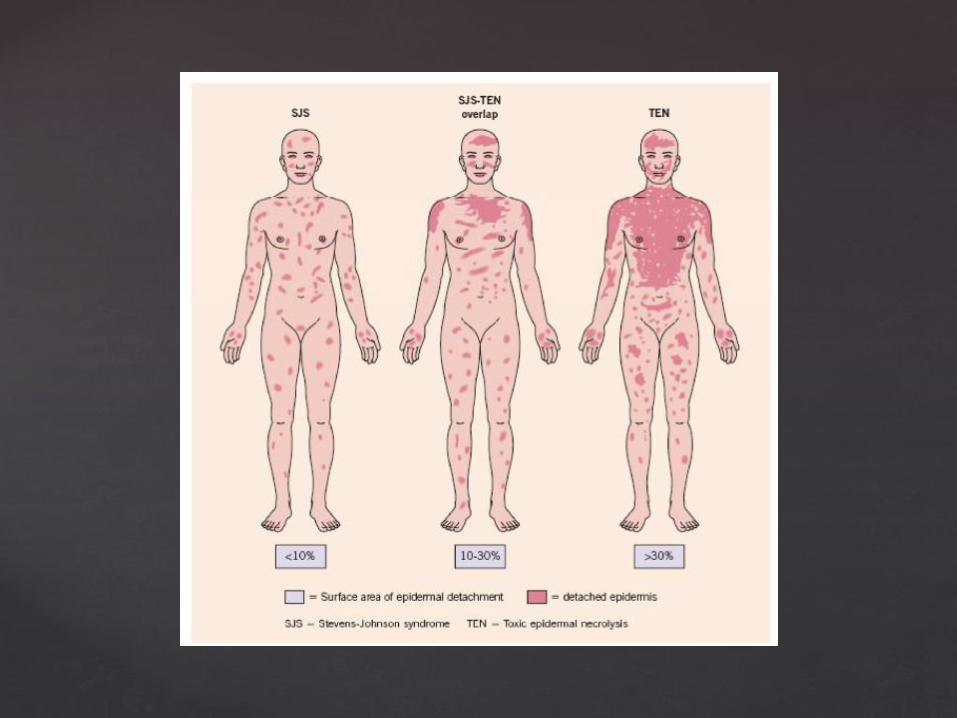

A, Erythema multiforme: typical targets, with regular round shape, well-defined borders, 3 different zones, predominant on the extremities. B, Stevens-Johnson syndrome: erythematous or purpuric macules with irregular shape and size. Blisters often occur on all or part of the macule. Lesions are widespread. Confluence of individual lesions remains limited, involving less than 10% of the body surface area. C, Overlap Stevens-Johnson syndrome–toxic epidermal necrolysis: confluent blisters result in detachment of the epidermis and erosions on 10% to 29% of the body surface area. D, Toxic epidermal necrolysis: widespread detachment of epidermis on more than 30% of the body surface area.

Mucosal involvement is prominent and severe, although not forming actual blisters. At least 2 mucosal surfaces are affected including: • Eyes (conjunctivitis) – red, sore, sticky • Lips/mouth (cheilitis, stomatitis) – red crusted lips,
mouth ulcers • Oesophagus – causing difficulty eating • Upper respiratory tract (trachea and bronchi) – causing
cough and respiratory distress • Genital area and urinary tract – ulcers • Gastrointestinal tract – causing diarrhoea.

SCORTEN is an illness severity score that has been developed to predict mortality in SJS and TEN cases. One point is scored for each of seven criteria present at the time of admission. The SCORTEN criteria are: • Age >40 years • Presence of a malignancy (cancer) • Heart rate >120 • Initial percentage of epidermal detachment >10% • Serum urea level >10 mmol/L • Serum glucose level >14 mmol/L • Serum bicarbonate level <20 mmol/L • The risk of dying from SJS/TEN depends on the score.
SCORTEN predicted mortality rates SCORTEN 0-1 >3.2% SCORTEN 2 >12.1% SCORTEN 3 >35.3% SCORTEN 4 >58.3% SCORTEN 5 or more >90%

Infectious
Drug-induced
Malignancy-related
Idiopathic
AETIOLOGY

Viral diseases that have been reported to cause Stevens-Johnson syndrome include the following:
Herpes simplex virus (possibly; remains a debated
issue) AIDS Coxsackie viral infections Influenza Hepatitis Mumps In children, Epstein-Barr virus and enteroviruses have
been identified. More than half of the patients with Stevens-Johnson syndrome report a recent upper respiratory tract infection.
INFECTION

Bacterial etiologies include the following:
Group A beta-hemolytic streptococci Diphtheria Brucellosis Lymphogranuloma venereum Mycobacteria Mycoplasma pneumoniae[11, 12] Rickettsial infections Tularemia Typhoid Possible fungal causes include
coccidioidomycosis, dermatophytosis, and histoplasmosis. Malaria and trichomoniasis have been reported as protozoal causes.

MEDICINE
Antibiotics are the most common cause of Stevens-Johnson syndrome, followed by analgesics, cough and cold medication, NSAIDs, psychoepileptics, and antigout drugs. Of antibiotics, penicillins and sulfa drugs are prominent; ciprofloxacin has also been reported

The following anticonvulsants have been implicated: • Phenytoin • Carbamazepine • Oxcarbazepine (Trileptal) • Valproic acid • Lamotrigine • Barbiturates Mockenhapupt et al stressed that most anticonvulsant-induced SJS occurs in the first 60 days of use.

Antiretroviral drugs implicated in Stevens-Johnson syndrome include nevirapine and possibly other non-nucleoside reverse transcriptase inhibitors. Indinavir has been mentioned

Stevens-Johnson syndrome has also been reported in patients taking the following drugs: • Modafinil (Provigil) • Allopurinol • Mirtazapine[17] • TNF-alpha antagonists (eg, infliximab, etanercept,
adalimumab) • Cocaine

GENETIC FACTORS
There is strong evidence for a genetic predisposition to severe cutaneous adverse drug reactions such as Stevens-Johnson syndrome. Carriage of the following human leukocyte antigens has been associated with increased risk:
• HLA-B*1502 • HLA-B*5801 • HLA-B*44 • HLA-A29 • HLA-B12 • HLA-DR7 • HLA-A2 • HLA-B*5801 • HLA-A*0206 • HLA-DQB1*0601

EPIDEMIOLOGY
In the United States, the annual frequency of TENS is reported to be 0.22-1.23 cases per 100,000 population. In the HIV-positive population, the incidence of TENS increases to 1 case per thousand per year.
Worldwide, the average annual incidence of TENS is 0.4-1.3 cases per million population. In 1992, the cumulative incidence of TENS and SJS in Germany was 1.9 cases per million population. A French survey of dermatologists and health care facilities reported an annual incidence of 1 case per million population.

For unclear reasons, TENS appears to have a predilection for females. The female-to-male ratio is 1.5:1.
TENS may occur in all age groups; however, the mean age of patients with TENS is reported to be between 46 and 63 years. Infection is more commonly implicated as an etiology in children, whereas medication exposure is more common in adults. Elderly persons may be at greater risk because of their tendency to use multiple medications.

The mortality is almost 10% for patients with SJS, approximately 30% for patients with SJS/TENS-overlap and almost 50% for patients with TENS. For SJS, SJS/TEN-overlap and TENS together the mortality rate is almost 25

TENS is believed to be an immune-related cytotoxic reaction aimed at destroying keratinocytes that express a foreign antigen.
TENS mimics a hypersensitivity reaction, with its characteristic delayed reaction to an initial exposure and an increasingly rapid reaction with repeated exposure.
PATHOPHYSIOLOGY

Exact mechanism is unknown; however, one theory holds that altered drug metabolism in some patients causes formation of reactive metabolites that bind to and alter cell proteins, triggering a T-cell–mediated cytotoxic reaction to drug antigens in keratinocytes.

CLINICALLY MANIFESTS IN THREE STAGES:
Crescendo
Critical
Convalescent
CLINICAL MANIFESTATION

Lasts for 2-3 days before onset
Generalised complaints:
Fever
Malaise
Rhinitis
Anorexia
Pharyngitis
CRESCENDO PHASE

Persistent fever
Mucosal & Cutaneous sloughing
Erythema and blisters
Generalised sloughing
CRITICAL PHASE: (8-12 DAYS)

OCULAR (40-50%)
• Conjunctivitis
• Photophobia
• Alternations in visual acuity
• Corneal opacities
• Corneal ulcerative lesions
• Scarring around eyelids
• Blindness
MULTISYSTEM MANIFESTATIONS

PULMONARY:
• Sore throat
• Pharyngitis
• Pneumonitis
• Pneumonia
• ARDS
• Respiratory failure
• Pulmonary embolism

GASTRO-INTESTINAL:
• Anorexia
• Gastritis
• Mucositis
• Stomatitis
• Gastro-intestinal bleeding
• Necrotic bowel

GENITO-URINARY
• Dysuria
• Anuria
• Urinary retention
• Urinary tract infection
• Renal failure

INTEGUMENTARY: (Most affected system)
• Loss of skin integrity
• Wound infections
• Hypopigmentation
• Hyperpigmentation
• Hypertrophic scarring
• Wound contracture

METABOLIC:
Increase in basal metabolic rate
IMMUNE:
• Fever
• Localised infections
• Systemic infections
• Systemic inflammatory response syndrome
• Multiple organ dysfunction syndrome

PSYCHOSOCIAL:
• Anxiety
• Pain
• Fear
• Powerlessness
• Altered body image
• Knowledge deficit of disease process

Cessation of sloughing
Re-epithelialization
Healing of skin and mucous membranes
CONVALESCENT PHASE

Duration of convalescent phase dependent on:
• Location and extent of involvement
• Presence of systemic infections
• Trauma to sloughing tissue
• Other complications

• Early recognition
• Referral to burn centre
• Cessation of use of drug
• Highly specialised nursing care
• Interdisciplinary team approach
TREATMENT

Fluid resuscitation
Treatment for alterations in skin
Control of infection and sepsis
Pain management
Nutritional therapy
Psychosocial support
Rehab services
ESSENTIAL ASPECTS

• Corticosteroids
• IV Immunoglobulin
• Immuno-modulating agents
• N-acetylcystine
Controversial

• Meticulous eye care every 1-2 hrs
• Saline rinses
• Liberal lubrication of conjuntiva
• Daily visits by opthalmologist
OCULAR

• Aggressive hygiene
suctioning
turning
incentive spirometry
therapeutic bronchoscopy
• Endotracheal intubation
• Mechanical ventilation to improve oxygenation
PULMONARY

• Nutritional consultation
• Soft diet
• Frequent oral rinses
• Antacid therapy
• Soft, flexible tubing
• Stool softeners
GASTRO-INTESTINAL

• Renal failure
• UTI
• Urinary catheterization
GENITO-URINARY

Prompt debridement
• prevent infection
• facilitate healing
Meticulous wound care
• cleansing
• mechanical debridement
• dressing
• antimicrobial dressings
• biological dressings
INTEGUMENTARY

Risks of hypothermia
Control of pain and anxiety
• extensive sloughing
• exposed nerve endings

Pigment change – patchwork of increased and decreased pigmentation Skin scarring, especially at sites of pressure or infection Loss of nails with permanent scarring (pterygium) and failure to regrow Joint contractures Scarred genitalia – phimosis (constricted foreskin which cannot retract)
and vaginal synechiae (occluded vagina) Serious eye problems, which can lead to blindness. This is the most
important of the longterm complications. These include: Dry and/or watery eyes, which may burn and sting when exposed to
light Conjunctivitis: red, crusted, or ulcerated conjunctiva Corneal ulcers, opacities and scarring Symblepharon: adhesion of conjunctiva of eyelid to eyeball Ectropion or entropion: turned-out or turned-in eyelid Trichiasis: inverted eyelashes Synechiae: iris sticks to cornea
Long term implications

36-year old, female patient
15 year history of depression
Fourteen day history of use of: Tegretol 200mg bd
Alzam
Remeron 30mgs nocte
Seroquel 50 mgs nocte
Zopidem 7.5mgs nocte
CLINICAL CASE STUDY

Presents with: 2 day history of rash
Temperature 39 degrees
Nausea
Swelling and Redness of eyes
Redness of Throat
Admitted to Surgical ICU on 12 September 2003

Shortly after admission:
• Diffuse bullae
• Consistent fever
• Marked desquamation of skin


14th September (Day 2)
• Heparin (DVT)
• Enteral feeds
• Rehydration
• Blood-stained urine
• Eyes: artificial tears + Voltaren drops
• Ulceration of the mouth

Marked diarrhea
• CVP inserted
Plastic surgeon involved
Confirmed diagnosis:
• Stevens-Johnson Syndrome
• Drug reaction to Tegretol
• TENS
17th September (Day 5)

21 September 2003 (Day 9)


• Tracheotomy performed • Debrided • Acticoat • New CVP • New A-line
30 September 2003 (Day 18)


6th October (Day 24)
• Candida Prapsilosis • Fever • Oozing from mouth • Lungs affected • Muscle relaxant to improve
ventilation • Coagulase neg Staphylococcus • Pseudomonas specimen • Tracheotomy tube changed • To larger diameter • Debride • Acticoat


17 October 2003 (Day 35)
• Debride • Acticoat • Bactroban face, lower legs


18th of October (Day 36)
3 Further units of packed cells 23% band forms 6% metamyelocytes 5% meylocytes 1% promyelocytes CVP 14-15 Ongoing septicaemia

24th October (Day 42)
• Hb 7 • Oozing settled • Nasogastric feeding
• Debride • Acticoat • Bactroban


27th October (Day 45)
• E.coli • Beta group Streptococcus
• No active bleeding • Urine output maintained • Temperature normal


12th November (Day 61)
• Temperature • Pseudomonas
• Generalised weakness • Poor vital capacity • Continued ventilation
• Profuse diarrhea • Distended abdomen

13 November (Day 62)
• Abdominal pain • Distension • Enlarged liver • Pancreatitis with raised lipase

16 November (Day 65)
• Lasix for oedema • Jaundiced
• Stenotrophomonas • Pseudomonas

2 December 2003 (Day 81)
• CJ tube removed • Nasal oxygen • Chest clear left, improved right • Temperature normal • Blood pressure normal • Lost peripheral oedema • Abdomen softer • Jaundiced • 4 finger hepatomegaly • Nasogastric feeding • No diarrhea • Difficulty swallowing • Fully mobilised

19 December 2003 (Day 98)
• Discharged from hospital • Left vocal cord paralysis • Mild degree laryngeal incoordination • Soft diet • Cough reflex good


