tdp-43 regulates cancer-associated micrornas - springer · tdp-43 binds to ug repeat sequences in...
TRANSCRIPT
RESEARCH ARTICLE
TDP-43 regulates cancer-associatedmicroRNAs
Xiaowei Chen1,3,6, Zhen Fan1,3, Warren McGee5, Mengmeng Chen4,5, Ruirui Kong4, Pushuai Wen4,Tengfei Xiao1, Xiaomin Chen1, Jianghong Liu4, Li Zhu4, Runsheng Chen1,2,6&, Jane Y. Wu4,5&
1 CAS Key Laboratory of RNA Biology, Institute of Biophysics, Chinese Academy of Sciences, Beijing 100101, China2 Research Network of Computational Biology, RNCB, Beijing 100101, China3 Core Facility for Protein Research, Institute of Biophysics, Chinese Academy of Sciences, Beijing 100101, China4 State Key Laboratory of Brain and Cognitive Science, Institute of Biophysics, Chinese Academy of Sciences, Beijing 100101,China
5 Department of Neurology, Center for Genetic Medicine, Lurie Cancer Center, Northwestern University Feinberg School ofMedicine, Chicago, IL 60611, USA
6 Guangdong Geneway Decoding Bio-Tech Co. Ltd, Foshan 528316, China& Correspondence: [email protected] (R. Chen), [email protected] (J. Y. Wu)
Received August 15, 2017 Accepted August 31, 2017
ABSTRACT
Aberrant regulation of miRNA genes contributes topathogenesis of a wide range of human diseases,including cancer. The TAR DNA binding protein 43 (TDP-43), a RNA/DNA binding protein associated with neu-rodegeneration, is involved in miRNA biogenesis. Here,we systematically examined miRNAs regulated by TDP-43 using RNA-Seq coupled with an siRNA-mediatedknockdown approach. TDP-43 knockdown affected theexpression of a number of miRNAs. In addition, TDP-43down-regulation led to alterations in the patterns of dif-ferent isoforms of miRNAs (isomiRs) and miRNA armselection, suggesting a previously unknown role of TDP-43 in miRNA processing. A number of TDP-43 associ-ated miRNAs, and their candidate target genes, areassociated with human cancers. Our data reveal highlycomplex roles of TDP-43 in regulating different miRNAsand their target genes. Our results suggest that TDP-43may promote migration of lung cancer cells by regulat-ing miR-423-3p. In contrast, TDP-43 increases miR-500a-3p expression and binds to the mature miR-500a-3psequence. Reduced expression of miR-500a-3p isassociated with poor survival of lung cancer patients,
suggesting that TDP-43 may have a suppressive role incancer by regulating miR-500a-3p. Cancer-associatedgenes LIF and PAPPA are possible targets of miR-500a-3p. Our work suggests that TDP-43-regulated miRNAsmay play multifaceted roles in the pathogenesis ofcancer.
KEYWORDS TDP-43, miRNA, cancer, migration,prognosis
INTRODUCTION
MicroRNAs (miRNAs) are small non-protein-coding RNAs(ncRNAs) with important regulatory function in biological andpathogenic processes by modulating mRNA decay ortranslational control (Ambros, 2004). Since their discoverytwo decades ago, miRNAs have been identified in nearlyevery eukaryotic organism examined (Ambros, 2011; Bartel,2009). Extensive studies have begun to reveal the complexroles that miRNAs play in various diseases, including neu-rodegenerative disorders and cancer (Esquela-Kerscher andSlack, 2006; Gascon and Gao, 2014). Specifically, aberrantexpression of miRNA is found in different types of cancer,and multiple miRNAs have been shown to contribute tocancer development and progression (Kong et al., 2012;Parpart and Wang, 2013).
Production of miRNAs is a highly regulated, multi-stepprocess (Czech and Hannon, 2011). Briefly, the full-lengthprimary transcript of a miRNA gene (pri-miRNA) forms a
Xiaowei Chen and Zhen Fan have contributed equally to this work.
Electronic supplementary material The online version of thisarticle (doi:10.1007/s13238-017-0480-9) contains supplementary
material, which is available to authorized users.
© The Author(s) 2017. This article is an open access publication
Protein Cell 2018, 9(10):848–866https://doi.org/10.1007/s13238-017-0480-9 Protein&Cell
Protein
&Cell
hairpin structure that is trimmed by the Drosha complex. Theresulting pre-miRNA contains a 5p-arm, a 3p-arm, and ahairpin loop connecting them; this loop is removed by theDicer complex to release a miRNA/miRNA* complex. Onearm is then selected by RNA-induced silencing complexes(RISCs) as the mature miRNA. Furthermore, recent high-throughput sequencing data have shown that many maturemiRNAs have a number of isoforms referred to as isomiRs,which may have different function (Cummins et al., 2006;Landgraf et al., 2007; Morin et al., 2008; Ruby et al., 2006).Thus, arm selection and isomiRs are important aspects inthe formation of mature miRNAs.
The TAR DNA-binding protein 43 (TDP-43) contains twoRNA recognition motifs (RRMs) and a carboxyl-terminalglycine-rich domain (Lee et al., 2012). In addition to tran-scriptional regulation, TDP-43 plays multiple roles in post-transcriptional gene regulation, including pre-mRNA splicing,mRNA transport and translation (Ayala et al., 2011; Baralleet al., 2013; Lagier-Tourenne et al., 2010; Ratti and Buratti,2016). As a component of the Drosha and Dicer complexes,TDP-43 promotes miRNA biogenesis (Buratti et al., 2010;Freischmidt et al., 2013; Gregory et al., 2004; Kawahara andMieda-Sato, 2012; Kocerha et al., 2011; Zhang et al., 2013).TDP-43 binds to UG repeat sequences in various RNAs(Kuo et al., 2009; Buratti et al., 2010), including the terminalloops of pre-miRNAs (Kawahara and Mieda-Sato, 2012).Long noncoding and coding RNA targets of TDP-43 inhuman and mouse have been reported (Tollervey et al.,2011; Polymenidou et al., 2011; Sephton et al., 2011).However, though there were early studies of microRNA tar-gets of TDP-43 using microarrays, RNA-Seq has not beenused to systematically examine the role of TDP-43 inmicroRNA regulation. In addition, though previous studieshave documented association of TDP-43 expression withcancer, the underlying mechanisms of this associationremain to be elucidated (Fang et al., 2011; Postel-Vinayet al., 2012; Teittinen et al., 2012;Campos-Melo et al., 2014;Park et al, 2013).
Here, we report a systematic search of miRNAs that areregulated by TDP-43 using a knockdown-coupled RNAsequencing approach. A number of microRNAs regulated byTDP-43 have been identified. In addition, TDP-43 down-regulation altered the isomiR patterns and arm selection of asubset of miRNAs. Biochemical experiments showed thatTDP-43 directly binds the mature sequences of a subset ofthe TDP-43 regulated miRNAs, including miR-423-3p andmiR-500a-3p. We identified several putative TDP-43-regu-lated miRNAs that are closely associated with cancers,especially lung cancer. A functional annotation pipelinedesigned for this study identified TDP-43-regulated miRNAsthat may play a role in non-small cell lung cancer (NSCLC).Remarkably, miR-423-3p promoted migration of lung cancercells in vitro. In contrast, miR-500a-3p was significantlyassociated with longer survival of lung cancer patients andtargeted two cancer-associated genes, LIF (leukemia inhi-bitory factor) and PAPPA (pregnancy-associated plasma
protein A, pappalysin 1). Taken together, our study hasrevealed complex gene networks that may be regulated byTDP-43 in human cancers and suggests that TDP-43 maymodulate the expression of a subset of miRNAs associatedwith human cancers.
RESULTS
TDP-43 regulates the expression of a varietyof microRNAs
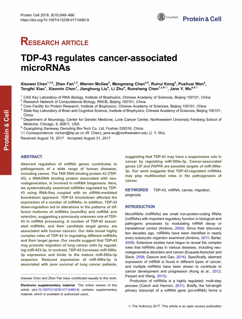
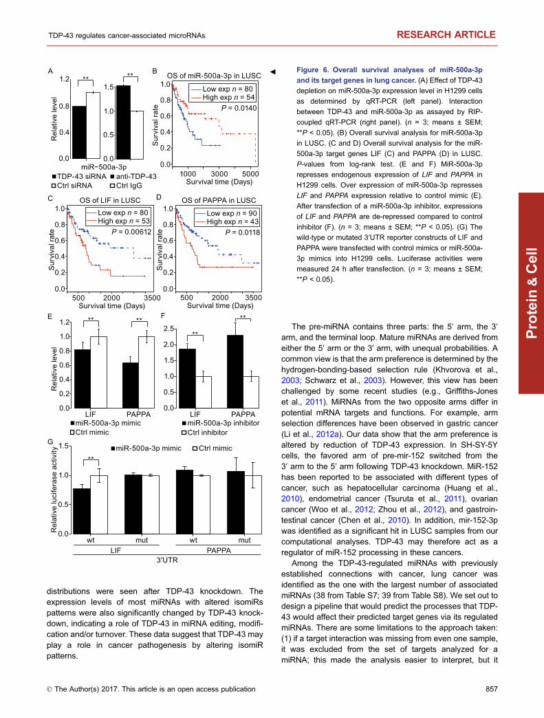
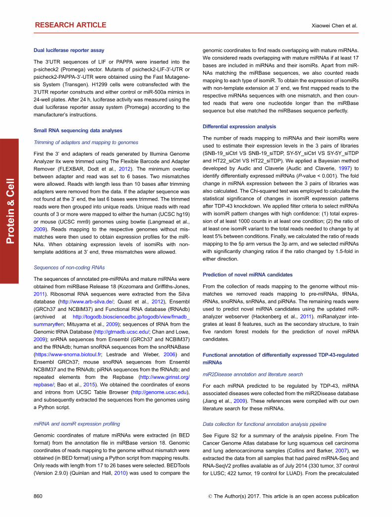
To systematically search for miRNAs that are modulated byTDP-43, we performed RNA interference (siRNA) mediatedTDP-43 knockdown in both human and mouse cells,specifically two human cell lines (neuroblastoma SH-SY5Yand glioblastoma SNB-19) and a mouse cell line with neu-ronal features, HT22 (see Fig. S1). Neuron-like cell lineswere chosen because of the known role of TDP-43 in neu-rodegenerative diseases. Small RNAs were extracted fromTDP-43 knockdown- and control cells and subjected to deepsequencing using an Illumina Genome Analyzer IIx system.From six sequenced libraries (one sample in each condition),we obtained 13.8–19.3 million reads for each sample of86-nucleotides (nt) in length (Table S1). Most sequencedreads contained the adapter sequences at their 3′ ends.After trimming adapters and removing reads containingambiguous bases, more than 95% of the total reads wereuseful and grouped into unique reads. The unique readswere analyzed and mapped to the human and mouse gen-omes (reference genomes hg19 and mm9, respectively).Approximately 55.6% of the total sequence reads could bemapped to the genomes without any sequence mismatch,and more than 27 million reads were usable for assessingRNA expression levels. The distributions of reads that couldbe aligned to tRNAs, rRNAs, snoRNAs, snRNAs, piRNAs,repeated sequences, and intron and exon sequences areshown in Table S1. The annotations of reads from TDP-43knockdown and control SH-SY5Y cell lines are shown inFig. 1A.
MicroRNAs whose expression was affected by TDP-43knock-down were carefully analyzed. The reads counts wereused to estimate the expression levels of miRNAs, as well asthe expression levels of the isomiRs and 3′ single-nucleotidemodified variants in the six libraries. Similar to a previousstudy (Morin et al., 2008), we used the most abundantvariant to assess the expression level of the correspondingmiRNA in the differential expression analysis.
Bayesian method (Audic and Claverie, 1997) was appliedto identify the miRNAs whose expression may have chan-ged. This method was originally designed to analyze differ-entially expressed genes through sequencing of their cDNAclones and has also been used to analyze small RNAsequencing data (Morin et al., 2008). In the SH-SY5Y cellline, TDP-43 knockdown resulted in 98 differentiallyexpressed miRNAs (P-value < 0.001), of which 68 miRNAswere down-regulated. Of these 98 miRNAs, 2 miRNAs were
TDP-43 regulates cancer-associated microRNAs RESEARCH ARTICLE
© The Author(s) 2017. This article is an open access publication 849
Protein
&Cell
up-regulated and 14 down-regulated after TDP-43 knock-down in the SNB-19 and HT22 cell lines. All of the differ-entially expressed miRNAs across the three cell lines arelisted in Table S2. Of the 14 miRNAs down-regulated in allthree cell lines from the RNA-Seq data, 11 were down-reg-ulated in at least two cell lines, as validated by quantitativeRT-PCR (Fig. 1B).
TDP-43 knockdown alters isomiR composition
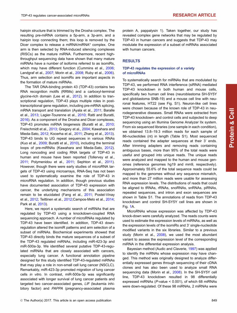
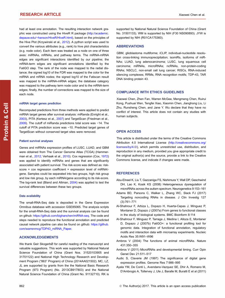
The generation of isomiRs is probably caused by positionalvariation when Drosha and/or Dicer cleave precursor miR-NAs (Morin et al., 2008). To compare the isomiR patternsbetween TDP-43 knockdown and control cell lines, wegrouped the isomiRs into four types (Fig. 2A): isomiR-5p(5′ end addition or trimming), isomiR-3p (3′ end addition ortrimming), isomiR-53p (simultaneous addition and/or trim-ming at both ends) and isomiR-3e (3′ end non-templatesingle-nucleotide extensions).
Applying the chi-square test to the expression levels ofthe 4 types of isomiRs based on mature miRNA sequencesfrom miRBase version 18 (Kozomara and Griffiths-Jones,2011), we identified 9, 9 and 26 miRNAsin the SH-SY5Y,
SNB19 and HT22 cells, respectively, that had significantlychanged isomiR patterns after TDP-43 knockdown, in com-parison to the control cells (Table S3; see MATERIALS ANDMETHODS). Changes in isomiR-3p were the most frequentamong the altered isomiR patterns. Reduced expressionlevels of isomiR-3p after TDP-43 knockdown were frequentlyaccompanied by increased expression of isomiR-3e, asobserved for hsa-miR-199a-5p, hsa-miR-301a-3p, andmmu-miR-199a-5p (Fig. 2B). In contrast, the expressionlevels of isomiR-5p and isomiR-53p are less frequentlyaltered after TDP-43 knockdown. Of note, isomiR-5p of miR-214-3p increased in both SH-SY-5Y and HT22 cells, andisomiR-53p of miR-31-3p increased in HT22 cells after TDP-43 knockdown (Fig. 2B).
TDP-43 knockdown also alters miRNA arm selection
In order to estimate the possible effect of TDP-43 on strandselection from themiRNAduplex, weobtained reads counts ofmiRNAs processed from each arm of all precursors. Mostprecursors were processed into one dominant mature miRNA(i.e., from one arm), and the dominant arm was the same in allsamples. Interestingly, we found several miRNAs where
miRNA Others tRNA
rRNA snoRNA snRNA
piRNA Genomic repeats
SH-SY-5Y TDP-43 siRNA
SH-SY-5Y Ctrl siRNA
BA
0.2
0.6
1.0
0.2
0.6
1.0
0.2
0.6
1.0
Let-7
d-3p
Let-7
i-3p
miR
-10a
-5p
miR
-15b
-3p
miR
-301
a-3p
miR
-16-
5pm
iR-2
1-3p
miR
-22-
3pm
iR-2
7a-3
pm
iR-2
7b-3
pm
iR-3
2-5p
miR
-125
b-1-
3pm
iR-1
30a-
3pm
iR-1
99a-
5p
***
*** ** *** *** *** *** **** ** ** *** ***
** ** ** **
*** *** *** ***** ** ** ** ** ** ** **
TDP-43 siRNA Ctrl siRNA
Rel
ativ
e le
vel
Rel
ativ
e le
vel
Rel
ativ
e le
vel
SH-SY-5Y
SNB-19
HT22
Figure 1. RNA-seq analyses of TDP-43 regulated miRNAs. Reads annotation and validation of small RNA sequencing in TDP-43
and control knockdown libraries. (A) Distribution of small RNA sequencing reads mapped to the human genome in SH-SY-5Y cells.
Components of TDP-43 knockdown small RNA library (upper) and control library (bottom) are shown. (B) qRT-PCR of miRNAs that
were differentially expressed following TDP-43 knockdown. The expression levels of mature miRNAs in SH-SY-5Y (upper panel),
SNB-19 (middle), and HT22 (bottom) cells transfected with either TDP-43 siRNA or control (n = 3). Statistical analyses were
performed using t-test. **P < 0.05; ***P < 0.01.
RESEARCH ARTICLE Xiaowei Chen et al.
850 © The Author(s) 2017. This article is an open access publication
Protein
&Cell
miRBase version 21 (Kozomara and Griffiths-Jones, 2014)annotates only one arm (either 5′ arm or 3′ arm) of their pre-cursors could be processed into mature miRNA. However, wefound that reads could map to both arms of their precursors,indicating a gap in its annotations (Table S4).
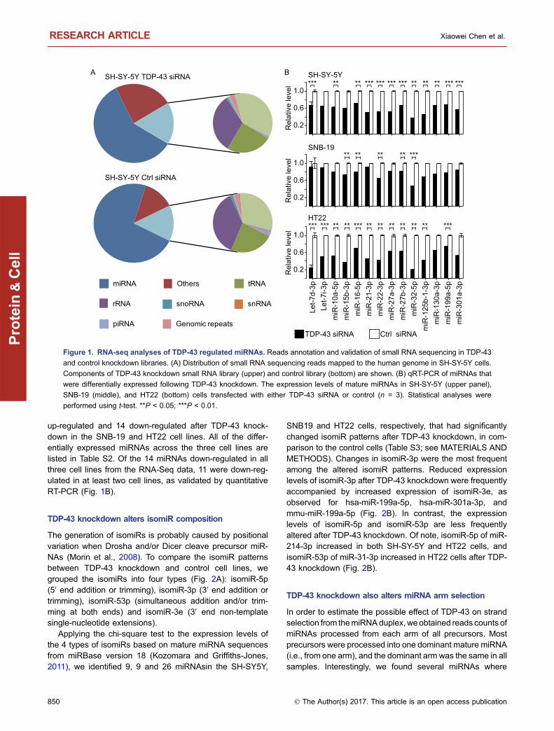
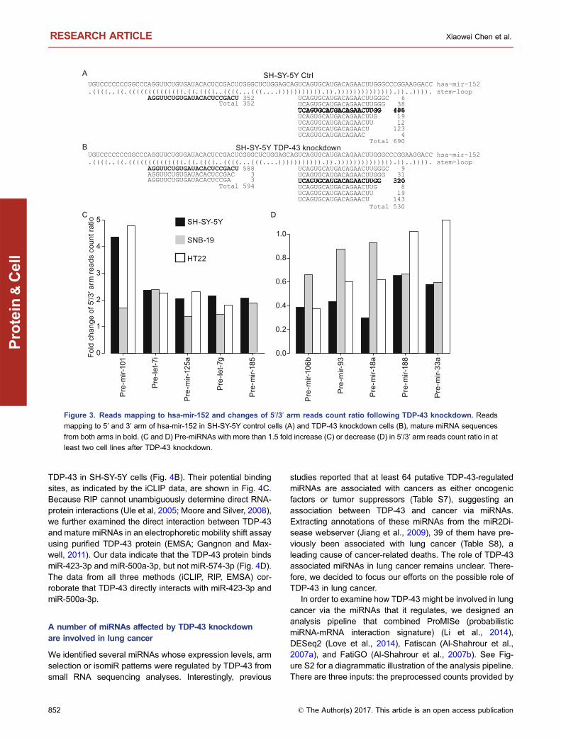
We calculated the 5′ arm/3′ arm reads count ratio forthese miRNA precursors. For a certain fraction of the pre-cursors, 5′ arm/3′ arm reads count ratio changed more than1.5-fold after TDP-43 knockdown. For example, in the SH-SY5Ycell line, 12 precursors showed increased and 12showed decreased 5′ arm/3′ arm reads count ratio afterTDP-43 knockdown. Similar results were obtained for theother two cell lines (Tables S4 and S5). The most abundantmiRNA with a changed arm ratio is miR-152, a miRNAknown to be involved in various types of cancer (see dis-cussion). In the SH-SY5Y control sample, the number ofreads deriving from the 3′ arm of pre-mir-152 was nearlytwice the number of reads deriving from the 5′ arm, whereas
after TDP-43 knockdown the number of reads from the5′ arm was about 12% higher than from the 3′ arm. Readsderived from the 5′ arm of pre-mir-152 after TDP-43 knock-down were of 3 different isoforms (Fig. 3A and 3B). Theratios of five pre-miRNAs (pre-let-7g, pre-let-7i, pre-mir-101,pre-mir-125a, and pre-mir-185) increased in at least two celllines after TDP-43 knockdown, whereas the ratios of fiveother pre-miRNAs (pre-mir-106b, pre-mir-188, pre-mir-18a,pre-mir-33a, and pre-mir-93) decreased in at least two celllines after TDP-43 knockdown (Fig. 3C and 3D). Together,these data clearly demonstrate that down-regulation of TDP-43 alters miRNA arm selection.
Novel predicted miRNAs and miRNA arms
Among the more than 100 million reads from high-throughputsequencing of the 6 libraries, there was also a large amount ofreads that mapped to un-annotated genomic regions. To identifynovel miRNA candidates among the unknown reads, we firstselected unique reads represented bymore than 3 reads countsandmapped these to genomic regions with a perfect match.Weapplied the updatedmiRanalyzer webserver (Hackenberg et al.,2011) to identify novel miRNA candidates. By including onlyunique reads with more than 50 reads counts, we predicted 8novel candidate miRNAs in human cell lines and 2 in the mousecell line (Table S6). The flanking sequences of these novelmiRNA candidates’ genome loci can be folded into stem-loopstructures compatible with those of known pre-miRNAs. Most ofthese novelmiRNAcandidateswere only detected after TDP-43knockdown, suggesting that TDP-43 may inhibit the expressionof these novel miRNA candidates. These novel miRNA candi-dates will be further investigated in future studies.
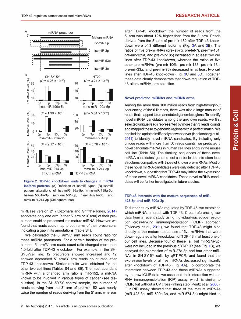
TDP-43 interacts with the mature sequences of miR-423-3p and miR-500a-3p
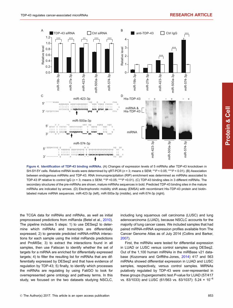
To further study miRNAs regulated by TDP-43, we examinedwhich miRNAs interact with TDP-43. Cross-referencing rawdata from a recent study using individual-nucleotide resolu-tion cross-linking immunoprecipitation (iCLIP) approach(Tollervey et al., 2011), we found that TDP-43 might binddirectly to the mature sequences of five miRNAs that weredown-regulated after knockdown of TDP-43 in at least one ofour cell lines. Because four of these (all but miR-27a-3p)were not included in the previous qRT-PCR (see Fig. 1B), weassayed the expression of miR-27a-3p and four other miR-NAs in SH-SY-5Y cells by qRT-PCR, and found that theexpression levels of all five miRNAs decreased significantlyafter knockdown of TDP-43 (Fig. 4A). To corroborate theinteraction between TDP-43 and these miRNAs suggestedby the raw iCLIP data, we assessed their interaction with anRNA immunoprecipitation (RIP) assay, which is similar toiCLIP, but without a UV cross-linking step (Peritz et al, 2006).Our RIP assay showed that three of the mature miRNAs(miR-423-3p, miR-500a-3p, and miR-574-3p) might bind to
A
B
miRNA precursorMature miRNA
isomiR 5p
isomiR 3p
isomiR 53p
isomiR 3e
5′
hsa-miR-199a-5p mmu-miR-199a-5p
10
70
40
5p 3p 53p 3e 5p 3p 53p 3e
22THY5-YS-HS
Rel
ativ
e le
vel (
%)
(P = 4.26 × 10-87) (P = 3.21 × 10-81)
Ctrl siRNA TDP-43 siRNAhsa-miR-214-3p mmu-miR-214-3p
Rel
ativ
e le
vel (
%)
(P = 5.34 × 10-56)
mmu-miR-31-3p5p 3p 53p 3e
20
60
40
Rel
ativ
e le
vel (
%)
(P = 2.17 × 10⁻³) (P = 5.78 × 10⁻⁵)
5p 3p 53p 3e 5p 3p 53p 3e
hsa-miR-301a-3p
0
80
40
(P = 1.90 × 10⁻²)
5p 3p 53p 3e
20
100
60
10
70
40
20
60
40
Figure 2. TDP-43 knockdown leads to changes in miRNA
isoform patterns. (A) Definition of isomiR types. (B) IsomiR
pattern alterations of hsa-miR-199a-5p, mmu-miR-199a-5p,
hsa-miR-301a-3p, mmu-miR-31-3p, hsa-miR-214-3p, and
mmu-miR-214-3p (Chi-square test).
TDP-43 regulates cancer-associated microRNAs RESEARCH ARTICLE
© The Author(s) 2017. This article is an open access publication 851
Protein
&Cell
TDP-43 in SH-SY-5Y cells (Fig. 4B). Their potential bindingsites, as indicated by the iCLIP data, are shown in Fig. 4C.Because RIP cannot unambiguously determine direct RNA-protein interactions (Ule et al, 2005; Moore and Silver, 2008),we further examined the direct interaction between TDP-43and mature miRNAs in an electrophoretic mobility shift assayusing purified TDP-43 protein (EMSA; Gangnon and Max-well, 2011). Our data indicate that the TDP-43 protein bindsmiR-423-3p and miR-500a-3p, but not miR-574-3p (Fig. 4D).The data from all three methods (iCLIP, RIP, EMSA) cor-roborate that TDP-43 directly interacts with miR-423-3p andmiR-500a-3p.
A number of miRNAs affected by TDP-43 knockdownare involved in lung cancer
We identified several miRNAs whose expression levels, armselection or isomiR patterns were regulated by TDP-43 fromsmall RNA sequencing analyses. Interestingly, previous
studies reported that at least 64 putative TDP-43-regulatedmiRNAs are associated with cancers as either oncogenicfactors or tumor suppressors (Table S7), suggesting anassociation between TDP-43 and cancer via miRNAs.Extracting annotations of these miRNAs from the miR2Di-sease webserver (Jiang et al., 2009), 39 of them have pre-viously been associated with lung cancer (Table S8), aleading cause of cancer-related deaths. The role of TDP-43associated miRNAs in lung cancer remains unclear. There-fore, we decided to focus our efforts on the possible role ofTDP-43 in lung cancer.
In order to examine how TDP-43 might be involved in lungcancer via the miRNAs that it regulates, we designed ananalysis pipeline that combined ProMISe (probabilisticmiRNA-mRNA interaction signature) (Li et al., 2014),DESeq2 (Love et al., 2014), Fatiscan (Al-Shahrour et al.,2007a), and FatiGO (Al-Shahrour et al., 2007b). See Fig-ure S2 for a diagrammatic illustration of the analysis pipeline.There are three inputs: the preprocessed counts provided by
A
D
1
2
3
4
5
0
Pre
-mir-
101
Pre
-let-7
i
Pre
-mir-
125a
Pre
-let-7
gSH-SY-5Y
SNB-19
HT22
Fold
cha
nge
of 5
′/3′ a
rm re
ads
coun
t rat
io
Pre
-mir-
185
0.2
0.4
0.6
0.8
1.0
0.0
Pre
-mir-
106b
Pre
-mir-
33a
Pre
-mir-
18a
Pre
-mir-
93
Pre
-mir-
188
B
C
SH-SY-5Y Ctrl
SH-SY-5Y TDP-43 knockdown
Figure 3. Reads mapping to hsa-mir-152 and changes of 5′/3′ arm reads count ratio following TDP-43 knockdown. Reads
mapping to 5′ and 3′ arm of hsa-mir-152 in SH-SY-5Y control cells (A) and TDP-43 knockdown cells (B), mature miRNA sequences
from both arms in bold. (C and D) Pre-miRNAs with more than 1.5 fold increase (C) or decrease (D) in 5′/3′ arm reads count ratio in at
least two cell lines after TDP-43 knockdown.
RESEARCH ARTICLE Xiaowei Chen et al.
852 © The Author(s) 2017. This article is an open access publication
Protein
&Cell
the TCGA data for miRNAs and mRNAs, as well as initialpreprocessed predictions from miRanda (Betel et al., 2010).The pipeline includes 5 steps: 1) to use DESeq2 to deter-mine which miRNAs and transcripts are differentiallyexpressed; 2) to generate predicted miRNA-mRNA interac-tions for each sample using the initial miRanda predictionsand ProMISe; 3) to extract the interactions found in allsamples, then use Fatiscan to identify whether the set oftargets for a miRNA are enriched for differentially expressedtargets; 4) to filter the resulting list for miRNAs that are dif-ferentially expressed by DESeq2 and that have evidence ofregulation by TDP-43; 5) finally, to identify which processesthe miRNAs are regulating by using FatiGO to look foroverrepresented gene ontology and pathway terms. In thisstudy, we focused on the two datasets studying NSCLC,
including lung squamous cell carcinoma (LUSC) and lungadenocarcinoma (LUAD), because NSCLC accounts for themajority of lung cancer cases. We included samples that hadpaired miRNA-mRNA expression profiles available from TheCancer Genome Atlas as of July 2014 (Collins and Barker,2007).
First, the miRNAs were tested for differential expressionin LUAD or LUSC versus control samples using DESeq2.Out of the 1,100 human miRNAs in the miRBase v21 data-base (Kozomara and Griffiths-Jones, 2014) 417 and 563miRNAs showed differential expression in LUAD and LUSCsamples, respectively, versus control samples. MiRNAsputatively regulated by TDP-43 were over-represented inthese groups (hypergeometric test P-value for LUAD (57/417vs. 83/1033) and LUSC (61/563 vs. 83/1037): 5.24 × 10⁻⁸
CmiR-423-3p
5′
3′
miR-500a-3p
5′
3′
miR-574-3p
5′
3′
0.0
0.2
0.4
0.6
0.8
1.0
1.2 *** *** ********
miR
-19b
-3p
miR
-27a
-3p
miR
-423
-3p
miR
-500
a-3p
miR
-574
-3p
0
1
2
3
4
***
***
***TDP-43 siRNA Ctrl siRNA anti-TDP-43 Ctrl IgGBA
Rel
ativ
e le
vel
Rel
ativ
e le
vel
miR
-19b
-3p
miR
-27a
-3p
miR
-423
-3p
miR
-500
a-3p
miR
-574
-3p
D
miR
-423
-3p
miR
-500
a-3p
miR
-574
-3p
His-TDP-43 +- - -
miRNA
miRNA &His-TDP-43
+ +
Figure 4. Identification of TDP-43 binding miRNAs. (A) Changes of expression levels of 5 miRNAs after TDP-43 knockdown in
SH-SY-5Y cells. Relative miRNA levels were determined by qRT-PCR (n = 3; means ± SEM; **P < 0.05; ***P < 0.01). (B) Association
between endogenous miRNAs and TDP-43. RNA Immunoprecipitation (RIP) enrichment was determined as miRNAs associated to
TDP-43 IP relative to control IgG (n = 3; means ± SEM; **P <0.05; ***P <0.01). (C) TDP-43 binding sites in 3 different miRNAs. The
secondary structures of the pre-miRNAs are shown, mature miRNAs sequences in bold. Predicted TDP-43 binding sites in the mature
miRNAs are indicated by arrows. (D) Electrophoretic mobility shift assay (EMSA) with recombinant His-TDP-43 protein and biotin-
labeled mature miRNA sequences. miR-423-3p (left), miR-500a-3p (middle), and miR-574-3p (right).
TDP-43 regulates cancer-associated microRNAs RESEARCH ARTICLE
© The Author(s) 2017. This article is an open access publication 853
Protein
&Cell
and 1.48 × 10⁻⁴, respectively). Because TDP-43 may notregulate all of these miRNAs, we examined the correlationbetween each miRNA and TDP-43 expression in lung can-cer samples. We performed Pearson correlation, and aftercorrecting for multiple hypothesis testing, identified 408 and467 miRNAs significantly correlated with TDP-43 in LUADand LUSC samples, respectively (FDR < 0.1). MiRNAsputatively regulated by TDP-43 had a trend for overrepre-sentation in these groups as well (hypergeometric testP-value for LUAD (39/408 vs. 83/1033) and LUSC (45/467vs. 83/1037): 0.091 and 0.059, respectively).
To identify which miRNAs showed enrichment for differ-entially expressed targets, we used the combination ofProMISe and Fatiscan. From the ProMISe step, out of the1033 and 1037 miRNAs that were expressed in LUAD andLUSC samples respectively, there were 213 and 274 miR-NAs that had at least 5 predicted targets in each LUAD orLUSC sample, respectively; miRNAs regulated by TDP-43were also over-represented in these groups (hypergeometrictest P-value for LUAD (67/213 vs. 83/1033) and LUSC(74/274 vs. 83/1037): 3.36 × 10⁻³⁵ and 1.89 × 10−36,respectively). A ranked list of the transcripts (using P-valuesof log fold change and log differential expression fromDESeq2) was submitted along with the predicted miRNA-mRNA interactions from ProMISe as custom annotations toFatiscan (part of the Babelomics v4 suite; Medina et al.,2010). This method uses a threshold-independent heuristicto identify whether an annotation term is over-represented inthe bottom or top of a ranked list; and in this case, we weresearching for miRNAs that had an enrichment for either up-regulated or down-regulated target transcripts.
In order to identify miRNAs that were both differentiallyexpressed and had enrichment for differentially expressedtargets, we combined the Fatiscan results with earlierDESeq2 results for the miRNAs to get a joint P-value. Weapplied three criteria to filter the list of miRNAs to the mostrelevant one: (A) the miRNA had to have a DESeq2 dif-ferential expression FDR < 0.1; (B) the targets of themiRNA had to be changing in the opposite direction (if themiRNA is up-regulated, the targets must be down-regu-lated, and vice versa); (C) the miRNA expression profilehad to have a statistically significant correlation with TDP-43 expression profile (FDR < 0.1). The results of this stepfor LUAD and LUSC are shown in Tables S9 and S10,respectively.
To determine what biological processes were related tothe identified targets, we extracted the unique transcriptsfrom the previous step and submitted them to FatiGO, alsopart of the Babelomics v4 suite; this tool performs an over-representation analysis for gene ontology and pathwayterms. Among the down-regulated transcripts, the most sig-nificant hits included integrin cell surface interactions andnegative regulation of cell proliferation; among the up-regu-lated transcripts, the most significant hits included nucleotidesynthesis, cell cycle checkpoints, and RNA processing. Thissuggests that TDP-43-regulated miRNAs may play a role in
promoting carcinogenesis and metastasis. The full list of hitscan be found in Table S11.
From this analysis pipeline, we defined “predicted causalinteractions” as those miRNA-mRNA interactions betweenputative TDP-43-regulated miRNAs and target mRNA tran-scripts with annotations in the processes discovered byFatiGO. See Figure S3A and S3B for the representativenetwork graph of up-regulated miRNAs and down-regulatedtranscripts in LUAD (one network of 7 up-regulated miRNAs,50 down-regulated transcripts, and 13 processes; anothernetwork of 4 down-regulated miRNAs, 62 transcripts, and 17processes), and Fig. S3C and S3D for the LUSC network,which was much larger. See Table S12 for the full node andedge lists.
In summary, our analysis pipeline identified a number ofputative TDP-43-regulated miRNAs which target severaltranscripts that have roles in cancer biology, including thetwo TDP-43 interacting miRNAs identified in this study, miR-423-3p and miR-500a-3p. These were experimentallyexamined further for their roles in lung cancer.
TDP-43 associated miR-423-3p promotes lung cancercell migration
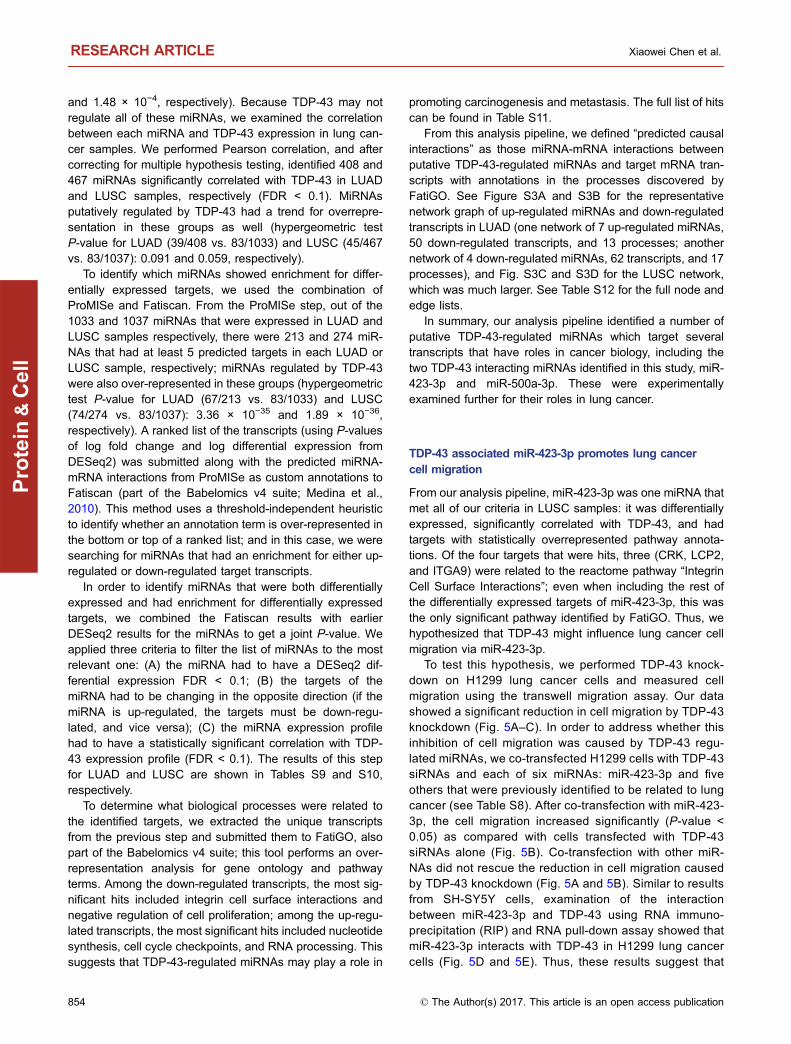
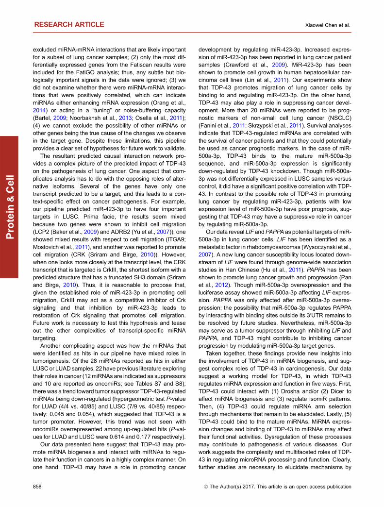
From our analysis pipeline, miR-423-3p was one miRNA thatmet all of our criteria in LUSC samples: it was differentiallyexpressed, significantly correlated with TDP-43, and hadtargets with statistically overrepresented pathway annota-tions. Of the four targets that were hits, three (CRK, LCP2,and ITGA9) were related to the reactome pathway “IntegrinCell Surface Interactions”; even when including the rest ofthe differentially expressed targets of miR-423-3p, this wasthe only significant pathway identified by FatiGO. Thus, wehypothesized that TDP-43 might influence lung cancer cellmigration via miR-423-3p.
To test this hypothesis, we performed TDP-43 knock-down on H1299 lung cancer cells and measured cellmigration using the transwell migration assay. Our datashowed a significant reduction in cell migration by TDP-43knockdown (Fig. 5A–C). In order to address whether thisinhibition of cell migration was caused by TDP-43 regu-lated miRNAs, we co-transfected H1299 cells with TDP-43siRNAs and each of six miRNAs: miR-423-3p and fiveothers that were previously identified to be related to lungcancer (see Table S8). After co-transfection with miR-423-3p, the cell migration increased significantly (P-value <0.05) as compared with cells transfected with TDP-43siRNAs alone (Fig. 5B). Co-transfection with other miR-NAs did not rescue the reduction in cell migration causedby TDP-43 knockdown (Fig. 5A and 5B). Similar to resultsfrom SH-SY5Y cells, examination of the interactionbetween miR-423-3p and TDP-43 using RNA immuno-precipitation (RIP) and RNA pull-down assay showed thatmiR-423-3p interacts with TDP-43 in H1299 lung cancercells (Fig. 5D and 5E). Thus, these results suggest that
RESEARCH ARTICLE Xiaowei Chen et al.
854 © The Author(s) 2017. This article is an open access publication
Protein
&Cell
TDP-43 may promote lung cancer cell migration throughregulation of miR-423-3p, corroborating the predictionfrom the functional annotation pipeline that TDP-43 is atumor-promoting factor.
TDP-43 regulated miRNAs, including miR-500a-3p, mayserve as prognostic markers of cancers
Among the TDP-43 affected miRNAs that we identified, theexpression levels of 17 miRNAs have previously beenassociated with NSCLC patient survival. Similarly, inglioblastoma multiforme (GBM), the expression levels of 18TDP-43 regulated miRNAs might have prognostic value forGBM (Table S13).
In order to test the association between TDP-43 regulatedmiRNAs and patient survival in cancer using samples inde-pendent from these previous studies, we collected miRNAexpression profiles of 134 lung squamous cell carcinoma(LUSC), 191 lung adenocarcinoma (LUAD) and 487 GBM,and gene expression profiles of 133 LUSC, 231 LUAD and538 GBM from The Cancer Genome of Atlas (TCGA),together with information on the patient survival. We foundthat 4 of the 17 TDP-43 regulated miRNAs in NSCLC werealso significantly associated with patient survival in theindependent cohort (let-7b-5p, miR-25-3p, miR-31-5p, andmiR-93-5p), and that 5 of the 18 miRNAs were confirmed tobe significantly associated with GBM patient survival in thisindependent cohort (miR-148a-3p, miR-17-5p, miR-20a-5p,miR-221-3p, and miR-31-5p) (Table S13). Low expression ofhsa-let-7b-5p and high expression of hsa-miR-31-5p wereassociated with poor survival in lung cancer, which is con-sistent with previous reports (Fig. S4; Tan et al., 2011;Yanaihara et al., 2006), and hsa-let-7b was identified as a hitfrom our pipeline. Low expression of miR-17-5p and miR-20a-5p and high expression of miR-148a-3p, miR-221-3p,and miR-31-5p were associated with poor prognostic out-come in GBM independent cohort (Fig. S4). The associa-tions of these 5 miRNAs with patient survival have all beenreported previously (Delfino et al., 2011; Srinivasan et al.,2011).
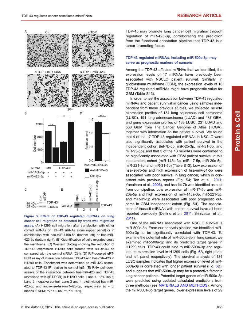
One of the miRNAs associated with NSCLC survival ismiR-500a-3p. From our analysis pipeline, we identified miR-500a-3p to be significantly correlated with TDP-43. Toexamine the potential role of miR-500a-3p in lung cancer, weexamined miR-500a-3p and its predicted target genes inH1299 cells. TDP-43 could bind to miR-500a-3p and regu-late its expression level in H1299 cells (Fig. 6A, right paneland left panel respectively). The survival analysis of 134LUSC samples indicates that higher expression level of miR-500a-3p is correlated with longer patient survival (Fig. 6B),and suggests that miR-500a-3p may be a protective factor inlung cancer patients. Potential target genes of miR-500a-3pwere predicted using updated calculated predictions fromthree methods (see MATERIALS AND METHODS). Amongthe miR-500a-3p target genes, lower expression levels of 29
A
0
400
800
1200
Cel
l num
ber
TDP-43miR-146b-5p
siRNA
miR-423-3p+
+-
---
--
Ctrl
Ctrl
siTD
P
TDP-43β-Tublin
0.0
0.5
1.0
1.5
hsa-miR-423-3p
***
Rel
ativ
e le
vel
Anti-TDP-43
Ctrl IgG
B
C
D
E
**
**
PDTislrtC
siTDP + miR-146b siTDP + miR-423
20×
55 kDa40 kDa TDP-43
1 2 3 4
Inpu
t
Ctrl
miR
-423
-3p
as-m
iR-4
23-3
p
40 kDa
Figure 5. Effect of TDP-43 regulated miRNAs on lung
cancer cell migration as detected by trans-well migration
assay. (A) H1299 cell migration after transfection with either
control siRNAs or TDP-43 siRNAs alone (upper panel) or in
combination with has-miR-146b-5p (bottom left) or has-miR-
423-3p (bottom right). (B) Quantification of cells migrated cross
the membrane. (C) Western blotting showing the reduction of
TDP-43 expression H1299 cells treated with siTDP-43 as
compared with the control siRNA (Ctrl). (D) RIP-coupled qRT-
PCR assay of interaction between TDP-43 and has-miR-423 in
H1299 cells. Enrichment was determined as miR-423 associ-
ated to TDP-43 IP relative to control IgG. (E) RNA pull-down
assays of the interaction between has-miR-423 and TDP-43
(combined with qRT-PCR) in H1299 cells. Lane 1, ∼3% input;
Lane 2, negative control; Lane 3 and 4, biotinylated has-miR-
423-3p and antisense-hsa-miR-423-3p, respectively. (n = 3;
means ± SEM; **P < 0.05; ***P < 0.01).
TDP-43 regulates cancer-associated microRNAs RESEARCH ARTICLE
© The Author(s) 2017. This article is an open access publication 855
Protein
&Cell
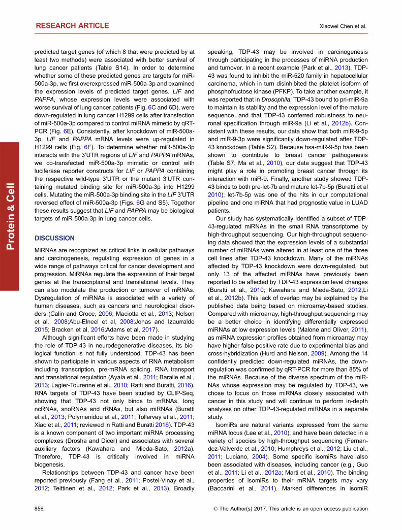
predicted target genes (of which 8 that were predicted by atleast two methods) were associated with better survival oflung cancer patients (Table S14). In order to determinewhether some of these predicted genes are targets for miR-500a-3p, we first overexpressed miR-500a-3p and examinedthe expression levels of predicted target genes. LIF andPAPPA, whose expression levels were associated withworse survival of lung cancer patients (Fig. 6C and 6D), weredown-regulated in lung cancer H1299 cells after transfectionof miR-500a-3p compared to control miRNA mimetic by qRT-PCR (Fig. 6E). Consistently, after knockdown of miR-500a-3p, LIF and PAPPA mRNA levels were up-regulated inH1299 cells (Fig. 6F). To determine whether miR-500a-3pinteracts with the 3′UTR regions of LIF and PAPPA mRNAs,we co-transfected miR-500a-3p mimetic or control withluciferase reporter constructs for LIF or PAPPA containingthe respective wild-type 3′UTR or the mutant 3′UTR con-taining mutated binding site for miR-500a-3p into H1299cells. Mutating the miR-500a-3p binding site in the LIF 3′UTRreversed effect of miR-500a-3p (Figs. 6G and S5). Togetherthese results suggest that LIF and PAPPA may be biologicaltargets of miR-500a-3p in lung cancer cells.
DISCUSSION
MiRNAs are recognized as critical links in cellular pathwaysand carcinogenesis, regulating expression of genes in awide range of pathways critical for cancer development andprogression. MiRNAs regulate the expression of their targetgenes at the transcriptional and translational levels. Theycan also modulate the production or turnover of mRNAs.Dysregulation of miRNAs is associated with a variety ofhuman diseases, such as cancers and neurological disor-ders (Calin and Croce, 2006; Maciotta et al., 2013; Nelsonet al., 2008;Abu-Elneel et al, 2008;Jonas and Izaurralde2015; Bracken et al, 2016;Adams et al, 2017).
Although significant efforts have been made in studyingthe role of TDP-43 in neurodegenerative diseases, its bio-logical function is not fully understood. TDP-43 has beenshown to participate in various aspects of RNA metabolismincluding transcription, pre-mRNA splicing, RNA transportand translational regulation (Ayala et al., 2011; Baralle et al.,2013; Lagier-Tourenne et al., 2010; Ratti and Buratti, 2016).RNA targets of TDP-43 have been studied by CLIP-Seq,showing that TDP-43 not only binds to mRNAs, longncRNAs, snoRNAs and rRNAs, but also miRNAs (Burattiet al., 2013; Polymenidou et al., 2011; Tollervey et al., 2011;Xiao et al., 2011; reviewed in Ratti and Buratti 2016). TDP-43is a known component of two important miRNA processingcomplexes (Drosha and Dicer) and associates with severalauxiliary factors (Kawahara and Mieda-Sato, 2012a).Therefore, TDP-43 is critically involved in miRNAbiogenesis.
Relationships between TDP-43 and cancer have beenreported previously (Fang et al., 2011; Postel-Vinay et al.,2012; Teittinen et al., 2012; Park et al., 2013). Broadly
speaking, TDP-43 may be involved in carcinogenesisthrough participating in the processes of miRNA productionand turnover. In a recent example (Park et al., 2013), TDP-43 was found to inhibit the miR-520 family in hepatocellularcarcinoma, which in turn disinhibited the platelet isoform ofphosphofructose kinase (PFKP). To take another example, itwas reported that in Drosophila, TDP-43 bound to pri-miR-9ato maintain its stability and the expression level of the maturesequence, and that TDP-43 conferred robustness to neu-ronal specification through miR-9a (Li et al., 2012b). Con-sistent with these results, our data show that both miR-9-5pand miR-9-3p were significantly down-regulated after TDP-43 knockdown (Table S2). Because hsa-miR-9-5p has beenshown to contribute to breast cancer pathogenesis(Table S7; Ma et al., 2010), our data suggest that TDP-43might play a role in promoting breast cancer through itsinteraction with miR-9. Finally, another study showed TDP-43 binds to both pre-let-7b and mature let-7b-5p (Buratti et al2010); let-7b-5p was one of the hits in our computationalpipeline and one miRNA that had prognostic value in LUADpatients.
Our study has systematically identified a subset of TDP-43-regulated miRNAs in the small RNA transcriptome byhigh-throughput sequencing. Our high-throughput sequenc-ing data showed that the expression levels of a substantialnumber of miRNAs were altered in at least one of the threecell lines after TDP-43 knockdown. Many of the miRNAsaffected by TDP-43 knockdown were down-regulated, butonly 13 of the affected miRNAs have previously beenreported to be affected by TDP-43 expression level changes(Buratti et al., 2010; Kawahara and Mieda-Sato, 2012,Liet al., 2012b). This lack of overlap may be explained by thepublished data being based on microarray-based studies.Compared with microarray, high-throughput sequencing maybe a better choice in identifying differentially expressedmiRNAs at low expression levels (Malone and Oliver, 2011),as miRNA expression profiles obtained from microarray mayhave higher false positive rate due to experimental bias andcross-hybridization (Hurd and Nelson, 2009). Among the 14confidently predicted down-regulated miRNAs, the down-regulation was confirmed by qRT-PCR for more than 85% ofthe miRNAs. Because of the diverse spectrum of the miR-NAs whose expression may be regulated by TDP-43, wechose to focus on those miRNAs closely associated withcancer in this study and will continue to perform in-depthanalyses on other TDP-43-regulated miRNAs in a separatestudy.
IsomiRs are natural variants expressed from the samemiRNA locus (Lee et al., 2010), and have been detected in avariety of species by high-throughput sequencing (Fernan-dez-Valverde et al., 2010; Humphreys et al., 2012; Liu et al.,2011; Luciano, 2004). Some specific isomiRs have alsobeen associated with diseases, including cancer (e.g., Guoet al., 2011; Li et al., 2012a; Marti et al., 2010). The bindingproperties of isomiRs to their mRNA targets may vary(Baccarini et al., 2011). Marked differences in isomiR
RESEARCH ARTICLE Xiaowei Chen et al.
856 © The Author(s) 2017. This article is an open access publication
Protein
&Cell
distributions were seen after TDP-43 knockdown. Theexpression levels of most miRNAs with altered isomiRspatterns were also significantly changed by TDP-43 knock-down, indicating a role of TDP-43 in miRNA editing, modifi-cation and/or turnover. These data suggest that TDP-43 mayplay a role in cancer pathogenesis by altering isomiRpatterns.
The pre-miRNA contains three parts: the 5′ arm, the 3′arm, and the terminal loop. Mature miRNAs are derived fromeither the 5′ arm or the 3′ arm, with unequal probabilities. Acommon view is that the arm preference is determined by thehydrogen-bonding-based selection rule (Khvorova et al.,2003; Schwarz et al., 2003). However, this view has beenchallenged by some recent studies (e.g., Griffiths-Joneset al., 2011). MiRNAs from the two opposite arms differ inpotential mRNA targets and functions. For example, armselection differences have been observed in gastric cancer(Li et al., 2012a). Our data show that the arm preference isaltered by reduction of TDP-43 expression. In SH-SY-5Ycells, the favored arm of pre-mir-152 switched from the3′ arm to the 5′ arm following TDP-43 knockdown. MiR-152has been reported to be associated with different types ofcancer, such as hepatocellular carcinoma (Huang et al.,2010), endometrial cancer (Tsuruta et al., 2011), ovariancancer (Woo et al., 2012; Zhou et al., 2012), and gastroin-testinal cancer (Chen et al., 2010). In addition, mir-152-3pwas identified as a significant hit in LUSC samples from ourcomputational analyses. TDP-43 may therefore act as aregulator of miR-152 processing in these cancers.
Among the TDP-43-regulated miRNAs with previouslyestablished connections with cancer, lung cancer wasidentified as the one with the largest number of associatedmiRNAs (38 from Table S7; 39 from Table S8). We set out todesign a pipeline that would predict the processes that TDP-43 would affect their predicted target genes via its regulatedmiRNAs. There are some limitations to the approach taken:(1) if a target interaction was missing from even one sample,it was excluded from the set of targets analyzed for amiRNA; this made the analysis easier to interpret, but it
1000 3000 5000Survival time (Days)
OS of miR-500a-3p in LUSC
0.0
0.2
0.4
0.6
0.8
1.0 Low exp n = 80High exp n = 54
P = 0.0140
miR−500a-3p0.0
0.4
0.8
1.2
Rel
ativ
e le
vel
0.0
0.5
1.0
1.5** **
TDP-43 siRNACtrl siRNA
anti-TDP-43Ctrl IgG
BA
Sur
viva
l rat
e
D
Low exp n = 90High exp n = 43
P = 0.0118
2000500 3500Survival time (Days)
OS of PAPPA in LUSC
0.0
0.2
0.4
0.6
0.8
1.0
Sur
viva
l rat
e
C
0.0
0.2
0.4
0.6
0.8
1.0
1.2
LIF PAPPA
** **
Rel
ativ
e le
vel
miR-500a-3p mimicCtrl mimic
0.0
0.5
1.0
1.5
2.0
2.5**
**
LIF PAPPAmiR-500a-3p inhibitorCtrl inhibitor
FE
G
Rel
ativ
e lu
cife
rase
act
ivity
0.0
0.5
1.0
1.5 miR-500a-3p mimic Ctrl mimic**
wt mutLIF
wt mutPAPPA
3′UTR
OS of LIF in LUSC
2000500 3500Survival time (Days)
0.0
0.2
0.4
0.6
0.8
1.0
Sur
viva
l rat
e
Low exp n = 80High exp n = 53
P = 0.00612
b Figure 6. Overall survival analyses of miR-500a-3p
and its target genes in lung cancer. (A) Effect of TDP-43
depletion on miR-500a-3p expression level in H1299 cells
as determined by qRT-PCR (left panel). Interaction
between TDP-43 and miR-500a-3p as assayed by RIP-
coupled qRT-PCR (right panel). (n = 3; means ± SEM;
**P < 0.05). (B) Overall survival analysis for miR-500a-3p
in LUSC. (C and D) Overall survival analysis for the miR-
500a-3p target genes LIF (C) and PAPPA (D) in LUSC.
P-values from log-rank test. (E and F) MiR-500a-3p
represses endogenous expression of LIF and PAPPA in
H1299 cells. Over expression of miR-500a-3p represses
LIF and PAPPA expression relative to control mimic (E).
After transfection of a miR-500a-3p inhibitor, expressions
of LIF and PAPPA are de-repressed compared to control
inhibitor (F). (n = 3; means ± SEM; **P < 0.05). (G) The
wild-type or mutated 3′UTR reporter constructs of LIF and
PAPPA were transfected with control mimics or miR-500a-
3p mimics into H1299 cells. Luciferase activities were
measured 24 h after transfection. (n = 3; means ± SEM;
**P < 0.05).
TDP-43 regulates cancer-associated microRNAs RESEARCH ARTICLE
© The Author(s) 2017. This article is an open access publication 857
Protein
&Cell
excluded miRNA-mRNA interactions that are likely importantfor a subset of lung cancer samples; (2) only the most dif-ferentially expressed genes from the Fatiscan results wereincluded for the FatiGO analysis; thus, any subtle but bio-logically important signals in the data were ignored; (3) wedid not examine whether there were miRNA-mRNA interac-tions that were positively correlated, which can indicatemiRNAs either enhancing mRNA expression (Orang et al.,2014) or acting in a “tuning” or noise-buffering capacity(Bartel, 2009; Noorbakhsh et al., 2013; Osella et al., 2011);(4) we cannot exclude the possibility of other miRNAs orother genes being the true cause of the changes we observein the target gene. Despite these limitations, this pipelineprovides a clear set of hypotheses for future work to validate.
The resultant predicted causal interaction network pro-vides a complex picture of the predicted impact of TDP-43on the pathogenesis of lung cancer. One aspect that com-plicates analysis has to do with the opposing roles of alter-native isoforms. Several of the genes have only onetranscript predicted to be a target, and this leads to a con-text-specific effect on cancer pathogenesis. For example,our pipeline predicted miR-423-3p to have four importanttargets in LUSC. Prima facie, the results seem mixedbecause two genes were shown to inhibit cell migration(LCP2 (Baker et al., 2009) and ADRB2 (Yu et al., 2007)), oneshowed mixed results with respect to cell migration (ITGA9;Mostovich et al., 2011), and another was reported to promotecell migration (CRK (Sriram and Birge, 2010)). However,when one looks more closely at the transcript level, the CRKtranscript that is targeted is CrkIII, the shortest isoform with apredicted structure that has a truncated SH3 domain (Sriramand Birge, 2010). Thus, it is reasonable to propose that,given the established role of miR-423-3p in promoting cellmigration, CrkIII may act as a competitive inhibitor of Crksignaling and that inhibition by miR-423-3p leads torestoration of Crk signaling that promotes cell migration.Future work is necessary to test this hypothesis and teaseout the other complexities of transcript-specific miRNAtargeting.
Another complicating aspect was how the miRNAs thatwere identified as hits in our pipeline have mixed roles intumorigenesis. Of the 28 miRNAs reported as hits in eitherLUSCor LUADsamples, 22 have previous literature exploringtheir roles in cancer (12miRNAs are indicated as suppressorsand 10 are reported as oncomiRs; see Tables S7 and S8);there was a trend toward tumor suppressor TDP-43-regulatedmiRNAs being down-regulated (hypergeometric test P-valuefor LUAD (4/4 vs. 40/85) and LUSC (7/9 vs. 40/85) respec-tively: 0.045 and 0.054), which suggested that TDP-43 is atumor promoter. However, this trend was not seen withoncomiRs overrepresented among up-regulated hits (P-val-ues for LUAD and LUSC were 0.614 and 0.177 respectively).
Our data presented here suggest that TDP-43 may pro-mote miRNA biogenesis and interact with miRNAs to regu-late their function in cancers in a highly complex manner. Onone hand, TDP-43 may have a role in promoting cancer
development by regulating miR-423-3p. Increased expres-sion of miR-423-3p has been reported in lung cancer patientsamples (Crawford et al., 2009). MiR-423-3p has beenshown to promote cell growth in human hepatocellular car-cinoma cell lines (Lin et al., 2011). Our experiments showthat TDP-43 promotes migration of lung cancer cells bybinding to and regulating miR-423-3p. On the other hand,TDP-43 may also play a role in suppressing cancer devel-opment. More than 20 miRNAs were reported to be prog-nostic markers of non-small cell lung cancer (NSCLC)(Fanini et al., 2011; Skrzypski et al., 2011). Survival analysesindicate that TDP-43-regulated miRNAs are correlated withthe survival of cancer patients and that they could potentiallybe used as cancer prognostic markers. In the case of miR-500a-3p, TDP-43 binds to the mature miR-500a-3psequence, and miR-500a-3p expression is significantlydown-regulated by TDP-43 knockdown. Though miR-500a-3p was not differentially expressed in LUSC samples versuscontrol, it did have a significant positive correlation with TDP-43. In contrast to the possible role of TDP-43 in promotinglung cancer by regulating miR-423-3p, patients with lowexpression level of miR-500a-3p have poor prognosis, sug-gesting that TDP-43 may have a suppressive role in cancerby regulating miR-500a-3p.
Our data reveal LIF andPAPPA as potential targets of miR-500a-3p in lung cancer cells. LIF has been identified as ametastatic factor in rhabdomyosarcomas (Wysoczynski et al.,2007). A new lung cancer susceptibility locus located down-stream of LIF were found through genome-wide associationstudies in Han Chinese (Hu et al., 2011). PAPPA has beenshown to promote lung cancer growth and progression (Panet al., 2012). Though miR-500a-3p overexpression and theluciferase assay showed miR-500a-3p affecting LIF expres-sion, PAPPA was only affected after miR-500a-3p overex-pression; the possibility that miR-500a-3p regulates PAPPAby interacting with binding sites outside its 3′UTR remains tobe resolved by future studies. Nevertheless, miR-500a-3pmay serve as a tumor suppressor through inhibiting LIF andPAPPA, and TDP-43 might contribute to inhibiting cancerprogression by modulating miR-500a-3p target genes.
Taken together, these findings provide new insights intothe involvement of TDP-43 in miRNA biogenesis, and sug-gest complex roles of TDP-43 in carcinogenesis. Our datasuggest a working model for TDP-43, in which TDP-43regulates miRNA expression and function in five ways. First,TDP-43 could interact with (1) Drosha and/or (2) Dicer toaffect miRNA biogenesis and (3) regulate isomiR patterns.Then, (4) TDP-43 could regulate miRNA arm selectionthrough mechanisms that remain to be elucidated. Lastly, (5)TDP-43 could bind to the mature miRNAs. MiRNA expres-sion changes and binding of TDP-43 to miRNAs may affecttheir functional activities. Dysregulation of these processesmay contribute to pathogenesis of various diseases. Ourwork suggests the complexity and multifaceted roles of TDP-43 in regulating microRNA processing and function. Clearly,further studies are necessary to elucidate mechanisms by
RESEARCH ARTICLE Xiaowei Chen et al.
858 © The Author(s) 2017. This article is an open access publication
Protein
&Cell
which TDP-43 act in the pathogenesis of various humandiseases, including cancer.
MATERIALS AND METHODS
Reagents, cell cultures, and transfection
The polyclonal anti-TDP-43 antibody (Abcam) and monoclonal anti-
beta-tubulin (BD Pharmingen) were obtained from corresponding
vendors.
Human neuroblastoma cells (SH-SY-5Y), human glioma cells
(SNB-19), human non-small cell lung cancer cells (H1299), and
mouse HT22 cells were cultured in DMEM supplemented with 10%
fetal bovine serum at 5% CO2 and 37°C. Human and mouse TDP-43
siGENOME SMARTpool oligos were purchased from Dharmacon.
The siRNAoligoswere transfected into the cells by lipofectamine2000
(Invitrogen) for 48 h followedby a second transfection for another 72 h.
SiGENOME Non-Targeting siRNA Pool #2 oligos (Dharmacon) were
used as a negative control. Mimetics and inhibitors of corresponding
miRNAs were purchased from Genepharma company, and trans-
fected into cells using lipofectamine2000 for 48 h.
Western blotting
Whole cell lysates from non-targeting siRNAs or TDP-43 siRNAs
transfected cells were obtained using lysis buffer (50 mmol/L Tris-
HCl (pH 6.8), 10% glycerol, 2.5% β-mercaptoethanol, 2% SDS,
0.1% Bromophenol Blue). Cell lysate was loaded onto 10% SDS-
PAGE and transferred onto Polyvinylidene Fluoride membranes.
Membranes were incubated with rabbit anti-TDP-43 antibody
(1:1000; Abcam). Mouse anti-β-tubulin (1:2500; BD Pharmingen)
was used as an internal control.
Total RNA extraction and small RNA sequencing
Following two rounds of transfection with the control or TDP-43 siR-
NAs, cellswere lysed in Trizol (Invitrogen) for total RNAextractionwith
DNA removed by treating samples using RNase-free DNase I
(Roche). The RNA yield was determined by UV absorbance spec-
troscopy (GE) andRNAquality was checked using 1% formaldehyde-
agarose gel electrophoresis. Small RNA libraries (16–52 nt) were
constructed using the TruSeq Small RNA Sample Prep Kit (Illumina)
and sequenced using an Illumina Genome Analyzer IIx platform (one
sample per lane), with one sample being sequenced from each con-
dition (TDP-43 siRNA or control) for each cell line.
Quantitative RT-PCR (qRT-PCR) analyses
Quantitative RT-PCR was performed using the Rotor-Q qRT-PCR
instrument (Qiagen). Total RNA treated with the Turbo DNA-free™
Kit (Ambion) was used. The levels of miRNAs were quantified using
the NCode™ VILO™ miRNA qRT-PCR kit (Invitrogen) and normal-
ized with the U6 small nuclear RNA (U6 snRNA). The expression of
corresponding miRNA target genes was measured using Trans-
Script II Green One-Step qRT-PCR SuperMix (Transgen) with beta-
actin and GAPDH as internal controls. All reactions, including
reverse transcriptions and PCRs, were run in triplicates in at least 3
independent experiments. Primers used are listed in Table S15.
RNA immunoprecipitation (RIP) assay
The RIP experiment protocol was described in our previously pub-
lished work (Fan et al., 2014). Briefly, 2 × 10⁷ SH-SY5Y cells were
collected, resuspended in 2 mL RIP lysis buffer [50 mmol/L Tris-HCl
(pH 7.4), 1 mmol/L EDTA (pH 8.0), 250 mmol/L NaCl, 0.5% NP-40,
1 mmol/L PMSF, 1× Phosphatase Inhibitor Cocktail (sigma), 1×
Protease Inhibitor Cocktail (sigma), and 0.1 U/mL of RNasin (Pro-
mega)] on ice for 15 min, and centrifuged at 4°C, 13,300 rpm for
30 min (Eppendorf). An antibody against TDP-43 (Abcam, IP grade)
or rabbit IgG control (Millipore) was added to the lysate with gentle
rotation at 4°C for 1 h. Protein A beads were added and incubated
for an additional 3 h at 4°C with gentle rotation. Beads were col-
lected at 4°C and centrifuged at 2,000 rpm for 2 min. They were then
washed for three times in RIP buffer and resuspended in 1 mLTrizol.
RNA was subsequently isolated for qRT-PCR analysis.
Electrophoretic mobility shift assay (EMSA)
BiotinylatedmiRNAs were purchased from Takara. The coding region
of TDP-43 was amplified with the following primers: TDP-FATGTCT-
GAATATATTCGGGT, TDP-R CTACATTCCCCAGCCAGAAG. The
full-length TDP-43 taggedwith His at theC-terminus (His-TDP-43wild
type) was constructed using the pEASY-E2 expression kit (Transgen).
The wild-type His-TDP-43 was expressed in Escherichia coli stain
Transetta (DE3) (Transgen) by incubation for 18 h at 16°C with
1mmol/L IPTG.The resultingproteinwas thenpurifiedwithNi-NTAFast
Start Kit (Qiagen) in accordance with the manufacturer’s instructions.
The biotin-labeled miRNAs were incubated with His-TDP-43 and the
assays were carried out using LightShift Chemiluminescent RNA
EMSA Kit (Thermo) following the manufacturer’s instruction.
RNA pull-down assay
Cells were lysed with RIP lysis buffer (same as above) and then
centrifuged. The lysate was split into four parts: one 100 μL aliquot
for input, and three 300 μL aliquots for negative control, sense strand
and antisense strand RNA, respectively. Biotin-labeled RNA was
added to the lysate and incubated at 4°C for 3 h with gentle agitation.
Streptavidin-coated magnetic beads (Invitrogen) were blocked for
2 h at 4°C in lysis buffer containing 1 mg/mL yeast tRNA (Ambion)
and 1 mg/mL BSA (Ambion), and then washed twice with 1 mL lysis
buffer. Then, the beads were suspended in the lysate for at least 3 h,
and subsequently washed 5 times with 1 Ml wash buffer (10 mmol/L
Tris (pH 8.0), 1 mmol/L EDTA, 0.5 mol/L NaCl). The beads were then
boiled for 5 min in 0.1% SDS for dissociation and then subjected to
Western blotting as described above.
Transwell migration assay
For the in vitro cell migration assay, 5 × 10⁴ cells were suspended in
0.5 mL DMEM without serum, and then plated into the transwell
inserts (BD Biosciences). To the bottom well, 0.75 mL DMEM with
serum was added. Cells were incubated for 12 h, fixed in 75%
ethanol for 10 min, and stained by crystal violet for 30 min. Cells that
migrated cross the membrane were counted under a microscope
from 6 randomly selected fields (at 200× magnification).
TDP-43 regulates cancer-associated microRNAs RESEARCH ARTICLE
© The Author(s) 2017. This article is an open access publication 859
Protein
&Cell
Dual luciferase reporter assay
The 3′UTR sequences of LIF or PAPPA were inserted into the
p-sicheck2 (Promega) vector. Mutants of psicheck2-LIF-3′-UTR or
psicheck2-PAPPA-3′-UTR were obtained using the Fast Mutagene-
sis System (Transgen). H1299 cells were cotransfected with the
3′UTR reporter constructs and either control or miR-500a mimics in
24-well plates. After 24 h, luciferase activity was measured using the
dual luciferase reporter assay system (Promega) according to the
manufacturer’s instructions.
Small RNA sequencing data analyses
Trimming of adapters and mapping to genomes
First the 3′ end adapters of reads generated by Illumina Genome
Analyzer IIx were trimmed using The Flexible Barcode and Adapter
Remover (FLEXBAR, Dodt et al., 2012). The minimum overlap
between adapter and read was set to 6 bases. Two mismatches
were allowed. Reads with length less than 10 bases after trimming
adapters were removed from the data. If the adapter sequence was
not found at the 3′ end, the last 6 bases were trimmed. The trimmed
reads were then grouped into unique reads. Unique reads with read
counts of 3 or more were mapped to either the human (UCSC hg19)
or mouse (UCSC mm9) genomes using bowtie (Langmead et al.,
2009). Reads mapping to the respective genomes without mis-
matches were then used to obtain expression profiles for the miR-
NAs. When obtaining expression levels of isomiRs with non-
template additions at 3′ end, three mismatches were allowed.
Sequences of non-coding RNAs
The sequences of annotated pre-miRNAs and mature miRNAs were
obtained from miRBase Release 18 (Kozomara and Griffiths-Jones,
2011). Ribosomal RNA sequences were extracted from the Silva
database (http://www.arb-silva.de/; Quast et al., 2012), Ensembl
(GRCh37 and NCBIM37) and Functional RNA database (fRNAdb)
(archived at http://togodb.biosciencedbc.jp/togodb/view/frnadb_
summary#en; Mituyama et al., 2009); sequences of tRNA from the
Genomic tRNA Database (http://gtrnadb.ucsc.edu/; Chan and Lowe,
2009); snRNA sequences from Ensembl (GRCh37 and NCBIM37)
and the fRNAdb; human snoRNA sequences from the snoRNABase
(https://www-snorna.biotoul.fr; Lestrade and Weber, 2006) and
Ensembl GRCh37; mouse snoRNA sequences from Ensembl
NCBIM37 and the fRNAdb; piRNA sequences from the fRNAdb; and
repeated elements from the Repbase (http://www.girinst.org/
repbase/; Bao et al., 2015). We obtained the coordinates of exons
and introns from UCSC Table Browser (http://genome.ucsc.edu),
and subsequently extracted the sequences from the genomes using
a Python script.
miRNA and isomiR expression profiling
Genomic coordinates of mature miRNAs were extracted (in BED
format) from the annotation file in miRBase version 18. Genomic
coordinates of reads mapping to the genome without mismatch were
obtained (in BED format) using a Python script from mapping results.
Only reads with length from 17 to 26 bases were selected. BEDTools
(Version 2.9.0) (Quinlan and Hall, 2010) was used to compare the
genomic coordinates to find reads overlapping with mature miRNAs.
We considered reads overlapping with mature miRNAs if at least 17
bases are included in miRNAs and their isomiRs. Apart from miR-
NAs matching the miRBase sequences, we also counted reads
mapping to each type of isomiR. To obtain the expression of isomiRs
with non-template extension at 3′ end, we first mapped reads to the
respective miRNAs sequences with one mismatch, and then coun-
ted reads that were one nucleotide longer than the miRBase
sequence but else matched the miRBases sequence perfectly.
Differential expression analysis
The number of reads mapping to miRNAs and their isomiRs were
used to estimate their expression levels in the 3 pairs of libraries
(SNB-19_siCtrl VS SNB-19_siTDP, SY-5Y_siCtrl VS SY-5Y_siTDP
and HT22_siCtrl VS HT22_siTDP). We applied a Bayesian method
developed by Audic and Claverie (Audic and Claverie, 1997) to
identify differentially expressed miRNAs (P-value < 0.001). The fold
change in miRNA expression between the 3 pairs of libraries was
also calculated. The Chi-squared test was employed to calculate the
statistical significance of changes in isomiR expression patterns
after TDP-43 knockdown. We applied filter criteria to select miRNAs
with isomiR pattern changes with high confidence: (1) total expres-
sion of at least 1000 counts in at least one condition; (2) the ratio of
at least one isomiR variant to the total reads needed to change by at
least 5% between conditions. Finally, we calculated the ratio of reads
mapping to the 5p arm versus the 3p arm, and we selected miRNAs
with significantly changing ratios if the ratio changed by 1.5-fold in
either direction.
Prediction of novel miRNA candidates
From the collection of reads mapping to the genome without mis-
matches we removed reads mapping to pre-miRNAs, tRNAs,
rRNAs, snoRNAs, snRNAs, and piRNAs. The remaining reads were
used to predict novel miRNA candidates using the updated miR-
analyzer webserver (Hackenberg et al., 2011). miRanalyzer inte-
grates at least 8 features, such as the secondary structure, to train
five random forest models for the prediction of novel miRNA
candidates.
Functional annotation of differentially expressed TDP-43-regulated
miRNAs
miR2Disease annotation and literature search
For each miRNA predicted to be regulated by TDP-43, miRNA
associated diseases were collected from the miR2Disease database
(Jiang et al., 2009). These references were compiled with our own
literature search for these miRNAs.
Data collection for functional annotation analysis pipeline
See Figure S2 for a summary of the analysis pipeline. From The
Cancer Genome Atlas database for lung squamous cell carcinoma
and lung adenocarcinoma samples (Collins and Barker, 2007), we
extracted the data from all samples that had paired miRNA-Seq and
RNA-SeqV2 profiles available as of July 2014 (330 tumor, 37 control
for LUSC; 422 tumor, 19 control for LUAD). From the precalculated
RESEARCH ARTICLE Xiaowei Chen et al.
860 © The Author(s) 2017. This article is an open access publication
Protein
&Cell

human target predictions from miRanda (Betel et al., 2010; http://
www.microrna.org/microrna/getDownloads.do), a matrix was gener-
ated using a python script reporting the number of binding sites for
each miRNA-mRNA interaction in humans. Only predicted sites with
a “good mirSVR” score were used, irrespective of conservation. A
perl script was then used to assign the TCGA raw miRNA counts
(*.isoform.quantification.txt files) to the mature miRNAs, as defined
by miRBase version 21 (Kozomara and Griffiths-Jones, 2014).
Another perl script was used to isolate the mRNA expression esti-
mates (*.isoforms.normalized_results files) for the next steps.
ProMISe analysis
ProMISe is a recently developed technique (Li et al., 2014) that
incorporates information about the number of binding sites a miRNA
has on a target gene as well as expression levels of both the miR-
NAs and the target genes. Unique to ProMISe, though, is the gen-
eration of a competition model of miRNAs competing for a particular
mRNA, and mRNAs competing to be inhibited by a particular
miRNA. The joint model of these two competition models outper-
forms all other available miRNA-mRNA interaction prediction tools,
and has the additional advantage of predicting these interactions
within a single sample. For our data, the matrix from the miRanda
predictions, the processed miRNA expression profiles, and the
normalized mRNA isoform expression profiles were used as input for
ProMISe, using the “joint model”, to generate for each sample a
“ProMISe signal” consisting of a probability matrix of any particular
miRNA targeting any particular gene. From the ProMISe signature
for each sample, all miRNA-mRNA interactions with non-zero
probability were counted as predicted miRNAs targets for that
sample. For each miRNA, only interactions seen in all samples were
included as a “predicted target” for downstream analyses.
Differential expression analysis and ranking transcripts
The isoform counts for miRNAs and mRNAs were submitted to
DESeq2 (Love et al., 2014) for differential expression analysis using
the standard settings. For miRNAs, the raw aggregated counts for
mature miRNAs were used. For mRNAs, the RSEM normalized
estimated counts were used; this is analogous to using salmon-
derived estimated transcript abundances, as described in a recent
paper (Soneson et al., 2016). In order to rank the transcripts for the
Fatiscan step, an “adjusted rank” was used to give the most weight
to transcripts that had the most expression, the most log-fold
change, and the most statistically significant change. If the transcript
had a base mean of 30 or less, then its rank was log10 of its base
mean plus the absolute value of its log2 fold change; otherwise, its
rank was those two items plus the absolute value of log10 of its
adjusted P-value. Then the rank was given the same sign as the
transcripts’ fold change (negative for down-regulated; positive for up-
regulated).
Fatiscan analysis
Fatiscan (Al-Shahrour et al., 2007a) is a tool that is threshold-inde-
pendent, using a heuristic to define a partition of a ranked list of
genes or transcripts to identify whether a set of them are overrep-
resented among the most up-regulated or most down-regulated. In
our case, we submitted a list of custom annotations based on the
ProMISe results, with each transcript annotated with the miRNAs
that target them, as well as the “adjusted rank” list generated in the
previous step. We then ran Fatiscan with the options “remove
duplicates”, “Fatiscan” model, “Two-tailed Fisher’s Exact Test”, and
our custom miRNA annotations as the database to test. The results
were downloaded and the adjusted P-values were extracted.
Selecting candidate miRNAs
From there, reminiscent of the technique used in SPIA (Tarca et al.,
2009) of combining two dimensions of data, we used their “normal
inversion” method to combine the DESeq2 adjusted P-value and the
Fatiscan adjusted P-value for each miRNA. This combined P-value
was then adjusted using the Benjamini-Hochberg method. We iden-
tified all miRNAs that had an adjusted combined P-valued < 0.05, and
then applied three criteria to select candidatemiRNAs: (A) themiRNA
had to have a DESeq2 differential expression FDR < 0.1; (B) the
targets of the miRNA had to be changing in the opposite direction (if
the miRNA is up-regulated, the targets must be down-regulated, and
vice versa); (C) the miRNA expression profile had to have a statisti-
cally significant correlation with the TDP-43 expression profile
(FDR < 0.1). To calculate the correlation, we extracted out the TDP-43
normalized gene counts from the TCGA data, and then performed a
Pearson correlation of the TDP-43 gene counts against each miR-
NA’s normalized counts (as calculated by DESeq2). We took the P-
value of that correlation, and adjusted it using the Benjamini-Hoch-
berg method. The resulting list of miRNAs for each combination of
miRNA-mRNA interactions (down-regulated miRNAs targeting up-
regulated mRNAs, and vice versa for LUAD and LUSC each) were
submitted for the FatiGO step.
FatiGO analysis
To generate a list of functional annotations, the transcripts identified
extracted from the targets of each candidate miRNA. Four functional
groups were tested separately: the down-regulated targets of up-
regulated miRNAs and the up-regulated targets of down-regulated
miRNAs for LUAD and LUSC each. The first step was to convert the
UCSC IDs to gene names. A Perl script with the June 2011 TCGA
human genome annotation (the annotation used at the time of data
generation; available at https://www.synapse.org/#!Synapse:
syn1356421), along with the current kgXref_table and the versions
5 and 6 from the UCSC database were used to construct a
table converting the UCSC transcript IDs to gene names, with some
manual updating of those names using the Ensembl and Unigene
databases. The resulting lists were submitted as gene lists to FatiGO
(Al-Shahrour et al., 2007b), as part of the Babelomics 4.3 suite (v4.
babelomics.org, Medina et al., 2010). Each gene list was compared
against the human genome; the gene ontology biological process,
gene ontology molecular function, BIOCARTA, KEGG, and Reac-
tome databases were tested using the default settings.
Construction of a predicted causal interaction network
From all of the above results, a network of predicted causal links,
from TDP-43 to lung cancer through TDP-43-regulated miRNAs and
their targets, was constructed based on the significant targets that
TDP-43 regulates cancer-associated microRNAs RESEARCH ARTICLE
© The Author(s) 2017. This article is an open access publication 861
Protein
&Cell
had at least one annotation. The resulting interaction network gra-
phic was constructed using the HiveR R package (http://academic.
depauw.edu/∼hanson/HiveR/HiveR.html), based on the principles of
the Hive Plot (Krzywinski et al., 2012). A python script was used to
convert the various attributes (e.g., rank) to hive plot characteristics
(e.g. node color). Each item was treated as a node on one of three
axes: miRNAs, mRNAs, and pathway terms. The miRNA-mRNA
edges are significant interactions identified by our pipeline; the
mRNA-term edges are significant annotations identified by the
FatiGO step. The rank of the node was mapped to the radial dis-
tance; the signed log10 of the FDR was mapped to the color for the
miRNA and mRNA nodes; the signed log10 of the Fatiscan result
was mapped to the miRNA-mRNA edges; the database category
was mapped to the pathway term node color and to the mRNA-term
edges; finally, the number of connections was mapped to the size of
each node.
miRNA target genes prediction
Recomputed predictions from three methods were applied to predict
miRNA target genes after survival analysis: miRanda (Enright et al.,
2003), PITA (Kertesz et al., 2007) and TargetScan (Friedman et al.,
2009). The cutoff of miRanda predictions total score was −14. Thecutoff of PITA prediction score was −10. Predicted target genes of
TargetScan without conserved target sites were removed.
Patient survival analyses
Genes and miRNAs expression profiles of LUSC, LUAD, and GBM
were obtained from The Cancer Genome Atlas (TCGA) (Hammer-
man et al., 2012; Verhaak et al., 2010). Cox regression (Cox, 1972)
was applied to identify miRNAs and genes that are significantly
associated with patient survival. The risk-score was defined as: risk-
score = cox regression coefficient × expression level of miRNA/-
gene. Samples could be separated into two groups, high risk group
and low risk group, by each miRNA/gene according to its risk-score.
The log-rank test (Bland and Altman, 2004) was applied to test the
survival differences between these two groups.
Data availability
The small-RNA-Seq data is deposited in the Gene Expression
Omnibus database with accession GSE85065. The analysis scripts
for the small-RNA-Seq data and the survival analysis can be found
on github: https://github.com/bighanchen/miRNA-seq. The code and
steps needed to reproduce the functional annotation and predicted
causal network pipeline can also be found on github: https://github.
com/warrenmcg/TDP43_miRNA_Paper.
ACKNOWLEDGEMENTS
We thank Geir SkogerbØ for careful reading of the manuscript and
valuable suggestions. This work was supported by National Natural
Science Foundation of China (Grant Nos. 31520103905 and
31701122) and National High Technology Research and Develop-
ment Program (“863” Program) of China (2014AA021502). MC, LZ,
JL are supported by grants from the the National Basic Research
Program (973 Program) (No. 2013CB917803) and the National
Natural Science Foundation of China (Grant No. 91132710). RK is
supported by National Natural Science Foundation of China (Grant
No. 31501133). WM is supported by NIH (F30 NS090893). JYW is
supported by NIH (R01CA175360).
ABBREVIATIONS
GBM, glioblastoma multiforme; iCLIP, individual-nucleotide resolu-
tion cross-linking immunoprecipitation; isomiRs, isoforms of miR-
NAs; LUAD, lung adenocarcinoma; LUSC, lung squamous cell
carcinoma; miRNAs, microRNAs; ncRNAs, non-protein-coding
RNAs; NSCLC, non-small cell lung cancer; RISCs, RNA-induced
silencing complexes; RRMs, RNA recognition motifs; TDP-43, TAR
DNA binding protein 43.
COMPLIANCE WITH ETHICS GUIDELINES
Xiaowei Chen, Zhen Fan, Warren McGee, Mengmeng Chen, Ruirui
Kong, Pushuai Wen, Tengfei Xiao, Xiaomin Chen, Jianghong Liu, Li
Zhu, Runsheng Chen, and Jane Y. Wu declare that they have no
conflict of interest. This article does not contain any studies with
human subjects.
OPEN ACCESS
This article is distributed under the terms of the Creative Commons
Attribution 4.0 International License (http://creativecommons.org/
licenses/by/4.0/), which permits unrestricted use, distribution, and
reproduction in any medium, provided you give appropriate credit to
the original author(s) and the source, provide a link to the Creative
Commons license, and indicate if changes were made.
REFERENCES
Abu-Elneel K, Liu T, Gazzaniga FS, Nishimura Y, Wall DP, Geschwind
DH, Lao K, Kosik KS (2008) Heterogeneous dysregulation of
microRNAs across the autism spectrum. Neurogenetics 9:153–161Adams BD, Parsons C, Walker L, Zhang WC, Slack FJ (2017)
Targeting noncoding RNAs in disease. J Clin Investig 127
(3):761–771Al-Shahrour F, Arbiza L, Dopazo H, Huerta-Cepas J, Mínguez P,
Montaner D, Dopazo J (2007a) From genes to functional classes
in the study of biological systems. BMC Bioinform 8:114
Al-Shahrour F, Minguez P, Tarraga J, Medina I, Alloza E, Montaner
D, Dopazo J (2007b) FatiGO+: a functional profiling tool for
genomic data. Integration of functional annotation, regulatory
motifs and interaction data with microarray experiments. Nucleic
Acids Res 35:W91–W96
Ambros V (2004) The functions of animal microRNAs. Nature
431:350–355Ambros V (2011) MicroRNAs and developmental timing. Curr Opin
Genet Dev 21:511–517Audic S, Claverie JM (1997) The significance of digital gene
expression profiles. Genome Res 7:986–995Ayala YM, De Conti L, Avendano-Vazquez SE, Dhir A, Romano M,
D’Ambrogio A, Tollervey J, Ule J, Baralle M, Buratti E et al (2011)
RESEARCH ARTICLE Xiaowei Chen et al.
862 © The Author(s) 2017. This article is an open access publication
Protein
&Cell
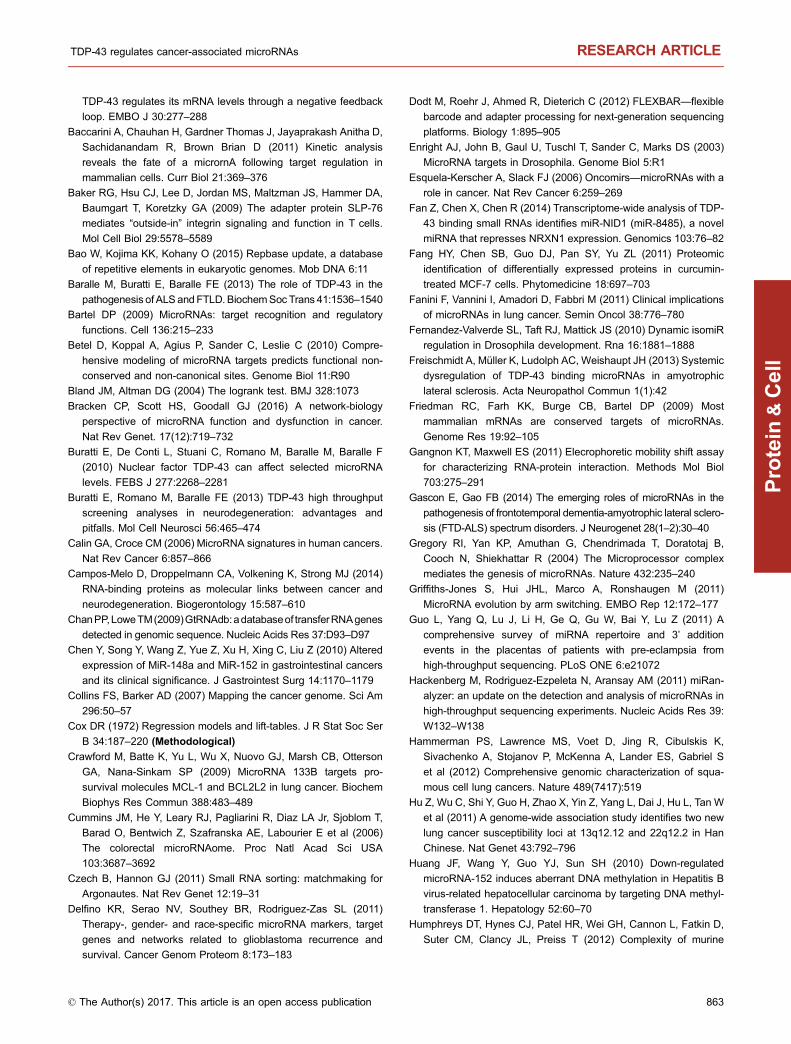
TDP-43 regulates its mRNA levels through a negative feedback
loop. EMBO J 30:277–288Baccarini A, Chauhan H, Gardner Thomas J, Jayaprakash Anitha D,
Sachidanandam R, Brown Brian D (2011) Kinetic analysis
reveals the fate of a micrornA following target regulation in
mammalian cells. Curr Biol 21:369–376Baker RG, Hsu CJ, Lee D, Jordan MS, Maltzman JS, Hammer DA,
Baumgart T, Koretzky GA (2009) The adapter protein SLP-76
mediates “outside-in” integrin signaling and function in T cells.
Mol Cell Biol 29:5578–5589Bao W, Kojima KK, Kohany O (2015) Repbase update, a database
of repetitive elements in eukaryotic genomes. Mob DNA 6:11
Baralle M, Buratti E, Baralle FE (2013) The role of TDP-43 in the
pathogenesis of ALS andFTLD. BiochemSoc Trans 41:1536–1540Bartel DP (2009) MicroRNAs: target recognition and regulatory
functions. Cell 136:215–233Betel D, Koppal A, Agius P, Sander C, Leslie C (2010) Compre-
hensive modeling of microRNA targets predicts functional non-
conserved and non-canonical sites. Genome Biol 11:R90
Bland JM, Altman DG (2004) The logrank test. BMJ 328:1073
Bracken CP, Scott HS, Goodall GJ (2016) A network-biology
perspective of microRNA function and dysfunction in cancer.
Nat Rev Genet. 17(12):719–732Buratti E, De Conti L, Stuani C, Romano M, Baralle M, Baralle F
(2010) Nuclear factor TDP-43 can affect selected microRNA
levels. FEBS J 277:2268–2281Buratti E, Romano M, Baralle FE (2013) TDP-43 high throughput
screening analyses in neurodegeneration: advantages and
pitfalls. Mol Cell Neurosci 56:465–474Calin GA, Croce CM (2006) MicroRNA signatures in human cancers.
Nat Rev Cancer 6:857–866Campos-Melo D, Droppelmann CA, Volkening K, Strong MJ (2014)
RNA-binding proteins as molecular links between cancer and
neurodegeneration. Biogerontology 15:587–610ChanPP,LoweTM(2009)GtRNAdb:adatabaseof transferRNAgenes
detected in genomic sequence. Nucleic Acids Res 37:D93–D97Chen Y, Song Y, Wang Z, Yue Z, Xu H, Xing C, Liu Z (2010) Altered
expression of MiR-148a and MiR-152 in gastrointestinal cancers
and its clinical significance. J Gastrointest Surg 14:1170–1179Collins FS, Barker AD (2007) Mapping the cancer genome. Sci Am
296:50–57Cox DR (1972) Regression models and lift-tables. J R Stat Soc Ser
B 34:187–220 (Methodological)
Crawford M, Batte K, Yu L, Wu X, Nuovo GJ, Marsh CB, Otterson
GA, Nana-Sinkam SP (2009) MicroRNA 133B targets pro-
survival molecules MCL-1 and BCL2L2 in lung cancer. Biochem
Biophys Res Commun 388:483–489Cummins JM, He Y, Leary RJ, Pagliarini R, Diaz LA Jr, Sjoblom T,
Barad O, Bentwich Z, Szafranska AE, Labourier E et al (2006)
The colorectal microRNAome. Proc Natl Acad Sci USA
103:3687–3692Czech B, Hannon GJ (2011) Small RNA sorting: matchmaking for
Argonautes. Nat Rev Genet 12:19–31Delfino KR, Serao NV, Southey BR, Rodriguez-Zas SL (2011)
Therapy-, gender- and race-specific microRNA markers, target
genes and networks related to glioblastoma recurrence and
survival. Cancer Genom Proteom 8:173–183
Dodt M, Roehr J, Ahmed R, Dieterich C (2012) FLEXBAR—flexible
barcode and adapter processing for next-generation sequencing
platforms. Biology 1:895–905Enright AJ, John B, Gaul U, Tuschl T, Sander C, Marks DS (2003)
MicroRNA targets in Drosophila. Genome Biol 5:R1
Esquela-Kerscher A, Slack FJ (2006) Oncomirs—microRNAs with a
role in cancer. Nat Rev Cancer 6:259–269Fan Z, Chen X, Chen R (2014) Transcriptome-wide analysis of TDP-
43 binding small RNAs identifies miR-NID1 (miR-8485), a novel
miRNA that represses NRXN1 expression. Genomics 103:76–82Fang HY, Chen SB, Guo DJ, Pan SY, Yu ZL (2011) Proteomic
identification of differentially expressed proteins in curcumin-
treated MCF-7 cells. Phytomedicine 18:697–703Fanini F, Vannini I, Amadori D, Fabbri M (2011) Clinical implications
of microRNAs in lung cancer. Semin Oncol 38:776–780Fernandez-Valverde SL, Taft RJ, Mattick JS (2010) Dynamic isomiR
regulation in Drosophila development. Rna 16:1881–1888Freischmidt A, Müller K, Ludolph AC, Weishaupt JH (2013) Systemic
dysregulation of TDP-43 binding microRNAs in amyotrophic
lateral sclerosis. Acta Neuropathol Commun 1(1):42
Friedman RC, Farh KK, Burge CB, Bartel DP (2009) Most
mammalian mRNAs are conserved targets of microRNAs.
Genome Res 19:92–105Gangnon KT, Maxwell ES (2011) Elecrophoretic mobility shift assay
for characterizing RNA-protein interaction. Methods Mol Biol
703:275–291Gascon E, Gao FB (2014) The emerging roles of microRNAs in the
pathogenesis of frontotemporal dementia-amyotrophic lateral sclero-
sis (FTD-ALS) spectrum disorders. J Neurogenet 28(1–2):30–40Gregory RI, Yan KP, Amuthan G, Chendrimada T, Doratotaj B,
Cooch N, Shiekhattar R (2004) The Microprocessor complex
mediates the genesis of microRNAs. Nature 432:235–240Griffiths-Jones S, Hui JHL, Marco A, Ronshaugen M (2011)
MicroRNA evolution by arm switching. EMBO Rep 12:172–177Guo L, Yang Q, Lu J, Li H, Ge Q, Gu W, Bai Y, Lu Z (2011) A
comprehensive survey of miRNA repertoire and 3’ addition
events in the placentas of patients with pre-eclampsia from
high-throughput sequencing. PLoS ONE 6:e21072
Hackenberg M, Rodriguez-Ezpeleta N, Aransay AM (2011) miRan-
alyzer: an update on the detection and analysis of microRNAs in
high-throughput sequencing experiments. Nucleic Acids Res 39:
W132–W138
Hammerman PS, Lawrence MS, Voet D, Jing R, Cibulskis K,
Sivachenko A, Stojanov P, McKenna A, Lander ES, Gabriel S
et al (2012) Comprehensive genomic characterization of squa-
mous cell lung cancers. Nature 489(7417):519
Hu Z, Wu C, Shi Y, Guo H, Zhao X, Yin Z, Yang L, Dai J, Hu L, Tan W
et al (2011) A genome-wide association study identifies two new
lung cancer susceptibility loci at 13q12.12 and 22q12.2 in Han
Chinese. Nat Genet 43:792–796Huang JF, Wang Y, Guo YJ, Sun SH (2010) Down-regulated
microRNA-152 induces aberrant DNA methylation in Hepatitis B
virus-related hepatocellular carcinoma by targeting DNA methyl-
transferase 1. Hepatology 52:60–70Humphreys DT, Hynes CJ, Patel HR, Wei GH, Cannon L, Fatkin D,
Suter CM, Clancy JL, Preiss T (2012) Complexity of murine
TDP-43 regulates cancer-associated microRNAs RESEARCH ARTICLE
© The Author(s) 2017. This article is an open access publication 863
Protein
&Cell
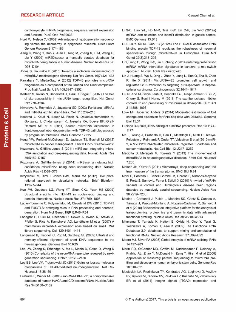
cardiomyocyte miRNA biogenesis, sequence variant expression
and function. PLoS One 7:e30933
Hurd PJ, Nelson CJ (2009) Advantages of next-generation sequenc-
ing versus the microarray in epigenetic research. Brief Funct
Genom Proteom 8:174–183Jiang Q, Wang Y, Hao Y, Juan L, Teng M, Zhang X, Li M, Wang G,
Liu Y (2009) miR2Disease: a manually curated database for
microRNA deregulation in human disease. Nucleic Acids Res 37:
D98–D104Jonas S, Izaurralde E (2015) Towards a molecular understanding of
microRNA-mediated gene silencing. Nat Rev Genet. 16(7):421–433Kawahara Y, Mieda-Sato A (2012) TDP-43 promotes microRNA
biogenesis as a component of the Drosha and Dicer complexes.
Proc Natl Acad Sci USA 109:3347–3352Kertesz M, Iovino N, Unnerstall U, Gaul U, Segal E (2007) The role
of site accessibility in microRNA target recognition. Nat Genet
39:1278–1284Khvorova A, Reynolds A, Jayasena SD (2003) Functional siRNAs
and miRNAs exhibit strand bias. Cell 115:209–216Kocerha J, Kouri N, Baker M, Finch N, DeJesus-Hernandez M,
Gonzalez J, Chidamparam K, Josephs KA, Boeve BF, Graff-
Radford NR et al (2011) Altered microRNA expression in
frontotemporal lobar degeneration with TDP-43 pathologycaused
by progranulin mutations. BMC Genome 12:527
Kong YW, Ferland-McCollough D, Jackson TJ, Bushell M (2012)
microRNAs in cancer management. Lancet Oncol 13:e249–e258Kozomara A, Griffiths-Jones S (2011) miRBase: integrating micro-
RNA annotation and deep-sequencing data. Nucleic Acids Res
39:D152–D157Kozomara A, Griffiths-Jones S (2014) miRBase: annotating high
confidence microRNAs using deep sequencing data. Nucleic
Acids Res 42:D68–D73Krzywinski M, Birol I, Jones SJM, Marra MA (2012) Hive plots-
rational approach to visualizing networks. Brief Bioinform
13:627–644Kuo PH, Doudeva LG, Wang YT, Shen CKJ, Yuan HS (2009)
Structural insights into TDP-43 in nucleic-acid binding and
domain interactions. Nucleic Acids Res 37:1799–1808Lagier-Tourenne C, Polymenidou M, Cleveland DW (2010) TDP-43
and FUS/TLS: emerging roles in RNA processing and neurode-
generation. Hum Mol Genet 19(R1):R46–R64Landgraf P, Rusu M, Sheridan R, Sewer A, Iovino N, Aravin A,
Pfeffer S, Rice A, Kamphorst AO, Landthaler M et al (2007) A
mammalian microRNA expression atlas based on small RNA
library sequencing. Cell 129:1401–1414Langmead B, Trapnell C, Pop M, Salzberg SL (2009) Ultrafast and
memory-efficient alignment of short DNA sequences to the
human genome. Genome Biol 10:R25
Lee LW, Zhang S, Etheridge A, Ma L, Martin D, Galas D, Wang K
(2010) Complexity of the microRNA repertoire revealed by next-
generation sequencing. RNA 16:2170–2180Lee EB, Lee VM, Trojanowski JQ (2012) Gains or losses: molecular
mechanisms of TDP43-mediated neurodegeneration. Nat Rev
Neurosci 13:38–50Lestrade L, Weber MJ (2006) snoRNA-LBME-db, a comprehensive
database of human H/ACA and C/D box snoRNAs. Nucleic Acids
Res 34:D158–D162
Li S-C, Liao Y-L, Ho M-R, Tsai K-W, Lai C-H, Lin W-C (2012a)
miRNA arm selection and isomiR distribution in gastric cancer.
BMC Genome 13:S13
Li Z, Lu Y, Xu XL, Gao FB (2012b) The FTD/ALS associated RNA
binding protein TDP-43 regulates the robustness of neuronal
specification through microRNA-9a in Drosophila. Hum Mol
Genet 22(2):218–225Li Y, Liang C, Wong K-C, Jin K, Zhang Z (2014) Inferring probabilistic
miRNA–mRNA interaction signatures in cancers: a role-switch
approach. Nucleic Acids Res 42(9):e76
Lin J, Huang S, Wu S, Ding J, Zhao Y, Liang L, Tian Q, Zha R, Zhan
R, He X (2011) MicroRNA-423 promotes cell growth and
regulates G1/S transition by targeting p21Cip1/Waf1 in hepato-
cellular carcinoma. Carcinogenesis 32:1641–1647Liu N, Abe M, Sabin Leah R, Hendriks G-J, Naqvi Ammar S, Yu Z,
Cherry S, Bonini Nancy M (2011) The exoribonuclease nibbler
controls 3′ end processing of micrornas in drosophila. Curr Biol
21:1888–1893Love MI, Huber W, Anders S (2014) Moderated estimation of fold
change and dispersion for RNA-seq data with DESeq2. Genome
Biol 15:31
Luciano DJ (2004) RNA editing of a miRNA precursor. Rna 10:1174–1177
Ma L, Young J, Prabhala H, Pan E, Mestdagh P, Muth D, Teruya-
Feldstein J, Reinhardt F, Onder TT, Valastyan S et al (2010) miR-
9, a MYC/MYCN-activated microRNA, regulates E-cadherin and
cancer metastasis. Nat Cell Biol 12:U247–U252Maciotta S, Meregalli M, Torrente Y (2013) The involvement of
microRNAs in neurodegenerative diseases. Front Cell Neurosci
7:265
Malone JH, Oliver B (2011) Microarrays, deep sequencing and the
true measure of the transcriptome. BMC Biol 9:34
Marti E, Pantano L, Banez-Coronel M, Llorens F, Minones-Moyano
E, Porta S, Sumoy L, Ferrer I, Estivill X (2010) A myriad of miRNA
variants in control and Huntington’s disease brain regions
detected by massively parallel sequencing. Nucleic Acids Res
38:7219–7235Medina I, Carbonell J, Pulido L, Madeira SC, Goetz S, Conesa A,
Tárraga J, Pascual-Montano A, Nogales-Cadenas R, Santoyo J
et al (2010) Babelomics: an integrative platform for the analysis of
transcriptomics, proteomics and genomic data with advanced
functional profiling. Nucleic Acids Res 38:W210–W213
Mituyama T, Yamada K, Hattori E, Okida H, Ono Y, Terai G,
Yoshizawa A, Komori T, Asai K (2009) The Functional RNA
Database 3.0: databases to support mining and annotation of
functional RNAs. Nucleic Acids Research 37:D89–D92Moore MJ, Silver PA (2008) Global Analysis of mRNA splicing. RNA
14:197–203Morin RD, O’Connor MD, Griffith M, Kuchenbauer F, Delaney A,
Prabhu AL, Zhao Y, McDonald H, Zeng T, Hirst M et al (2008)
Application of massively parallel sequencing to microRNA pro-
filing and discovery in human embryonic stem cells. Genome Res
18:610–621Mostovich LA, Prudnikova TY, Kondratov AG, Loginova D, Vavilov
PV, Rykova VI, Sidorov SV, Pavlova TV, Kashuba VI, Zabarovsky
ER et al (2011) Integrin alpha9 (ITGA9) expression and
RESEARCH ARTICLE Xiaowei Chen et al.
864 © The Author(s) 2017. This article is an open access publication
Protein
&Cell
epigenetic silencing in human breast tumors. Cell Adhes Migr
5:395–401Nelson PT, Wang W-X, Rajeev BW (2008) MicroRNAs (miRNAs) in
neurodegenerative diseases. Brain Pathol 18:130–138Noorbakhsh J, Lang AH, Mehta P (2013) Intrinsic noise of
microRNA-regulated genes and the ceRNA hypothesis. PLoS
ONE 8:e72676
Orang AV, Safaralizadeh R, Kazemzadeh-Bavili M (2014) Mecha-
nisms of miRNA-mediated gene regulation from common down-
regulation to mRNA-specific upregulation. Int J Genom 2014:1–15
Osella M, Bosia C, Corá D, Caselle M (2011) The role of incoherent
MicroRNA-mediated feedforward loops in noise buffering. PLoS
Comput Biol 7:e1001101
Pan H, Hanada S, Zhao J, Mao L, Ma MZ (2012) Protein secretion is
required for pregnancy-associated plasma protein-a to promote
lung cancer growth in vivo. PLoS ONE 7:e48799
Park YY, Kim SB, Han HD, Sohn BH, Kim JH, Liang J, Lu Y,
Rodriguez-Aguayo C, Lopez-Berestein G, Mills GB, Sood AK,
Lee JS (2013) Tat-activating regulatory DNA-binding protein
regulates glycolysis in hepatocellular carcinoma by regulating the
platelet isoform of phosphofructokinase through microRNA 520.
Hepatology 58(1):182–191Parpart S, Wang XW (2013) MicroRNA regulation and its conse-
quences in cancer. Curr Pathobiol Rep 1:71–79Peritz T, Zeng F, Kannanayakal TJ, Kilk K, Eiríksdóttir E, Langel U,
Eberwine J (2006) Immunoprecipitation of mRNA-protein com-
plexes. Nat Protoc 1:577–580Polymenidou M, Lagier-Tourenne C, Hutt KR, Huelga SC, Moran J,
Liang TY, Ling SC, Sun E, Wancewicz E, Mazur C et al (2011)
Long pre-mRNA depletion and RNA missplicing contribute to
neuronal vulnerability from loss of TDP-43. Nat Neurosci 14
(4):459–468Postel-Vinay S, Véron AS, Tirode F, Pierron G, Reynaud S, Kovar H,
Oberlin O, Lapouble E, Ballet S, Lucchesi C et al (2012) Common
variants near TARDBP and EGR2 are associated with suscep-
tibility to Ewing sarcoma. Nat Genet 44:323–327Quast C, Pruesse E, Yilmaz P, Gerken J, Schweer T, Yarza P,
Peplies J, Glockner FO (2012) The SILVA ribosomal RNA gene
database project: improved data processing and web-based
tools. Nucleic Acids Res 41:D590–D596Quinlan AR, Hall IM (2010) BEDTools: a flexible suite of utilities for
comparing genomic features. Bioinformatics 26:841–842Ratti A, Buratti E (2016) Physiological functions and pathobiology of
TDP-43 and FUS/TLS proteins. J Neurochem 138:95–111Ruby JG, Jan C, Player C, Axtell MJ, Lee W, Nusbaum C, Ge H,
Bartel DP (2006) Large-scale sequencing reveals 21U-RNAs and
additional microRNAs and endogenous siRNAs in C. elegans.
Cell 127:1193–1207Schwarz DS, Hutvagner G, Du T, Xu Z, Aronin N, Zamore PD (2003)
Asymmetry in the assembly of the RNAi enzyme complex. Cell
115:199–208Sephton CF, Cenik C, Kucukural A, Dammer EB, Cenik B, Han Y,
Dewey CM, Roth FP, Herz J, Peng J et al (2011) Identification of
neuronal RNA targets of TDP-43-containingribonucleoprotein
complexes. J Biol Chem 286:1204–1215
Skrzypski M, Dziadziuszko R, Jassem J (2011) MicroRNA in lung
cancer diagnostics and treatment. Mutat Res 717:25–31Soneson C, Love MI, Robinson MD (2016) Differential analyses for
RNA-seq: transcript-level estimates improve gene-level infer-
ences. F1000Research. doi:10.12688/f1000research.7563.2
Srinivasan S, Patric IR, Somasundaram K (2011) A ten-microRNA
expression signature predicts survival in glioblastoma. PLoS One
6:e17438
Sriram G, Birge RB (2010) Emerging roles for Crk in human cancer.
Genes Cancer 1:1132–1139Tan X, Qin W, Zhang L, Hang J, Li B, Zhang C, Wan J, Zhou F, Shao
K, Sun Y et al (2011) A 5-microRNA signature for lung squamous
cell carcinoma diagnosis and hsa-miR-31 for prognosis. Clin
Cancer Res 17:6802–6811Tarca AL, Drăghici S, Khatri P, Hassan SS, Mittal P, Kim J-S, Kim CJ,
Kusanovic JP, Romero R (2009) A novel signaling pathway
impact analysis. Bioinformatics 25:75–82 (Oxford, England)
Teittinen KJ, Kärkkäinen P, Salonen J, Rönnholm G, Korkeamäki H,
Vihinen M, Kalkkinen N, Lohi O (2012) Nucleolar proteins with
altered expression in leukemic cell lines. Leukemia Res 36:232–236
Tollervey JR, Curk T, Rogelj B, Briese M, Cereda M, Kayikci M,
Konig J, Hortobagyi T, Nishimura AL, Zupunski V et al (2011)
Characterizing the RNA targets and position-dependent splicing
regulation by TDP-43. Nat Neurosci 14:452–458Tsuruta T, Kozaki K, Uesugi A, Furuta M, Hirasawa A, Imoto I,
Susumu N, Aoki D, Inazawa J (2011) miR-152 is a tumor
suppressor microRNA that is silenced by DNA hypermethylation
in endometrial cancer. Cancer Res 71:6450–6462Ule J, Jensen K, Mele A, Darnell RB (2005) CLIP: A method for
identifying protein-RNA interaction sites in living cells. Methods
37:376–386Verhaak RG, Hoadley KA, Purdom E, Wang V, Qi Y, Wilkerson MD,
Miller CR, Ding L, Golub T, Mesirov JP et al (2010) Integrated
genomic analysis identifies clinically relevant subtypes of
glioblastoma characterized by abnormalities in PDGFRA, IDH1,
EGFR, and NF1. Cancer Cell 17:98–110Woo HH, Laszlo CF, Greco S, Chambers SK (2012) Regulation of
colony stimulating factor-1 expression and ovarian cancer cell
behavior in vitro by miR-128 and miR-152. Mol Cancer 11:58
Wysoczynski M, Miekus K, Jankowski K, Wanzeck J, Bertolone S,
Janowska-Wieczorek A, Ratajczak J, Ratajczak MZ (2007)
Leukemia inhibitory factor: a newly identified metastatic factor
in rhabdomyosarcomas. Cancer Res 67:2131–2140Xiao S, Sanelli T, Dib S, Sheps D, Findlater J, Bilbao J, Keith J,
Zinman L, Rogaeva E, Robertson J (2011) RNA targets of TDP-
43 identified by UV-CLIP are deregulated in ALS. Mol and Cell
Neurosci 47:167–180Yanaihara N, Caplen N, Bowman E, Seike M, Kumamoto K, Yi M,
Stephens RM, Okamoto A, Yokota J, Tanaka T et al (2006)
Unique microRNA molecular profiles in lung cancer diagnosis
and prognosis. Cancer Cell 9:189–198Yu J, Cao Q, Mehra R, Laxman B, Yu J, Tomlins SA, Creighton CJ,
Dhanasekaran SM, Shen R, Chen G et al (2007) Integrative
genomics analysis reveals silencing of β-adrenergic signaling by
polycomb in prostate Cancer. Cancer Cell 12:419–431
TDP-43 regulates cancer-associated microRNAs RESEARCH ARTICLE
© The Author(s) 2017. This article is an open access publication 865
Protein
&Cell
Zhang Z, Almeida S, Lu Y, Nishimura AL, Peng L, Sun D, Wu B,
Karydas AM, Tartaglia MC, Fong JC et al (2013) Downregulation
of microRNA-9 in iPSC-derived neurons of FTD/ALS patients with
TDP-43 mutations. PLoS One 8:e76055
Zhou X, Zhao F, Wang ZN, Song YX, Chang H, Chiang Y, Xu HM
(2012) Altered expression of miR-152 and miR-148a in ovarian
cancer is related to cell proliferation. Oncol Rep 27:447–454
RESEARCH ARTICLE Xiaowei Chen et al.
866 © The Author(s) 2017. This article is an open access publication
Protein
&Cell