taxonomy of corynebacterium plant pathogens, including a new
TRANSCRIPT
INTERNATIONAL JOURNAL O F SYSTEMATIC BACTERIOLOGY, July 1982, p. 315-326 0020-77 1 3/82/0303 15-1 2$02.00/0
Vol. 32. No. 3
Taxonomy of Corynebacterium Plant Pathogens, Including a New Pathogen of Wheat, Based on Polyacrylamide Gel
Electrophoresis of Cellular Proteins? RANDALL R. CARLSON A N D ANNE K. VIDAVER
Depart in en t q f Plant Pci t h ology , Un h e r s i ty (! f Ne bras X u , Lin c d n . N e h rci J Xu 68583 -0 722
The known extant plant pathogenic Ccrynehuc~terirm species were analyzed by polyacrylamide gel electrophoresis of their cellular proteins. The patterns of the protein bands of 13 species and a new corynebacterial wheat pathogen showed seven distinct groups. Five of these groups consisted of only one species each, one group contained four species, and the last group contained the new wheat pathogen and the remaining four species. The pathogens that could not be distinguished by the polyacrylamide gel analysis differed in phenotypic character- istics, including pathogenic specificity. Thus, for these bacteria we propose recognition of the following taxa: Corynehactrrium fnscians (Tilford) Dowson, Corynehacteriurn i1ici.s (Mandel et al.), Corynehac~teriitm tritici (ex Hutchinson) nom. rev., Coiynebac-teriirm iranicirm (ex Scharif) nom. rev., Corynehacterium ra thay i ( Smith) Dow son, Cory ne bac te riir in JIac*c.irmfa ciens sub sp . f r a cc‘u mjiiciens (Hedge s ) Do w s o n , Co ry n e b a c. t e r ii4 in frtr c c‘ 14 r n j k cis n s sub s p . poinsettia e ( S t a rr and Pirone) comb. nov., Corynehacter*izrm flaccirmjciciens subsp. hetae (Keyworth et al.) comb. nov., Corynebacteriiim flaccumnfiiciens subsp. oortii (Saaltink and Maas Geesteranus) comb. nov., Cor~nebactet-ii4in michiganense subsp. michi- garrense (Smith) Jensen, Copnebactcriuin michiganense subsp. nebraskense (Schuster et al.) comb. nov., Coi-ynehncterium michiganense subsp. insidiosum (McCulloch) comb. nov., Corynehacteriurn michiganense subsp. sepedcinicum (Spieckermann and Kotthom, comb. nov., and Coi-ynebacterium michigcrnense subsp. tessellariirs subsp. nov., the type strain of which is $train 78181 (= ATCC 33566 = PDDCC 7221).
An orange-pigmented coryneform bacterium was isolated from diseased wheat plants in 1976 and was subsequently identified as the causal agent of bacterial mosaic of wheat (5a). Studies were undertaken to determine the taxonomic relationship of this organism to other phytopath- ogenic corynebacteria, particularly the corn pathogen Corynebacterium nebruskense (Schus- ter et al. 1973) emend. Vidaver and Mandell974 (Approved Lists, 1980), since both of these pathogens are associated with plant diseases discovered in Nebraska. Preliminary results of this research have been presented elsewhere (Carlson and Vidaver, Phytopathology 69:1023, 1979; Carlson and Vidaver, Phytopathology 71:207, 1981).
The genus Corynebacterium contains bacteria with similar morphologies and a wide range of biochemical properties. Although attempts have been made to split the genus into homogeneous groups (3, 6, 11, 12, 22, 23, 33, 48, 57), no proposal has been generally accepted. In this
i- Paper 6591 in the journal series of the Nebraska Agricul- tural Experiment Station.
-~ - ~~
study, only the relationships among the phyto- pathogens traditionally included in the genus Corynebacterium were examined.
Kersters and De Ley (26) elegantly differenti- ated several gram-negative phytopathogenic bacteria by means of polyacrylamide tube gels; the results of these authors suggested that ex- amination of phytopathogenic gram-positive bacteria by similar techniques might be useful and productive. In contrast to conventional tests, several gene products from more than one bacterium can be compared simultaneously in a slab gel. The only previous electrophoretic stud- ies of proteins from phytopathogenic corynebac- teria showed that “Coiynebacteriirm tritici” (Hutchinson 1917) Burkholder 1948 and Coryne- bacterium fascians (Tilford 1936) Dowson 1942 (Approved Lists, 1980) differed from each other and from Corynehucterium betae Keyworth et al. 1956 (Approved Lists, 1980), Corynebacteri- um poinsettiae (Starr and Pirone 1942) Burk- holder 1948 (Approved Lists, 1980), and Coryne- bacterium flaccumfaciens (Hedges 1922) Dowson 1942 (Approved Lists, 1980) with re- spect to the patterns of esterase, catalase, and
31 5

316 CARLSON AND VIDAVER
peroxidase activities detected in starch gels (37) (names in quotation marks have no standing in bacterial nomenclature because they were not included on the Approved Lists of Bacterial Names [44] and have not been validly published since 1 January 1980). Polyacrylamide gel elec- trophoresis of 39 strains of Corynebacterium diphtheriae (Kruse 1886) Lehmann and Neu- mann 1896 (Approved Lists, 1980) showed dif- ferences among biovars, although extraction was poor and few distinct bands could be detect- ed either by visual inspection or densitometer tracings (31).
The relationships among the phytopathogenic corynebacteria were assessed by gradient poly- acrylamide gel electrophoresis (PAGE) of cellu- lar proteins. A rationale for modifying the cur- rent classification of Corynehacterium plant pathogens is presented below, along with data supporting the classification of the bacterial mo- saic pathogen as a subspecies of Corynebacteri- urn michiganense (Smith 1910) Jensen 1934 (Ap- proved Lists, 1980).
MATERIALS AND METHODS
Bacterial strains. A total of 80 isolates of the bacteri- al mosaic pathogen were obtained from diseased win- ter wheat cultivars; the isolation methods used are described elsewhere (5a). The specific strains of the bacterial mosaic pathogen (selected because of their differing bacteriocin production characteristics) and the other bacteria studied are listed in Table 1 .
Media. Stock cultures were maintained both at 4°C on nutrient broth yeast extract medium (NBY medi- um) (55) and in the freeze-dried condition. The follow- ing media were used to determine growth characteris- tics: NBY medium (55); modified Burkholder agar (35); tetrazolium chloride medium (25), as modified by Vidaver and Mandel (56); and C . nebrnskense selec- tive medium (CNS medium) (16). Unless otherwise stated, experiments were performed at 25 5 3°C.
Preparation of protein samples for PAGE. Cells were grown to early stationary phase (absorbance at 640 nm, ca. 1.0 U , as determined with a Spectronic 20 colorimeter) in 10 to 30 ml of NBY broth and were then washed three times by centrifugation in 0.2 M sodium phosphate buffer (pH 6.8). The growth medi- um, cell growth stage at harvest, and protein extrac- tion procedure were standardized to reduce the varia- tion in the gel patterns. The final pellet was suspended in 2.0 ml of the NaPO, buffer. An equal volume of lysozyme solution [l mg/ml in 0.05 M tris(hydroxy- methyl)aminomethane-0.02 M ethylenediaminetetraa- cetate buffer, pH 8.01 was added, and the mixture was incubated for 15 min at 37°C. Sodium lauryl sulfate (lo%, wt/vol) was added to a final concentration of 5%. This treatment lysed most strains; if lysis was not apparent by increased viscosity and decreased turbidi- ty, the cells were frozen (-20°C) and thawed three times. Cellular proteins were extracted from the disin- tegrated cells essentially as described by Van Etten et al. (54). An equal volume of water-saturated phenol
INT. J . SYST. BACTERIOL.
TABLE 1. Bacterial strains examined by PAGE Organism Strains“
C. michiganense
C. nebrnskense
C. insidiosum
C. sepedonicutn
C . Jlucmrnfuciens
C . poinsettiue
C . betae
C. oortii C. ilicis
C . fuscians
C . ruthuyi
“C. tritici”
“C. iranicum” C. bovis
M . luteus Bacterial mosaic pathogen
14-4 13-3 15-6 1379 3D21 829s 15-2 156-2 CN74-1 CN78-1 CN48-1 CN76-1 298 173 N7A CN18-6 30 239 MIB P2 CSca CSmt ATCC 6887 Ne21 c v 7 ICPB CPl ICPB CP13 ICPB CBlOl ICPB CB102A ICPB COl0l PDDCC 2607 PDDCC 2608 PDDCC 2609 6D21 CF79-1 ICPB CF107 ICPB CR1 ICPB CRlOl NCPPB 1857 ICPB CTlOl ICPB CT102 NCPPB 2253 ATCC 7715 1 ATCC 4698 78141 78151 78181 (= ATCC 33566) 78221 78222 78271
” Strains were obtained from the American Type Culture Collection (ATCC), International Collection of Phytopathogenic Bacteria (ICPB), Plant Disease Division Culture Collection (PDDCC), and National Collection of Plant Pathogenic Bacteria (NCPPB). If not otherwise identified, strains were obtained from other investigators or are Nebraska isolates.
(85 g of phenol, 16 ml of water, 1 ml of 1 N NaOH) was mixed with the lysate by blending with a Vortex mixer, the suspension was centrifuged at low speed, and the
VOL. 32, 1982 CORYNEBACTERIUM PLANT PATHOGENS 317
phenol layer (lower layer) was removed without dis- turbing the water phase (upper layer) or the interface. The phenol-soluble proteins were precipitated from the phenol phase by adding 5 volumes of methanol containing 0.1 M ammonium acetate, mixing well, and then incubating the preparation at -20°C for at least 2 h. The precipitate was collected by low-speed centrifu- gation and was washed once with methanol (10 mi); the final pellet was dried under a vacuum. The cellular proteins were dissolved in 100 pl of sample buffer [2.5 g of Ficoll (Pharmacia), 1 ml of 0.1% crystal violet, 5 ml of 10% (wt/vol) sodium lauryl sulfate, 0.5 ml of 3 M tris(hydroxymethy1)aminomethane (pH 8.8), 16 ml of water] by boiling for 1 min or more. Dithioerythritol (77 mg/ml) was added at 1 part per 100 just before boiling. Reference proteins were dissolved in sample buffer and electrophoresed in the outside lanes of the gel; 2.5 pg of a reference protein was detectable when the preparation was stained. The residual lysozyme in each sample served as an internal marker.
PAGE. The gradient slab gels (vertical) used were supported in a Studier apparatus (51). The gels were made as described by Ames (l), and the buffer system of Laemmli (29) was used. The separating gel (lower gel) was composed of a linear 8 to 20% polyacrylamide gradient, and the stacking gel (upper gel) contained 4.2% polyacrylamide (53). Samples (5 to 25 pl: opti- mum quantity determined empirically) were electro- phoresed at 60 V (4 Vkm) until the crystal violet in the sample buffer eluted from the gel (about 20 h). The gels were strained for 1 h (0.5 g of Coomassie brilliant blue R, 455 ml of methanol, 455 ml of water, 90 ml of glacial acetic acid) in a container placed on a slow-moving platform in a 50°C water bath shaker. The gels then were destained against several changes of destaining solution (35% [vol/vol] methanol and 10% [vol/vol] glacial acetic acid in water) at 50°C in a water bath shaker. The destained gels were photographed with Polaroid type 665 film through an orange filter. The gels could be stored for reference at room temperature in destaining solution.
G+C content of the DNA. The guanine-plus-cytosine (G+C) content of the deoxyribonucleic acid (DNA) was determined as previously described (42, 56).
Tetrazolium salt susceptibility. Growth on tetrazoli- um chloride medium with any of several tetrazolium derivatives substituted for triphenyltetrazolium chlo- ride was scored as positive if single colonies grew on a streak plate within 2 weeks. The following derivatives were tested: iodonitrotetrazolium, o-tolyl tetrazolium, rn-Nitro Blue Tetrazolium, neotetrazolium, tetrazoli- um blue, and tetrazolium violet. Ethanol was some- times added to stock solutions to solubilize the deriva- tives.
Plasmid DNA detection. Bacterial DNA was extract- ed, purified, and assayed for the presence of plasmids as described previously (18).
Bacteriocin production and assay. The conditions used for the production and assay of bacteriocins are described elsewhere (17).
Plant inoculation methods. Winter wheat (Tr i t ium aestivum L. ‘Centurk’), maize (Zea mays L. ‘Golden Cross Bantam’), and tomato (Lycopersicon escufen- turn Mill. ‘Mocross Surprise’) were inoculated by the partial vacuum technique (15) with strains of the bacterial mosaic pathogen, C. nebraskense, and C. rnichiganense in all host-pathogen combinations to
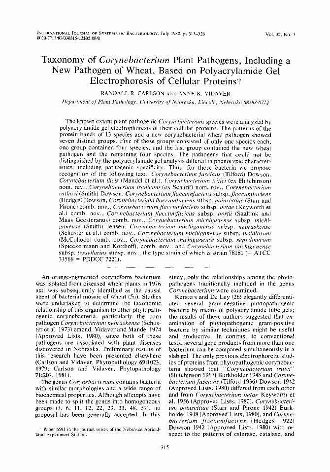
FIG. 1. Electron micrographs of the bacterial rno- saic pathogen. (A) Strain 78181. (B) Strain 78222. (C) Strain 78141. Bars = 1 p,m.
determine host specificity (C. michiganense and C. nebraskense are pathogens of tomato and corn, re- spectively). The pathogen concentrations in the inocu- la were adjusted to lo6 colony-forming units per ml.
RESULTS
Morphology and growth characteristics of the bacterial mosaic pathogen. Cells of the bacterial mosaic pathogen were gram-positive, nonmotile (between 20 and 28°C) rods with a pleomorphism characteristic of coryneform bacteria. Cells measured approximately 0.5 by 1.5 pm (Fig. 1). Growth on NBY medium, modified Burkholder agar, and CNS medium was slow, with colonies becoming visible in 2 to 3 days. Full colony development required 5 to 7 days. Colonies were circular (diameter, 2 to 4 mm), convex, glisten- ing, and butyrous and had entire margins. Strains grew at 10°C but not at 37°C. The tem- perature for optimal growth was between 24 and 28°C. The intensity of pigmentation varied with the medium and the age of the culture, but all strains produced an apricot orange pigment. The colony colors on tetrazolium chloride medium (see below) varied from pale blue to red, de- pending on the derivative of tetrazolium includ- ed in the agar. C. michiganense, C . nebras- kense, Corynebac-tei-iurn insidiosum (McCulloch 1925) Jensen 1934 (Approved Lists, 1980), and Corynebacteriurn sepedonicurn (Spieckermann and Kotthoff 1914) Skaptason and Burkholder 1942 (Approved Lists, 1980) could be differenti- ated from each other by their reactions to two tetrozolium salts, triphenyltetrazolium chloride and iodonitrotetrazolium; C. rnichiganense and the bacterial mosaic pathogen gave identical reactions (Table 2). The inability of C. sepedoni- cum to grow on iodonitrotetrazolium and tri- phenyltetrazolium chloride was not consistent with previously reported results (56) , possibly because of strain variation. C. nebraskense was unable to grow on triphenyltetrazolium chloride, a characteristic that distinguished this species from both C . michiganense and the bacteria1

318 CARLSON AND VIDAVER INT. J . SYST. BACTERIOL.
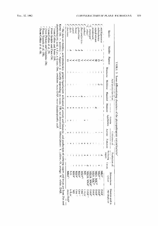
TABLE 2. Some differentiating characters of C. micliiganrnsr, C . nrbrmhrnse, C . insidiosutn, C. JepPdonicum, and the bacterial mosaic pathogen
Growth on: G + C
(mol 96)‘ Organism CNS INT TTC Pigment“ content Ho4t Specificity’ Colony type’
medium” agaP agar‘ - - _ _ ____ ~ ~ ~ _ _ _ _ _ _ _ _ ~ - - - -~ ________ ___
C. seprdonicutn - - - White 72 Potato Fluidal C. insidiosum - - + Yellow 73 Alfalfa Fluidal C . nebraskense + + - Orange 73.5 Corn Domed, mucoid C. tnichiganense + + + Yellow 73 Tomato, pepper Fluidal Bacterial mosaic pathogen + + + Orange 74 Wheat Domed, mucoid
‘’ See reference 16.
‘ TTC agar, Agar containing 50 Fg of triphenyltetrazolium chloride per ml. ‘‘ Colony pigmentation on NBY agar. ‘’ Data from Vidaver and Mandel (561, except for the data for the bacterial mosaic pathogen.
INT agar, Agar containing 10 pg of iodonitrotetrazolium per ml.
See text for references.
mosaic pathogen. Neither C . sepedonicum nor C. insidiosum grew on selective medium CNS, in contrast to C. michiganense, C. nehraskense, and the bacterial mosaic pathogen. Table 3 lists additional comparative characters for the Cory- nebacterium plant pathogens.
G+C contents of DNAs. The G+C contents of DNAs were as follows: strain 77113,73.5 mol%; strain 7661, 74.0 mol%; and strain 77143, 74.5 mol% (1.732 to 1.733 g/cm3). Comparative G+C values for C. rnichiganense, C. nebraskense, C . insidiosum, and C. seprdonicum are listed in Table 2 .
PAGE of cellular proteins. The patterns of the protein bands clearly differentiated some of the species but not others. The use of a gradient gel enabled resolution of proteins ranging in molec- ular weight from 10,000 to 120,000 in a gel 15 cm long. For any given strain, the patterns were reproducible throughout analyses of the same sample in successive gels; independent extrac- tions of the same strain also gave indistinguish- able patterns. The strains of each species gave similar overall patterns, but the patterns could differ in specific bands. All of the strains listed in Table 1 were analyzed by this method. Different strains of C. nehraskense (Fig. 2) and C. michi- ganense (Fig. 3) showed nearly identical protein patterns. The variation in the protein patterns was slightly greater among the strains of C. michiganense than among the strains of C. ne- braskense. The protein bands of the bacterial mosaic pathogen (Fig. 3 , lane F), C. rnichigan- ense (lane D), C. nehraskense (lane E), C. insidiosum (lane C ) , and C. sepedonicurn (lane B) were very similar. Likewise, the patterns for the nomenspecies C . JIaccumfaciens (Fig. 5 , lane C), C. hetae (lane D), C. poinsettiae (lane B), and Corynebacterium oortii Saaltink and Maas Geesteranus 1969 (Approved Lists, 1980) (lane E) were very similar. In both Fig. 4 and 5 ,
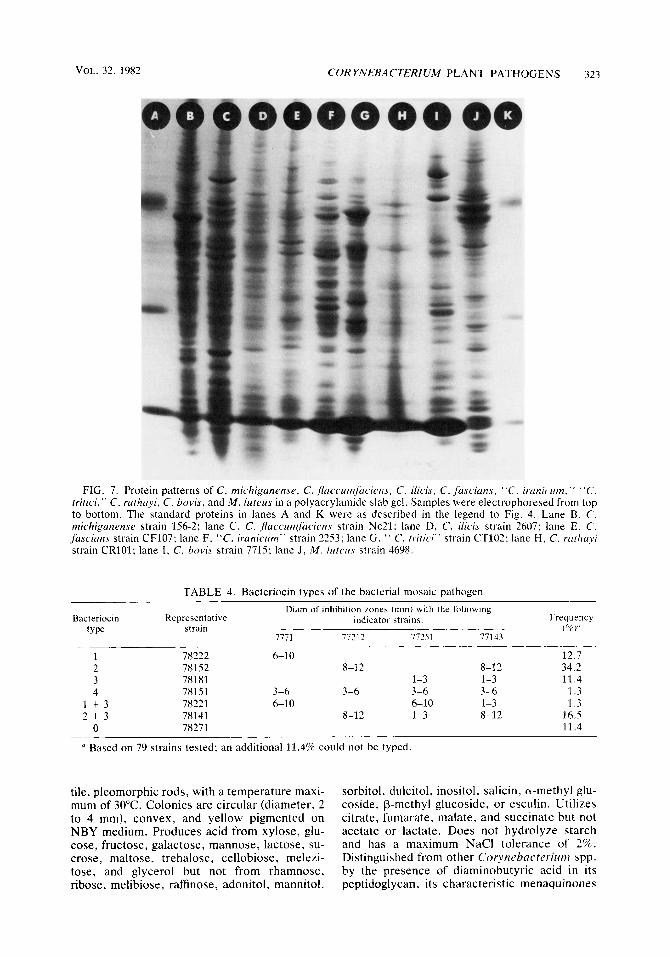
the variation among the species of each group was minor, little more than the variation encoun- tered among strains of a single species (Fig. 2 and 3). The four Corynebacterium pathogens of wheat, “C. tritici” (Fig. 6, lane E), “Corynebac- terium iranicum” Scharif 1961 (lane D), Coryne- bacterium rafhayi (Smith 1913) Dowson 1942 (Approved Lists, 1980) (lane F), and the bacteri- al mosaic pathogen (lane C), were distinctly different. The mammal-associated species Cory- nebacterium bovis Bergey et al. 1923 (Approved Lists, 1980) (Fig. 7, lane I) and the gram-positive coccus Micrococcus luteus (Schroeter 1872) Cohn 1872 (Approved Lists, 1980) (lane J) had protein patterns that were clearly different from those of the phytopathogens (lanes B through H) .
Bacteriocins of the bacterial mosaic pathogen. Most strains of the bacterial mosaic pathogen produced a single bacteriocin, but a few strains produced two bacteriocins simultaneously and some strains produced none at all (Table 4). Four distinct bacteriocins were detected; three of these were unique to the bacterial mosaic pathogen (Fig. S), as shown by a second set of indicator strains (17). The fourth bacteriocin could not be differentiated from the bacteriocin produced by C . nehraskense CN74-1 (Fig. 8). The type of bacteriocin produced by the bacteri- al mosaic pathogen could not be correlated with geographic source, wheat cultivar, or year of isolation (data not shown); e.g., all four bacte- riocins were produced by strains isolated from adjacent plots in a cultivar field trial. The pro- duction of bacteriocin was used to type the strains, as this was a more reliable character than susceptibility to bacteriocins.
Disease reactions. Greenhouse-grown wheat inoculated with strains of the bacterial mosaic pathogen readily developed the scattered yellow lesions characteristic of the disease after 3 to 5
TA
BL
E 3. Som
e differentiating characters of the phytopathogenic corynebacteria" A
cid production from:
Utilization of
Production of M
enaquinone D
iarnino acid in -
Gelatin
-~
~
-
~
Motility
Pigment
peptidoglycan R
harnnose M
elibiose M
annitol M
annose liqLlefaction
Acetate
Funxirate R
educing Levan
system
Species
~-
sugars ~
__
_
_ - -
~-
~
C. m
ichigunense -
Y
d -
+ +
+ +
d -
MK
9h D
AB
' C
. nebru
skense
-
0
-
d -
+ -
+ +
+ +
MK9'."
DA
B"
C. srpedonicum
-
W
"C. iranicum
" -
Y
-
-
-
+ -
-
+ -
-
MK
10. MK
@
DA
Bh
C. rathayi
-
Y
-
-
+ -
+ -
+ +
+ N
D
L-LYS'
C. j7uccum
fcirc.ien.s d
Y
+ +
d +
-
+ +
-
-
MK
~"c/ O
rn' ,'' +
+ +
-
+ -
-
-
MK
Y
Orn' .''
-
-
-
MK
Y
Om
' .'' C
. poinsettiae d
0
C. betcie
d Y
+
+ +
+ C
. oortii +
Y
+ +
+ +
-
-
-
-
-
MK
~"
N
D
+ +
+ +
+ +
+ -
ND
L-LY
S' C
. ilicis d
Y
C. fiw
cians -
-
0
Kem
p (14). f, positive; -, negative; d, variable depending on strain; N
D, not determ
ined. Abbreviations: Y
, yellow; 0
, orange; W, w
hite; DA
B,
diaminobutyric acid; L
-LY
S, L-lysine: Orn, ornithine; m
eso-DA
P, meso-diam
inopimelic acid.
-
-
+ +
+ -
MK
9,MK
7h D
AB
' +
d -
-
-
-
-
-
-
-
MK
9,MK
7" D
AB
' +
Y
-
-
-
-
+ +
+ d
MK
10, MK
@
DA
B".'
+ Y
-
-
+ C
. insidiosum
"C. tritici"
+
+ +
-
-
MK8f:R
in es 0
- D
AP'
-
-
-
+ +
+ +
-
I' The data on m
otility, acid production, gelatin liquefaction, utilization of acetate and fumarate, and production of reducing sugars and levan are from
Dye and
From C
ollins and Jones (10). ' From
Keddie and C
ure (24). " From
Collins et al. (9).
'' From Perkins and C
umm
ins (36). From
Yam
ada et al. (58). From
Collins et al. (8).
N
m

320 CARLSON AND VIDAVER INT. J . SYST. BACTERIOL.
FIG. 2. Protein patterns of C . nebraskense in a polyacrylamide slab gel. Samples were electrophor- esed from top to bottom. Lane A, Strain CN76-1; lane B, strain CN74-1; lane C, strain CN74-1 (independent extraction); lane D, strain CN48-1; lane E, strain 298; lane F, strain N7A; lane G, strain 173.
FIG. 3. Protein patterns of C . rnichiganense in a polyacrylamide slab gel. Samples were electrophor- esed from top to bottom. Lane A, strain 156-2; lane €3, strain 14-4; lane C, strain 3D21; lane D, strain 15-6; lane E, strain 15-2; lane F, strain 829s; lane G , strain 1379; lane H, strain 13-3.
days of incubation. The relative populations of the bacterial mosaic pathogen, C. michiganense, and C . nebraskense in inoculated wheat, corn, and tomato plants showed that the highest popu- lation levels occurred in natural hosts, as shown elsewhere (5a). Neither C. michiganense nor C. nebraskense produced symptoms in wheat.
Plasmid analysis. Seven strains were exam- ined for the presence of plasmids; of these, only strain 78151 contained detectable plasmid DNA. The plasmid was about 32 megadaltons long, as determined by a comparison with a 34-megadal- ton plasmid of C. nebraskense (18). Since plas- mids are rare, they are unlikely to determine major properties of the bacterial mosaic patho- gen.
DISCUSSION
The initial recognition of plant pathogens as distinct species of Corynebacterium was made on the presumption that pathogens of diverse
plant hosts would be quite different; recognition of the similarity of these pathogens was delayed due to the lack of appropriate comparative anal- yses of groups of pathogens. The current edition of Bergey ' s Manual of Determinative Bacteriol- ogy (4) lists 12 species of plant pathogens in the genus Corynebacterium. Dye and Kemp (14) have proposed that all of the corynebacterial phytopathogens should be assigned to four spe- cies (C. michiganense, c. jaccumfaciens, Cory- nebacterium ilicis, and C. fascians) on the basis of their numerical taxonomy study. With some notable exceptions, our study generally supports the taxonomic groupings of these authors. Thus, we propose revising the nomenclature of the phytopathogenic corynebacteria as discussed below.
The nomenspecies C . michiganense, C . ne- braskense, C . insidiosum, and C. sepedonicum were nearly identical when they were analyzed by PAGE; on this basis, we concur with the proposal of Dye and Kemp (14) to combine these

VOL. 32. 1982 CORYNEBACTERIUM PLANT PATHOGENS 321
FIG. 4. Protein patterns of C. sepedonicurn, C. insidiosum, C. rnichiganense, C. nebraskense, and the bacterial mosaic pathogen in a polyacrylamide slab gel. Samples were electrophoresed from top to bot- tom. Samples containing three standard proteins were electrophoresed in lanes A and G. These proteins were (from top to bottom) bovine serum albumin (molecular weight, 66,300), carbonic anhydrase B (28,000), and lysozyme (14,300). Each standard protein band con- tained approximately 2.5 pg of protein. Lane B, C. sepedonicurn strain 30; lane C, C. insidiosum strain P2; lane D, C. rnichiganense strain 156-2; lane E, C. nebraskense strain CN74-1; lane F, bacterial mosaic pathogen strain 78221.
organisms into a single species. Dye and Kemp concluded that there are insufficient differences among these bacteria to justify recognition of them as distinct taxa at even the subspecies level; they argued for infrasubspecific status for these bacteria as pathovars of C. rnichiganense. The classification of these bacteria as pathovars would retain the traditional names of pathogens that cause specific and differentiable diseases. In its recommendations for infrasubspecific terms, the International Code of Nomenclature of Bac- teria (30) defines pathovars as bacteria that differ in “pathogenic reactions in one or more hosts.” Dye and Kemp (14) contend that patho-
FIG. 5 . Protein patterns of C. poinsettiue, C. Jac- curnufaciens, C. hetae, and C . oortii in a polyacryl- amide slab gel. Samples were electrophoresed from top to bottom. The standard proteins in lanes A and F were as described in the legend to Fig. 4. Lane B, C. poinsettiae strain CP1; lane C , C. Jaccurnfuciens strain Ne21; lane D, C. betue strain CB102A; lane E, C. oortii strain COlOl.
vars can also differ in a limited number of biochemical and physiological properties. How- ever, we believe . that numerous differences among these bacteria require classification at the higher level of subspecies. We show that these taxa differ with respect to tetrazolium salt toler- ances, colony morphology and pigmentation, and overall bacteriocin production. These bacte- ria also differ in their reactions to many bio- chemical tests (Tables 2 and 3), and C. insidio- sum and C . sepedonicurn differ from C . nebraskense and C. rnichiganense in their mena- quinone systems (Table 3 ) . All of these charac- ters are independent of pathogenic specificity. The designation of these organisms as subspe- cies would retain the traditional names of these bacteria, would avoid the implication of the term pathovar that pathogenicity is an overriding tax- onomic character, and would allow the rational identification of related saprophytes and aviru-
322 CARLSON AND VIDAVER INT. J . SYST. BACTERIOL.
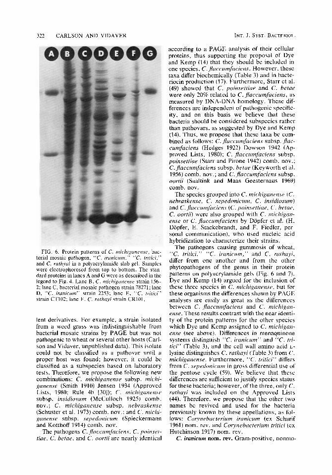
FIG. 6. Protein patterns of C. rnichiganense, bac- terial mosaic pathogen, “C. iranicrtm,” “C. tritici,” and C. rarhyai in a polyacrylamide slab gel. Samples were electrophoresed from top to bottom. The stan- dard proteins in lanes A and G were as described in the legend to Fig. 4. Lane B , C. michiganense strain 156- 2; lane C, bacterial mosaic pathogen strain 78271; lane D, “C. iranicum” strain 2253; lane E, “C. tritici” strain CT102; lane F, C. rathayi strain CR101.
lent derivatives. For example, a strain isolated from a weed grass was indistinguishable from bacterial mosaic strains by PAGE but was not pathogenic to wheat or several other hosts (Carl- son and Vidaver, unpublished data). This isolate could not be classified as a pathovar until a proper host was found; however, it could be classified as a subspecies based on laboratory tests. Therefore, we propose the following new combinations: C. michiganense subsp. michi- ganense (Smith 1910) Jensen 1934 (Approved Lists, 1980; Rule 4b 1301); C. michiganense subsp. insidiosum (McCulloch 1925) comb. nov. ; C. michiganense subsp. nebraskense (Schuster et al. 1973) comb. nov.; and C. michi- ganense subsp. sepedonic-um (Spieckermann and Kotthoff 1914) comb. nov.
The pathogens C. flaccumfaciens, C. poinset- tiue, C. betae, and C. oortii are nearly identical
according to a PAGE analysis of their cellular proteins, thus supporting the proposal of Dye and Kemp (14) that they should be included in one species, C . Jlaccumfaciens. However, these taxa differ biochemically (Table 3) and in bacte- riocin production (17). Furthermore, Starr et al. (49) showed that C. poinsettiue and C. betae were only 20% related to C. Jaccumfaciens, as measured by DNA-DNA homology. These dif- ferences are independent of pathogenic specific- ity, and on this basis we believe that these bacteria should be considered subspecies rather than pathovars, as suggested by Dye and Kemp (14). Thus, we propose that these taxa be com- bined as follows: C. flaccumfaciens subsp. f lac- cumfuciens (Hedges 1922) Dowson 1942 (Ap- proved Lists, 1980); C. flaccumfaciens subsp. poinsettiae (Starr and Pirone 1942) comb. nov.; C. jlaccumfuciens subsp. betae (Keyworth et al. 1956) comb. nov.; and C. jlaccumfaciens subsp. oortii (Saaltink and Maas Geesternaus 1969) comb. nov.
The species grouped into C. michiganense (C. nebraskense, C. sepedonicum, C . insidiosum) and C. flaccumfaciens (C. poinsettiae, C. hetar, C. oortii) were also grouped with C . michigan- ense or C. fluccumfaciens by Dopfer et al. (H. Dopfer, E. Stackebrandt, and F. Fiedler, per- sonal communication), who used nucleic acid hybridization to characterize their strains.
The pathogens causing gummosis of wheat, “C. tritici,” “C. iranicum,” and C. rathayi, differ from one another and from the other phytopathogens of the genus in their protein patterns on polyacrylamide gels (Fig. 6 and 7). Dye and Kemp (14) argued for the inclusion of these three species in C . michiganense, but for these organisms the differences shown by PAGE analyses are easily as great as the differences between C. jhccumfaciens and C . michigan- ense. These results contrast with the near identi- ty of the protein patterns for the other species which Dye and Kemp assigned to C. michigan- ense (see above). Differences in menaquinone systems distinguish “C. iranicum” and “ C . tri- tici” (Table 3) , and the cell wall amino acid L- lysine distinguishes C. rathayi (Table 3 ) from C. michigunense. Furthermore, “C. tritici” differs from C. sepedonicum in gross differential use of the pentose cycle (59). We believe that these differences are sufficient to justify species status for these bacteria; however, of the three, only C. ruthayi was included on the Approved Lists (44). Therefore, we propose that the other two names be revived and used for the bacteria previously known by these appellations, as fol- lows: Corynebacterium iranicum (ex Scharif 1961) nom. rev. and Corynebacterium tritici (ex Hutchinson 1917) nom. rev.
C. iranicum nom. rev. Gram-positive, nonmo-
VOL. 32, 1982 CURYNEBACTERICTM PLANT PATHOGENS 323
FIG. 7. Protein patterns of C. rnichiganense, C. .pcic.cr~t~~lfnc.ien.rrcie~i.~, C. i lkis, C. jiiscirins. “ C . irunic rim,” “C. tritici,” C. rartzayi, C. hovis, and M . lirtrus in a polyacrylamide slab gel. Samples were electrophoresed from top to bottom. The standard proteins in lanes A and K were a s described in the legend to Fig. 4. Lane B, C. rnichiganense strain 1.56-2: lane C, C . jlaccurrzfiiciens strain Ne21: lane D, C. iiicis strain 2607; lane E, C. fascians strain CF107; lane F, “C. irunicum” strain 22.53: lane G , ‘‘ C. tririci” strain CT102: lane H , C. rrithiiyi strain CRlOl; lane I , C. bovis strain 7715; lane J , M . lriterrs strain 4698.
TABLE 4. Bacteriocin types of the bacterial mosaic pathogen
Bacteriocin type
1 2
4 1 + 3 2 + 3
0
Representative strain -
78222 781.52 78181 781.51 78221 78141 78271
Diam of inhibition zones (mm) with the following indicator strains:
7771 7721 2 77251 77143 -
-~~
6-1 0 8-1 2 8-1 2
1-3 1-3 3-6 3-6 3-6 3-6 6-1 0 6-10 1-3
8-1 2 1-3 8-1 2
Frequency (% S‘
12.7 34.2 11.4 1.3 1.3
16.5 11.4
a Based on 79 strains tested; an additional 11.4% could not be typed.
tile, pleomorphic rods, with a temperature maxi- mum of 30°C. Colonies are circular (diameter, 2 to 4 mm), convex, and yellow pigmented on NBY medium. Produces acid from xylose, glu- cose, fructose, galactose, mannose, lactose, su- crose, maltose, trehalose, cellobiose, melezi- tose, and glycerol but not from rhamnose, ribose, melibiose, raffinose, adonitol, mannitol.
sorbitol, dulcitol, inositol, salicin, a-methyl glu- coside, p-methyl glucoside, or esculin. Utilizes citrate, fumarate, malate, and succinate but not acetate or lactate. Does not hydrolyze starch and has a maximum NaCl tolerance of 2%. Distinguished from other Coiynebacterii4m spp. by the presence of diaminobutyric acid in its peptidoglycan, its characteristic menaquinones

324 CARLSON AND VIDAVER I N T . J . SYST. BACTERIOL.
FIG. 8. Differentiation of the bacteriocins of phyto- pathogenic corynebacteria on NBY medium. The pro- ducers and indicators were strains of C. nebruskense (superscript a) , C. michiganense (superscript b), C. insidiosum (superscript c), C. sepedonicurn (super- script d), bacterial mosaic pathogen (superscript e), and “C. iranicum.” (superscript 0.
(MK10 and MK8), and its distinct pattern of protein bands in polyacrylamide gels. Isolated from wheat (T . aestivurn L.) showing a gumming disease of the seedheads. The type strain is strain NCPPB 2253, the reference strain of Dye and Kemp (14). As the preceding description is based on a single strain, the type strain, the species description also serves as a description of the type strain.
C . tritici nom. rev. Gram-positive, nonmotile, pleomorphic rods, with a temperature maximum of 34°C. Colonies are circular (diameter, 2 to 4 mm), convex, and yellow pigmented on NBY medium. Produces acid from xylose, glucose, fructose, galactose, mannose, sucrose, maltose, trehalose, cellobiose, glycerol, and mannitol but not from rhamnose, ribose, lactose, melibiose, raffinose, melezitose, adonitol, sorbitol, dulci- tol, inositol, salicin, a-methyl glucoside, p- methyl glucoside, or esculin. Utilizes acetate, citrate, fumarate, and succinate but not formate, lactate, or propionate. h e s not hydrolyze starch and has a maximum NaCl tolerance of 3 to 4%. Distinguished from other Corynebacteri- urn spp. by the presence of diaminobutyric acid in its peptidolyglycan, its characteristic mena- quinones (MK10 and MKS), and its distinct pattern of protein bands in polyacrylamide gels. Isolated from wheat (T . aestivum L.) showing a gumming disease of the leaves, stems, and seed- heads. The type strain is ATCC 11403 (= NCPPB 1857), the reference strain of Dye and Kemp (14). The preceding description is based
on six strains (including the type strain) charac- terized by Dye and Kemp (14). As the tests for these six strains were uniform, the species de- scription also serves as a description of the type strain.
The other two plant pathogens traditionally included in the genus Corynebacterium, C. fas- cians (Tilford 1936) Dowson 1942 and C. ilicis (Mandel et al. 1961 [M. Mandel, E. F. Guba, and W. Litsky, Bacteriol. Proc., p. 61, 1961]), have protein patterns in polyacrylamide gels that distinguish them from each other and from the rest of the phytopathogens (Fig. 7). On this basis, we agree with Dye and Kemp (14) that these bacteria are legitimate species.
The bacterial mosaic pathogen clearly belongs in the species C . michiganense on the basis of the near identity of its protein bands in poly- acrylamide gels with those of other C. rnichigan- ense strains (Fig. 4 and 5 ) , the presence of diaminobutyric acid in its cell wall (M. Davis, personal communication) (Table 3), and similar G+C values (Table 2). However, the bacterial mosaic pathogen can be distinguished from the other above-mentioned subspecies of C . rnichi- ganense by pigmentation and morphology on NBY medium, growth on CNS medium, tetrazo- lium salt tolerances, overall bacteriocin produc- tion, and comparative pathogenicity data. In our opinion, these differences warrant the establish- ment of a new subspecies of C. michiganense. Therefore, we propose that the name of the bacterial mosaic pathogen be Corynebacterium rnichiganense subsp. tessellarius. A description of this new taxon follows.
Corynebacterium rnichiganense subsp. tessellar- ius subsp. nov. (tes.sel.lar.’i.us. L. mas. noun tessellarius a mosaic stone maker). Gram-posi- tive, pleomorphic rods that are nonmotile be- tween 20 and 28°C and measure approximately 0.5 by 1.5 pm. Colonies are circular (diameter, 2 to 4 mm), convex, glistening, and butyrous and have entire margins. Optimal growth occurs between 24 and 28°C; strains grow at 10°C but not at 37°C. All strains produce an apricot orange pigment, the intensity of which varies with the growth medium and the age of the culture. Strains grow on NBY medium. This taxon is distinguished from other corynebacteria on the basis of the G + C content (74 mol%) of its DNA, the presence of diaminobutyric acid in its peptidoglycan, and its distinct pattern of protein bands in polyacrylamide gels. It is distinguished from the other subspecies of C. michiganense by colony pigment, colony morphology, ability to grow on the CNS medium and tetrazolium chlo- ride medium (containing either triphenyltetrazo- lium chloride or iodonitrotetrazolium), and its host specificity (pathogenic to wheat [T . aesti- vum L.], causing foliar lesions in a mosaic
VOL. 32, 1982 COR YNEBACTERZUM PLANT PATHOGENS 325
pattern). The type strain is strain 78181 (= ATCC 33566 = PDDCC 7221). The species description is based on six strains (Table 1) which have different bacteriocin production characteristics, but cannot otherwise be distin- guished. Thus, the species description is also the type strain description.
ACKNOWLEDGMENTS
We thank Les Lane for advice and encouragement, Manlcy Mandel for performing the G + C determinations. Douglas Dye for constructive criticism and comments. and George Morris for the electron micrographs.
REPRINT REQUESTS
Address reprint requests to: Anne K. Vidaver, Department of Plant Pathology, 406 Plant Sciences Hall, University of Nebraska, Lincoln, NE 68583-0722.
1.
2.
3.
4.
5.
5a.
6.
7.
8.
9.
10.
11.
12.
13.
14.
15.
LITERATURE CITED
Ames, G. F.-L. 1974. Resolution of bacterial proteins by polyacrylamide gel electrophoresis on slabs. J . Biol. Chem. 249:634-644. Bergey, D. H., F. C. Harrison, R. S. Breed, B. W. Ham- mer, and F. M. Huntoon. 1923. Bergey's manual of deter- minative bacteriology, 1st ed. The Williams & Wilkins Co., Baltimore. Bousfield, I. J. 1972. A taxonomic study of some coryne- form bacteria. J . Gen. Microbiol. 71:441-455. Buchanan, R. E., and N. E. Gibbons (ed.). 1974. Bergey's manual of determinative bacteriology, 8th ed. The Wil- liams & Wilkins Co., Baltimore. Burkholder, W. H. 1948. Genus 1. Corynebacterium Leh- mann and Neumann, p. 381-408. I n R. S. Breed, E. G. D. Murray, and A. P. Hitchens (ed.), Bergey's manual of determinative bacteriology, 6th ed. The Williams & Wil- kins Co., Baltimore. Carlson, R. R., and A. K. Vidaver. 1982. Bacterial mosa- ic, a new corynebacterial disease of wheat. Plant Dis.
Clark, F. E. 1952. The generic classification of the soil corynebacteria. Int. Bull. Bacteriol. Nomencl. Taxon. 2:45-56. Cohn, F. 1872. Untersuchungen iiber Bakterien. Beitr. Biol. Pflanz. 1:127-224. Collins, M. D., M. Goodfellow, and D. E. Minnikin. 1979. Isoprenoid quinones in the classification of coryneform and related bacteria. J . Gen. Microbiol. 110:127-136. Collins, M. D., M. Goodfellow, and D. E. Minnikin. 1980. Fatty acid, isoprenoid quinone and polar lipid composi- tion in the classification of Curtobacterium and related taxa. J. Gen. Microbiol. 118:29-37. Collins, M. D., and D. Jones. 1980. Lipids in the classifica- tion and identification of coryneform bacteria containing peptidoglycans based on 2,4-diaminobutyric acid. J . Appl. Bacteriol. 48:459-470. Conn, H. J., and I. Dimick. 1947. Soil bacteria similar in morphology to Mycobacterium and Corynebacterium. J. Bacteriol. 54:291-303. Da Silva, G. A. N., and J . G. Holt. 1965. Numerical taxonomy of certain coryneform bacteria. J . Bacteriol.
Dowson, W. J. 1942. On the generic name of the gram- positive bacterial plant pathogens. Trans. Br. Mycol. Soc.
Dye, D. W., and W. J . Kemp. 1977. A taxonomic study of plant pathogenic Corynebacterium species. N . Z. J . Agric. Res. 20563-582. Gross, D. C., and J. E. Devay. 1977. Population dynamics and pathogenesis of Pseudomonas syringae in maize and cowpea in relation to in i,itro production of syringomycin. Phytopathology 67:475-483.
66: 76-79.
90~921-927.
2531 1-31 4.
16. Gross, D. C., and A. K. Vidaver. 1979. A selective medi- um for isolation of Corynehacteriiim nebraskense from soil and plant parts. Phytopathology 69:82-87.
17. Gross, D. C., and A. K. Vidaver. 1979. Bacteriocins of phytopathogenic Corynebacterium species. Can. J. Mi- crobiol. 25367-374.
18. Gross, D. C., A. K. Vidaver, and M. B. Keralis. 1979. Indigenous plasmids from phytopathogenic Corynebacte- rium species. J. Gen. Microbiol. 115479-489.
19. Hedges, F. 1922. A bacterial wilt of the bean caused by Bacterium flaccumfciciens nov. sp. Science 55433-434.
20. Hutchinson, C. M. 1917. A bacterial disease of wheat in the Punjab. Mem. Dep. Agric. India Bacteriol. Ser. 1:169- 175.
21. Jensen, H. L. 1934. Studies on saprophytic mycobacteria and corynebacteria. Proc. Linnean SOC. N . S .W. 59:19- 61.
22. Jones, D. 1975. A numerical taxonomy study of coryne- form and related bacteria. J . Gen. Microbiol. 87:52-96.
23. Keddie, R. M. 1978. What do we mean by coryneform bacteria?, p. 1-12. I n I. J. Bousfield and A. G. Calley (ed.), Coryneform bacteria. Academic Press, Inc., Lon- don.
24. Keddie, R. M., and G. L. Cure. 1978. Cell wall composi- tion of coryneform bacteria, p. 47-83. In I. J . Bousfield and A. G. Calley (ed.), Coryneform bacteria. Academic Press, Inc., London.
25. Kelman, A. 1954. The relationship of pathogenicity in Pseudomonns solanacearum to colony appearance on a tetrazolium medium. Phytopathology 44:693-695.
26. Kersters, K., and J. De Ley. 1975. Identification and grouping of bacteria by numerical analysis of their electro- phoretic protein patterns. J . Gen. Microbiol. 87:333-342.
27. Keyworth, W. G., J. S. Howell, and W. J. Dowson. 1956. Corynebcicterium hetae (sp. nov.) the causal organism of silvering disease of red beet. Plant Pathol. 588-90.
28. Kruse, W. 1896. Systematik der Streptothricheen und Bakterien, p. 185-526. I n C. Fliigge (ed.), Die Mikroor- ganismen, 3rd ed. , vol. 2. F. C. W. Vogel, Leipzig.
29. Laemmli, U. K. 1970. Cleavage of structural proteins during the assembly of the head of bacteriophage T4. Nature (London) 227:680-685.
30. Lapage, S. P., P. H. A. Sneath, E. F. Lessel, V. B. D. Skerman, H. P. R. Seeliger, and W. A. Clark (ed.). 1975. International code of nomenclature of bacteria. 1975 revi- sion. American Society for Microbiology, Washington, D.C.
31. Larsen, S. A., S. T. Bickham, T. M. Buchanan, and W. L. Jones. 1971. Polyacrylamide gel electrophoresis of Cory- nebacterium diphtheriae: a possible epidemiological aid. Appl. Microbiol. 229385-890.
32. Lehmann, K. B., and R. 0. Neumann. 1896. Atlas and Grundriss der Bakteriologie und Lehrbuch der speziellen bakteriologischen Diagnostik, 1st ed. J . F. Lehmann, Miinchen.
33. Masuo, E., and T. Nakagawa. 1969. Numerical classifica- tion of bacteria. 11. Computer analysis of coryneform bacteria. (2) Comparison of group formations obtained on two different methods of scoring data. Agric. Biol. Chem.
34. McCulloch, L. 1925. Apl(rnob(itter insidiosiim n. sp. the cause of an alfalfa disease. Phytopathology 15496-497.
35. Nelson, G. A., and G. Semeniuk. 1964. An antagonistic variant of Corynebacterium insidiosum and some proper- ties of the inhibitor. Phytopathology 54:330-335.
36, Perkins, H. R., and C. S. Cummins. 1964. Chemical struc- ture of bacterial cell walls. Nature (London) 201:1105- 1107.
37. Robinson, K. 1966. An examination of Corynebucterium spp. by gel electrophoresis. J . Appl. Bacteriol. 29:179- 184.
38. Saaltink, G. J., and H. P. Maas Geesteranus. 1969. A new disease in tulip caused by Corynebacterium oorti i nov. spec. Neth. J. Plant Pathol. 75123-128.
33~1124-1133.

326 CARLSON AND VIDAVER INT. J . SYST. BACTERIOL.
39. Scharif, G. 1961. CotyrlPbac,tcriirm ivmnicrrm sp. nov. on wheat (Triticwn vu/gar.e L.) in Iran, and a comparative study of it with C. tritici and C . rothuyi. Entomol. Phytopathol. Appl. 19:l-24.
40. Schroeter, J. 1872. Ueber einige durch Bakterien gebildete Pigmente, p. 109-126. I n F. Cohn (1875). Beitr. Biol. Pflanz., J . U. Kern’s Verlag, Breslau.
41. Schuster, M. L., B. Hoff, M. Mandel, and I. Lazar. 1973. Leaf freckles and wilt, a new corn disease, p. 176-191. 111 Proceedings of the 27th Annual Corn and Sorghum Ke- search Conference. American Seed Trade Association, Washington, D.C.
42. Schuster, M. L., A. K. Vidaver, and M. Mandel. 1968. A purple-pigment-producing bean wilt bacterium, Conjne- bacterium Jlaccuinfuciens var. violuceum, n. var. Can. J . Microbiol. 14:423-427.
43. Skaptason, J. B., and W. H. Burkholder. 1942. Classifica- tion and nomenclature of the pathogen causing ring rot of potatoes. Phytopathology 32:439-441.
44. Skerman, V. B. D., V. McGowan, and P. H. A . Sneath (ed.). 1980. Approved lists of bacterial names. Int. J . Syst. Bacteriol. 30:225-420.
45. Smith, E. F. 1910. A new tomato disease of economic importance. Science 31:794-796.
46. Smith, E. F. 1913. A new type of bacterial disease. Science 38:926.
47. Spieckermann, A., and P. Kotthoff. 1914. Untersuchungen uber die Kartoffelpflanze und ihre Krankheiten. Land- wirtsch. Jahrb. 46559-732.
48. Stackebrandt, E., B. J . Lewis, and C. R . Wuese. 1980. The phylogenetic structure of the coryneform group of bacte- ria. Zentralbl. Bakteriol. Parasitenkd. Infektionskr. Hyg. Abt. 1 Orig. Reihe C 1:137-149.
49. Starr, M. P., M. Mandel, and N. Murata. 1975. The
phytopathogenic coryneform bacteria in the light of DNA base composition and DNA-DNA segmental homology. J. Gen. Appl. Microbiol. 21:13-26.
50. Starr, M. P., and P. P. Pirone. 1942. Phytomonns poinset- tiue n. sp., the cause of bacterial disease of poinsettia. Phytopathology 32:1076-1081.
51. Studier, F. W. 1973. Analysis of bacteriophage T7 early RNAs and proteins on slab gels. J . Mol. Biol. 79:237-248.
52. Tilford, P. E. 1936. Fasciation of sweet pea caused by Phytornonas jascians n. sp. J. Agric. Res. 53:383-394.
53. Van Etten, J. L., and S. N. Freer. 1978. Polyadenylate- containing RNA in dormant and germinating sporangio- spores of Rhizopus stolonifer. Exp. Mycol. 2:301-312.
54. Van Etten, J. L., S. N. Freer, and B. K. McCune. 1979. Presence of a major (storage?) protein in dormant spores of Bottyodiplodia theobromae. J . Bacteriol. 138:hS0-652.
55. Vidaver, A. K. 1967. Synthetic and complex media for the rapid detection of fluorescence of phytopathogenic pseu- domonads: effect of the carbon source. Appl. Microbiol. 15:1523-1524.
56. Vidaver, A. K., and M. Mandel. 1974. Corynebac~terirri?~ nebraskense, a new, orange-pigmented ph ytopat hogenic species. Int. J. Syst. Bacteriol. 24:482-485.
57. Yamada, K., and K. Komagata. 1972. Taxonomic studies on coryneform bacteria. V. Classification of coryneform bacteria. J . Gen. Appl. Microbiol. 18:417-431.
58. Yamada, Y., G. Inouye, Y. Tahara, and K. Kondo. 1976. The menaquinone system in the classification of coryne- form and nocardioform bacteria and related organisms. J . Gen. Appl. Microbiol. 22:203-214.
59. Zagallo, A. C., and C. H. Wang. 1967. Comparative car- bohydrate catabolism in corynebacteria. J . Gen. Micro- biol. 47:347-357.