taxonomicreviewofthreejapanesespeciesofedible … jellyfish (scyphozoa: rhizostomeae) makotoomori1...
TRANSCRIPT

Plankton Biol. Ecol. 51 (1): 36-51, 2004
plankton
biology & ecology*■:■ The Plankton Society of Japan 2(MU
Taxonomic review of three Japanese species of edible
jellyfish (Scyphozoa: Rhizostomeae)
MAKOTO OMORI1 & MlNORU KlTAMURA2
1 Akqjima Marine Science Laboratory, 179 Aka, Zamamison. Okinawa, 901-33 It. Japan
2 Tokyo University of Fisheries, 4-5-7 Konan, Minatoku. Tokyo 1QX H477, Japan. Present address: Marine Ecosystem Research
Department, Japan Marine Science and Technology Center (JAMSTEC), 2-15 Natsushima-cho. Yokosuka. Kanagawa, 237-0061.
Japan
Received 24 April 2003; accepted 19 August 2003
Abstract: Specimens of three edible jellyfish from Japan, i.e. "Bizen kurage", "Hizen kurage", and
"Echizen kurage", are re-examined and re-described so that their nomenclature is stabilized. The
"Bizen kurage" is Rhopilema esculentum Kishinouye 1891, and the "Hizen kurage" is Rhopilema
hispidum (Vanhoffen 1888). The "Echizen kurage" is a distinct species of the genus Nemopilema. We
propose to revive the original name Nemopilema nomurai Kishinouye 1922 from the more com
monly used Stomolophus nomurai. The taxonomic position of N. nomurai within the Scapulatae is
discussed.
Key words: edible jellyfish, taxonomy, Bizen kurage, Hizen kurage, Echizen kurage, Rhizostomeae.
Introduction
Some jellyfish in the order Rhizostomeae arc exploited
for human consumption in Japan, China, Korea, and South
east Asian countries. The commercial value of the semi-
dried products imported from abroad to Japan is more than
5 billion JPN Yen annually (Omori & Nakano 2001). In
spite of their importance as a commodity, only a little is
known about the taxonomy of these jellyfish. Their ex
tremely large size, often extending more than 50 cm in di
ameter of the umbrella, heavy weight, and difficulty of
preservation for taxonomic study hinder studies by special
ists, and therefore the taxonomy is considerably confused.
It has been said that there are three species that have tra
ditionally been exploited in Japan, namely the "Bizen
kurage", "Hizen kurage", and "Echizen kurage" (Omori
1981). "Kurage" means jellyfish in Japanese, whereas
"Bizen", "Hizen" and "Echizen" are the old names of the
provinces, respectively, where occurrence of the jellyfish
was first reported. The "Bizen kurage" was originally de
scribed in Japanese by Kishinouye (1890) and later given
the species name, Rhopilema esculenta, in a brief note
(Kishinouye 1891). Kishinouye (1899) re-described R. es-
culenta in English, and western specialists cited this in their
studies but not the original description. However, there is a
Corresponding author: Mukoto Omori; e-mail. makoniori(«:amsl.or.jr>
certain disagreement in color and morphology between the
original description of the specimens and re-description
such as: in the former, they are deep blue specimens with
out appendages at the bottom of the fused part of the
mouth-arms, whereas the latter include reddish-brown spec
imens with whip-shaped appendages. Such similarities and
differences between the original description and re-descrip
tion have not been fully discussed as yet.
The "Hizen kurage" was reported by Kishinouye (1897)
as a new species, Rhopilema verntcosa. As the original de
scription written in Japanese was brief, he re-described the
species in English later but again without sufficient draw
ings (Kishinouye, 1899). Prior to this, Vanhoffen (1888) es
tablished a new species Rhizostoma hispidum based on
specimens collected near Hong Kong. Maas (1903) trans
ferred this species to the genus Rhopilema, and suggested
that Rhopilema verntcosa was a synonym of Rho.
hispidum. Kishinouye (1922) agreed with him later. Ac
cording to Vanhoffen (1888), Rhi. hispidum carries 4 whip-
shaped appendages at the bottom of the fused part of the
mouth-arms, however, both Kishinouye (1899) and Maas
(1903) did not mention these appendages.
The "Echizen kurage" was also described only in Japan
ese by Kishinouye (1922) as a new genus and new species.
Nemopilema nomurai. Uchida (1954) later referred to this
species as Stomolophus nomurai without sufficient reason
for transferring the genus, and Japanese researchers have so

Taxonomic Review of Three Japanese Species of Edible Jellyfish (Scyphozoa: Rhizostomeae) 37
far followed suit (e.g. Shimomura 1959; Yamaji 1966; Ya-
mada 1997). Furthermore, Kramp (1961) and Hon et al.
(1978) regarded this species as a synonym of Stomolophus
meleagris. One of the present authors has previously com
mented, however, that they are not the same species and
proposed to refer to Stomolophus nomurai by its original
name Nemopilema nomurai (Omori & Nakano 2001).
Unfortunately, Mayer (1910), Kramp (1961) and Cor
nelius (1997) who revised the Rhizostomeae of the world
neither examined these Japanese species nor read the origi
nal descriptions. As translation difficulties have prevented
them from giving an accurate appraisal of these species,
taxonomic confusion has not been resolved.
In order to stabilize the nomenclature of these edible jel
lyfish, in the present study, we collected specimens at vari
ous localities in Japan, re-examined morphological features
in detail, and re-described the three species.
Materials and Methods
The "Bizen kurage" and "Hizen kurage" occur concur
rently and are commercially exploited from June to Sep
tember at the head of the Ariake Sea, a small bay in Kyushu
(Fig. 1), using mainly set-nets on the bottom perpendicular
to the tidal current. They are called "Aka (red) kurage" and
"Shiro (white) kurage", respectively, at the local fishing
market. We collected both species on June 15 and August 4,
2000, and again on August 22 and 23, 2002. In addition, we
collected a blue medusa, morphologically resembling
"Aka kurage", at Kaseda on the East China Sea side of
Kagoshima Prefecture, Kyushu (Fig. 1), on August 3, 2001.
The "Echizen kurage" specimens were collected on Octo
ber 2, 2000, when a dense aggregation of the species was
trapped in a large set-net for the yellowtail fishery off
Udagou, Yamaguchi Prefecture, Honshu on the Sea of
Japan side (Fig. 1).
All specimens were examined immediately at the place
of collection. Then, they were fixed in formalin-seawater
for detailed morphological study in the laboratory.
Re-description
Rhopilema esculentum Kishinouye, 1891
"Bizen kurage" (Figs. 2, 3,4)
Rhopilema sp. Kishinouye 1890, p. 47, pi. 2.
Rhopilema esculenta Kishinouye 1891, p. 53; 1899, p. 205,
taf. 13, figs. 1-5; Mayer 1910, p. 704, fig. 423; Uchida
35° 00'
34° 00'
33° 00'-
32° 001
31° 00'
129° 00' 130° 00' 131° 00' 132° 00' 133° 00'E
Fig. 1. Map of sampling sites.

38 1. OMORI & M. KlTAMURA
1936, p. 77, fig. 46; 1965, p. 239, fig. 250; Wli 1955, p.
36, figs. 1-3.
Rhopilema esculentum—Krmnp 1961, p. 380; Hsu & Chin
1962, p. 214, figs. 22, 23; Hon et al. 1978, pp. 2. 13, 1
plate, figs. 1-5; Huang & Wang 1985, p. 1, figs. 1-5;
Gaoetal. 2002,p. 222, fig. 134.
Rhopilema esculenta var. asamushi Uchida, 1927, pp. 216,
233.
Rhopilema asamushi Uchida, 1936, p. 78, figs. 47, 48;
1938a, p. 45; 1938b, p. 149; 1954, pp. 211, 214, 216, fig.
2; 1965, p. 239, fig. 251; Yasuda & Suzuki 1992, p. 147.
The original name of species esculenta has been changed
to esculentum as it must agree in gender with the generic
name.
Material
From Ariake Sea: 2 specimens (diameter of umbrella 8.3
and 12.5 cm) collected on June 15, 2000, 1 specimen (44
cm) on August 4, 2000, and 3 specimens (40-58cm) on
August 23, 2002. From Kaseda, Kagoshima Prefecture: 14
specimens (8-13 cm) collected on August 3, 2001.
Type specimens
None of the early papers by Kishinouye (1890, 1891,
1899) mention if type specimens were located somewhere.
In spite of our survey at museums and universities where
the specimens might be lodged, they were not found. There
fore, we selected a specimen collected from the Ariake Sea
on June 15, 2000, to serve as the neotype (NSMT-Col378).
It is lodged with two other specimens from the Ariake Sea
(NSMT-Col379, 1380) and 14 specimens from Kaseda
(NSMT-CoI386 to 1391) in the National Science Museum,
Tokyo.
Description
Umbrella about half as high as broad when swimming;
rigid: thick in centra] part while thin in margin; diameter up
to 70 cm when Rattened (Fig. 2). Exumbrclla smooth, long
and short marginal grooves occurring alternately but some
times with no regular arrangement in small specimens.
Eight rhopalia located in umbrella margin, lip of each
rhopalium slightly swollen. Marginal lappets rounded; their
number in eacli octant varied from 16 to 18 in the speci
mens larger than 40 cm in umbrella diameter from the Ari
ake Sea, while it varied from 7 to I I in smaller specimens
(<13cm) including those from Kaseda. Surface on exum-
brella side oi" the lappets in the specimens from Kaseda
has vertical stripes like narcomcdusan otoporpae while no
stripes exist in specimens from the Ariake Sea. Bight
rhopalia located in umbrella margin, tip of each rhopalium
slightly swollen. Rhopalar lappets pointed and laterally nar
row (Fig. 3). Sensory pits located on exumbrclla just above
the rhopalia, narrow, with three points in the lower margin.
Fig. 2. Rhopilema esculentum. Whole body of the largest speci
men from Ariake Sea (umbrella diameter 58 cm and pale-blue in
color, whereas mouih-arms reddish-brown) collected on August
23, 2002. A. Exumbrellar view. B. Subumbrellar view.
Circular muscle interrupted on rhopalar canals but not
interrupted on inter-rhopalar canals, so that the muscles
formed eight separate groups. Inner margin of the circular
muscle traces a circle. The muscle of the four larger speci
mens from the Ariake Sea well developed so that anasto
mosing canal systems invisible, while that of other speci
mens not so developed.
Radial canals 16; eight rhopalar and eight inter-rhopalar
canals. A ring canal runs to connect the mid points of the
radial canals. The rhopalar canals reach the umbrella mar
gin; distal ends divide into two and enter into the rhopalar
lappets. The inter-rhopalar canals recognized between base
of fused mouth-arms and ring canal; distal parts not recog
nized because they join into anastomosing canal system.
The net like anastomosis peripherally decreases in size. The
anastomosing canal system on the proximal side of the ring
canal communicates with the rhopalar and ring canals but
not with the inter-rhopalar canals.
Gonads belt-shaped and folded complexly on lower
stomach wall. Four subgenital cavities distinct and are sepa-

Taxonomic Review of Three Japanese Species of Edible Jellyfish {Scyphozoa: Rhizostomeae) 39
1 cm
5 mmn
Fig. 3. Rhopilema esciilentum. Rhopalar lappets and a sensory pit, exumbrellar view. Rhopalar lappets of specimen collected
from Kaseda (B) have otoporpae-Hke stripes in exumbrellar surface although those from Ariake Sea (A), the umbrella diameter of
which is 44 cm, have not.
rated from one another by partitions. An opening of sub-
genital cavity located between neighboring 2 inter-rhopalar
canals. A rigid projection exists at front of each opening; its
surface rough excepting for the areas close to the rhopalar
canals.
Mouth-arms fused with each other for the proximal one-
fourth of their length (from the base, where they connect
with the subumbrella, to the distal level of the scapulets)
only and divided into eight arms distally. Each arm has
three wings, two of them face away from the central axis
(outer wings) and the other faces the central axis (inner
wing). No window in either inner or outer wings. Each
wing divided several times with numerous mouthlets. Mar
gins of the wings form thin membranes, folded complexly
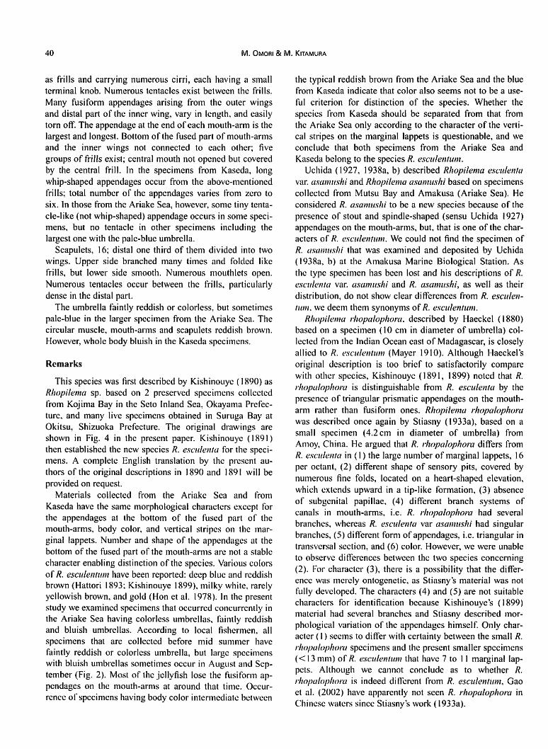
40 M. OMORI & M. KlTAMURA
as frills and carrying numerous cirri, each having a small
terminal knob. Numerous tentacles exist between the frills.
Many fusiform appendages arising from the outer wings
and distal part of the inner wing, vary in length, and easily
torn off. The appendage at the end of each mouth-arm is the
largest and longest. Bottom of the fused part of mouth-arms
and the inner wings not connected to each other; five
groups of frills exist; central mouth not opened but covered
by the central frill. In the specimens from Kaseda, long
whip-shaped appendages occur from the above-mentioned
frills; total number of the appendages varies from zero to
six. In those from the Ariake Sea, however, some tiny tenta
cle-like (not whip-shaped) appendage occurs in some speci
mens, but no tentacle in other specimens including the
largest one with the pale-blue umbrella.
Scapulets, 16; distal one third of them divided into two
wings. Upper side branched many times and folded like
frills, but lower side smooth. Numerous mouthlets open.
Numerous tentacles occur between the frills, particularly
dense in the distal part.
The umbrella faintly reddish or colorless, but sometimes
pale-blue in the larger specimen from the Ariake Sea. The
circular muscle, mouth-arms and scapulets reddish brown.
However, whole body bluish in the Kaseda specimens.
Remarks
This species was first described by Kishinouye (1890) as
Rhopilema sp. based on 2 preserved specimens collected
from Kojima Bay in the Seto Inland Sea, Okayama Prefec
ture, and many live specimens obtained in Suruga Bay at
Okitsu, Shizuoka Prefecture. The original drawings are
shown in Fig. 4 in the present paper. Kishinouye (1891)
then established the new species R. escuienta for the speci
mens. A complete English translation by the present au
thors of the original descriptions in 1890 and 1891 will be
provided on request.
Materials collected from the Ariake Sea and from
Kaseda have the same morphological characters except for
the appendages at the bottom of the fused part of the
mouth-arms, body color, and vertical stripes on the mar
ginal lappets. Number and shape of the appendages at the
bottom of the fused part of the mouth-arms are not a stable
character enabling distinction of the species. Various colors
of/?, esculentum have been reported: deep blue and reddish
brown (Hattori 1893; Kishinouye 1899), milky white, rarely
yellowish brown, and gold (Hon et al. 1978). In the present
study we examined specimens that occurred concurrently in
the Ariake Sea having colorless umbrellas, faintly reddish
and bluish umbrellas. According to local fishermen, all
specimens that are collected before mid summer have
faintly reddish or colorless umbrella, but large specimens
with bluish umbrellas sometimes occur in August and Sep
tember (Fig. 2). Most of the jellyfish lose the fusiform ap
pendages on the mouth-arms at around that time. Occur
rence of specimens having body color intermediate between
the typical reddish brown from the Ariake Sea and the blue
from Kaseda indicate that color also seems not to be a use
ful criterion for distinction of the species. Whether the
species from Kaseda should be separated from that from
the Ariake Sea only according to the character of the verti
cal stripes on the marginal lappets is questionable, and we
conclude that both specimens from the Ariake Sea and
Kaseda belong to the species R. esculentum.
Uchida (1927, 1938a, b) described Rhopilema escuienta
var. asamushi and Rhopilema asamushi based on specimens
collected from Mutsu Bay and Amakusa (Ariake Sea). He
considered R. asamushi to be a new species because of the
presence of stout and spindle-shaped (sensu Uchida 1927)
appendages on the mouth-arms, but, that is one of the char
acters of/?, esculentum. We could not find the specimen of
R. asamushi that was examined and deposited by Uchida
(1938a, b) at the Amakusa Marine Biological Station. As
the type specimen has been lost and his descriptions of /?.
escuienta var. asamushi and /?. asamushi, as well as their
distribution, do not show clear differences from R. esculen
tum. we deem them synonyms of/?, esculentum.
Rhopilema rhopalophora, described by Haeckel (1880)
based on a specimen (10 cm in diameter of umbrella) col
lected from the Indian Ocean east of Madagascar, is closely
allied to R. esculentum (Mayer 1910). Although Haeckel's
original description is too brief to satisfactorily compare
with other species, Kishinouye (1891, 1899) noted that R.
rhopalophora is distinguishable from /?. escuienta by the
presence of triangular prismatic appendages on the mouth-
arm rather than fusiform ones. Rhopilema rhopalophora
was described once again by Stiasny (1933a), based on a
small specimen (4.2cm in diameter of umbrella) from
Amoy, China. He argued that /?. rhopalophora differs from
/?. escuienta in (1) the large number of marginal lappets, 16
per octant, (2) different shape of sensory pits, covered by
numerous fine folds, located on a heart-shaped elevation,
which extends upward in a tip-like formation, (3) absence
of subgenital papillae, (4) different branch systems of
canals in mouth-arms, i.e. /?. rhopalophora had several
branches, whereas R. escuienta var asamushi had singular
branches, (5) different form of appendages, i.e. triangular in
transversal section, and (6) color. However, we were unable
to observe differences between the two species concerning
(2). For character (3), there is a possibility that the differ
ence was merely ontogenetic, as Stiasny's material was not
fully developed. The characters (4) and (5) are not suitable
characters for identification because Kishinouye's (1899)
material had several branches and Stiasny described mor
phological variation of the appendages himself. Only char
acter (1) seems to differ with certainty between the small /?.
rhopalophora specimens and the present smaller specimens
(<13mm) of/?, esculentum that have 7 to 11 marginal lap
pets. Although we cannot conclude as to whether R.
rhopalophora is indeed different from /?. esculentum, Gao
et al. (2002) have apparently not seen /?. rhopalophora in
Chinese waters since Stiasny's work (1933a).

Taxonomic Review of Three Japanese Species of Edible Jellyfish (Scyphozoa: Rhizostomeae} 41
M
Fig. 4. Rhopilema esculentum. After original drawings by Kishinouye (1890).
A.R. Adradius, a. Scapulet, b. Lower mouth-arm, c.c. Circular canal, E.U Exumbrella, f. Gastric cirri, G. Stomach, gn. Gonad,
l.R. Interradius, K. Gelatinous projection, M. Mouth cavity, O. "Hearing organ", O.R Oral pillar, RR. Perradius, r. Sense organ,
S. "Smell organ", s.c. Subgenital cavity.
1, Whole body, 2. Vertical section, left—section of adradial line, right—section of perradial line, 3. Umbrella, left half—lower
view, right half—upper view, 4. Sense organ, 5. A fold of "smell organ", 6. Small tentacle around mouth opening, 7. Gastric cirri,
8. Male sexual organ, 9. Sperm with two mature ones, 10. Ovum.
The life cycle of/?, esculentum has been revealed. In the
sea the medusae need 2-3 months of growth to develop
from ephyra to maturity with an umbrella of 25—45 cm in
diameter in Liaodong Bay, China (Ding & Chen 1981). In
the Minjiang estuary, China, the growth after the ephyra is
divided into three phases, i.e. slower first phase before early
May, faster second phase to end of July, and body contrac
tion final phase after August (Lu et al. 1999a, b). Under
laboratory conditions a few individuals of the scyphistomae
lived more than one year and strobilated repeatedly up to 18
42 M. OMORI & M. KlTAMURA
times in one year and 32 times in two years. Each scyphis-
toma could produce 7-8 ephyrae on average (Chen & Ding
1983). Resource enhancement experiments through releas
ing ephyrae were successfully done off Liaoning Province,
northern Yellow Sea (Chen et al. 1994).
Distribution
Rhopilema esculentum has been recorded in the Seto In
land Sea (Kishinouye 1890), Suruga Bay (Kishinouye
1890), Karatsu Bay (Hattori 1893), Ariake Sea (Hattori
1893, Kishinouye 1899, Uchida 1938b, present study),
Wakasa Bay (Yasuda & Suzuki 1992), and East China Sea
off Kagoshima Pref. (present study). Also from the Chinese
coast from Liaotung Bay to Kwangtung Province and
Moppo, Korea (Hon et al. 1978; Gao et al. 2002). In the
Ariake Sea very large blooms of this species occurred from
1977 to 1979. There is however no record of occurrence in
the Seto Inland Sea or Suruga Bay in recent decades.
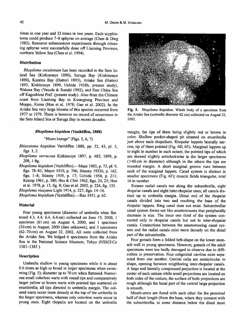
Fig. 5. Rhopilema hispidum. Whole body of a specimen from
the Ariake Sea (umbrella diameter 62 cm) collected on August 22,
2002.
Rhopilema hispidum (Vanhoffen, 1888)
"Hizen kurage" (Figs. 5, 6, 7)
Rhizostoma hispidum Vanhoffen 1888, pp. 32, 43, pi. 5,
figs. 1,2.
Rhopilema verrucosa Kishinouye 1897, p. 485; 1899, p.
208, 1 fig.
Rhopilema hispidum (Vanhoffen).—Maas 1903, p. 73, pi. 9,
figs. 78-81; Mayer 1910, p. 706; Stiasny 1933b, p. 162,
figs. 1-8; Stiasny 1939, p. 17; Uchida 1954, p. 211;
Kramp 1961, p. 380; Hsu & Chin 1962, figs. 24, 25; Hon
et al. 1978, p. 13, fig. 8; Gao et al. 2002, p. 224, fig. 135.
Rhopilema visayana Light 1914, p. 227, figs. 14-16.
Rhopilema hispidium (Vanhoffen).—Rao 1931, p. 62.
Material
Four young specimens (diameter of umbrella when flat
tened 4.3, 4.4, 6.4, 6.4 cm) collected on June 15, 2000, 1
specimen (61 cm) on August 4, 2000, and 1 specimen
(50 cm) in August, 2000 (date unknown), and 3 specimens
(62-70 cm) on August 22, 2002. All were collected from
the Ariake Sea. We lodged 6 specimens from the Ariake
Sea in the National Science Museum, Tokyo (NSMT-Co
1381-1385).
Description
Umbrella shallow in young specimens while it is about
0.6 times as high as broad in larger specimens when swim
ming (Fig. 5); diameter up to 70 cm when flattened. Numer
ous small colorless warts with round tips and comparatively
larger yellow or brown warts with pointed tips scattered on
exumbrella; all tips directed to umbrella margin. The col
ored warts occur more densely at the top of the umbrella in
the larger specimens, whereas only colorless warts occur in
young ones. Eight rhopalia are located on the umbrella
margin; the tips of them being slightly red or brown in
color. Shallow pocket-shaped pit situated on exumbrella
just above each rhopalium. Rhopalar lappets laterally nar
row, tip of them pointed (Fig. 6D, 6E). Marginal lappets up
to eight in number in each octant; the pointed tips of which
are skewed slightly anticlockwise in the larger specimens
(>60cm in diameter) although in the others the tips are
rounded margin. A short marginal groove runs between
each of the marginal lappets. Canal system is distinct in
smaller specimens (Fig. 6F); muscle fields triangular, total
16 in number.
Sixteen radial canals run along the subumbrella, eight
rhopalar canals and eight inter-rhopalar ones; all canals dis
tinct up to umbrella margin. Distal end of the rhopalar
canals divided into two and reaching the base of the
rhopalar lappets. Ring canal does not exist. Subumbrellar
canal system forms net like anastomouses that peripherally
decrease in size. The inner one third of the system con
nected only to rhopalar canals but not to inter-rhopalar
canals. Connections between the anastomosing canal sys
tem and the radial canals exist more densely on the distal
part of the subumbrella.
Four gonads form a folded belt-shape on the lower stom
ach wall in young specimens. However, gonads of the adult
specimens were too badly damaged to observe due to diffi
culties in preservation. Four subgenital cavities exist sepa
rated from one another. Genital ostia are semicircular in
shape, opening between neighboring inter-rhopalar canals.
A large and laterally compressed projection is located at the
center of each ostium while small projections are located on
both sides of the ostium; the surface of both projections are
rough although the basal part of the central large projection
is smooth.
Mouth-arms are fused with each other for the proximal
half of their length (from the base, where they connect with
the subumbrella, to some distance below the distal most

Taxonomic Review of Three Japanese Species of Edible Jellyfish (Scyphozoa: Rhizostomeae) 43
5 cm
lcm
1 mmD F,
B F
Fig. 6. Rhopilema hispidum. A. Mouth-arm and a pair of scapulets, lateral view. Hatched area of the lower distal margin is the
lower wing and that of the left margin is the inner wing. No large club-shaped appendages are present on the mouth-arms of this
specimen. Large appendages are probably torn off during collection or lost ontogenetically. B. Mouth-arm, lateral view. A large
club-shaped appendage exists at the point of contact of the three wings. C. Scapulet. lateral view. D and E. Rhopalar lappets and a
sensory pit. exumbrellar view (D) and subumbrellar view (E). One of the two lappets is broken at its tip. F. Anastomosing canal
system of subumbrella. A and C are sketched based on a large specimen collected on August 4, 2000, whereas B, D and E are
based on a small specimen collected on June 15, 2000.

44 M. OmORI & M. KlTAMURA
rsi
ant
pbr
rpr
ex
Fig. 7. Rhopilcma hispidum. After original driiwings by Vanhoflen (1888). (Permission for reprinting was given by the source
"Senkcnbcrgische Bibliothck, Frankfurt am Main".)
Taxonomic Review of Three Japanese Species of Edible Jellyfish (Scyphozoa: Rhizostomeae) 45
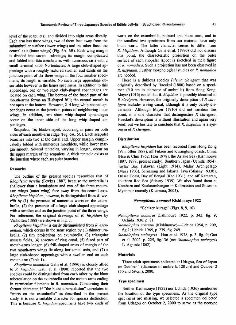
level of the scapulets), and divided into eight arms distally.
Each arm has three wings, two of them face away from the
subumbrellar surface (lower wings) and the other faces the
central axis (inner wings) (Fig. 6A, 6B). Each wing margin
is divided into several subwings; its margin complicated
and folded into thin membranes with numerous cirri with a
small terminal knob. No tentacles. A large club-shaped ap
pendage with a roughly textured swollen end exists on the
junction point of the three wings in the four smaller speci
mens; its length is variable. No such large appendage ob
servable however in the larger specimens. In addition to this
appendage, one or two short club-shaped appendages are
located on each wing. The bottom of the fused part of the
mouth-arms forms an H-shaped frill; the central mouth is
not open at the bottom. However, 2-4 long whip-shaped ap
pendages occur at the junction points of neighboring inner
wings; in addition, two short whip-shaped appendages
occur on the inner side of the long whip-shaped ap
pendages.
Scapulets, 16; blade-shaped, occurring in pairs on both
sides of each mouth-arm ridge (Fig. 6A, 6C). Each scapulet
branches into two at the distal end. Upper margin compli-
catedly folded with numerous mouthlets, while lower mar
gin smooth. Several tentacles, varying in length, occur on
the upper margin of the scapulets. A thick tentacle exists at
the junction where each scapulet branches.
Remarks
The outline of the present species resembles that of
Rhopilema verrilli (Fewkes 1887) because the umbrella is
shallower than a hemisphere and two of the three mouth-
arm wings (outer wing) face away from the central axis.
Rhopilema hispidum, however, is distinguished from R. ver
rilli by (1) the presence of numerous warts on the exum-
brella, (2) the presence of a large club-shaped appendage
with a swollen end on the junction point of the three wings.
For reference, the original drawings of R. hispidum by
Vanhdffen (1888) are shown in Fig. 7.
Rhopilema hispidum is easily distinguished from R. escu-
lentum, which occurs in the same region by (1) thinner um
brella, (2) tiny projections on exumbrella, (3) triangular
muscle fields, (4) absence of ring canal, (5) fused part of
mouth-arms longer, (6) frill-shaped areas of margin of the
two mouth-arm wings lie along horizontal axis, and (7) a
large club-shaped appendage with a swollen end on each
mouth-arm (Table 1).
Rhopilema nomadica Galil et al. (1990) is closely allied
to R. hispidum. Galil et al. (1990) reported that the two
species could be distinguished from each other by the blunt
tuberculation on the exumbrella and the mouth-arms ending
in vermicular filaments in R. nomadica. Concerning their
former character, if "the blunt tuberculation" correlates to
"warts on the exumbrella" as described in the present
study, it is not a suitable character for species distinction.
This is because R. hispidum specimens have two kinds of
warts on the exumbrella, pointed and blunt ones, and in
the smallest two specimens from our material have only
blunt warts. The latter character seems to differ from
R. hispidum. Although Galil et al. (1990) did not discuss
this point, the characteristic projection on the outer
surface of each rhopalar lappet is sketched in their figure
of/?, nomadica. Such a projection has not been observed in
R. hispidum. Further morphological studies on R. nomadica
are needed.
There is a dubious species Pilema clavigera that was
originally described by Haeckel (1880) based on a speci
men (9.0 cm in diameter of umbrella) from Hong Kong.
Mayer (1910) noted that/?, hispidum is possibly identical to
P. clavigera. However, the originally description of P. clav
igera includes a ring canal, although it is only barely dis-
cernable. Although Mayer (1910) did not mention this
point, it is one character that distinguishes P. clavigera.
Haeckel's description is without illustration and again very
brief, but we hesitate to conclude that R. hispidum is a syn
onym of/? clavigera.
Distribution
Rhopilema hispidum has been recorded from Hong Kong
(Vanhoffen 1888), off Fukien and Kwangtung coasts, China
(Hsu & Chin 1962; Hon 1978), the Ariake Sea (Kishinouye
1897, 1899; present study), Southern Japan (Uchida 1954),
Taytay Bay, Palawan (Light 1914), Malay Archipelago
(Maas 1903), Semarang and Jakarta, Java (Stiasny 1933b),
Orissa Coast, Bay of Bengal (Rao 1931), and off Kamaran,
southern Red Sea (Stiasny 1939). We also found them at
Kotabaru and Kualatambangan in Kalimantan and Sittwe in
Myanmar recently (Kitamura, 2003).
Nemopilema nomurai Kishinouye 1922
"Echizen kurage" (Figs. 8, 9, 10)
Nemopilema nomurai Kishinouye 1922, p. 343, fig. 9;
Uchida 1936, p. 81.
Stomolophus nomurai (Kishinouye)—Uchida 1954, p. 209,
fig.2; Uchida 1965, p. 239, fig. 249.
Stomolophus meleagris—Hon et al. 1978, p. 3, fig. 9; Gao
et al. 2002, p. 225, fig. 136 (not Stomolophus meleagris
L. Agassiz 1862).
Materials
Three adult specimens collected at Udagou, Sea of Japan
on October 1 (diameter of umbrella 120 cm) and October 2
(50 and 69 cm), 2000.
Type specimen
Neither Kishinouye (1922) nor Uchida (1936) mentioned
the location of the type specimens. As the original type
specimens are missing, we selected a specimen collected
from Udagou on October 2, 2000 to serve as the neotype
If) I. OmOR] & M. KlTAMURA
Table 1. Morphological comparison of three species of edible jellyfish in Japan.
Rkopilema esculentum Rhopilema hispidum Nemopilema nomurai
Iixumbrella smooth
Projections at front of suhgenkal cavity presence
Coalescent part of mouth-arms
Windows in mouth-arms and scapulets
Types of appendages on month-arm
basal one-fourth of the length
absence
fusiform
with two types of warts,
liny and comparatively
larger
presence
basal half of the length
absence
club-shaped
with granule-like warts
absence
basal one-fourth of the length
presence
whip-shaped
and lodged it at the National Science Museum, Tokyo
(NSMT-Co 1392J.
Description
Umbrella rigid and thick, about half as high as broad
when swimming; diameter up to 120 cm. Whole of the ex-
umbrella covered by colorless granular warts; marginally
located warts of smaller diameter than central ones. Mar
ginal lappets 8 in number in each octant; each lappet di
vided into two round sub-lappets. Eight rhopalia located on
umbrella margin. Rhopalar lappets short and slender (Fig.
9F). A sensory pit narrow groove in shape, located on ex-
umbrella just above each rhopalium. Subumbrellar muscle
well developed, divided into 16 sector groups, located be
tween adjuccnt radial canals. The muscle run across the dis
tal half of the inter-rhopalar canals but not the rhopalar
canals.
Radial canals 16; S rhopalar and 8 inter-rhopalar canals.
Both rhopalar and inter-rhopalar canals easily distinguish
able right up to the umbrella margin. Because the well-de
veloped muscles cover almost all area of the subumbrella,
anastomosing canal system not visible, and circular canal
unknown whether present or not.
Four subgenital cavities separated from each other by
partitions. Genital ostia elliptical without projections at the
entrance.
Mouth-arms, J-shaped in lateral view, fused with each
other for the proximal one-fourth of their length (from the
base, where they connect with the subumbrella, to distal
end level of the scapulets), and divided into eight arms
distally. Each of the eight arms has three wings, two of
them face outwards (outer wing) and the other faces central
axis (inner wing) (Fig. 9A, 9B). Each wing complexly
branched; margin of the wings formed by a thin membrane,
with numerous cirri, and is folded complexly into frills
(Fig. 9C). Each cirrus has a small terminal knob. Windows
open in the outer wings in 2-3 rows. Mouthlets open
among the folds. Numerous whip-shaped appendages occur
on the wings. The appendages on the lower part of each
wing longer and denser than those on the upper part. At the
bottom of the fused part of the mouth-arms, the upper ends
of the inner wings connect to each other via a thin H-
sliaped membrane (Fig. 9D); center of the fused part
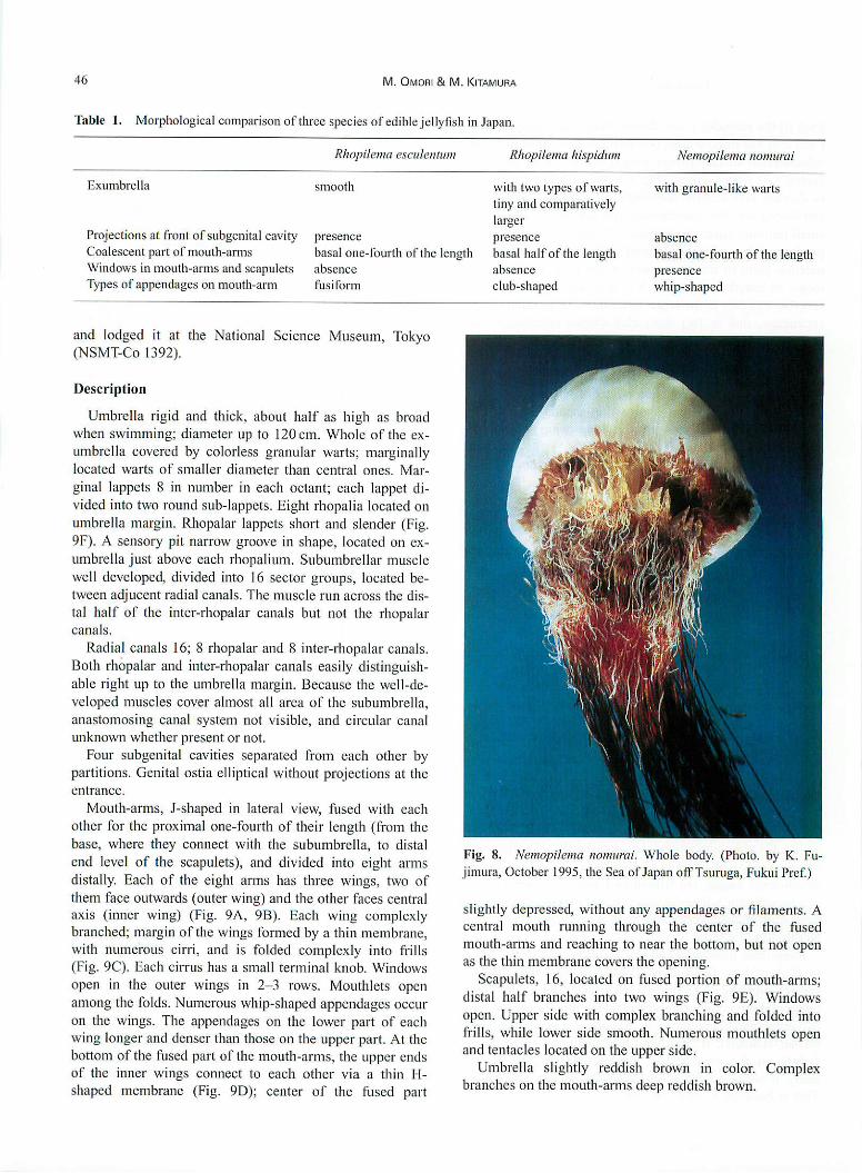
Fig. 8. Nemopilema nomurai. Whole body. (Photo, by EC. Fu-
jimura, October 1995, the Sea of Japan off Tsuruga, Fukui Pref.)
slightly depressed, without any appendages or filaments. A
central mouth running through the center of the fused
mouth-arms and reaching to near the bottom, but not open
as the thin membrane covers the opening.
Scapulets, 16, located on fused portion of mouth-arms;
distal half branches into two wings (Fig. 9E). Windows
open. Upper side with complex branching and folded into
frills, while lower side smooth. Numerous mouthlets open
and tentacles located on the upper side.
Umbrella slightly reddish brown in color. Complex
branches on the mouth-arms deep reddish brown.

Taxonomic Review of Three Japanese Species of Edible Jellyfish (Scyphozoa: Rhizostomeae) 47
A B
Fig. 9. Nemopilema nomurai. A. Mouth-arm, lateral view. The left margin is the outer wing on which tentacles occur densely
and the right margin is the inner wing. B. Mouth-arm, outer view. A and B are sketched based on the same mouth-arm. C. Frill-
like thin membrane with numerous cirri at the mouth-arm margin. D. Bottom of the coalescent mouth-arms. An H-shaped mem
brane connects the upper ends of the inner wings. E. Scapulet, lateral view. Distal half is divided into two wings. F. Rhopalar lap
pets and a sensory pit, exumbrellar view. All figures are sketched based on the specimen with an umbrella diameter of 69 cm.

48 M. OMORI & M. KlTAMURA
r-r -]■■:
a
\ ■'
a
Fig. 10. Nemopilema nommai. After original drawings by Kishinouye (1922).
1. Whole body, 2. Body in cross section, a. horizontal section along the line a, b. the same along the line b, c. the same along the
line c, the same along the line d.
Remarks
Western scientists, probably due to language difficulties,
have not so far seriously assessed the taxonomic status of
Nemopilema nominal. English translation and illustration
of the original description (Kishinouye 1922) are as fol
lows. We attempted to write just as he had described.
"Nemopilema nomurai Kishinouye: The type specimen
was collected in December at Fukui Prefecture facing to
the Sea ofJapan. With scapulets: mouth-arms without club-
shaped appendages, but with numerous filaments; genital
ostia without projections; mouth-arms and scapulets with
numerous windows varyingly in size.
Umbrella, hemispherical and rigid (Fig. 10-1). Bell shal
lower andjellv thinner than in Rhopilema esculenta. Exum-
brella with numerous small warts. The warts, that are lo
cated towards the center of the umbrella, longer than the
marginal ones. Umbrella margin with total of about 120
lappets, about 12 lappets in each octant. Marginal clefts

Taxonomic Review of Three Japanese Species of Edible Jellyfish (Scyphozoa: Rhizostomeae) 49
deep and shallow alternately. At a glance, each octant
seems to have 6 marginal lappets, and each lappet has a
shallow depression. Circular muscle well-developed. Cen
tral part of subumbrella divided by 16 radial canals (Fig.
10-1).
Mouth-pillars, 4, short and narrow; their outer dimen
sions widen and the outer margin undulates as three waves.
The mouth pillar longer than wide. Mid mouth-pillar is
about one-fourth of width ofsubgenital cavity. Wide groove
presents on upper surface. Inner ends fused to each other
andform a horizontal shelf, the shelf is thin, and its outer
margin pointed. Entrances ofgenital ostia located on inter-
radial line; ellipsoid in shape, flat and with no projection.
Mouth-arm disc, wide and thick. Distal end of the disc
divided into 8 mouth-arms. Four interradial sinuses located
at depressed bottom of the disc; these sinuses directed to
the outer and upper sides. Scapulets, 16, located adradially.
They are depressed laterally and branched into two at the
distal end; half-moon in shape in lateral view.
Mouth-arms, long, extending vertically; each branches
into two at distal end, which is triangular in shape with
three margins containing mouth-folds in cross section. Two
of the three margins face outwards and the other margin
faces inward. Mouth-folds well developed and extend later
ally and upward, forming numerous triangular projections.
The mouth-arms contain numerous windows varying in
size, as with Lobonema smithi described by Mayer (1910)
from Manila Harbor, Philippines. Each window is slender
and pointed on the upper bell-ward side, but slightly broad
on the lower side. The windows are arranged in three rows
vertically along the central axis of(he mouth-arms, and two
rows along the mouth-folds. Usually, the upper windows
are larger than the lower ones. Larger specimens have
larger windows. In the scapulets rounded windows usually
in 2 rows located along the mouth-folds; in this case the
lower windows are larger than the upper ones, although
both are smaller than those located on the mouth-arms
(Fig. 10-2). Both mouth-arms and scapulets with numerous
filaments. The filaments on the scapulets are thick and
short, whereas those on the lower part of the mouth-arms
become gradually longer, and longest at the distal end
which is purple in color.
Gastric cavity, slightly depressed vertically. Its outline
cross-shaped, 16 straight canals running, perradially, inter-
radially, and adradially, from gastric cavity to bell margin.
There is a network of anastomosing canals between the
above-mentioned 16 canals. Ring canal unclear. From the
bottom of the gastric cavity, four perradial canals and a
central mouth develop. In horizontal section the perradial
canals laterally depressed, whereas the central mouth very
narrow and hard to see (Fig. I0-2a, b, c, d). Thefour perra
dial canals slightly curve outwards and dichotomize at the
umbrella-wards edge of the mouth-arm disk, then forming
the adradial canals. Each adradial canal branches into two
narrow canals, which enter the scapulets, branching exten
sively, and finally open into the mouth-folds of the
scapulets. After producing two branches, the adradial
canals continue into the mouth-arms. Before entering the
mouth-arms, one of the adradial canals produce thin canal
that is directed inward- and downward to open at the cross-
shaped mouth-folds on the bottom of the mouth-arm disc.
The adradial canals, after entering the mouth-arms, branch
several times, and open at the mouth-folds. The adradial
canals divided into a main canal and many branches that
directed three sides ofthe mouth-folds.
Umbrella colorless, margins of the lappets brown. Oral
pillar, mouth-arm disc, scapulets, and mouth-arms are
slightly brown. Mouth-folds chocolate bmwn or yellowish-
brown.
Although the umbrella diameter of the specimens is
about 90 cm, many become larger than this. The largest
medusa is about 150 kg in weight. At Fukui Prefecture, this
species occurs post-August, with highest abundances in Oc
tober and November. All medusae are swept offshore or die
during December. Small specimens have not yet been ob
served. Perhaps the present species dwells in bottom waters
during its early developmental stages.
The present species exhibits many characters found in
members of the genus Rhopilema. However, it can be dis
tinguished from members of that genus by the absence of
club-shaped appendages on the mouth-arms, and its lack of
a projection near the genital ostium. I propose the name
Nemopilema nomurai, n. gen. n. sp. for this species after
Mr. Kan-ichi Nomura, Director of the Fukui Prefecture
Fisheries Experimental Station."
This original description by Kishinouye falls within the
bounds of our redescription based on specimens from
Udagou.
Because of the presence of 16 scapulets, the present
species is included in the superfamily Scapulatae, which is
composed of two families, the Rhizostomatidae and the
Stomolophidae. The Rhizostomatidae are characterized by
(1) mouth-arms fused along the basal part only, (2) without
a central mouth, and (3) the distal part of the mouth-arms
forms 3-winged structure (see Cornelius 1997; Mianzan &
Cornelius 1999). The present species is included in this
family. Kramp (1961) considered that the family Rhizos
tomatidae includes three genera, i.e. Eupilema, Rhizostoma,
and Rhopilema. Eupilema species have no appendage. Rhi
zostoma species have a club-shaped terminal appendage on
each mouth-arm, precluding the placement of the present
species within either of these genera. Rhopilema species re
semble the present species in the presence of large
scapulets and of numerous appendages. However, the pre
sent species is distinguished from members of the genus
Rhopilema by; 1) presence of windows in the outer wings
of the mouth-arms and scapulets, (2) absence of a projec
tion in front of the subgenital ostium, and (3) absence
of large appendages such as club-shaped or fusiform
appendages on the mouth-arms. Apparently, judgments
by Uchida (1936) and Kramp (1961) synonymizing

50 M. OMORI & M. KlTAMURA
Nemopilema into the genus Stomolophus were in error. We
suggest that the present species be retained in the genus
Nemopilema.
Generic diagnosis of Nemopilema is as follows: size of
exumbrellar warts increases towards the center; with a nar
row central mouth covered by a membrane at the bottom of
the fused portion of the mouth-arms; no projection at the
entrance of the genital ostia; mouth-arms fused along the
basal one-fourth of their length, J-shaped, with scapulatae,
with windows in 2-3 rows; with whip-shaped appendages
on the mouth-arms but lacking fusiform or club-shaped ap
pendages (Table 1).
People in Japan sometimes use parts of this medusa as
bait for sea bream fishing.
Distribution
Sea of Japan off Kyushu and Honshu, Japan (Kishinouye
1922; Shimomura 1959, present study). Liao-tung Bay, Po
hai, and Yellow Sea coasts to off Shoushan, Zhejiang
Province, China (Hon et al. 1978) and northern part of the
East China Sea (Gao et al. 2002). Its distribution is perhaps
restricted to the marginal seas of the northwestern Pacific
around Korea, China and Japan. Its life history is not well
known, but we assume that scyphistoma of this species may
be produced along the coasts of the Korean Peninsula and
northern China in spring. Liberated medusae grow rapidly
and drift from the western reaches of the Sea of Japan to ar
rive along the coast of Japan after summer.
Nemopilema nomurai sometimes occurs so abundantly in
the Sea of Japan that set-nets are broken and fish in the nets
are damaged due to by-catches of this species. In 1958 the
very large occurrence extended from the Sea of Japan even
to the Tsugaru Strait and drifted south down along the Pa
cific coast of Japan (Shimomura 1959). Yasuda (1995) re
ported a fresh mass occurrence of the present species along
the Japanese coast of the Sea of Japan in 1995. In 2000 and
2003, other very large occurrence, which were a serious
fisheries nuisance, occurred along the Sea of Japan from
end of August to November.
Acknowledgements
The authors would like to express their gratitude to
Messrs. M. Eguchi, M. Hayashi, J. Hirose, K. Hori, Y.
Kawamura, K. Nakahata and S. Ueno for assisting with
specimen collection. Dr. H.-Ch. John and Dr. H. Tiemann
of the Zoological Institute of Hamburg kindly translated the
German literature by Haeckel, Vanhoffen, Maas, and Sti-
asny for us. Dr. P. Cornelius provided valuable suggestions
during the early period of this study. Dr. T. Yasuda kindly
provided us with a photo of Nemopilema nomurai that was
taken by Mr. K. Fujimura. Without their help the present
study could not have been successful. We express our sin
cere thanks to Dr. Mary N. Arai and Dr. D. J. Lindsay
for their kind reading of the manuscript and for providing
useful suggestions. This study was partially supported by
grants-in-aid to M.K. from the Research Institute of Marine
Invertebrates, Tokyo. M.O. and M.K. contributed equally to
this work.
Literature Cited
Agassiz, L. 1862. Contributions to the Natural History of the
United States of America, 4: 1-380, pis. 20-34.
Chen, J. & G. Ding 1983. Effect of temperature on the strobilation
of jellyfish (Rhopilema esculenta Kishinouye—Scyphozoa,
Rhizostomeae). Acta zooi. Sinica 29: 195-206, pis. 1&2. (in
Chinese.)
Chen, J., N. Lu, C. Liu, S. Jiang & X. Lu 1994. Resource enhance
ment experiments on the edible medusae Rhopilema esculenta
Kishinouye in the coastal waters of northern Yellow Sea. Mar.
Fish. Res. 15: 103-113. (in Chinese.)
Cornelius, P. F. S. 1997. Keys to the genera of Cubomedusae and
Scyphomedusae (Cnidaria), p. 109-122. In Proceedings of the
6th International Conference on Coelenterate Biology.
Ding, G. & J. Chen 1981. The life history of Rhopilema esculenta
Kishinouye. J. Fish. China 5: 93-102, pis. 1 &2. (in Chinese.)
Fevvkes, J. W. 1887. A new rhizostomatous medusa from New
England. Amer. J. ScL, Ser. 3, 33: 119-125, pi. 1.
Galil, B. S., E. Spanier & W. W Ferguson 1990. The Scyphome
dusae of the Mediterranean coast of Israel, including two
Lessepsian migrants new to the Mediterranean. Zooi Meded.
64: 95-105.
Gao, S., H. Hong & S. Zhang 2002. Fauna Sinica Invertebrata 27.
Phylum Cnidaria, Class Hydmzoa, Subclass Siphonophorae.
Class Scyphomedusae. Science Press, Beijing, 272 pp. (in Chi
nese.)
Haeckel, E, 1880. System der Acraspeden. Zweite Halfte des Sys
tem der Medusen. p. 361-672. 20 pis. Jena.
Hattori, S. 1893. Edible jellyfish in Saga Prefecture. Dobutsugaku
Zasshi. Tokyo 5(59): 342-345. (in Japanese.)
Hon, H.-C, S.-M. Chang & C.-C. Wang. 1978. Haitsue
(Rhopilema esculentum). Science Publications, Beijing, 70 pp.
(in Chinese.)
Hsu, C.-T. & T.-G. Chin 1962. Studies on the medusae from the
Fukien coast. J. Xiamen Univ., Natural Science 9: 206-224. (in
Chinese.)
Huang, M.-X. & Y.-S. Wang 1985. The morph-observation of
Rhopilema esculentum Kishinouye. Chinese J. Zooi 20(2): 1-3.
{in Chinese.)
Kishinouye, K. 1890. Bizen kurage. Rhopilema sp. Dobutsugaku
Zasshi. Tokyo 2( 16): 47-54, pi. 2. (in Japanese.)
Kishinouye, K. 1891, Bizen kurage. Rhopilema esculenta. Dobut
sugaku Zasshi, Tokyo 3(28): 53. (in Japanese.)
Kishinouye, K, 1897. New species of edible jellyfish. Dobutsug
aku Zasshi, Tokyo 9: 483. (in Japanese.)
Kishinouye, K., 1899, Edible medusae. Zooi Jahrbiicher. 12:
205-210, 1 fig.
Kishinouye, K. 1922. Echizen kurage. Nemopilema nomurai.
Dobutsugaku Zasshi, Tokyo 34: 343-346, pi. 9. (in Japanese.)
Kitamura, M. 2003. Taxonomy of edible jellyfish in Southeast
Asia. Umiushi-tsuushin 38: 2-5. (in Japanese.)
Kramp, P. L. 1961. Synopsis of the medusae of the world. J. mar.
Taxonomic Review of Three Japanese Species of Edible Jellyfish (Scyphozoa: Rhizostomeae) 51
biol. Ass. U.K. 40: 1-469.
Light, S. F. 1914. Some Philippine Scyphomedusae. including two
new genera, five new species, and one new variety. Philipp. J.
Sci., Sec. D9\\95-23\.
Lu, Z., Q. Dai, Y. Yan, L. Chen & J. Chen 1999a. A study on
growth of Rhopilema esculeuia in Minjian Estuary. ./.
Oceanogr. Taiwan Strait 18: 314-319. (in Chinese.).
Lu, Z., Y. Yan, Q. Dai, L. Chen & J. Chen 1999b. Fishery ecology
of jellyfish at Minjian River estuary. Chinese J. Appl. Ecol. 10:
341-344. (in Chinese.).
Maas, O. 1903. Die Scyphomedusen der Siboga Exped. 1901.
11(9). 91 pp.. 12 pis.
Mayer, A. G. 1910. Medusae of the World. Vol. 3. The Scyphome
dusae. Carnegie Inst., Wash. D.C.. p.495-735, pis. 56-76.
Mianzan, H. W. & P. Cornelius 1999. Cubomedusae and
Scyphomedusae. p. 513-559. In South Atlantic Zooplankton, 1.
(ed. Boltovskoy, D.). Backhuys Publishers, Leiden.
Omori, M. 1981. Edible jellyfish (Scyphomedusae: Rhizostomeae)
in the Far East waters: a brief review of the biology and fishery.
Bull. Plankton Soc. Japan 28: 1-11. (in Japanese.)
Omori, M. & E. Nakano 2001. Jellyfish fisheries in Southeast
Asia. Hydrobiologia 451: 19-26.
Rao, H. S. 1931. Notes on Scyphomedusae in the Indian Museum.
Rec. Indian Mus. 33: 25-62, pis. 3, 4.
Shimomura, T. 1959. On the unprecedented flourishing of
"Echizen-kurage", Stomolophus nomurai (Kishinouye), in the
Tsushima warm current regions in Autumn, 1958. Bull. Japan
Sea Reg. Fish. Res. Lab. 7: 85-107. (in Japanese.)
Stiasny, G. 1933a. Ober Rhopilema rhopalophora Haeckel. Zoo I.
Meded. 15: 149-155.
Stiasny, G. 1933b. Ober einige Entwicklungsstadien von
Rhopilema hispidum (Vanhoffen) Maas. Zool. Meded. 15: 162—
174.
Stiasny, G. 1939. Uber einige Scyphomedusen von Kamaran
(Rotes Meer). Zool. Anz. 128: 17-23.
Uchida. T. 1927. Report of the biological survey of Mutsu Bay. 2.
Medusae of Mutsu Bay. 5c/. Rep. Tohoku Univ.. Biol. 2: 215-
238.
Uchida, T. 1936. Class Scyphozoa. Fauna Nipponica, 3(2), p.
1-94. Sanseido, Tokyo, (in Japanese.)
Uchida. T. 1938a. Report of the biological survey of Mutsu Bay
32. Medusae from Mutsu Bay (revised report). Sci. Rep. Tohoku
Univ., Sec. 4, 13: 37-46.
Uchida, T. 1938b. Medusae in the vicinity of the Amakusa Marine
Biological Station. Bull. Biogeogr Soc. Japan 8: 143-149.
Uchida, T. 1954. Distribution of Scyphomedusae in Japanese and
its adjacent waters. J. Fac. Sci., Hokkaido Univ., Ser. 6, Zool.
12:209-219.
Uchida, T. 1965. Scyphozoa. p. 230-239. In New Illustrated Ency
clopedia ofthe Fauna ofJapan (eds. Okada, K., S. Uchida & T.
Uchida). Hokuryukan, Tokyo, (in Japanese.)
Vanhoffen, E. 1888. Untersuchungen iiber semastome und rhizos-
tome Medusen. Bibliotheca Zoologica. Stuttgart 1(3): 1-52, 6
pis.
Wu, B.-L. 1955. Haitsue. Rhopilema esculenta. Sheng Wu Hsueh
T'ung Pao (Biological Newsletter) 4: 35-40. (in Chinese.)
Yamada, M. 1997. Scyphozoa. p. 541-553. In An Illustrated
Guide to Marine Plankton in Japan (eds. Chihara. M. & M.
Murano). Tokai University Press, Tokyo, (in Japanese.)
Yamaji, 1. 1966. Illustrations of the Marine Plankton of Japan.
Hoikusha Publication, Tokyo, 372 pp. (in Japanese.)
Yasuda, T. & Y. Suzuki 1992, Notes on an edible medusa,
Rhopilema asamushi Uchida, caught in Wakasa Bay, Japan. J.
Plankton Soc. Japan 38: 147-148.
Yasuda, T. 1995. Once more, mass occurrence of Echizen kurage.
Umiushi-tsuushin 9: 6-8. (in Japanese.)