taurine transporter (taut) deficiency impairs …taurine transporter (taut) deficiency impairs...
TRANSCRIPT
Taurine transporter (TauT) deficiency impairs ammoniadetoxification in mouse liverNatalia Qvartskhavaa, Cheng Jun Jina, Tobias Buschmanna, Ute Albrechta, Johannes Georg Bodea, Niloufar Monhaserya,Jessica Oenartoa, Hans Jürgen Bidmonb, Boris Görga, and Dieter Häussingera,1
aClinic for Gastroenterology, Hepatology and Infectious Diseases, Heinrich Heine University, 40225 Düsseldorf, Germany; and bCécile and Oskar VogtInstitute for Brain Research, Heinrich Heine University, 40225 Düsseldorf, Germany
Edited by Arthur J. L. Cooper, New York Medical College, Valhalla, NY, and accepted by Editorial Board Member David J. Mangelsdorf February 11, 2019(received for review July 30, 2018)
Hepatic ammonia handling was analyzed in taurine transporter(TauT) KO mice. Surprisingly, hyperammonemia was present at anage of 3 and 12 months despite normal tissue integrity. This wasaccompanied by cerebral RNA oxidation. As shown in liver perfusionexperiments, glutamine production from ammonia was diminishedin TauT KO mice, whereas urea production was not affected. Inlivers from 3-month-old TauT KO mice protein expression andactivity of glutamine synthetase (GS) were unaffected, whereasthe ammonia-transporting RhBG protein was down-regulated byabout 50%. Double reciprocal plot analysis of glutamine synthesisversus perivenous ammonia concentration revealed that TauT KOhad no effect on the capacity of glutamine formation in 3-month-old mice, but doubled the ammonia concentration required for half-maximal glutamine synthesis. Since hepatic RhBG expression isrestricted to GS-expressing hepatocytes, the findings suggest thatan impaired ammonia transport into these cells impairs glutaminesynthesis. In livers from 12-, but not 3-month-old TauT KO mice,RhBG expression was not affected, surrogate markers for oxidativestress were strongly up-regulated, and GS activity was decreased by40% due to an inactivating tyrosine nitration. This was also reflectedby kinetic analyses in perfused liver, which showed a decreasedglutamine synthesizing capacity by 43% and a largely unaffectedammonia concentration dependence. It is concluded that TauT de-ficiency triggers hyperammonemia through impaired hepatic gluta-mine synthesis due to an impaired ammonia transport via RhBG at 3months and a tyrosine nitration-dependent inactivation of GS in 12-month-old TauT KO mice.
glutamine | hyperammonemia | taurine | scavenger cells | oxidative stress
The liver plays a central role for ammonia homeostasis in thebody. A sophisticated structural and functional organization
of urea and glutamine synthesis as well as glutaminase hasevolved, which not only allows for efficient ammonia detoxicationby the liver, but also to adjust urea synthesis to the needs of sys-temic acid base homeostasis (1, 2). In this organization, gluta-minase augments urea synthesis by amplifying the ammoniaconcentration in the periportal compartment, whereas glutaminesynthetase (GS), which is restricted to a small perivenous cellpopulation (3) acts as a high-affinity detoxification system for theammonia, which escapes periportal urea synthesis (1, 2, 4). Due tothis important function, the small perivenous subpopulation ofhepatocytes has been termed “scavenger cells” (2). These cellsexclusively express GS, the glutamate transporter 1 (Glt1) and theNH3/NH4
+ transporter Rhesus type glycoprotein B (RhBG) aswell as uptake systems for dicarboxylates (3, 5, 6). Pioneer workwith the isolated perfused rat liver suggested already decades agothat intact scavenger cells are essential for maintenance of phys-iologically low ammonia concentrations in the blood (1, 2, 7), andliver-specific GS deletion results in systemic hyperammonemia inmice (8). This study also confirmed that chronic hyperammonemiain the absence of liver damage is sufficient to induce cerebraloxidative stress and to impair behavior and locomotion, which arehallmarks of hepatic encephalopathy (HE) (8).
The amino acid taurine is highly abundant in brain, liver, andother organs (9). Intracellular taurine concentrations are main-tained at high levels through uptake by the taurine transporterTauT (10). Functions of taurine in the body are remarkably di-verse: taurine acts as an osmolyte, an antioxidant, and a molecularchaperone, and promotes antiinflammatory effects and modulatesneurotransmission (11–13). Studies from TauT KO mice showedthat low taurine levels cause a variety of malfunctions in differentorgans in an age-dependent way (14–20). Within 1 y after birthTauT KO mice lose vision, suffer from auditory, olfactory, andmuscle dysfunction, and show altered synaptic transmission inbrain (for review see ref. 21). At higher age (beyond 15 mo), livermanifestations develop, such as fibrosis, unspecific hepatitis, andtumor formation (17, 21). However, the pathogenetic events thatprecede such structural liver dysfunction in aged TauT KO miceremained unclear until now.In the present study, we analyzed effects of TauT deficiency on
hepatic ammonia handling in 3- and 12-mo-old TauT KO mice.Our results suggest that TauT deficiency impairs ammonia de-toxification by perivenous scavenger cells in the liver, thereby in-ducing systemic hyperammonemia. This is explained by down-regulation of the ammonium transporter RhBG at 3 mo of age anda tyrosine nitration-induced inactivation of GS at 12 mo of age dueto oxidative/nitrosative stress. The data also suggest that RhBGexpression can be a site of control of hepatic glutamine synthesis.
ResultsTaurine Transporter Knockout Triggers Hyperammonemia WithoutAffecting Liver Integrity. TauT deficiency was shown to triggerliver fibrosis in mice beyond an age of 15 mo (17). However, asshown by Sirius red staining (SI Appendix, Fig. S1A) and immu-nofluorescence analysis of collagens I and IV (SI Appendix, Fig.S1B), no signs of fibrosis were found in livers from 3- and 12-mo-oldTauT KO and WT mice and expression of these surrogate markers
Significance
Taurine transporter deficiency in mice results in hyperammonemiaand reduced glutamine formation in hepatic scavenger cells dueto an impaired ammonia transport at 3 months of age and a ty-rosine nitration-dependent inactivation of glutamine synthetasein 12-month-old mice. The data suggest that down-regulation ofthe ammonia transporter RhBG can be rate limiting for the syn-thesis of glutamine in mouse liver.
Author contributions: D.H. designed research; N.Q., C.J.J., T.B., U.A., N.M., J.O., H.J.B., andB.G. performed research; J.G.B. contributed new reagents/analytic tools; N.Q., C.J.J., T.B.,U.A., N.M., J.O., H.J.B., B.G., and D.H. analyzed data; and B.G. and D.H. wrote the paper.
The authors declare no conflict of interest.
This article is a PNAS Direct Submission. A.J.L.C. is a guest editor invited by theEditorial Board.
Published under the PNAS license.1To whom correspondence should be addressed. Email: [email protected].
This article contains supporting information online at www.pnas.org/lookup/suppl/doi:10.1073/pnas.1813100116/-/DCSupplemental.
Published online March 12, 2019.
www.pnas.org/cgi/doi/10.1073/pnas.1813100116 PNAS | March 26, 2019 | vol. 116 | no. 13 | 6313–6318
MED
ICALSC
IENCE
S
Dow
nloa
ded
by g
uest
on
Feb
ruar
y 21
, 202
0
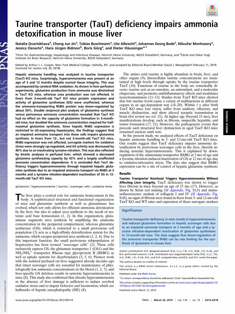
of fibrosis was not different from respective wild-type controls.However, a strong Sirius red staining was noted in liver slices frombile duct ligated mice (21 d postsurgery), which served as a positivecontrol (SI Appendix, Fig. S1A). These data suggest that taurinetransporter knockout does not induce liver fibrosis or structuralliver damage in TauT KO mice up to an age of 12 mo. However,unexpectedly, blood ammonia levels were significantly elevated toabout 115 μmol/L in both, 3- and 12-mo-old TauT KO mice com-pared with WT mice, which presented blood ammonia levels ofabout 70 μmol/L (Fig. 1).
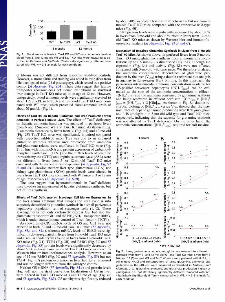
Effects of TauT KO on Hepatic Glutamine and Urea Production fromAmmonia in Perfused Mouse Liver. The effect of TauT deficiencyon hepatic ammonia handling was analyzed in perfused liversfrom 3- and 12-mo-old WT and TauT KO mice. As shown in Fig.2, ammonia clearance by livers from 3- (Fig. 2A) and 12-mo-old(Fig. 2B) TauT KO mice was significantly impaired comparedwith respective wild-type mice. This was due to an impairedglutamine synthesis, whereas urea production from ammoniaand glutamate release were unaffected in TauT KO mice (Fig.2). In line with this, mRNA and protein expression of carbamoyl-phosphate synthetase 1 (CPS1) and the mRNA levels of ornithinetranscarbamylase (OTC) and argininosuccinate lyase (ASL) werenot different in livers from 3- or 12-mo-old TauT KO micecompared with the respective wild-type mice (SI Appendix, Fig. S2A and B). Likewise, neither liver type glutaminase (LGA) norkidney type glutaminase (KGA) protein levels were altered inlivers from TauT KO mice compared with WT mice at 3 or 12 moof age, respectively (SI Appendix, Fig. S2B).The data suggest that hyperammonemia in TauT-deficient
mice involves an impairment of hepatic glutamine synthesis, butnot of urea synthesis.
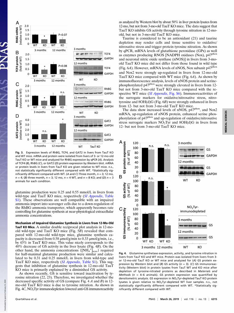
Effects of TauT Deficiency on Scavenger Cell Marker Expression. Inthe liver acinus ammonia that escapes the urea cycle is sub-sequently detoxified by glutamine synthesis in a small perivenoushepatocyte population termed scavenger cells (1, 2). Thesescavenger cells not only exclusively express GS, but also theglutamate transporter Glt1 and the NH3/NH4
+ transporter RhBG,which is under transcriptional control of T cell factor 4 (TCF4).As shown by qPCR, mRNA levels of GS and Glt1 were not
affected in both, 3- and 12-mo-old TauT KO mice (SI Appendix,Figs. S3A and S4A), whereas mRNA levels of RhBG were sig-nificantly down-regulated in livers from 3-mo-old TauT KO miceand a similar tendency was found in livers from 12-mo-old TauTKO mice (Fig. 3A). TCF4 (Fig. 3B) and RhBG (Fig. 3C and SIAppendix, Fig. S5) protein levels were significantly decreased byabout 50% in livers from 3-mo-old TauT KO mice as shown byWestern blot or immunofluorescence analysis. However, at anage of 12 mo RhBG (Fig. 3C and SI Appendix, Fig. S5) but notTCF4 (Fig. 3B) protein expression in liver had fully recoveredand was no longer different from the wild-type control.Neither GS mRNA (SI Appendix, Fig. S4A) and protein levels
(Fig. 4A) nor the strict perivenous localization of GS in liverwere altered in TauT KO mice at 3 and 12 mo of age (Fig. 4Aand SI Appendix, Fig. S4B). GS activity was significantly reduced
by about 40% in protein lysates of livers from 12- but not from 3-mo-old TauT KO mice compared with the respective wild-typemice (Fig. 4B).Glt1 protein levels were significantly increased by about 80%
in livers from 3-mo-old and about fourfold in livers from 12-mo-old TauT KO mice as shown by Western blot and immunoflu-orescence analysis (SI Appendix, Fig. S3 B and C).
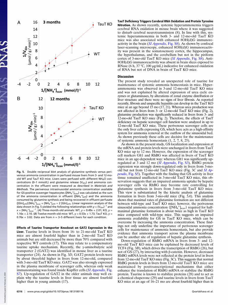
Mechanism of Impaired Glutamine Synthesis in Livers from 3-Mo-OldTauT KO Mice. As shown above, in perfused livers from 3-mo-oldTauT KO mice, glutamine synthesis from ammonia at concen-trations up to 0.5 mmol/L is diminished (Fig. 2A), although GSexpression (Fig. 4A) and activity (Fig. 4B) were not affectedcompared with 3-mo-old wild-type mice. We therefore analyzedthe ammonia concentration dependence of glutamine pro-duction by the liver (VGln) using a double-reciprocal plot analysisin analogy to Lineweaver–Burk blotting. In this approach, theperivenous intrasinusoidal ammonia concentration available forGS-positive scavenger hepatocytes {[NH4
+]scav} can be esti-mated as the sum of the ammonia concentration in effluent{[NH4
+]effl} and the ammonia consumed by glutamine synthesisand being recovered in effluent perfusate {[Gln]effl)}: [NH4
+
]scav = [NH4+]effl + 2 [Gln]effl. As shown in Fig. 5A double re-
ciprocal blotting of [NH4+]scav versus VGln showed that the max-
imal rates of hepatic glutamine production were 0.50 μmol/g/minand 0.48 μmol/g/min in 3-mo-old wild-type and TauT KO mice,respectively, indicating that the capacity for glutamine synthesiswas not affected by TauT deficiency. On the other hand, theammonia concentrations {[NH4
+]scav} required for half-maximal
Fig. 2. Urea, glutamine, ammonia, and glutamate release into effluent ofperfused livers from 3- and 12-mo-old WT and TauT KO mice. Livers from 3(A)- and 12 (B)-mo-old WT and TauT KO mice were perfused with 0, 0.2, or0.5 mmol/L NH4Cl and concentrations of urea, glutamine, ammonia, andglutamate in the effluent were measured as described in Materials andMethods. Urea, glutamine, ammonia, and glutamate production is given asμmol/g/min. n.s., not statistically significantly different compared with WT.*Statistically significantly different compared with WT. n = 3–5 animals foreach condition.
Fig. 1. Blood ammonia levels in TauT KO and WT mice. Ammonia levels inblood from 3- and 12-mo-old WT and TauT KO mice were measured as de-scribed in Materials and Methods. *Statistically significantly different com-pared with WT. n = 3–8 animals for each condition.
6314 | www.pnas.org/cgi/doi/10.1073/pnas.1813100116 Qvartskhava et al.
Dow
nloa
ded
by g
uest
on
Feb
ruar
y 21
, 202
0
glutamine production were 0.25 and 0.55 mmol/L in livers fromwild-type and TauT KO mice, respectively (SI Appendix, TableS1). These observations are well compatible with an impairedammonia import into scavenger cells due to a down-regulation ofthe RhBG ammonia transporter, which apparently becomes ratecontrolling for glutamine synthesis at near-physiological extracellularammonia concentrations.
Mechanism of Impaired Glutamine Synthesis in Livers from 12-Mo-OldTauT KO Mice. A similar double reciprocal plot analysis in 12-mo-old wild-type and TauT KO mice (Fig. 5B) revealed that com-pared with 12-mo-old wild-type mice, glutamine synthesis ca-pacity is decreased from 0.58 μmol/g/min to 0.33 μmol/g/min, i.e.,by 43% in TauT KO mice. This value nicely corresponds to the40% decrease of GS activity in the liver lysate (Fig. 4B). On theother hand, the ammonia concentrations {[NH4
+]scav} requiredfor half-maximal glutamine production were similar and calcu-lated to be 0.31 and 0.25 mmol/L in livers from wild-type andTauT KO mice, respectively (SI Appendix, Table S1). This sug-gests that inhibition of glutamine synthesis in 12-mo-old TauTKO mice is primarily explained by a diminished GS activity.As shown recently, GS is sensitive toward inactivation by ty-
rosine nitration (22, 23). Therefore, we investigated whether thedecreased specific activity of GS (compare Fig. 4 A and B) in 12-mo-old TauT KO mice is due to tyrosine nitration. As shown inFig. 4C, NO2Tyr immunodepletion lowered anti-GS immunoreactivity
as analyzed byWestern blot by about 50% in liver protein lysates from12-mo, but not from 3-mo-old TauT KOmice. The data suggest thatTauT KO inhibits GS activity through tyrosine nitration in 12-mo-old, but not in 3-mo-old TauT KO mice.Taurine is considered to be an antioxidant (21) and taurine
depletion may render cells and tissue sensitive to oxidative/nitrosative stress and trigger protein tyrosine nitration. As shownby qPCR, mRNA levels of glutathione peroxidase (GPx) as wellas enzymes producing RNOS [NADPH oxidases (Nox), p47phox
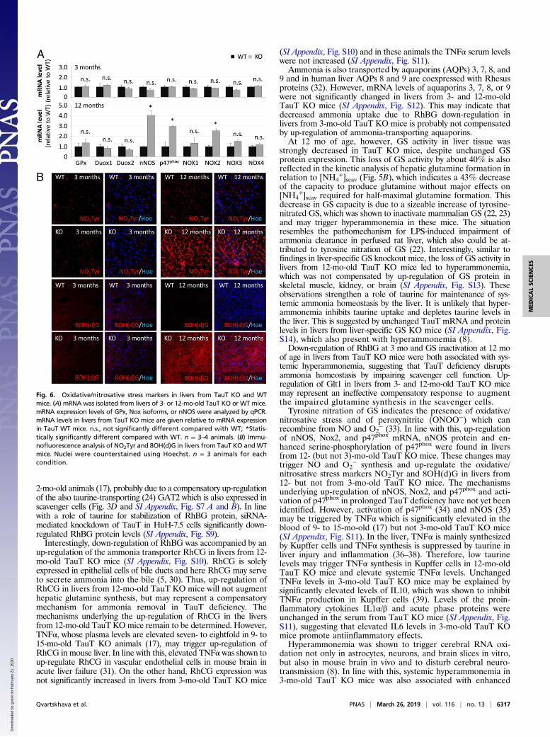
and neuronal nitric oxide synthase (nNOS)] in livers from 3-mo-old TauT KO mice did not differ from those found in wild type(Fig. 6A). However, mRNA levels of nNOS, Nox subunit p47phox
and Nox2 were strongly up-regulated in livers from 12-mo-oldTauT KO mice compared with WT mice (Fig. 6A). As shown byimmunofluorescence analysis, levels of nNOS protein and serine-phosphorylated p47phox were strongly elevated in livers from 12-but not from 3-mo-old TauT KO mice compared with the re-spective WT mice (SI Appendix, Fig. S6). Immunoreactivities ofthe surrogate markers for oxidative/nitrosative stress, nitro-tyrosine and 8OH(d)G (Fig. 6B) were strongly enhanced in liversfrom 12- but not from 3-mo-old TauT KO mice.The data show increased levels of nNOS, p47phox, and Nox2
mRNA, up-regulation of nNOS protein, enhanced serine phos-phorylation of p47phox and up-regulation of oxidative/nitrosativestress surrogate markers NO2Tyr and 8OH(d)G in livers from12- but not from 3-mo-old TauT KO mice.
Fig. 3. Expression levels of RhBG, TCF4, and GAT2 in livers from TauT KOandWT mice. mRNA and protein were isolated from livers of 3- or 12-mo-oldTauT KO or WT mice and analyzed for RhBG expression by qPCR (A). Analysisof TCF4 (B), RhBG (C), or GAT2 (D) protein expression by Western blot. mRNAor protein levels in livers from TauT KO are given relative to WT mice. n.s.,not statistically significantly different compared with WT. *Statistically sig-nificantly different compared with WT. (A and C) Three month, n = 3; 12 mo,n = 4; (B) three month, n = 3; 12 mo, n = 4 WT, and n = 8 KO; and (D) n = 3animals for each condition.
Fig. 4. Glutamine synthetase expression, activity, and tyrosine nitration inlivers from TauT KO and WT mice. Protein was isolated from livers from 3-or 12-mo-old TauT KO or WT mice and analyzed for (A) GS protein ex-pression by Western blot and (B) GS activity (n = 3). (C ) GS immunoreac-tivity (Western blot) in protein lysates from TauT WT and KO mice afterdepletion of tyrosine-nitrated proteins as described in Materials andMethods (n = 4–6 animals). GS protein expression was quantified bydensitometric analysis. GS expression in NO2Tyr-depleted TauT KO proteinlysates is given relative to NO2Tyr-depleted WT liver samples. n.s., notstatistically significantly different compared with WT. *Statistically sig-nificantly different compared with WT.
Qvartskhava et al. PNAS | March 26, 2019 | vol. 116 | no. 13 | 6315
MED
ICALSC
IENCE
S
Dow
nloa
ded
by g
uest
on
Feb
ruar
y 21
, 202
0
Effects of Taurine Transporter Knockout on GAT2 Expression in theLiver. Taurine levels in livers from 16- to 21-mo-old TauT KOmice are almost fourfold higher than in 2-mo-old TauT KOanimals but are still decreased by more than 70% compared withrespective WT controls (17). This may relate to a compensatorytaurine uptake mechanism. Recently, the γ-aminobutyric acidtransporter 2 (GAT2) was identified in mouse liver as a taurinetransporter (24). As shown in Fig. 3D, GAT2 protein levels wereby about threefold higher in livers from 12-mo-old, comparedwith 3-mo-old TauT KO mice. GAT2 was also strongly expressedin the plasma membrane of scavenger cells, whereas only weakimmunostaining was found inside Kupffer cells (SI Appendix, Fig.S7). Up-regulation of GAT2 in the older animals may well ex-plain why the taurine levels in liver tissue are almost fourfoldhigher than in young animals (17).
TauT Deficiency Triggers Cerebral RNA Oxidation and Protein TyrosineNitration. As shown recently, systemic hyperammonemia triggerscerebral RNA oxidation in mouse brain where it was suggestedto disturb cerebral neurotransmission (8). In line with this, sys-temic hyperammonemia in both 3- and 12-mo-old TauT KOmice was also associated with enhanced 8OH(d)G immunore-activity in the brain (SI Appendix, Fig. S8). As shown by confocallaser-scanning microscopy, enhanced 8OH(d)G immunoreactiv-ity was present in the somatosensory cortex, the hippocampus,the hypothalamus, and the cerebellum but not in the piriformcortex of 3-mo-old TauT KO mice (SI Appendix, Fig. S8). Anti-8OH(d)G immunoreactivity was absent in brain slices exposed toRNase (6 h, 37 °C, 100 μg/mL) indicative for enhanced oxidationof RNA but not of DNA in brain of TauT KO mice.
DiscussionThe present study revealed an unexpected role of taurine formaintenance of systemic ammonia homeostasis in mice. Hyper-ammonemia was observed in 3-and 12-mo-old TauT KO miceand was not explained by altered expression of urea cycle en-zymes or glutaminases, by alterations of zonal enzyme distribution orliver structure and there were no signs of liver fibrosis. As reportedrecently, fibrosis and unspecific hepatitis can develop in the TauT KOmice at an age beyond 15 mo (17, 21). Whereas urea production wasnot affected in livers from 3- or 12-mo-old TauT KO mice (Fig. 2),glutamine production was significantly reduced in livers from 3- and12-mo-old TauT KO mice (Fig. 2). Therefore, the effects of TauTdeficiency on hepatic scavenger cell function were analyzed in up to12-mo-old TauT KO mice. These perivenous scavenger cells arethe only liver cells expressing GS, which here acts as a high-affinitysystem for ammonia removal at the outflow of the sinusoidal bed.As shown previously these cells are decisive for the maintenanceof systemic ammonia homeostasis (1, 2, 7, 8, 25).As shown in the present study, GS localization and expression at
the mRNA and protein levels were unchanged in livers from TauTKO mice up to 12 mo. However, the expression of the scavengercell markers Glt1 and RhBG was affected in livers of TauT KOmice in an age-dependent way: whereas Glt1 was significantly up-regulated at 3 and 12 mo (SI Appendix, Fig. S3), RhBG proteinexpression was strongly down-regulated only in livers from 3-mo-old but not from 12-mo-old TauT KO mice (Fig. 3C and SI Ap-pendix, Fig. S5). Together with the finding that GS activity in livertissue remained unaffected in 3-mo-old TauT KO mice, this ob-servation suggests that an impaired ammonia uptake by hepaticscavenger cells via RhBG may become rate controlling forglutamine synthesis in livers from 3-mo-old TauT KO mice.This view is substantiated by the kinetic analysis of glutamineformation in livers from 3-mo-old mice (Fig. 5A). This analysisshows that maximal rates of glutamine formation are not differentbetween wild-type and TauT KO mice; however, the perivenoussinusoidal ammonia concentration {[NH4
+]scav} required for half-maximal glutamine formation is about twice as high in TauT KOmice compared with wild-type mice. This suggests an impairedammonia availability for GS in TauT KO mice, which can beovercome by increasing the ammonia concentration. These find-ings not only underline the importance of perivenous scavengercells for maintenance of ammonia homeostasis, but also provideevidence that ammonia transport across the plasma membranecan be another site of regulation of hepatic glutamine synthesis.Down-regulation of RhBG mRNA in livers from 3- and 12-
mo-old TauT KO mice can be explained by decreased levels ofTCF4 (Fig. 3B), which drives the transcription of RhBG (26), butnot of GS (27), by interacting with β-catenin. However, decreasedRhBG mRNA levels were not reflected at the protein level in liversfrom 12-mo-old TauT KOmice (Fig. 3C). This suggests that normalRhBG protein levels in livers from 12-mo-old TauT KO mice aremaintained by posttranscriptional mechanisms that eitherenhance the translation of RhBG mRNA or stabilize the RhBGprotein. Taurine is known to stabilize proteins (28) and to act asa chemical chaperone (29) and taurine levels in livers from TauTKO mice at an age of 16–21 mo are about fourfold higher than in
Fig. 5. Double reciprocal blot analysis of glutamine synthesis versus peri-venous ammonia concentration in perfused mouse livers from 3- and 12-mo-old WT and TauT KO mice. Livers were perfused with different NH4Cl con-centrations (0–1 mmol/L) and glutamine release (VGln) and ammonia con-centration in the effluent were measured as described in Materials andMethods. The perivenous intrasinusoidal ammonia concentration availablefor GS-positive scavenger hepatocytes {[NH4
+]scav} was calculated as the sumof the ammonia concentration in effluent {[NH4
+]effl} and the ammoniaconsumed by glutamine synthesis and being recovered in effluent perfusate{[Gln]effl}:[NH4
+]scav = [NH4+]effl + 2 [Gln]effl. Linear regression analysis of the
data shown in Fig. 5 yielded the following relationships with y = (VGln)−1 and
x = [NH4+]scav
−1. (A) Three-month-old animals: WT, y = 0.49x + 2.01; KO, y =1.16x + 2.10. (B) Twelve-month-old mice: WT, y = 0.53x + 1.73; TauT KO, y =0.76x + 3.02. Data are from n = 3–5 different livers for each condition.
6316 | www.pnas.org/cgi/doi/10.1073/pnas.1813100116 Qvartskhava et al.
Dow
nloa
ded
by g
uest
on
Feb
ruar
y 21
, 202
0
2-mo-old animals (17), probably due to a compensatory up-regulationof the also taurine-transporting (24) GAT2 which is also expressed inscavenger cells (Fig. 3D and SI Appendix, Fig. S7 A and B). In linewith a role of taurine for stabilization of RhBG protein, siRNA-mediated knockdown of TauT in HuH-7.5 cells significantly down-regulated RhBG protein levels (SI Appendix, Fig. S9).Interestingly, down-regulation of RhBG was accompanied by an
up-regulation of the ammonia transporter RhCG in livers from 12-mo-old TauT KO mice (SI Appendix, Fig. S10). RhCG is solelyexpressed in epithelial cells of bile ducts and here RhCG may serveto secrete ammonia into the bile (5, 30). Thus, up-regulation ofRhCG in livers from 12-mo-old TauT KO mice will not augmenthepatic glutamine synthesis, but may represent a compensatorymechanism for ammonia removal in TauT deficiency. Themechanisms underlying the up-regulation of RhCG in the liversfrom 12-mo-old TauT KOmice remain to be determined. However,TNFα, whose plasma levels are elevated seven- to eightfold in 9- to15-mo-old TauT KO animals (17), may trigger up-regulation ofRhCG in mouse liver. In line with this, elevated TNFα was shown toup-regulate RhCG in vascular endothelial cells in mouse brain inacute liver failure (31). On the other hand, RhCG expression wasnot significantly increased in livers from 3-mo-old TauT KO mice
(SI Appendix, Fig. S10) and in these animals the TNFα serum levelswere not increased (SI Appendix, Fig. S11).Ammonia is also transported by aquaporins (AQPs) 3, 7, 8, and
9 and in human liver AQPs 8 and 9 are coexpressed with Rhesusproteins (32). However, mRNA levels of aquaporins 3, 7, 8, or 9were not significantly changed in livers from 3- and 12-mo-oldTauT KO mice (SI Appendix, Fig. S12). This may indicate thatdecreased ammonia uptake due to RhBG down-regulation inlivers from 3-mo-old TauT KO mice is probably not compensatedby up-regulation of ammonia-transporting aquaporins.At 12 mo of age, however, GS activity in liver tissue was
strongly decreased in TauT KO mice, despite unchanged GSprotein expression. This loss of GS activity by about 40% is alsoreflected in the kinetic analysis of hepatic glutamine formation inrelation to [NH4
+]scav (Fig. 5B), which indicates a 43% decreaseof the capacity to produce glutamine without major effects on[NH4
+]scav required for half-maximal glutamine formation. Thisdecrease in GS capacity is due to a sizeable increase of tyrosine-nitrated GS, which was shown to inactivate mammalian GS (22, 23)and may trigger hyperammonemia in these mice. The situationresembles the pathomechanism for LPS-induced impairment ofammonia clearance in perfused rat liver, which also could be at-tributed to tyrosine nitration of GS (22). Interestingly, similar tofindings in liver-specific GS knockout mice, the loss of GS activity inlivers from 12-mo-old TauT KO mice led to hyperammonemia,which was not compensated by up-regulation of GS protein inskeletal muscle, kidney, or brain (SI Appendix, Fig. S13). Theseobservations strengthen a role of taurine for maintenance of sys-temic ammonia homeostasis by the liver. It is unlikely that hyper-ammonemia inhibits taurine uptake and depletes taurine levels inthe liver. This is suggested by unchanged TauT mRNA and proteinlevels in livers from liver-specific GS KO mice (SI Appendix, Fig.S14), which also present with hyperammonemia (8).Down-regulation of RhBG at 3 mo and GS inactivation at 12 mo
of age in livers from TauT KO mice were both associated with sys-temic hyperammonemia, suggesting that TauT deficiency disruptsammonia homeostasis by impairing scavenger cell function. Up-regulation of Glt1 in livers from 3- and 12-mo-old TauT KO micemay represent an ineffective compensatory response to augmentthe impaired glutamine synthesis in the scavenger cells.Tyrosine nitration of GS indicates the presence of oxidative/
nitrosative stress and of peroxynitrite (ONOO−) which canrecombine from NO and O2
− (33). In line with this, up-regulationof nNOS, Nox2, and p47phox mRNA, nNOS protein and en-hanced serine-phosphorylation of p47phox were found in liversfrom 12- (but not 3)-mo-old TauT KO mice. These changes maytrigger NO and O2
− synthesis and up-regulate the oxidative/nitrosative stress markers NO2Tyr and 8OH(d)G in livers from12- but not from 3-mo-old TauT KO mice. The mechanismsunderlying up-regulation of nNOS, Nox2, and p47phox and acti-vation of p47phox in prolonged TauT deficiency have not yet beenidentified. However, activation of p47phox (34) and nNOS (35)may be triggered by TNFα which is significantly elevated in theblood of 9- to 15-mo-old (17) but not 3-mo-old TauT KO mice(SI Appendix, Fig. S11). In the liver, TNFα is mainly synthesizedby Kupffer cells and TNFα synthesis is suppressed by taurine inliver injury and inflammation (36–38). Therefore, low taurinelevels may trigger TNFα synthesis in Kupffer cells in 12-mo-oldTauT KO mice and elevate systemic TNFα levels. UnchangedTNFα levels in 3-mo-old TauT KO mice may be explained bysignificantly elevated levels of IL10, which was shown to inhibitTNFα production in Kupffer cells (39). Levels of the proin-flammatory cytokines IL1α/β and acute phase proteins wereunchanged in the serum from TauT KO mice (SI Appendix, Fig.S11), suggesting that elevated IL6 levels in 3-mo-old TauT KOmice promote antiinflammatory effects.Hyperammonemia was shown to trigger cerebral RNA oxi-
dation not only in astrocytes, neurons, and brain slices in vitro,but also in mouse brain in vivo and to disturb cerebral neuro-transmission (8). In line with this, systemic hyperammonemia in3-mo-old TauT KO mice was also associated with enhanced
Fig. 6. Oxidative/nitrosative stress markers in livers from TauT KO and WTmice. (A) mRNA was isolated from livers of 3- or 12-mo-old TauT KO or WT mice.mRNA expression levels of GPx, Nox isoforms, or nNOS were analyzed by qPCR.mRNA levels in livers from TauT KO mice are given relative to mRNA expressionin TauT WT mice. n.s., not significantly different compared with WT; *Statis-tically significantly different compared with WT. n = 3–4 animals. (B) Immu-nofluorescence analysis of NO2Tyr and 8OH(d)G in livers from TauT KO andWTmice. Nuclei were counterstained using Hoechst. n = 3 animals for eachcondition.
Qvartskhava et al. PNAS | March 26, 2019 | vol. 116 | no. 13 | 6317
MED
ICALSC
IENCE
S
Dow
nloa
ded
by g
uest
on
Feb
ruar
y 21
, 202
0
8OHG immunoreactivity in the somatosensory cortex, the hip-pocampus, the hypothalamus, and the cerebellum but not in thepiriform cortex. This pattern of regional RNA oxidation in thebrain resembled that observed in the liver-specific glutaminesynthetase knockout mouse, which exhibits a similar degree ofsystemic hyperammonemia as the TauT KO mouse (8). Thesefindings suggest that at least some of the neuropathologiesreported in TauT KO mice such as impaired plasticity in corti-costriatal neurotransmission (40) which are also found in liver-specific GS KO (41) mice may be attributed to systemic hyper-ammonemia. On the other hand, taurine was shown to diminishcerebral oxidative stress (42) and to counteract ammonia-induced edema in cerebrocortical brain slices (43). This clearlyindicates that apart from effects on hepatic ammonia, handlingother neuroprotective effects of taurine can come into play.Taken together, the present study shows that taurine is essential
for scavenger cell function in the liver and for the maintenance ofsystemic ammonia homeostasis.TauT deficiency-induced hyperammonemia triggers cerebral
oxidative stress, which provides another aspect of the role oftaurine as neuroprotectant. Furthermore, prolonged TauT de-ficiency triggers oxidative/nitrosative stress in liver through in-duction of NADPH oxidase and nitric oxide synthases.Serum taurine levels are frequently reduced in patients with
liver cirrhosis (44), but it is currently unclear to what extent this
will contribute to liver dysfunction. It remains also to be estab-lished, whether single nucleotide polymorphisms in the TauTgene exist that underlie an increased individual susceptibility fordeveloping liver disease or its complications. Further research isrequired to identify the mechanisms underlying the beneficialactions of taurine in liver and brain.
Materials and MethodsDetailed information on genotyping, Sirius red staining, immunofluores-cence, Western blot, qPCR analysis, and NO2-Tyr immunodepletion can befound in SI Appendix. GS activity was measured using a colorimetric trans-ferase assay (SI Appendix), which is more sensitive compared with thesynthetase assay. All experiments were approved by the Landesamt fürNatur- und Verbraucherschutz, North Rhine-Westphalia.
Experiments were carried out with the indicated number of mice. Resultsare presented as arithmetric means ± SEM. Statistical testing was performedusing two-sided unpaired Student’s t test (Excel, Microsoft) or one-wayanalysis of variance (ANOVA) followed by Tukey’s post hoc test (Prism,GraphPad). A P value of ≤0.05 was considered significant.
ACKNOWLEDGMENTS. The authors are grateful for expert technical assis-tance provided by Michaela Fastrich, Nicole Eichhorst, Vanessa Herbertz, andTorsten Janssen. This work was funded by the Deutsche Forschungsgemein-schaft Projektnummer 190586431–SFB 974 “Communication and SystemsRelevance in Liver Injury and Regeneration” (Düsseldorf, Germany).
1. Häussinger D (1983) Hepatocyte heterogeneity in glutamine and ammonia metabolismand the role of an intercellular glutamine cycle during ureogenesis in perfused rat liver.Eur J Biochem 133:269–275.
2. Häussinger D (1990) Nitrogen metabolism in liver: Structural and functional organi-zation and physiological relevance. Biochem J 267:281–290.
3. Gebhardt R, Mecke D (1983) Heterogeneous distribution of glutamine synthetaseamong rat liver parenchymal cells in situ and in primary culture. EMBO J 2:567–570.
4. Cooper AJ, Nieves E, Coleman AE, Filc-DeRicco S, Gelbard AS (1987) Short-term met-abolic fate of [13N]ammonia in rat liver in vivo. J Biol Chem 262:1073–1080.
5. Weiner ID, Miller RT, Verlander JW (2003) Localization of the ammonium transporters,Rh B glycoprotein and Rh C glycoprotein, in the mouse liver. Gastroenterology 124:1432–1440.
6. Stoll B, Häussinger D (1991) Hepatocyte heterogeneity in uptake and metabolism ofmalate and related dicarboxylates in perfused rat liver. Eur J Biochem 195:121–129.
7. Häussinger D, Gerok W (1984) Hepatocyte heterogeneity in ammonia metabolism:Impairment of glutamine synthesis in CCl4 induced liver cell necrosis with no effect onurea synthesis. Chem Biol Interact 48:191–194.
8. Qvartskhava N, et al. (2015) Hyperammonemia in gene-targeted mice lacking func-tional hepatic glutamine synthetase. Proc Natl Acad Sci USA 112:5521–5526.
9. Huxtable RJ (1992) Physiological actions of taurine. Physiol Rev 72:101–163.10. Uchida S, et al. (1992) Molecular cloning of the cDNA for an MDCK cell Na(+)- and
Cl(-)-dependent taurine transporter that is regulated by hypertonicity. Proc NatlAcad Sci USA 89:8230–8234.
11. Lambert IH, Kristensen DM, Holm JB, Mortensen OH (2015) Physiological role oftaurine–From organism to organelle. Acta Physiol (Oxf) 213:191–212.
12. Bhattarai JP, Park SJ, Chun SW, Cho DH, Han SK (2015) Activation of synaptic andextrasynaptic glycine receptors by taurine in preoptic hypothalamic neurons. NeurosciLett 608:51–56.
13. Kim C, Cha YN (2014) Taurine chloramine produced from taurine under inflammationprovides anti-inflammatory and cytoprotective effects. Amino Acids 46:89–100.
14. Ito T, Yoshikawa N, Ito H, Schaffer SW (2015) Impact of taurine depletion on glucosecontrol and insulin secretion in mice. J Pharmacol Sci 129:59–64.
15. Ito T, et al. (2010) Cardiac and skeletal muscle abnormality in taurine transporter-knockout mice. J Biomed Sci 17(Suppl 1):S20.
16. Huang DY, et al. (2006) Impaired ability to increase water excretion in mice lackingthe taurine transporter gene TAUT. Pflugers Arch 451:668–677.
17. Warskulat U, et al. (2006) Chronic liver disease is triggered by taurine transporterknockout in the mouse. FASEB J 20:574–576.
18. Ito T, et al. (2014) Tissue depletion of taurine accelerates skeletal muscle senescenceand leads to early death in mice. PLoS One 9:e107409.
19. Ito T, et al. (2018) Mass spectrometry-based metabolomics to identify taurine-modifiedmetabolites in heart. Amino Acids 50:117–124.
20. Jong CJ, Ito T, Prentice H, Wu JY, Schaffer SW (2017) Role of mitochondria and en-doplasmic reticulum in taurine-deficiency-mediated apoptosis. Nutrients 9:E795.
21. Warskulat U, et al. (2007) Phenotype of the taurine transporter knockout mouse.Methods Enzymol 428:439–458.
22. Görg B, Wettstein M, Metzger S, Schliess F, Häussinger D (2005) Lipopolysaccharide-induced tyrosine nitration and inactivation of hepatic glutamine synthetase in therat. Hepatology 41:1065–1073.
23. Görg B, et al. (2007) Reversible inhibition of mammalian glutamine synthetase bytyrosine nitration. FEBS Lett 581:84–90.
24. Zhou Y, et al. (2012) Deletion of the γ-aminobutyric acid transporter 2 (GAT2 andSLC6A13) gene in mice leads to changes in liver and brain taurine contents. J BiolChem 287:35733–35746.
25. Häussinger D, Schliess F (2007) Glutamine metabolism and signaling in the liver. FrontBiosci 12:371–391.
26. Merhi A, De Mees C, Abdo R, Victoria Alberola J, Marini AM (2015) Wnt/β-cateninsignaling regulates the expression of the ammonium permease gene RHBG in humancancer cells. PLoS One 10:e0128683.
27. Gebhardt R, Hovhannisyan A (2010) Organ patterning in the adult stage: The role ofWnt/beta-catenin signaling in liver zonation and beyond. Dev Dyn 239:45–55.
28. Arakawa T, Timasheff SN (1985) The stabilization of proteins by osmolytes. Biophys J47:411–414.
29. Yancey PH (2005) Organic osmolytes as compatible, metabolic and counteractingcytoprotectants in high osmolarity and other stresses. J Exp Biol 208:2819–2830.
30. Weiner ID, Verlander JW (2003) Renal and hepatic expression of the ammoniumtransporter proteins, Rh B glycoprotein and Rh C glycoprotein. Acta Physiol Scand179:331–338.
31. WangW, et al. (2018) Effect of tumor necrosis factor-α on the expression of the ammoniatransporter Rhcg in the brain in mice with acute liver failure. J Neuroinflammation 15:234.
32. Litman T, Søgaard R, Zeuthen T (2009) Ammonia and urea permeability of mam-malian aquaporins. Handb Exp Pharmacol 327–358.
33. Beckman JS, Koppenol WH (1996) Nitric oxide, superoxide, and peroxynitrite: Thegood, the bad, and ugly. Am J Physiol 271:C1424–C1437.
34. Dang PM, et al. (2006) Anti-inflammatory effect of interleukin-10 on human neu-trophil respiratory burst involves inhibition of GM-CSF-induced p47PHOX phosphor-ylation through a decrease in ERK1/2 activity. FASEB J 20:1504–1506.
35. Stasko SA, Hardin BJ, Smith JD, Moylan JS, Reid MB (2013) TNF signals via neuronal-type nitric oxide synthase and reactive oxygen species to depress specific force ofskeletal muscle. J Appl Physiol 114:1629–1636.
36. Wettstein M, Häussinger D (1997) Cytoprotection by the osmolytes betaine andtaurine in ischemia-reoxygenation injury in the perfused rat liver. Hepatology 26:1560–1566.
37. Seabra V, Stachlewitz RF, Thurman RG (1998) Taurine blunts LPS-induced increases inintracellular calcium and TNF-alpha production by Kupffer cells. J Leukoc Biol 64:615–621.
38. Kincius M, et al. (2007) Taurine protects from liver injury after warm ischemia in rats:The role of kupffer cells. Eur Surg Res 39:275–283.
39. Grewe M, Gausling R, Gyufko K, Hoffmann R, Decker K (1994) Regulation of themRNA expression for tumor necrosis factor-alpha in rat liver macrophages. J Hepatol20:811–818.
40. Sergeeva OA, et al. (2003) Taurine-induced long-lasting enhancement of synaptictransmission in mice: Role of transporters. J Physiol 550:911–919.
41. Chepkova AN, et al. (2017) Impaired novelty acquisition and synaptic plasticity incongenital hyperammonemia caused by hepatic glutamine synthetase deficiency. SciRep 7:40190.
42. Aydın AF, et al. (2016) Carnosine and taurine treatments diminished brain oxidativestress and apoptosis in D-galactose aging model. Metab Brain Dis 31:337–345.
43. Albrecht J, Wegrzynowicz M (2005) Endogenous neuro-protectants in ammonia tox-icity in the central nervous system: Facts and hypotheses. Metab Brain Dis 20:253–263.
44. Agouza IE, Fouad R, Ahmed R, El-Sayed M, Menshawy A (2017) Comparison betweenfibroscan and serum taurine for early diagnosis of liver fibrosis in Egyptian patientsinfected with HCV. Clin Med Biochem 3:127.
6318 | www.pnas.org/cgi/doi/10.1073/pnas.1813100116 Qvartskhava et al.
Dow
nloa
ded
by g
uest
on
Feb
ruar
y 21
, 202
0