tânia alexandrina frequency and antibiotic resistance of ... tract infection, cystitis, antibiotic...
TRANSCRIPT

Universidade de Aveiro 2014
Departamento de Biologia
Tânia Alexandrina Ribeiro Costa
Frequency and antibiotic resistance of bacteria implicated in community urinary tract infections in North Aveiro (2011-2014)
Incidência e resistência a antibióticos em bactérias implicadas nas infeções urinárias no distrito de Aveiro Norte (2011-2014)

DECLARAÇÃO
Declaro que este relatório é integralmente da minha autoria, estando
devidamente referenciadas as fontes e obras consultadas, bem como
identificadas de modo claro as citações dessas obras. Não contém, por isso,
qualquer tipo de plágio quer de textos publicados, qualquer que seja o meio
dessa publicação, incluindo meios eletrónicos, quer de trabalhos académicos.

Universidade de Aveiro
2014
Departamento de Biologia
Tânia Alexandrina Ribeiro Costa
Frequency and antibiotic resistance of bacteria implicated in community urinary tract infections in North Aveiro (2011-2014)
Incidência e resistência a antibióticos em bactérias implicadas nas infeções urinárias no distrito de Aveiro Norte (2011-2014)
Dissertação apresentada à Universidade de Aveiro para cumprimento dos requisitos necessários à obtenção do grau de mestre em microbiologia, realizada sob a orientação científica da Professora Doutora Maria Adelaide de Pinho Almeida (CESAM) departamento de biologia da Universidade de Aveiro, e sob a coorientação do Dr. Ricardo Filipe Romão Ferreira, especialista no Centro Médico da Praça, Lda.

Dedico este trabalho à minha família pelo incansável apoio e a todos que acreditaram que era possível.

o júri
Presidente Prof. Doutora Maria Ângela Sousa Dias Alves Cunha
Professora auxiliar do Departamento de Biologia da Universidade de Aveiro.
Arguente principal Prof. Doutora Isabel da Silva Henriques Estagiária de Pós-Doutoramento, Universidade de Aveiro – Centro de Estudos do Ambiente e do Mar (cesam)
Co-orientador Licenciado Ricardo Filipe Romão Ferreira Especialista, Centro Médico da Praça, Lda.

agradecimentos
Agradeço à Universidade de Aveiro pelos recursos técnicos e humanos concedidos durante todo o meu percurso académico. À Prof.ª Doutora Ângela Cunha, por presidir a mesa, e à Prof.ª Doutora Isabel Henriques por se disponibilizar a arguir a minha defesa de dissertação. Agradeço à Professora Doutora Adelaide Almeida pela inteira disponibilidade e compreensão. Por todos os anos de orientação, ensinamento e superação dos meus limites, será para sempre uma referência de exigência. Um sincero agradecimento à Inês Linhares pelas imprescindíveis contribuições, pelas horas de escuta e dedicação. Agradeço ao Centro Médico da Praça, em especial ao Dr. Fausto Sá por ter permitido a elaboração da minha dissertação no seio da sua instituição, por toda a sua amabilidade e disponibilidade. Ao Dr. Ricardo Ferreira e Dr.ª Jasmim Neves pela valiosa orientação e por partilharem comigo o que têm de mais rico, o seu conhecimento. A toda equipa do centro médico por terem contribuído para o meu crescimento pessoal e profissional. Agradeço aos meus pais, por me terem concebido, por me amarem incondicionalmente e por tornarem real todos os meus sonhos. Admiro e respeito todo o vosso esforço, amo-vos. Sou eternamente responsável por não vos desapontar. Agradeço a minha avó, ao meu irmão, ao meu primo Fábio Ribeiro, à minha tia Fernanda e ao meu tio João por comemorarem com entusiamos todas as minha conquistas. A vocês o meu carinho inestimável. Ao Marco Brandão, mais que um namorado o meu equilíbrio, sou grata pelo seu amor, paciência e compreensão. E porque nas dificuldades é que o amor prospera, hoje amo-te mais um bocadinho. As melhores amigas e companheiras, Marina Silva, Patrícia Marques, Catarina Amorim, Cátia Fernandes, Cláudia Jesus, Sara Mesquita, Juliana Tavares e Susana Margarida sou para sempre grata por todo o carinho, companheirismo, força e momentos ímpares. Não é preciso ser-se poeta para dizer o importante, que não somos esquecidos. A vós inesquecíveis, dedico o meu carinho e respeito. Agradeço à Luz que ilumina o meu caminho e reúne todas as condições para eu ser mais feliz a cada dia que passa. Por me inspirar a seguir em diante das dificuldades, e por ter colocado todas estas boas pessoas no meu caminho.

palavras-chave
Infeção do trato urinário, cistite, tratamento, resistência
resumo
A infeção do trato urinário é a segunda infeção mais comum na comunidade e a mais comum no contexto hospitalar a nível mundial. As crianças, grávidas, idosos, diabéticos, pacientes com deformidades urológicas, cateterizados e imunodeficientes são considerados população de risco e por isso são mais propensos a desenvolver infeção do trato urinário. As amostras estudadas foram colhidas em regime de ambulatório no laboratório de análises clínicas, Centro Médico da Praça Lda, no município de São João da Madeira, distrito de Aveiro (Portugal), durante o período de estudo entre junho de 2011 a junho de 2014. E. coli (64%) foi a bactéria patogénica mais frequente, seguida da Klebsiella spp (12%), de Enterococcus spp (7%) e P. mirabilis (6%). Das 4270 urinas analisadas, 3561 (83%) foram colhidas em mulheres e apenas 709 (17%) em homens, num intervalo de idade entre 0 e os 104 anos. Desta amostra 1537 (37%) eram multirresistentes, entre elas 1099 foram colhidas de mulheres e 437 de homens. As bactérias patogénicas multirresistentes foram em média resistentes a 6 antibióticos e a 5 classes de antibióticos. Na generalidade os homens foram mais resistentes que as mulheres. Os resultados do estudo mostraram que dos antibióticos de primeira linha de tratamento apenas a nitrofurantoína é apropriado no tratamento empírico para ambos os sexos. Dos antibióticos sugeridos pela EAU como segunda linha de tratamento, a Amoxicilina - ácido clavulânico e o sulfametoxazole - trimetoprim pode ser considerado apenas para tratar mulheres, por fim, a ampicilina não é adequada para aos pacientes deste estudo. Deste modo, é sugerido como alternativa os antibióticos imipenem e gentamicina para o tratamento empírico de ambos os sexos.


keywords
Urinary tract infection, cystitis, antibiotic therapy, resistance.
abstract
The urinary tract infection is the second most common infection in community and the most common nosocomial infection worldwide. Specific subpopulations are more likely to have UTI, such as, infants, pregnant women, elderly, diabetics, patients with urologic abnormalities, patients with catheters and immunodeficients. All the samples were collected at Centro Médico da Praça Lda on ambulatory system, located in São João da Madeira municipality, District of Aveiro north (Portugal) from June 2011 to June 2014. From 4270 analysed urine samples, 3561 (83%) were collected from women and only 709 (17%) were collected from men, in a range age from 0 to 104 years old. E. coli was (64%) the most frequent uropathogen, followed by Klebsiella spp (12%), Enterococcus spp (7%) and P. mirabilis (6%). From all samples, 1537 (37%) were multidrug resistant (MDR), 1099 were from women and 437 from men. The MDR uropathogens were resistant on average to a 6 antimicrobials and to a 5 antimicrobial classes of drugs. In general, men were more resistant to antimicrobials than women. According the results of this study, among the first line drugs recommended by EUA for empirical treatment of UTI the antimicrobials only nitrofurantoin is suitable for both sexes and ciprofloxacin may be only considered to treat women. From EAU recommended second line therapy, ampicillin is not appropriated to empirical treatment for both sexes, amoxicillin-clavulanic acid and trimethoprim-sulfamethoxazole should not be considered for men UTI empirical treatment, due the local high incidence of resistance. Thus, it is suggested imipenem and gentamicin as alternatives to treat both sexes.


Universidade de Aveiro Departamento de Biologia
2014
TABLE OF CONTENTS
CHAPTER I – INTRODUCTION .................................................................................................. 27
1. HISTORY ....................................................................................................................... 29
2. FACTORS OF SUSCEPTIBILITY ........................................................................................ 32
2.1. Infants ...................................................................................................................... 32
2.2. Adults ....................................................................................................................... 33
2.3. Pregnancy ................................................................................................................. 34
2.4. Elderly ...................................................................................................................... 35
2.5. Catheter-associated UTI ............................................................................................ 36
2.6. Diabetes-associated UTI ............................................................................................ 38
3. DIAGNOSIS ................................................................................................................... 39
3.1. Specimen collection .................................................................................................. 39
3.2. Urinalisys .................................................................................................................. 40
3.3. VITEK 2 diagnosis tool ............................................................................................... 42
3.3.1. VITEK 2 Identification .................................................................................................... 43
3.3.2. VITEK 2 Antimicrobial Susceptibility Test (AST) ............................................................ 43
4. TREATMENT OF UTI ...................................................................................................... 47
4.1. Empirical treatment .................................................................................................. 47
4.2. Exceptional treatment .............................................................................................. 48

Universidade de Aveiro Departamento de Biologia
2014
4.2.1. Childhood recommended treatment ............................................................................ 48
4.2.2. Pregnancy recommended treatment ............................................................................ 49
4.2.3. Diabetes recommended treatment .............................................................................. 49
4.2.4. Elderly recommended treatment .................................................................................. 50
5. ETIOLOGY ..................................................................................................................... 50
6. UROPATHOGEN RESISTANCE TO ANTIMICROBIALS ....................................................... 51
6.1. Classes and antimicrobial agents ............................................................................... 52
6.2. Mechanisms of resistance ......................................................................................... 55
6.3. Antibiotic stewardship .............................................................................................. 57
7. PREVENTION ................................................................................................................ 60
7.1. Preventive antimicrobial measures ........................................................................... 60
7.2. Preventive non-antimicrobial formulations ............................................................... 60
8. RESISTANCE AND FUTURE PERSPECTIVES ...................................................................... 62
9. REFERENCES - CHAPTER I .............................................................................................. 64
CHAPTER II - FREQUENCY AND ANTIBIOTIC RESISTANCE OF BACTERIA IMPLICATED IN
COMMUNITY URINARY TRACT INFECTIONS IN NORTH AVEIRO BETWEEN 2011 AND 2014 .................. 73
1. ABSTRACT .................................................................................................................... 75
2. INTRODUCTION ............................................................................................................ 76
3. METHODS .................................................................................................................... 79
Universidade de Aveiro Departamento de Biologia
2014
3.1. Data samples ............................................................................................................ 79
3.2. Sampling .................................................................................................................. 79
3.3. Urinalysis .................................................................................................................. 79
3.4. Identification of bacterial isolates ............................................................................. 80
3.5. Antimicrobial Susceptibility Test (AST) ...................................................................... 80
3.6. Statistical analysis ..................................................................................................... 81
4. RESULTS ....................................................................................................................... 82
4.1. Characterization of the UTI samples .......................................................................... 82
4.2. Bacterial implied in uti .............................................................................................. 84
4.3. Antimicrobial resistance pattern of the main bacteria implied in UTI ......................... 88
4.4. Multidrug resistant bacteria implied in uti ................................................................ 96
5. DISCUSSION ............................................................................................................... 100
6. CONCLUSIONS ............................................................................................................ 108
7. CHAPTER II - REFERENCES ........................................................................................... 110
Universidade de Aveiro Departamento de Biologia
2014
Universidade de Aveiro Departamento de Biologia
2014
LIST OF FIGURES
CHAPTER I
Figure 1 Urinary tract systems and their anatomic singular characteristics. Adapted from:
http://healthcare.utah.edu/healthlibrary/related/doc, retrieved 9 24, 2014. .......................................... 31
Figure 2 Bladder compression during pregnancy. Adapted from:
http://my.clevelandclinic.org/health/diseases_conditions/hic_Am_I_Pregnant/hic_Coping_with_the_Ph
ysical_Changes_and_Discomforts_of_Pregnancy/hic-pregnancy-childbirth-bladder-control, retrieved 9
24, 2014. ...................................................................................................................................................... 35
Figure 3 The four stages of the urinary catheter lifecycle to decrease catheter use and CA-UTI.
Adapted from (Meddings, et al., 2014). ...................................................................................................... 38
Figure 4 VITEK® 2 identification and antimicrobial susceptibility test cards. Adapted from:
http://www.biomerieux-diagnostics.com/vitek-2-identification-cards, retrieved 10 17, 2014. ................ 43
Figure 5 Antibiotic resistance is increasing while the number of antibiotics is decreasing.
Adapted from Brooks (2014). ...................................................................................................................... 53
CHAPTER II
Figure 1 a - The incidence of UTI by age groups considering the genders; d Significant statistically
differences between age groups distribution (Chi-square, p < 0.05); Percentage in relation to all UTI
episodes (N=4270). b – d Significant statistically ......................................................................................... 83
Figure 2 Study of aetiology of UTI isolates by age groups considering the genders; d significant
differences between age groups (Kruskal-Wallis test, p<0.05); d Significant statistically differences
between sexes (Mann-Whitney U test, p< 0.05). ........................................................................................ 85
Universidade de Aveiro Departamento de Biologia
2014
Figure 3 Etiology over the study period; a Percentage scale; b Logarithmic scale; d significant
statistical differences between the main bacteria throughout the study period (Kruskal-Wallis test, p<
0.05). ............................................................................................................................................................ 87
Figure 4 Etiology over the study period considering the genders; d etiology significantly
statistically differences between genders into the years 2011 and 2013 (Mann-Whitney U test, p< 0.05).
..................................................................................................................................................................... 88
Figure 5 Variation of antimicrobial resistance pattern of bacteria during the study period (2011-
2014). ........................................................................................................................................................... 93
FIGURE 6 Variation of antimicrobial resistance pattern of bacteria over the age groups (children -
adolescence - young adults - adults - elderly). .......................................................................................... 94
Figure 7 Variation of MDR isolates by age groups; d statistical significant differences between
genders (Mann–Whitney U test, p <0.05); .................................................................................................. 97
Figure 8 Variation of MDR isolates by age groups; d statistical significant differences between
genders (Mann–Whitney U test, p <0.05); d statistical significant differences over the age groups
(Kruskal-Wallis test, p < 0.05) ...................................................................................................................... 98
Figure 9 Variation of MDR isolates over the study period; d statistical significant differences
between genders (Mann–Whitney U test, p <0.05); d statistical significant differences throughout the
study period (Kruskal-Wallis test, p < 0.05). ................................................................................................ 99
Figure 10 Distribution of MDR isolates resistance to the antimicrobials tested - total number of
bacteria multidrug resistant; Total MDR, N=1536; Female MDR, n=1099; Male MDR, n=437; d statistical
significant different between genders (Mann–Whitney U test, p<0.05). .................................................100
Universidade de Aveiro Departamento de Biologia
2014
LIST OF TABLES
CHAPTER I
Table 1 Classification of urinary tract infection episodes in six categories (Al-Badr, et al., 2013).. 29
Table 2 Content of VITEK® 2 AST gram-negative most used cards for UTI diagnosis (bioMérieux
Portugal, 2014) ............................................................................................................................................ 45
Table 3 Content of VITEK® 2 AST Gram Positive (GP) most used cards for UTI diagnosis
(bioMérieux Portugal, 2014) ........................................................................................................................ 46
Table 4 Antimicrobials classification and targets (bioMérieux, Inc, 2008; Range, et al., 2007). ..... 54
Table 5 Resistance mechanisms found in common bacteria pathogens (Poole, 2007; Vishwas, et
al., 2012; Mandell, et al., 2004) ................................................................................................................... 55
CHAPTER II
Table 1 Frequency of isolates responsible for at least 1%; N – frequency of isolates; Total % -
percentage of isolates in relation to N; Female (%) - percentage of isolates in relation to n women; Male
(%) - percentage of isolates in relation to n men. ...................................................................................... 84
Table 2 Arithmetic averaged of antimicrobial resistance in the main bacteria for women and
men; TMP-SMX, trimethoprim-sulfamethoxazole; AMX-CA, amoxicillin-clavulanic acid; n total number of
resistant bacteria; F – Female; M- Male; ● Higher than the recommendable value (>20%)...................... 91
Table 3 Weighted average of bacterial resistance considering the weight of the total incidence of
each uropathogens for women and men ; - Not considered, natural resistance; T- Total average
resistance; F – Female’s average resistance; M- Male’s average resistance; CIPRO – ciprofloxacin; FOS -
fosfomycin; NITRO - Nitrofurantoin; AMP – ampicillin ; TMP-SMX – trimethoprim-sulfamethoxazole;
Universidade de Aveiro Departamento de Biologia
2014
AMX-CA – amoxicillin-clavulanic acid; FOS – fosfomycin; IMP - imipenem; GEN – gentamicin;● Higher
than the recommendable value (>20%). ..................................................................................................... 95
Universidade de Aveiro Departamento de Biologia
2014
MAIN ACRONYMS
AES Advanced Expert System
AMP Ampicillin
AMX Amoxicillin
AMX-CA Amoxicillin-Clavulanic Acid
ASB Asymptomatic Bacteriuria
AST Antimicrobial Sensibility Test
CA-UTI Catheter Associated Urinary Tract Infection
CFU Colony-forming Unit
CIPRO Ciprofloxacin
CLSI Clinical and Laboratory Standards Institute
C-UTI Complicated Urinary Tract Infection
DA-UTI Diabetes-associated Urinary Tract Infection
DM Diabetes mellitus
EAU European Association of Urology
ESCMID European Society of Clinical Microbiology and Infectious Diseases
ESBL Extended Spectrum β-lactamases
EUCAST European Committee on Antimicrobial Susceptibility Testing
FOS Fosfomycin
GEN Gentamicin
GN Gram-Negative
GP Gram-Positive
ID Identification
Universidade de Aveiro Departamento de Biologia
2014
IDSA Infection Disease Society of America
IMCI Integrated Management of Childhood Illness
IMP Imipenem
MIC Minimal Inhibitory Concentration
MSU Midstream Sample of Urine
NASA National Aeronautics and Space Administration
NIT Nitrofurantoin
QIRs Quiescent Intracellular Reservoirs
R-UTI Recurrent Urinary Tract Infection
SMX Sulfamethoxazole
SWAB Stichting Werkgroep Antibioticabeleid
TMP-SMX Trimethoprim-Sulfamethoxazole
UPEC Uropathogenic Escherichia coli
UTI Urinary Tract Infection
U-UTI Uncomplicated Urinary Tract Infection
WHO World Health Organization

Universidade de Aveiro Departamento de Biologia
2014
27
CHAPTER I – INTRODUCTION

Universidade de Aveiro Departamento de Biologia
2014
28
Universidade de Aveiro Departamento de Biologia
2014
29
CHAPTER I - INTRODUCTION
1. HISTORY
Urinary tract infection (UTI) is the second most common infection in
community and the most common nosocomial infection worldwide (Girard, et al.,
2002; Roriz-Filho, et al., 2010; Al-Badr, et al., 2013; Ott, et al., 2013; Sujatha, et al.,
2014). Due to its prevalence, it has high impact on ambulatory healthcare costs,
caused by many visits to a physician, diagnostic tests and prescriptions (Kapoor, et al.,
2012; Al-Badr, et al., 2013).
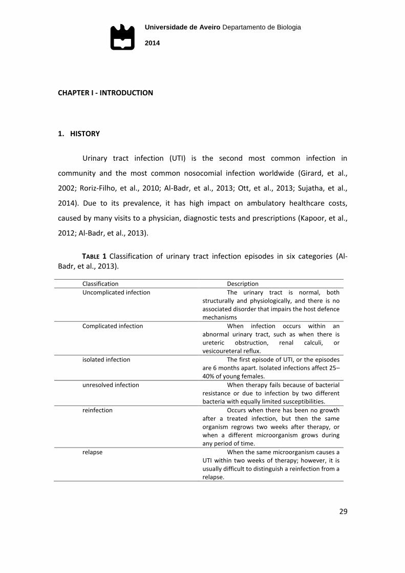
TABLE 1 Classification of urinary tract infection episodes in six categories (Al-Badr, et al., 2013).
Classification Description
Uncomplicated infection The urinary tract is normal, both structurally and physiologically, and there is no associated disorder that impairs the host defence mechanisms
Complicated infection When infection occurs within an abnormal urinary tract, such as when there is ureteric obstruction, renal calculi, or vesicoureteral reflux.
isolated infection The first episode of UTI, or the episodes are 6 months apart. Isolated infections affect 25–40% of young females.
unresolved infection When therapy fails because of bacterial resistance or due to infection by two different bacteria with equally limited susceptibilities.
reinfection Occurs when there has been no growth after a treated infection, but then the same organism regrows two weeks after therapy, or when a different microorganism grows during any period of time.
relapse When the same microorganism causes a UTI within two weeks of therapy; however, it is usually difficult to distinguish a reinfection from a relapse.
Universidade de Aveiro Departamento de Biologia
2014
30
The episode of UTI is classified as lower and upper urinary tract infection
according to where it occurs: urethritis in urethra, cystitis in bladder, bacteriuria in
urine and pyelonephritis in kidney and ureters (Barber, et al., 2013). UTI may involve
either lower and upper urinary tracts, or most often only the lower urinary tract
(Rowe, et al., 2013). It is denominated as uncomplicated urinary tract infection (U-UTI)
when it occurs in a young women non-pregnant with a normal genitourinary tract.
Whereas the complicated urinary tract infection (C-UTI) occurs in a genitourinary tract
with structural or functional abnormalities, including catheterized patients (Roriz-Filho,
et al., 2010).
Typical UTI symptoms are pain, fever, urgency and frequency of micturition,
dysuria (painful micturition), suprapubic cramping pain and sense of weight, turbid or
cloudy urines, sometimes with an unpleasant smell, nocturia and haematuria. Usually
the symptoms of lower urinary tract infection are slight, or absent in case of
asymptomatic bacteriuria (ASB). In women, ASB is defined as the presence of two
consecutive urine specimens positive for the same bacterial strain in quantities equal
or superior to 105 CFU/ mL (Rowe, et al., 2013). In men, the microbiologic criteria for
diagnosis of ASB is not as well rendered valid, some authors define as a single voided
specimen with one bacterial isolate in quantities equal or superior to 104 CFU/mL
(Grabe, et al., 2014).
Ascending infection causing pyelonephritis (upper urinary tract infection),
causes back pain and costovertebral angle tenderness and can also be accompanied
with symptoms of malaise, fever, nausea and vomiting in severe infections. If bacteria
enters the blood stream it may lead to severe complications, including septicaemia,
shock and, rarely, death (Kapoor, et al., 2012; Al-Badr, et al., 2013)
The UTI prevalence is significantly higher in women than men, likely as a result
of anatomic differences, due the proximity of urethra to anus which provides the self-
colonization by gastrointestinal pathogens (Al-Badr, et al., 2013). Also, the shorter
Universidade de Aveiro Departamento de Biologia
2014
31
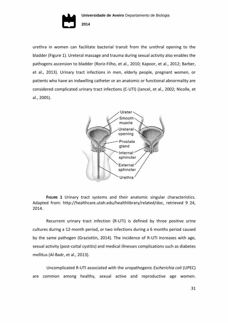
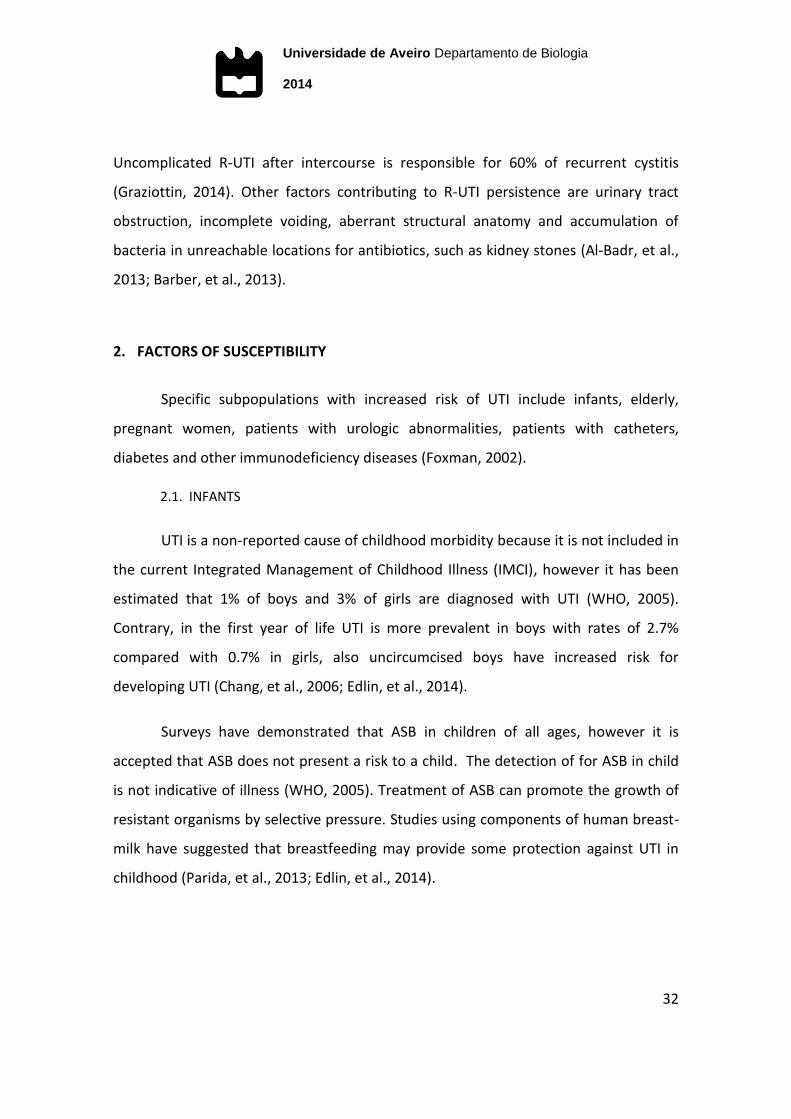
urethra in women can facilitate bacterial transit from the urethral opening to the
bladder (Figure 1). Ureteral massage and trauma during sexual activity also enables the
pathogens ascension to bladder (Roriz-Filho, et al., 2010; Kapoor, et al., 2012; Barber,
et al., 2013). Urinary tract infections in men, elderly people, pregnant women, or
patients who have an indwelling catheter or an anatomic or functional abnormality are
considered complicated urinary tract infections (C-UTI) (Jancel, et al., 2002; Nicolle, et
al., 2005).
FIGURE 1 Urinary tract systems and their anatomic singular characteristics. Adapted from: http://healthcare.utah.edu/healthlibrary/related/doc, retrieved 9 24, 2014.
Recurrent urinary tract infection (R-UTI) is defined by three positive urine
cultures during a 12-month period, or two infections during a 6 months period caused
by the same pathogen (Graziottin, 2014). The incidence of R-UTI increases with age,
sexual activity (post-coital cystitis) and medical illnesses complications such as diabetes
mellitus (Al-Badr, et al., 2013).
Uncomplicated R-UTI associated with the uropathogenic Escherichia coli (UPEC)
are common among healthy, sexual active and reproductive age women.
Universidade de Aveiro Departamento de Biologia
2014
32
Uncomplicated R-UTI after intercourse is responsible for 60% of recurrent cystitis
(Graziottin, 2014). Other factors contributing to R-UTI persistence are urinary tract
obstruction, incomplete voiding, aberrant structural anatomy and accumulation of
bacteria in unreachable locations for antibiotics, such as kidney stones (Al-Badr, et al.,
2013; Barber, et al., 2013).
2. FACTORS OF SUSCEPTIBILITY
Specific subpopulations with increased risk of UTI include infants, elderly,
pregnant women, patients with urologic abnormalities, patients with catheters,
diabetes and other immunodeficiency diseases (Foxman, 2002).
2.1. INFANTS
UTI is a non-reported cause of childhood morbidity because it is not included in
the current Integrated Management of Childhood Illness (IMCI), however it has been
estimated that 1% of boys and 3% of girls are diagnosed with UTI (WHO, 2005).
Contrary, in the first year of life UTI is more prevalent in boys with rates of 2.7%
compared with 0.7% in girls, also uncircumcised boys have increased risk for
developing UTI (Chang, et al., 2006; Edlin, et al., 2014).
Surveys have demonstrated that ASB in children of all ages, however it is
accepted that ASB does not present a risk to a child. The detection of for ASB in child
is not indicative of illness (WHO, 2005). Treatment of ASB can promote the growth of
resistant organisms by selective pressure. Studies using components of human breast-
milk have suggested that breastfeeding may provide some protection against UTI in
childhood (Parida, et al., 2013; Edlin, et al., 2014).
Universidade de Aveiro Departamento de Biologia
2014
33
2.2. ADULTS
The gender vulnerability is clear for UTI, women have 50 times more chances of
acquiring UTI than men over all age groups (Graziottin, 2014). Most U-UTI occurs in
women without any anatomic or functional abnormality and the seriousness varies
from mild to severe. It is estimated that 50-60% of all women after puberty experience
at least one UTI episode during their lifetime and in 8% of UTI episodes the pathogens
may stay silent (Kapoor, et al., 2012; Al-Badr, et al., 2013; Barber, et al., 2013; Rowe, et
al., 2013).
The UTI rates normally increase with the beginning of sexual activity in young
patients, the post-coital symptoms in women last till 6 days on average (Graziottin,
2014). The major cause of U-UTI post-coital is due to the ascension of vaginal bacteria
to the urinary tract (Rowe, et al., 2013). Also post-menopausal women have higher
rates of UTI due to pelvic prolapse, lack of estrogen, loss of lactobacilli in the vaginal
flora (Graziottin, 2014). Additional risk factors include comorbidity associations that
increase UTI susceptibility, however the majority of UTI occurs in healthy women (Al-
Badr, et al., 2013; Barber, Norton, et al., 2013).
The large gender difference in the UTI prevalence is caused by many factors
including: the greater distance between urethra and anus the usual source of
uropathogens; the drier environment surrounding the male urethra; the greater length
of the male urethra; and the antibacterial activity of the prostatic fluid. The UTI in a
healthy adult men between the ages of 15 and 50 years old is very uncommon,
however, it is more common the UTI sepsis evolution in men than in women. The exact
reasons for UTI infections in healthy men are not clear, however some sex-related
behaviours, such as, unprotected intercourse with an infected partner, unprotected
anal intercourse and even the lack of circumcision seem to be associated (Nicolle, et
al., 2005; Roriz-Filho, et al., 2010; Grabe, 2008).
Universidade de Aveiro Departamento de Biologia
2014
34
Usually, UTI in men are generally viewed as complicated because most of UTI
episodes occur in new-born, infant or elderly and they are related to urological
abnormalities, bladder outlet obstruction, instrumentation of genitourinary or several
other comorbidities, such as, diabetes and HIV (Nicolle, et al., 2005; Roriz-Filho, et al.,
2010; SIGN, 2012).
Among men the UTI incidence increases after the age of 50 and it is associated
with the prostatic disease and urinary catheterization (Nicolle, et al., 2005; Roriz-Filho,
et al., 2010). Conditions like prostatitis, chlamydial infection and epididymitis should
be considered in the differential diagnosis of men with acute dysuria (SIGN, 2012).
In 90% of men the appearance of febrile UTI is associated to prostatitis, thus
the urological evaluation should be carried out routinely in men whenever a
complicating factor is suspected. Between 52% and 90% of men with a UTI have been
reported to have prostatic involvement in the infection, which can result in prostatic
abscesses or prostatitis (Grabe, et al., 2008).
2.3. PREGNANCY
Pregnant women in particular are vulnerable to UTI because pregnancy itself is
an immunocompromised state (Kapoor, et al., 2012; Sujatha, et al., 2014). Pregnancy-
UTI incidence elevates up to 20% and frequently causes of premature delivery. It also
increases the risk between 25 to 40% chance of ascending to upper urinary tract
infection, hypertensive disease, anaemia, postpartum complications and foetal
mortality (Foxman, 2002; Kapoor, et al., 2012; Sujatha, et al., 2014)
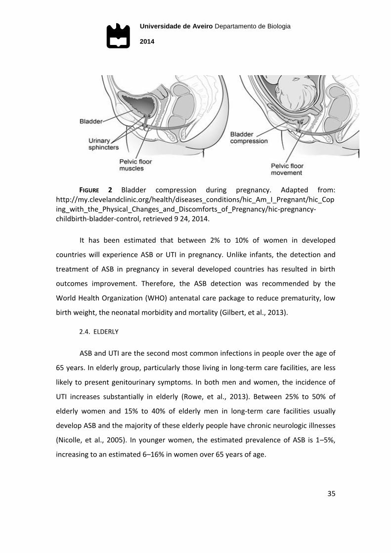
Anatomical factors like the pressure of the enlarged uterus on the bladder, the
physiological hormonal relaxant effect of progesterone on the smooth muscle of the
urinary tract, the vesicoureteral reflux, may predispose to recurrent UTI (Figure 2)
(Kapoor, et al., 2012).
Universidade de Aveiro Departamento de Biologia
2014
35
FIGURE 2 Bladder compression during pregnancy. Adapted from: http://my.clevelandclinic.org/health/diseases_conditions/hic_Am_I_Pregnant/hic_Coping_with_the_Physical_Changes_and_Discomforts_of_Pregnancy/hic-pregnancy-childbirth-bladder-control, retrieved 9 24, 2014.
It has been estimated that between 2% to 10% of women in developed
countries will experience ASB or UTI in pregnancy. Unlike infants, the detection and
treatment of ASB in pregnancy in several developed countries has resulted in birth
outcomes improvement. Therefore, the ASB detection was recommended by the
World Health Organization (WHO) antenatal care package to reduce prematurity, low
birth weight, the neonatal morbidity and mortality (Gilbert, et al., 2013).
2.4. ELDERLY
ASB and UTI are the second most common infections in people over the age of
65 years. In elderly group, particularly those living in long-term care facilities, are less
likely to present genitourinary symptoms. In both men and women, the incidence of
UTI increases substantially in elderly (Rowe, et al., 2013). Between 25% to 50% of
elderly women and 15% to 40% of elderly men in long-term care facilities usually
develop ASB and the majority of these elderly people have chronic neurologic illnesses
(Nicolle, et al., 2005). In younger women, the estimated prevalence of ASB is 1–5%,
increasing to an estimated 6–16% in women over 65 years of age.

Universidade de Aveiro Departamento de Biologia
2014
36
The treatment for ASB is only recommended in elderly prior to transurethral
resection of the prostate or any urologic procedures for which mucosal bleeding is
anticipated. The differentiation of UTI from ASB must be looked at very carefully,
because misclassification may occurs (Rowe, et al., 2013).
Age-associated changes in immune function, exposure to nosocomial
pathogens in case of institutionalized patients and the increasing number of
comorbidities put the elderly at an increased risk for developing infection. Medical
comorbidities, such as stroke and dementia, may predispose individuals to bowel and
bladder incontinence, which have been associated with symptomatic UTI and
persistent ASB (Rowe, et al., 2013).
In older women, some studies suggest an association with menopause, due to
the loss of estrogens and the worsening of constipation with age (Graziottin, 2014). In
older men, prostatic hypertrophy causing obstruction to the normal flow of urine leads
to high post void residual and it has been postulated to be a risk factor for UTI in old-
aged (Rowe, et al., 2013).
2.5. CATHETER-ASSOCIATED UTI
The catheter-associated UTI (CA-UTI) is a very common nosocomial infection
and indwelling urethral catheter is a procedure exceedingly used in health care
facilities, with 17.5% of patients in 66 European hospitals having a catheter (Nicolle,
2014). Indwelling urethral catheter is generally considered to be short term if it is in
situ for less than 30 days and chronic or long term when it is in situ for 30 days or
more, in case of institutionalized adults (Nicolle, 2014).
Catheterization is also a major cause of hospital-acquired UTI, which may be
associated with enhanced nosocomial mortality rates (Parida, et al., 2013). The risk
increases between 3% to 10% per day of catheterization, and at the 30th day of
catheterization the infection rates are about 100%, which is an additional cost per
Universidade de Aveiro Departamento de Biologia
2014
37
admission (Meddings, et al., 2014) (Parida, et al., 2013; Nicolle, 2014). For indwelling
urethral catheter patients, ASB is defined as a positive urinary culture for one bacterial
isolate in quantities ≥102 CFU/mL, in the absence of symptoms (Rowe, et al., 2013).
Prevention of CA-UTI has recently become an important goal of health-care
infection prevention programs. These programs criteria include aseptic insertion of
urinary catheters, minimizing the use and duration of catheters, which has led to a
decrease in the incidence of CA-UTI. In adults who require catheterization, the use of
antimicrobial-coated catheters may delay bacterial colonization and thus decreases
the incidence of CA-UTI (Rowe, et al., 2013; Nicolle, 2014)
Potential risk factors resulting from prolonged catheterization and catheter
insertion outside a protected environment, also put the patient at an increased risk
(Parida, et al., 2013). A recent study about cleaning the urinary catheter prior to the
catheterization showed significant evidences that the use of water or saline solution
reduces rates of UTI and suggests the realization and implementation of a new
guideline to prevent CA-UTI. The aseptic insertion and maintenance of urinary catheter
and system and its correct removal are relevant from the point of view of prevention
and control of UTI (Figure 3). Also guidelines and previous studies indicate that urinary
meatus should be cleaned with water or saline solution (Cunha, et al., 2013). Alternate
voiding management strategies such as intermittent catheterization or/and, external
condom catheters for men, should be used when possible (Nicolle, 2014).

Universidade de Aveiro Departamento de Biologia
2014
38
FIGURE 3 The four stages of the urinary catheter lifecycle to decrease catheter use and CA-UTI. Adapted from (Meddings, et al., 2014).
2.6. DIABETES-ASSOCIATED UTI
Patients with Diabetes mellitus (DM) have a higher risk of ASB, R-UTI and
pyelonephritis (Al-Badr, et al., 2013). DM is a progressive disease that is characterized
by a state of chronic hyperglycaemia. Findings suggest that UTI are more commonly
experienced by those with DM compared to those without DM. The exact mechanism
is unknown, but several possibilities have been proposed to explain the association
between these two diseases. The presence of higher glucose concentrations in the
urine might promote UTI development by amplifying bacterial reproduction and
creating a favourable environment for infections (Fu, et al., 2014). Women are more
prone to ASB than men with diabetes, but in both sexes the progression to clinical
pyelonephritis is more probable than in normal individuals. Women with type 1
diabetes are particularly at risk if they have had diabetes for a long time or have
developed complications, particularly peripheral neuropathy and proteinuria. The risk
factors for developing ASB differ between type 1 and type 2 diabetes, usually the
Universidade de Aveiro Departamento de Biologia
2014
39
patients with type 2 diabetes were old age, proteinuria, a low body mass index and a
past history of R-UTI (Grabe, et al., 2014).
Diabetes-associated UTI (DA-UTI) increases the risk of acute pyelonephritis by
Enterobacteriaceae that usually infects the lower tract, also Klebsiella infection is
particularly common (25% compared with 12% in non-diabetics). Other patient-related
factors such as age, metabolic control and duration of diabetes have also been
suggested as increasing the risk of infection among those with diabetes (Fu, et al.,
2014).
3. DIAGNOSIS
Urine contains an enormous amount of information, despite being a waste
product. Urine bladder culture and microscopic urine sediment analysis have been the
gold standards to urinalysis. Well-standardized procedures are the basis of an effective
diagnostic strategy for urinalysis (Delanghe, et al., 2014; Sujatha, et al., 2014).
The laboratory is responsible for giving the correct information to the patient
about the best sampling procedures, including biological collection, conservation,
storage and specimen transport to the laboratory. Since the patients themselves often
collect the urine specimen, a flawless pre-analytical urinalysis depends on well-
standardized procedures. Therefore, for being a very susceptible procedure, the
requirements of urinalysis have gained importance and have become stricter
(Delanghe, et al., 2014).
3.1. SPECIMEN COLLECTION
To avoid contamination with urogenital flora the collection of urine specimen
should be achieved by cleaning the hands and washing the glans penis of men or the
introitus of women (Demilie, et al., 2014). Due to the influence on the viability of
Universidade de Aveiro Departamento de Biologia
2014
40
bacteria, the use of soap or antiseptics is not recommended. Clean-catch urine or
midstream portions of first morning urine samples (not less than 4 hours storage in the
bladder) should be collected to a sterile urine container in order to avoid
contaminations. The volume should be between 15 ml and 20 ml in a sterile urine
container provided by the laboratory or purchased in a pharmacy. Samples are
transported in the primary containers and stored at 4°C until be processed, the lack of
temperature control can lower the quality of urinary test results (European
Confederation of Laboratory Medicine, 2000).
Catheter-urine specimens for culture should be collected directly from the
catheter or tubing, to maintain a closed drainage system. These may be collected
either through the catheter collection port or through puncture of the tubing with a
needle (Nicolle, 2014).
In case of an incorrect specimen collection, the urine collection must be
repeated. The conservation procedure is also a limiting factor for the over-all
diagnostic accuracy, urine samples must be refrigerated so that a precise urinalysis can
be processed within 24 hours (Delanghe, et al., 2014). A proper pre-analytical
procedure is crucial to urinalysis.
3.2. URINALISYS
The experimental and analytical procedures follow the norms from the Clinical
and Laboratory Standards Institute (CLSI, 2010).
On arrival at the laboratory the samples are triaged and 10-15 mL from the
original sample are transferred into one examination tube to the dipstick analysis in
order to detect nitrite (indicative of microbial activity) and leucocyte esterase
(indicative of pyuria).
Universidade de Aveiro Departamento de Biologia
2014
41
Microscopic analysis of the urine sediment is performed after centrifugation at
1500 rpm for 5 minutes, haematuria (erythrocytes) and pyuria (neutrophils) are
searched, if superior to 10 cells per high power field may be diagnosed with
bacteriuria. This optional analysis contributes to the diagnosis of UTI and it may
suggest abnormalities and vulnerabilities that could lead to a complicated UTI (Barber,
et al., 2013).
Then, gram stain is used to differentiate bacterial species into gram-positive
and gram-negative. Dipstick test, that detects nitrite and leucocyte esterase, is also
used to diagnose UTI (Gieteling, et al., 2014).
The urine culture procedure is performed by vertical immersion with a
disposable plastic loop of 1 µl into the original sterile container and spread horizontally
on the surface of the chromID™ CPS® plates (bioMérieux SA, 2013). Then, plates are
incubated at 37 °C in aerobic atmosphere for 18 hours. After incubation, all the
negative cultures, if growth is inferior to 103 CFU/mL, are excluded.
ChromID™ CPS® is an isolation and identification medium that is used for
urinary samples and enables the microbial count of the sample through the
standardized seeding method. The colonies of E.coli are pink and red; Enterococcus are
turquoise colonies; Klebsiella, Enterobacter, Serratia and Citrobacter are brown-
greenish colonies; Proteus, Providencia and Morganella are dark brown colonies
expressing desaminase.
The microbial criteria in urine culture is defined as a positive result to UTI, If
growth is equal or superior to 105 CFU/mL in mid-stream sample of urine (MSU), with a
maximum of 2 isolated microbial species. It is considered contamination if it has been
observed 3 or more specimens in the culture, or if growth is equal or inferior to 103
CFU/mL (Girard, et al., 2002; Roriz-Filho, et al., 2010). ASB is defined by the presence
of bacteria in urine culture, if growth is inferior to 105 CFU/mL in MSU, without clinical
Universidade de Aveiro Departamento de Biologia
2014
42
symptoms of infection and should not be treated in young non-pregnant women
(Roriz-Filho, et al., 2010). To the subpopulations with increased risk of UTI and
symptomatic should be considered positive culture, if growth is equal or superior to
104 UFC/mL, such as, catheter samples, pregnant women, children and elderly. For
suprapubic aspirated urine should be considered positive culture, if growth is equal or
superior to 102 UFC/mL (Kapoor, et al., 2012).
3.3. VITEK 2 DIAGNOSIS TOOL
VITEK 2 system (bioMérieux) is an accurate and reproducible diagnostic tool
that allows the determination of the exact etiology of patient’s infectious. With great
automation, safety and minimal manual operations provides microbial identification in
vitro on short time compared with conventional methods (Khardori, 2014)
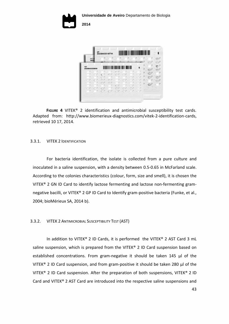
Nowadays Vitek 2 system is directed to clinical laboratories professionals, but it
was originated with the National Aeronautics and Space Administration (NASA) space
program to identify infections in astronauts (bioMérieux SA, 2009). The VITEK 2 system
cards are the size and shape of a playing card and contain 64 microwells (Figure 4).
Each well contains identification substrates or known antimicrobials in different
concentrations in association with mass spectrometry and bioinformatics make the
identification possible (bioMérieux SA, 2014 b; Khardori, 2014).
Universidade de Aveiro Departamento de Biologia
2014
43
FIGURE 4 VITEK® 2 identification and antimicrobial susceptibility test cards. Adapted from: http://www.biomerieux-diagnostics.com/vitek-2-identification-cards, retrieved 10 17, 2014.
3.3.1. VITEK 2 IDENTIFICATION
For bacteria identification, the isolate is collected from a pure culture and
inoculated in a saline suspension, with a density between 0.5-0.65 in McFarland scale.
According to the colonies characteristics (colour, form, size and smell), it is chosen the
VITEK® 2 GN ID Card to identify lactose fermenting and lactose non-fermenting gram-
negative bacilli, or VITEK® 2 GP ID Card to Identify gram-positive bacteria (Funke, et al.,
2004; bioMérieux SA, 2014 b).
3.3.2. VITEK 2 ANTIMICROBIAL SUSCEPTIBILITY TEST (AST)
In addition to VITEK® 2 ID Cards, it is performed the VITEK® 2 AST Card 3 mL
saline suspension, which is prepared from the VITEK® 2 ID Card suspension based on
established concentrations. From gram-negative it should be taken 145 µl of the
VITEK® 2 ID Card suspension, and from gram-positive it should be taken 280 µl of the
VITEK® 2 ID Card suspension. After the preparation of both suspensions, VITEK® 2 ID
Card and VITEK® 2 AST Card are introduced into the respective saline suspensions and
Universidade de Aveiro Departamento de Biologia
2014
44
incubated at 36°C in the VITEK® 2 incubator, the results are available within 10 hours
(bioMérieux SA, 2014 a).
The antimicrobial susceptibility testing (AST) results are validated by the
Advanced Expert System (AES) program, and follow the European Committee on
Antimicrobial Susceptibility Testing (EUCAST) program. The determination of the
phenotypic AST depends on minimal inhibitory concentration (MIC) breakpoints. The
breakpoints allow the bacteria grouping into the categories: susceptible, intermediate,
and resistance. Meanwhile it is also performed the ESBL-confirmation tests
(bioMérieux SA, 2014 a; Stokkou, et al., 2014).
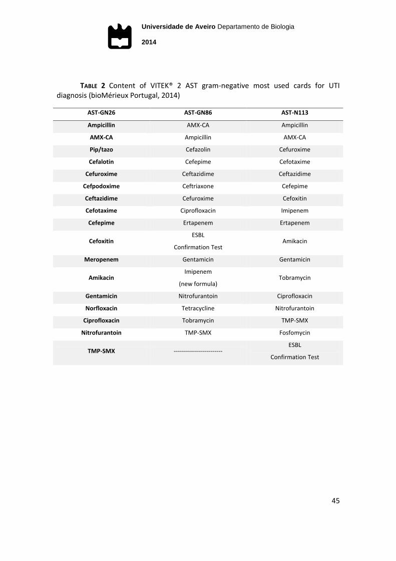
The VITEK® 2 AST Cards most used in Portugal urinalysis are: VITEK® 2 AST-
GN26 (Portuguese card of urines), VITEK® 2 AST-GN86 VITEK® 2 AST-N113 for gram
negative bacteria with resistance to majority of the antibiotics from the VITEK® 2 AST-
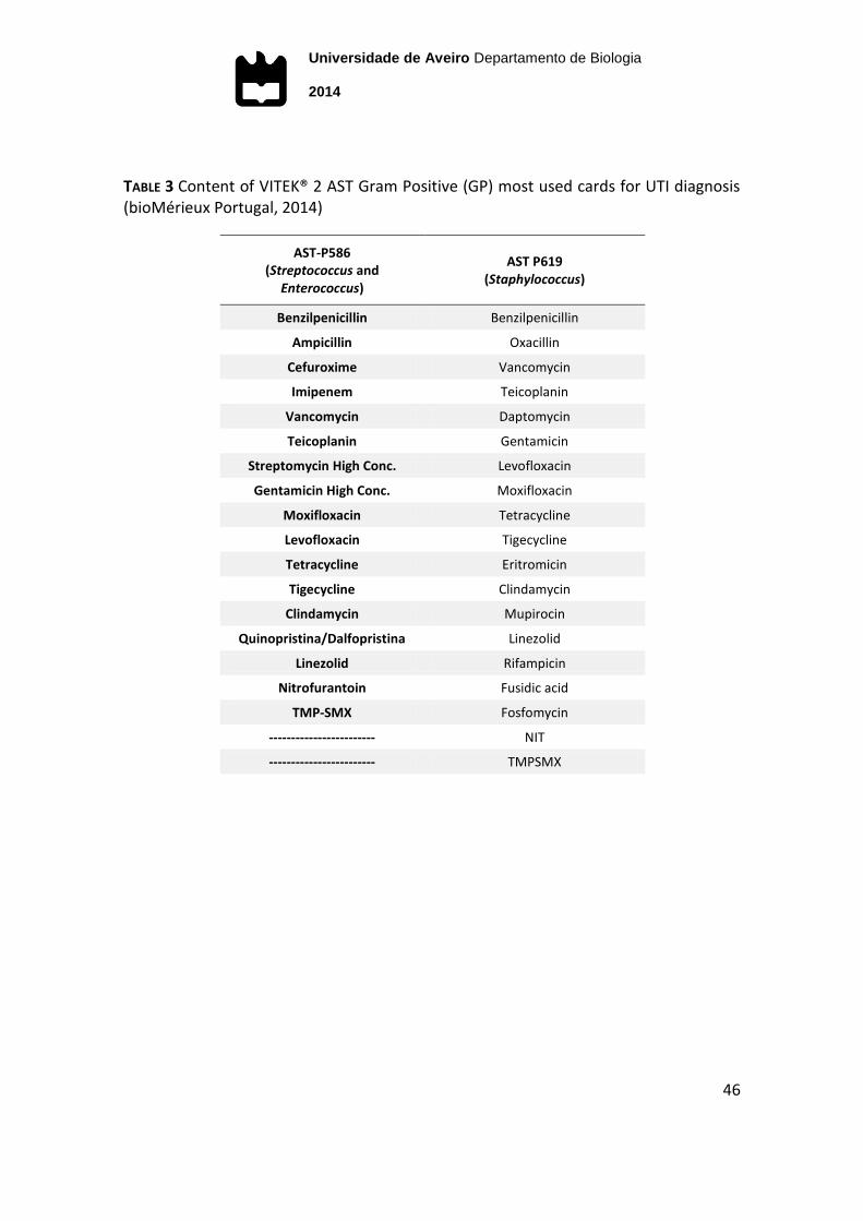
GN26 (Table 2); VITEK® 2 AST-P586 for gram positive Enterococcus and Streptococcus
and VITEK® 2 AST P619 for Gram positive Staphylococcus (Table 3) (bioMérieux
Portugal, 2014).
Universidade de Aveiro Departamento de Biologia
2014
45
TABLE 2 Content of VITEK® 2 AST gram-negative most used cards for UTI diagnosis (bioMérieux Portugal, 2014)
AST-GN26 AST-GN86 AST-N113
Ampicillin AMX-CA Ampicillin
AMX-CA Ampicillin AMX-CA
Pip/tazo Cefazolin Cefuroxime
Cefalotin Cefepime Cefotaxime
Cefuroxime Ceftazidime Ceftazidime
Cefpodoxime Ceftriaxone Cefepime
Ceftazidime Cefuroxime Cefoxitin
Cefotaxime Ciprofloxacin Imipenem
Cefepime Ertapenem Ertapenem
Cefoxitin ESBL
Confirmation Test Amikacin
Meropenem Gentamicin Gentamicin
Amikacin Imipenem
(new formula) Tobramycin
Gentamicin Nitrofurantoin Ciprofloxacin
Norfloxacin Tetracycline Nitrofurantoin
Ciprofloxacin Tobramycin TMP-SMX
Nitrofurantoin TMP-SMX Fosfomycin
TMP-SMX ------------------------ ESBL
Confirmation Test
Universidade de Aveiro Departamento de Biologia
2014
46
TABLE 3 Content of VITEK® 2 AST Gram Positive (GP) most used cards for UTI diagnosis (bioMérieux Portugal, 2014)
AST-P586 (Streptococcus and
Enterococcus)
AST P619 (Staphylococcus)
Benzilpenicillin Benzilpenicillin
Ampicillin Oxacillin
Cefuroxime Vancomycin
Imipenem Teicoplanin
Vancomycin Daptomycin
Teicoplanin Gentamicin
Streptomycin High Conc. Levofloxacin
Gentamicin High Conc. Moxifloxacin
Moxifloxacin Tetracycline
Levofloxacin Tigecycline
Tetracycline Eritromicin
Tigecycline Clindamycin
Clindamycin Mupirocin
Quinopristina/Dalfopristina Linezolid
Linezolid Rifampicin
Nitrofurantoin Fusidic acid
TMP-SMX Fosfomycin
------------------------ NIT
------------------------ TMPSMX
Universidade de Aveiro Departamento de Biologia
2014
47
4. TREATMENT OF UTI
The aim of antimicrobial UTI treatment is the eradication of the current
microbial infection by applying effective antimicrobial therapy. To relief from
recurrence and minimize collateral damages, it is strongly advised to whenever
possible avoid the empirical treatment and perform the antimicrobial susceptibility
testing.
4.1. EMPIRICAL TREATMENT
The early treatment of UTI is crucial to reduce the rate of morbidity, empirical
antimicrobial therapy guided by local resistance rates should be the primary influence
on clinician’s choices (Rodrigues, et al., 2011; Barber, Norton, et al., 2013; Linhares, et
al., 2013).
The WHO Global Strategy recommends that the choice of empirical treatment
should be guided by local or national resistance surveillance data, in the absence of
community regional level surveillance study. Also the local surveillance data should be
used to guide the drugs management, educate prescribers and guide infection control
policies. But unfortunately, there are few publications about the main uropathogens in
the community-acquired UTI and their antimicrobial resistance profile, when
compared with UTI acquired at hospital level (WHO, 2001; Girard, et al., 2002; WHO,
2014)
The Infectious Diseases Society of America (IDSA) Practice Guidelines in
collaboration with European Society for Microbiology and Infectious Diseases
(ESCMID) and European Association of Urology (EAU) recommends as first-line therapy
of U-UTI, fosfomycin due to minimal resistance and propensity for collateral damage
(Gupta, et al., 2011). Ciprofloxacin is suggested as an effective treatment, but among
Universidade de Aveiro Departamento de Biologia
2014
48
men the treatment should be extended to 10-14 days (Roriz-Filho, et al., 2010).
Nitrofurantoin is also an appropriate choice for therapy due to minimal resistance and
secondary effects (Gupta, et al., 2011).
The second-line therapy recommended is fluoroquinolones, which contain 6-
fluoro substituents when compared to quinolones and it is highly efficacious in 3-day
regimens, but have a propensity for collateral damages and resistance, so they should
only be used if sensitivity testing is performed (Rowe, et al., 2013). The β-lactam
agents are appropriate choices for therapy when other recommended agents cannot
be used, because of their inferior efficacy and more adverse effects. Trimethoprim-
Sulfamethoxazole (TMP-SMX) in spite of being widely diffused in the international
guidelines should be based on AST and only prescribed if local resistance rates of
uropathogens do not exceed 20%, due to its high resistance rates in E. coli bacterium
(Linhares, et al., 2013). According to the 1999 guidelines, amoxicillin or ampicillin
should not be used for empirical treatment given the relatively poor efficacy and the
very high prevalence of resistance to these agents worldwide (Gupta, et al., 2011)
In placebo-controlled studies of U-UTI spontaneous clinical cure rate was from
25% to 42%, it was significantly higher compare to symptomatic and bacteriological
cure rates, however the patients treated with antibiotic therapy had a better
prevention of reinfection (Wagenlehner, et al., 2011).
4.2. EXCEPTIONAL TREATMENT
4.2.1. CHILDHOOD RECOMMENDED TREATMENT
Children with U-UTI are likely to respond to amoxicillin, sulphonamides, TMP-
SMX or cephalosporins, due to their good concentration in the lower urinary tract
(WHO, 2005).
Universidade de Aveiro Departamento de Biologia
2014
49
4.2.2. PREGNANCY RECOMMENDED TREATMENT
In pregnant women, treatment of UTI deserves special attention because of the
perinatal risks involved. During pregnancy treatment is mandatory and the AST should
guide the treatment rather than empirical treatment (Calderón-Jaimes, et al., 2013;
Kapoor, et al., 2012).
The first-line antibiotics include all β-lactam agents, nitrofurantoin. Second-line
antibiotics include fosfomycin, trimethoprim (TMP) and the fluoroquinolones, and
should never be considered during pregnancy antibiotics such as sulphonamides,
chloramphenicol and tetracycline (Roriz-Filho, et al., 2010; Kapoor, et al., 2012).
In the first trimester of pregnancy, TMP and fluoroquinolones cannot be
considered because they affect the normal growth and formation of the foetus.
Nitrofurantoin is restricted during the last few weeks due to the risk of haemolytic
anaemia of foetus or neonate (Kapoor, et al., 2012).
4.2.3. DIABETES RECOMMENDED TREATMENT
For DA-UTI patients are recommended two weeks (7-14 days) of oral
antimicrobial therapy and hyperglycaemic control (Mnif, et al., 2014). Fosfomycin,
TMP-SMX and nitrofurantoin are safe and effective antimicrobial methods to cure and
prevent UTI in patients with DM, and fosfomycin is associated with rarely recurrence
of UTI (Ruxer, et al., 2007).
Antibiotic treatment of ASB significantly increases the risk of adverse events
without significant clinical benefit, and also increases resistance (SIGN, 2012). However
ASB is common in women with DM, and if left untreated, it may lead to renal
Universidade de Aveiro Departamento de Biologia
2014
50
functional impairment, so the clinicians must measure the cost-benefit according to
each patient criteria (Grabe, et al., 2014).
4.2.4. ELDERLY RECOMMENDED TREATMENT
TMP-SMX should have the preference for treatment of UTI in old-aged (Rowe,
et al., 2013). Among elderly males it is recommend the prostate examination and
antimicrobial sensibility test (AST) (Roriz-Filho, et al., 2010).
Nitrofurantoin has low resistance rates in E. coli, although other
Enterobacteriaceae species are more common in elderly and may have intrinsic
resistance to nitrofurantoin. In addition, this antimicrobial is contra-indicated in
patients with chronic kidney disease (Rowe, et al., 2013).
5. ETIOLOGY
The literature is unanimous, E. coli is the most common UTI bacterium. This
bacterium is responsible for 75% to 95% of community-acquired infections and for
50%-60% of the hospital-acquired UTI (Roriz-Filho, et al., 2011; Gilbert, et al., 2013;
Landry, et al., 2014; Nicolle, 2014). The uropathogenic Escherichia coli (UPEC) is known
to invade urothelium cells and form quiescent intracellular reservoirs (QIRs). It is
thought that QIRs may provide a source for bacterial persistence and R-UTI (Hickling,
et al., 2013)
Other significant common bacteria that can cause UTI are Staphylococcus
saprophyticus, Staphylococcus epidermidis, also other Enterobacteriaceae, such as
Proteus mirabilis, Klebsiella and Providentia species. Gram-positive organisms, like
methicillin-resistant Staphylococcus aureus and Enterococcus, are less common overall,
but are seen with increasing frequency in healthcare settings and in adults with chronic
Universidade de Aveiro Departamento de Biologia
2014
51
indwelling (Roriz-Filho, et al., 2010; Al-Badr, et al., 2013; Gilbert, et al., 2013; Rowe, et
al., 2013).
For susceptible subpopulations, such as DM patients, the incidence of Klebsiella
spp and Streptococcus sp infections are more common. However Pseudomonas sp
infections are more common in chronically-catheterised patients (Al-Badr, et al., 2013).
In the C-UTI patients and R-UTI are very frequent the appearance of multidrug
resistant E. coli with extended spectrum β-lactamases (ESBL), which difficult the
treatment (Roriz-Filho, et al., 2010). For men, coagulase-negative staphylococci are
also common, in addition to gram-negative bacilli and Enterococcus sp and Proteus
mirabilis (Nicolle, et al., 2005).
6. UROPATHOGEN RESISTANCE TO ANTIMICROBIALS
The emergence of antimicrobial resistance is a growing problem in medicine
worldwide (WHO, 2001). The consumption and misuse of antimicrobials are directly
related to the increased of bacteria resistance, infected patients morbidity and
mortality, and consequently to the rising of health-care costs (Edlin, et al., 2014;
Landry, et al., 2014).
The nosocomial ASB constitutes a major pool of antibiotic-resistant strains of
pathogens (Parida, et al., 2013). Multi-resistant gram-negative bacteria accounted for
a higher number of nosocomial infections than resistant gram-positive bacteria (Ott, et
al., 2013).
In community, in spite of being well known that excessive use of antimicrobials
compromise its efficacy and lead to its resistance, the continuous antibiotic
prophylaxis therapy is usually used as an effective measure to prevent UTI (Kapoor, et
al., 2012).
Universidade de Aveiro Departamento de Biologia
2014
52
E. coli resistance rates have been reported to ampicillin (39-45%), TMP-SMX
(14-31%), nitrofurantoin (1.8-16%) and fluoroquinolones (0.7-10%) (WHO, 2005).
According to WHO in the Global Report on Surveillance, the E.coli is resistant to the 3rd
generation of cephalosporins and fluoroquinolones and also multidrug resistant
Klebsiella spp (Girard, et al., 2002; WHO, 2014). The ciprofloxacin is in continuous
decrease for urine isolates from outpatients (from 90% to 88%) and inpatients (from
85% to 82%) (Landry, et al., 2014). Among C-UTI patients and R-UTI it is very common
the appearance of E. coli with extended spectrum β-lactamases (ESBL), which implies a
difficult treatment with broad-spectrum antibiotic (Roriz-Filho, et al., 2010). Even in
childhood antimicrobial resistance is alarming. Edling and his colleagues reported in a
study about antimicrobial resistance in paediatric urology that in 2009 TMP-SMX
resistance rates for E. coli pediatric UTI increased in both sexes, boys (from 23% up to
31%) and girls (from 20% up to 23%) (Edlin, et al., 2014).
TMP-SMX is widely diffused in international guides, however, they should only
be prescribed after antimicrobial sensibility tests and not as an empirical treatment,
due to its high rates of resistance in E. coli isolates (Roriz-Filho, et al., 2010).
Fluoroquinolones have played an important role in the treatment of infectious disease,
with their wide spectrum of activity, convenient dosing, and good patient tolerability
(Landry, et al., 2014).
6.1. CLASSES AND ANTIMICROBIAL AGENTS
Antibiotics were discovered in the middle of the 19th century and soon after
the discovery of penicillin a number of treatment failures with some bacteria, such as
staphylococci, which were no longer sensitive to penicillin (Byarugaba, et al., 2009).
Over the years, the continued use of various antimicrobial agents lead to the
development of bacteria resistance mechanisms (Giedraitienė, et al., 2011). The
multidrug resistance emergence contributes to a global economic and health-care
Universidade de Aveiro Departamento de Biologia
2014
53
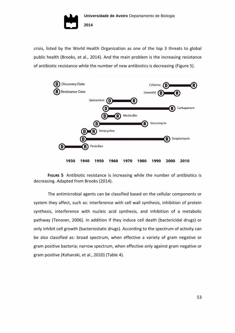
crisis, listed by the World Health Organization as one of the top 3 threats to global
public health (Brooks, et al., 2014). And the main problem is the increasing resistance
of antibiotic resistance while the number of new antibiotics is decreasing (Figure 5).
FIGURE 5 Antibiotic resistance is increasing while the number of antibiotics is decreasing. Adapted from Brooks (2014).
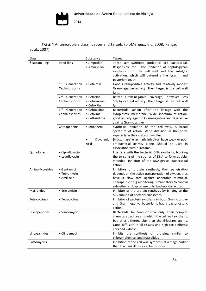
The antimicrobial agents can be classified based on the cellular components or
system they affect, such as: interference with cell wall synthesis, inhibition of protein
synthesis, interference with nucleic acid synthesis, and inhibition of a metabolic
pathway (Tenover, 2006). In addition if they induce cell death (bactericidal drugs) or
only inhibit cell growth (bacteriostatic drugs). According to the spectrum of activity can
be also classified as: broad spectrum, when effective a variety of gram negative or
gram positive bacteria; narrow spectrum, when effective only against gram negative or
gram positive (Kohanski, et al., 2010) (Table 4).
Universidade de Aveiro Departamento de Biologia
2014
54
TABLE 4 Antimicrobials classification and targets (bioMérieux, Inc, 2008; Range, et al., 2007).
Class Substance Target
β-lactam Ring Penicillins • Ampicillin • Amoxicillin
These semi-synthetic antibiotics are bactericidal. Responsible for the inhibition of peptidoglycan synthesis from the cell wall and the autolytic activation, which will determine the lyses and posteriori death.
1st Generation Cephalosporins
• Cefalotin Good Gram-positive activity and relatively modest Gram-negative activity. Their target is the cell wall lysis.
2nd Generation Cephalosporins
• Cefaclor • Cefuroxime • Cefoxitin
Better Gram-negative coverage, however less Staphylococcal activity. Their target is the cell wall lysis.
3rd Generation Cephalosporins
• Cefotaxime • Cefixime • Ceftazidime
Bactericidal action after the linkage with the cytoplasmic membrane. Wide spectrum of action, good activity against Gram-negative and less active against Gram-positive.
Carbapenems • Imipenem Synthesis inhibition of the cell wall. A broad spectrum of action. Wide diffusion in the body, especially in the cerebrospinal fluid.
• Clavulanic Acid
β-lactamase1 enzymatic inhibitor, have weak or poor antibacterial activity alone. Should be used in association with β-lactams.
Quinolones • Ciprofloxacin • Levofloxacin
Interfere with the bacterial DNA synthesis, blocking the twisting of the strands of DNA to form double-stranded, inhibitor of the DNA-girase. Bactericidal action.
Aminoglycosides • Gentamicin • Tobramycin • Amikacin
Inhibitors of protein synthesis, their penetration depends on the active transportation of oxygen, thus have a slow rate against anaerobic microbial. Therapeutic drug monitoring is mandatory to control side effects. Hospital use only, bactericidal action.
Macrolides • Eritromicin Inhibitor of the protein synthesis by binding to the 50S subunit of bacterial ribosomes.
Tetracyclines • Tetracycline Inhibitor of protein synthesis in both Gram-positive and Gram-negative bacteria. It has a bacteriostatic action.
Glycopeptides • Vancomycin Bactericidal for Gram-positive only. Their complex chemical structure also Inhibit the cell wall synthesis, but at a different site than the β-lactam agents. Good diffusion in all tissues and high toxic effects: ears and kidneys.
Lincosamides • Clindamycin Inhibits the synthesis of proteins, similar to chloramphenicol and macrolides.
Fosfomycins Inhibition of the cell wall synthesis at a stage earlier than the penicillins or cephalosporins.
Universidade de Aveiro Departamento de Biologia
2014
55
6.2. MECHANISMS OF RESISTANCE
In order to survive, the bacteria develop mechanisms that enable them to
respond to selective pressure exerted by various environments and competitive
challenges. Antimicrobial resistance is a natural biological phenomenon and is often a
consequence of microbial adaptation to antimicrobials exposure (Byarugaba, et al.,
2009).
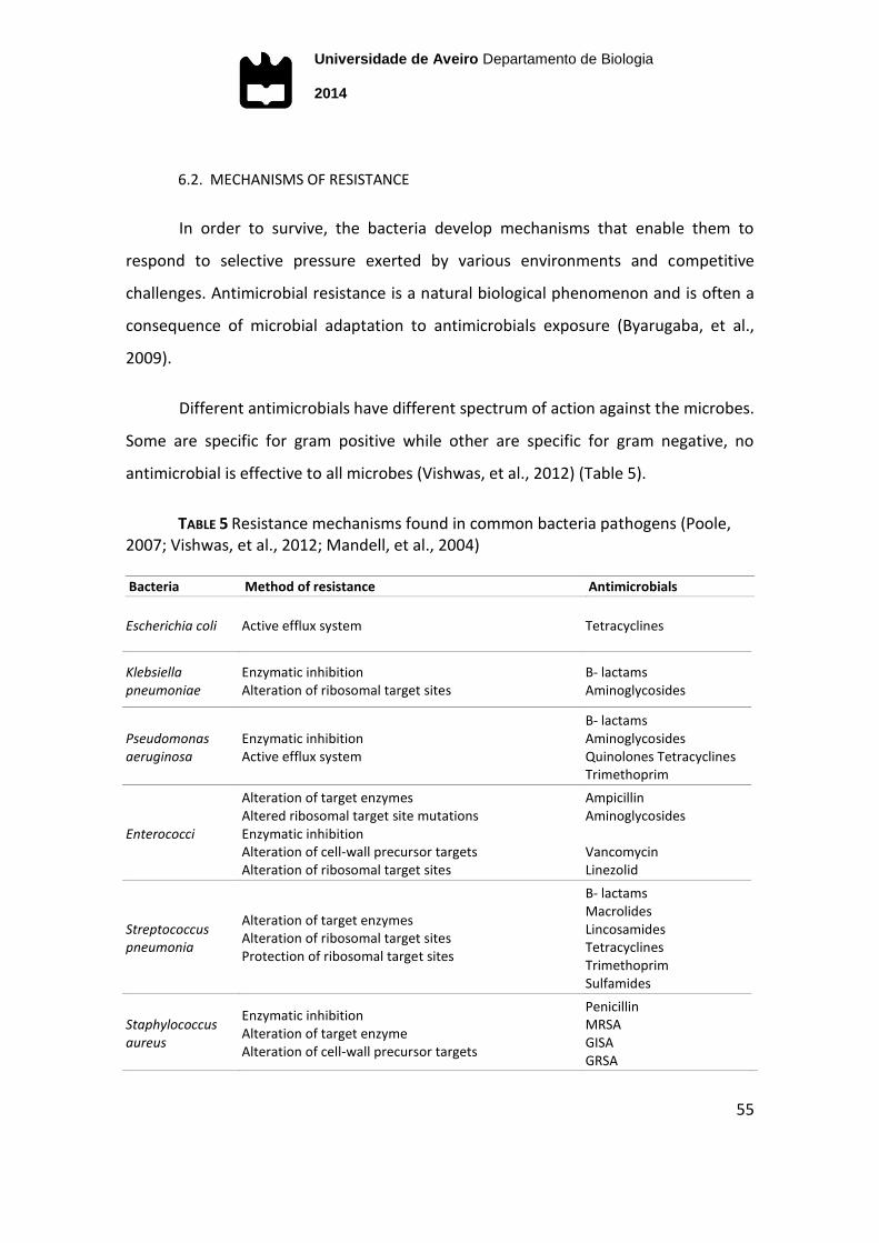
Different antimicrobials have different spectrum of action against the microbes.
Some are specific for gram positive while other are specific for gram negative, no
antimicrobial is effective to all microbes (Vishwas, et al., 2012) (Table 5).
TABLE 5 Resistance mechanisms found in common bacteria pathogens (Poole, 2007; Vishwas, et al., 2012; Mandell, et al., 2004)
Bacteria Method of resistance Antimicrobials
Escherichia coli Active efflux system Tetracyclines
Klebsiella pneumoniae
Enzymatic inhibition Alteration of ribosomal target sites
Β- lactams Aminoglycosides
Pseudomonas aeruginosa
Enzymatic inhibition Active efflux system
Β- lactams Aminoglycosides Quinolones Tetracyclines Trimethoprim
Enterococci
Alteration of target enzymes Altered ribosomal target site mutations Enzymatic inhibition Alteration of cell-wall precursor targets Alteration of ribosomal target sites
Ampicillin Aminoglycosides Vancomycin Linezolid
Streptococcus pneumonia
Alteration of target enzymes Alteration of ribosomal target sites Protection of ribosomal target sites
Β- lactams Macrolides Lincosamides Tetracyclines Trimethoprim Sulfamides
Staphylococcus aureus
Enzymatic inhibition Alteration of target enzyme Alteration of cell-wall precursor targets
Penicillin MRSA GISA GRSA
Universidade de Aveiro Departamento de Biologia
2014
56
Bacteria may be naturally resistant (vertical evolution) by the lack of transport
system for an antibiotic, the lack of the target of the antibiotic molecule, or the cell
wall is covered with an outer membrane that establishes a permeability barrier against
the antibiotic (gram negative).
Or may develop resistance to antibiotics by mutating existing genes (vertical
gene transfer). When exposed to unfavourable environmental conditions the bacteria
enter into a “hypermutable state” model, which the rate of mutations increase from
10 to 50 up to 10 000 times during a prolonged nonlethal selection of microorganisms.
This adaptive process is the only and main source of the antibiotic-resistant mutants to
originate under normal conditions and they are called “adaptive mutations”
(Giedraitienė, et al., 2011; Vishwas, et al., 2012; Brooks, et al., 2014).
The acquiring resistance (horizontal gene transfer) beyond spontaneous
mutation requires either the modification of existing genetic material or the
acquisition of new genetic material from another source, strain or specie. The
horizontal gene transfer occurs by many different mechanisms, mobile genetic
elements including: phages, plasmids and transposons (Giedraitienė, et al., 2011;
Vishwas, et al., 2012).
There are at least three possible mechanisms: transduction, (via
bacteriophages and integrons) occurs when DNA are transferred between two closely
related bacteria; transformation (via plasmids and conjugative transposons) is a
process where parts of DNA are taken up by the bacteria normally present in the
external environment due to the death and lysis of another bacterium; conjugation
(via incorporation of chromosomal DNA, plasmids into a chromosome) occurs when
there is direct cell-cell contact between two bacteria closely related and transfer small
pieces of DNA (Giedraitienė, et al., 2011; Vishwas, et al., 2012).
Universidade de Aveiro Departamento de Biologia
2014
57
The transfer of resistance genes is more effective than chromosomal mutation.
Most plasmids are double-stranded circular DNA whose size may vary from 2–3 kb,
which encode up to 10% of the host cell chromosome and confer resistance to main
classes of antimicrobial agents (cephalosporins, fluoroquinolones, and
aminoglycosides) (Giedraitienė, et al., 2011).
Transposons can be integrated into plasmids or the host’s chromosome,
encompass small elements called insertion sequence elements and transposing
bacteriophages. They have terminal repeat sequences that play a role in
recombination and recognize a protein (for example, transposase or recombinase) that
is necessary to insert or remove a transposon from specific genome regions.
Transposons are transferred by conjugation, transformation, or transduction and
spread quicker than genes in chromosomes. Conjugative transposons have
characteristic features of plasmids and can help to transfer endogenic plasmids from
one microorganism to another (Giedraitienė, et al., 2011).
When resistance determinants are on plasmids, they will spread quickly within
the genus or even unrelated bacterial genera. When resistance is associated with
genes on chromosomes, resistant microorganisms will spread more slowly
(Giedraitienė, et al., 2011). Also, the presence of low levels of the antibiotic in the
environment promotes gene transfer.
The combined effects of fast growth rates to large densities of cells, genetic
processes of mutation and selection, and the ability to exchange genes, account for
the extraordinary rates of adaptation and evolution of bacteria (Vishwas, et al., 2007).
6.3. ANTIBIOTIC STEWARDSHIP
Bacterial resistance is closely associated with the use of antimicrobial agents in
clinical practice. Prolonged therapy with antibiotics may lead to the development of
Universidade de Aveiro Departamento de Biologia
2014
58
resistance in a microorganism that initially is sensitive to antibiotics, but later it can
adapt gradually and develop resistance to antibiotics (Giedraitienė, et al., 2011). Also,
the use in nonhuman niches is another important reason for the spread of resistant
bacteria, such for growth promotion, feed efficiency, and routine disease prevention
purposes in animal agriculture it is a factor of increasing resistance (IDSA, 2011;
Giedraitienė, et al., 2011). For example, Salmonella and Campylobacter acquire
resistance to antibiotics and have transferred antibiotic resistance to natural human
flora. The E. coli resistance to ciprofloxacin is associated with the use of
fluoroquinolones in aviculture (Giedraitienė, et al., 2011).
Some conditions that may promote the acquiring of antimicrobial resistance
that should be avoided, such as:
Exposure to non-lethal levels of antimicrobials;
Exposure to bacteria with acquired resistance genes;
Over- the-counter purchases without medical supervision;
Antimicrobials prescriptions for non-bacterial infections;
Antimicrobials self-medication without medical supervision;
Growth promotion, feed efficiency, and routine disease prevention in
animal agriculture without veterinarian prescription;
Considering the rising of multidrug resistant bacteria, preventative measures
are required (Byarugaba, et al., 2009). To minimize the medication errors in clinical
practice were suggested the 10 tips for safe prescribing (National Prescribing Centre,
2011):
Universidade de Aveiro Departamento de Biologia
2014
59
1. Keep yourself up-to-date in your knowledge of therapeutics, especially
for the conditions you commonly see;
2. Before prescribing, make sure you have all the information you need
about the patient, including co-morbidities and allergies;
3. Before prescribing, make sure you have all the information you need
about the drug(s) you are considering to prescribe, including side effects
and interactions;
4. Sometimes the risks of prescribing outweigh the benefits and before
doing so, think: ‘Do I need to prescribe this drug at all?;
5. Check computerised alerts in case you have missed an important
interaction or drug allergy;
6. Always carefully check prescriptions for errors before signing them;
7. Involve patients in prescribing decisions and give them the information
they need in order to take the medicine as prescribed, to recognise
important side-effects and to know when to return for monitoring;
8. Have systems in place for ensuring that patients receive essential
laboratory test monitoring for the drugs they are taking, and that they
are reviewed at appropriate intervals;
9. Make sure that high levels of safety are built into your repeat
prescribing system;
Universidade de Aveiro Departamento de Biologia
2014
60
10. Make sure you have safe and effective ways of communicating
medicines information between primary and secondary care, and act on
medication changes suggested by secondary care clinicians;
7. PREVENTION
7.1. PREVENTIVE ANTIMICROBIAL MEASURES
Appropriate prescribing is essential to improve patient outcomes and to help
prevent the emergence of resistant organisms. Risk versus benefits should be weighed
carefully before using antibiotics. Poor empiric prescribing practices, lack of urine
testing, and nonselective use of prophylaxis exacerbate this problem. It is also strongly
recommended some practice patterns, such as: urine antimicrobial susceptibility
testing in order to only treat when indicated; high selective application of antibiotic
prophylaxis; local surveillance programs with inpatient versus outpatient data should
be implemented (Edlin, et al., 2014).
The adoption of conservative measures by subpopulations at increased risk is
very important to counter the prevalence of community-acquired UTI. Some hygiene
modifications, such as: limiting of spermicide use; urinating and washing the genitals
pre- and post-coital; wiping from front to back after toilet and every 3-4 hours; shower
instead of immersion bath and avoid the perfumed soaps; acquisition of cotton
underwear instead nylon. These preventive behavioural actions can avoid many
episodes of R-UTI in the long-term (Kapoor, et al., 2012).
7.2. PREVENTIVE NON-ANTIMICROBIAL FORMULATIONS
Prevention strategies for R-UTI in subpopulations with increased risk have been
studied using non-antimicrobial therapies in place of antibiotic prophylaxis, such as:
Universidade de Aveiro Departamento de Biologia
2014
61
estrogen replacement therapy, lactobacilli formulation and cranberry formulations
(SIGN, 2012; Hickling, et al., 2013).
Estrogen is thought to play an important role in maintaining a low vaginal pH in
pre-menopausal women. The low estrogen in postmenopausal women promotes
changes in the vaginal flora and the predominant flora in younger women often
disappear. This leads to an increase in vaginal pH and promotes colonization of
uropathogens (Rowe, et al., 2013). The application of intravaginal estrogen, as topical
vaginal estrogen cream, proved to be effective and safe (Hickling, et al., 2013).
Oral lactobacilli formulation has been tested as a prevention strategy in
postmenopausal women with recurrent UTI. The hypothesis was that oral lactobacilli
may repopulate the vagina with premenopausal vaginal flora, thereby preventing UTI
(Rowe, et al., 2013).
The intake of Cranberry products is a non-antimicrobial approach, which has
been used for prevention and treatment of UTI. Cranberry proanthocyanidin is the
active ingredient in cranberries that inhibits adherence of P-fimbriated E.coli to the
bladder epithelium. In contrast, cranberry formulations interfere with certain
medications and should be taken after medical advice only (SIGN, 2012; Hickling, et al.,
2013; Jepson, et al., 2013; Rowe, et al., 2013).
Other formulations, such as ascorbic acid (vitamin C) intake to acidify urine and
make it hostile for bacterial growth, in vitro data suggest a bacteriostatic effect
(Hickling, et al., 2013). Another example is methenamine salts intake, these salts are
hydrolysed in the urine to form ammonia and formaldehyde. Formaldehyde is widely
bacteriostatic and does not allow the development of bacterial resistance. In a study,
they have compared continuous methenamine hippurate and continuous antimicrobial
prophylaxis in healthy adult women and found no significant difference between
prophylactic trimethoprim and methenamine hippurate (Hickling, et al., 2013).
Universidade de Aveiro Departamento de Biologia
2014
62
Fluid intake, especially water, helps dilute urine and promotes frequently
voiding, which allows bacteria to be flushed from your urinary tract before an infection
can begin (Beetz, 2003). Well as the limitation of the intake of substances that irritate
the bladder epithelium, like caffeine and chocolates may help to prevent UTI (Kapoor,
et al., 2012).
8. RESISTANCE AND FUTURE PERSPECTIVES
The antimicrobial resistance is a global problem that calls for a global response,
and the implementation of a global strategy and preventive guidelines to slow down
the antimicrobial resistance is crucial to its success (WHO, 2014).
The local surveillance implementation is very important to promote effective
empirical treatments. However, the main clinical diagnostic dilemma persists, the
distinguishing of symptomatic UTI from ASB and which patients to treat with
antibiotics (Barber, et al., 2013; Hickling, et al., 2013; Rowe, et al., 2013).
Future studies to evaluate non-antimicrobial formulations in order to stop the
increasing resistance rates remain important to prevention of UTI (Barber, et al., 2013;
Hickling, et al., 2013; Rowe, et al., 2013). Like the implementation of CA-UTI
prevention guidelines, which should be included into an effective program. Including
indications of catheter selection, catheter insertion/maintenance and dates of
insertion and removal should be established. And alternative strategies, such as
substitution of indwelling urethral catheters for external condom catheters in men,
and the staff education in health care facilities are essential to minimize the risk of CA-
UTI (Nicolle, 2014).

Universidade de Aveiro Departamento de Biologia
2014
63
The application of preventive practices and renewed treatment approaches can
reduce significantly the prevalence of UTI, even as the resistance rates worldwide in
the future.
Universidade de Aveiro Departamento de Biologia
2014
64
9. REFERENCES - CHAPTER I
Al-Badr A & Al-Shaikh G (2013). Recurrent Urinary Tract Infections Management in
Women: A review. Sultan Qaboos University Med J, 13(3), 359-367.
Barber A E, Norton J P, Spivak A M & Mulvey, M A (2013). Urinary Tract Infections:
Current and Emerging Management Strategies. Clinical Pratice 57(5), 719-724.
Beetz R (2003). Mild dehydration: a risk factor of urinary tract infection? Eur J Clin Nutr
57(2), 52-58.
bioMérieux Inc (2008). Antibiotic Classification and Modes of Action: Customer
Education. Retrieved 9 18, 2014, from BioMérieux, Inc:
http://www.biomerieux-usa.com/upload/VITEK-Bus-Module-1-Antibiotic-
Classification-and-Modes-of-Action-1.pdf
bioMérieux Portugal (2014). bioMériux Portugal. Retrieved 10 17, 2014, from Lista de
produtos bioMérieux 2014:
http://www.biomerieux.pt/upload/Biomerieux_vers%C3%A3o_3___definitiva
bioMérieux SA (2009). bioMérieux. Retrieved 10 17, 2014, from VITEK 2 Bacterial
Identification Cards, Antibiotic Susceptibility Testing Cards:
http://www.biomerieux-
usa.com/servlet/srt/bio/usa/dynPage?open=USA_PRD_LST&doc=USA_PRD_LST
_G_PRD_USA_4&pubparams.sform=2&lang=en
Universidade de Aveiro Departamento de Biologia
2014
65
bioMérieux SA (2013). chromID to directly identify micro-organisms. Retrieved 10 21,
2014, from bioMérieux: http://www.biomerieux-culturemedia.com/product/9-
chromid-cps-elite
bioMérieux SA (2014 a). VITEK® 2 Advanced Expert System. Retrieved 10 17, 2014,
from bioMérieux: http://www.biomerieux-diagnostics.com/vitek-2-advanced-
expert-system
bioMérieux SA (2014 b). VITEK® 2 Identification Cards. Retrieved 10 17, 2014, from
bioMérieux: http://www.biomerieux-diagnostics.com/vitek-2-identification-
cards
Brooks B D & Brooks A E (2014). Therapeutic strategies to combat antibiotic resistance.
Advanced Drug Delivery Reviews.
Byarugaba D K, Sosa A J, Amabile C & Hsueh PR (2010). Mechanisms of Antimicrobial
Resistance. Antimicrobial Resistance in Developing Countries, XXIII, 15-26.
Calderón-Jaimes E, Casanova-Román G, Galindo-Fraga A, Gutiérrez-Escoto P, Landa-
Juárez S, Moreno-Espinosa S, Rodríguez-Covarrubias F, Simón-Pereira L &
Valdez-Vázquez R (2013). Diagnosis and treatment of urinary tract infections: a
multidisciplinary approach for uncomplicated cases. Bol Med Hosp Infant Mex
70(1), 3-10.
Chang S L & Shortliffe L D (2006). Pediatric Urinary Tract Infections. Pediatr Clin N Am
53, 379– 400.
CLSI (2010). Clinical and Laboratory Standards Institute. Performance standard for
antimicrobial susceptibility. Document M100–S20–U.
Universidade de Aveiro Departamento de Biologia
2014
66
Cunha M, Santos E, Andrade A, Jesus R, Aguiar C, Marques F, Enes F, Santos M,
Fernandes R & Soares S (2013). Effectiveness of cleaning or disinfecting the
urinary meatus before urinary catheterization: a systematic review. Rev Esc
Enferm USP 47(6), 1407-1413.
Delanghe J & Speeckaert M (2014). Preanalytical requirements of urinalysis. Biochem
Med 24(1), 89–104.
Demilie T, Beyene G, Melak S & Tsegaye W (2014). Diagnostic accuracy of rapid urine
dipstick test to predict urinary tract infection among pregnant women in Felege
Hiwot Referral Hospital, Bahir Dar, North West Ethiopia. BMC Research Notes
7(481), 1-5.
Edlin R S, & Copp H L (2014). Antibiotic resistance in pediatric urology. Ther Adv Urol
6(2), 54–61.
European Confederation of Laboratory Medicine. (2000). European Urinalysis
Guidelines 60, 1-96.
Foxman B (2002). Epidemiology of urinary tract infections: incidence, morbidity, and
economic costs. Am J Med 113(1A).
Fu A Z, Iglayemail K, Qiu Y, Engel S, Shankar R & Brodovicz K (2014). Risk
Characterization for Urinary Tract Infections in Subjects With Newly Diagnosed
Type 2 Diabetes. J Diabetes Complications 14, 00189-5.
Funke G & Funke-Kissling P (2004). Evaluation of the New VITEK 2 Card for
Identification of Clinically Relevant Gram-Negative Rods. Journal of Clinical
Microbiology 42(9), 4067–4071.
Universidade de Aveiro Departamento de Biologia
2014
67
Giedraitienė A, Vitkauskienė A, Naginienė3 R & Pavilonis A (2011). Antibiotic
Resistance Mechanisms of Clinically. Important Bacteria Medicina (Kaunas).
47(3), 137-46.
Gieteling E, Leur J V, Stegeman C & Groeneveld P (2014). Accurate and fast diagnostic
algorithm for febrile urinary tract infections in humans. The Netherlands
Journal of Medicine 77(7).
Gilbert N M, O’Brien V P, Hultgren S, Macones G, Lewis W G & Lewis A L (2013).
Urinary Tract Infection as a Preventable Cause of Pregnancy Complications:
Opportunities, Challenges, and a Global Call to Action. Global Advances in
Health and Medicine 2(5), 59-69.
Girard R, Perraud M, Prüss A, Savey A, Tikhomirov E, Thuriaux M, & Vanhems P (2002).
Prevention of hospital-acquired infections: A practical guide. Retrieved 9 18,
2014, from World Health Organization:
http://www.who.int/csr/resources/publications/whocdscsreph200212.pdf
Grabe M M J (2008). The Management of Male Urinary and Genital Tract Infections.
Retrieved 10 22, 2014 from European Association of Urology:
http://www.uroweb.org/fileadmin/user_upload/Guidelines/The%20Managem
ent%20of%20Male%20Urinary%20and%20Genital%20Tract%20Infections.pdf
Grabe M, Bjerklund-Johansen T E, Botto H, Çek M, Naber K G, Pickard R S, Tenke P,
Wagenlehner F & Wullt B (2014). Guidelines on Urological Infections. Retrieved
10 20, 2014, from European Association of Urology (EAU):
http://www.uroweb.org/guidelines/online-guidelines/
Universidade de Aveiro Departamento de Biologia
2014
68
Graziottin A (2014). Recurrent cystitis after intercourse: why the gynaecologist has a
say. TreeLife Media 2, 319-336.
Gupta K, Hooton T M, Naber K G, Wullt B, Colgan R, Miller L G, Moran G J, Nicolle L E,
Raz R, Schaeffer A J & Soper D E (2011). International Clinical Practice
Guidelines for the Treatment of Acute Uncomplicated Cystitis and
Pyelonephritis in Women: A 2010 Update by the Infectious Diseases Society of
America and the European Society for Microbiology and Infectious Diseases.
Clinical Practice Guidelines 52, e103-e120.
Hickling D R & Nitti V W (2013). Management of Recurrent Urinary Tract Infections in
Healthy Adult Women. Reviews in Urology 15(6), 41-48.
IDSA (2011). Combating Antimicrobial Resistance: Policy Recommendations to Save
Lives. Retrieved 11 9, 2014, from Infectious Diseases Society of America:
http://www.idsociety.org/uploadedFiles/IDSA/Policy_and_Advocacy/Current_T
opics_and_Issues/Advancing_Product_Research_and_Development/Vaccines/S
tatements/Combating%20Antimicrobial%20Resistance-
Policy%20Recommendations%20to%20Save%20Lives.pdf
Jancel T & Dudas V (2002). Management of uncomplicated urinary tract infections.
West J Med 176, 51-55.
Jepson R G, Williams G & Craig J C (2013). Cranberries for preventing urinary tract
infections. Sao Paulo Med J. 131(5), 363.
Kapoor K G & Sinha T (2012). Section 4: Renal Diseases in Pregnancy. In A. DeCherney,
L. Nathan, T. M. Goodwin, N. Laufer, & A. Roman, Treatment and Prognosis in
Obstetrics & Gynecology (pp. 137-143). McGraw-Hill Medical.
Universidade de Aveiro Departamento de Biologia
2014
69
Khardori N (2014). Future of diagnostic microbiology. Indian J Med Microbiol 32(4),
371-377.
Kohanski M A, Dwyer D J & Collins J J (2010). How antibiotics kill bacteria: from targets
to networks. Nat Rev Microbiol. 8(6), 423–435.
Landry E, Sulz L, Bell A, Rathgeber L & Balogh H (2014). Urinary Tract Infections:
Leading Initiatives in Selecting Empiric Outpatient Treatment (UTILISE). Can J
Hosp Pharm 67(2), 116-125.
Linhares I, Raposo T, Rodrigues A & Almeida A (2013). Frequency and antimicrobial
resistance patterns implicated in community urinary tract infections: a ten-year
surveillance study (2000–2009). BCM Infectious Diseases 13(19).
Mandell G, Owen R, Bennet J, Dolin R & Martin J (2004). Principles and practice of
infections disease online. Retrieved 11 9, 2014, from Elsevier
https://books.google.pt/books?id=73pYBAAAQBAJ&pg=PA244&lpg=PA244&dq
=Tetracycline+Active+efflux+from+the+cell+E.+coli&source=bl&ots=UYctcBUvq
9&sig=kFMM7EkSoTWW8nK90nf6DDHeldI&hl=pt-
PT&sa=X&ei=i3WHVOnuMcy0UczdgcgB&ved=0CFQQ6AEwBg#v=onepage&q&f
=false
Meddings J, Rogers M A, Krein S L, Fakih M G, Olmsted R N & Saint S (2014). Reducing
unnecessary urinary catheter use and other strategies to prevent catheter-
associated urinary tract infection. BMJ Qual Saf 23, 277-289.
Mnif M F, Kamoun M, Kacem F H, Bouaziz Z, Charfi N, Mnif F, Naceur B B, Rekik N &
Abid M (2014). Complicated urinary tract infections associated with diabetes
Universidade de Aveiro Departamento de Biologia
2014
70
mellitus: Pathogenesis, diagnosis and management. Indian Journal of
Endocrinology and Metabolism 17(3), 442-445.
Nacional Prescribing Centre (2011). 10 Top Tips for GPs – Strategies for safer
prescribing. National Institute for Health & Clinical Excellence, 1-16. Retrieved
12 14, 9 from National Prescribing Centre
http://www.npc.nhs.uk/evidence/resources/10_top_tips_for_gps.pdf.
Nicolle L E (2014). Catheter associated urinary tract infections. Antimicrobial
Resistance and Infection Control 3(23), 1-8.
Nicolle L E, Bradley S, Colgan R, Rice J C, Schaeffer A & Hooton T M (2005). Infectious
Diseases Society of America Guidelines for the Diagnosis and Treatment of
Asymptomatic Bacteriuria in Adults. Clinical Infectious Diseases 40, 643–654.
Ott E, Saathoff S, Graf K, Schwab F & Chaberny I F (2013). The Prevalence of
Nosocomial and Community Acquired Infections in a University Hospital.
Deutsches Ärzteblatt International 110(31–32), 533−540.
Parida S & Mishra S K (2013). Urinary tract infections in the critical care unit: A brief
review. Indian Journal of Critical Care Medicine November-December 2013
17(6), 370-374.
Poole K (2007). Efflux pumps as antimicrobial resistance mechanisms. Annals of
Medicine. 39, 162–176
Rang H P, Dale M M (2007). Antibióticos. Farmacologia. Elsevier: tradução da 5a edição
Americana. 9, 1-2.
Universidade de Aveiro Departamento de Biologia
2014
71
Rodrigues F J & Barroso A P (2011). Etiologia e sensibilidade bacteriana em infecções
do tracto urinário. Rev Port Saúde Pública 123-131.
Roriz-Filho, J S, Vilar F C, Mota L M, Leal C L & Pisi P C (2010). Infecção do trato
urinário. Condutas em enfermaria de clínica médica de hospital de média
complexidade. Medicina 118-125. Ribeirão Preto: Medicina.
Rowe T A & Juthani-Mehta M (2013). Urinary tract infection in older adults. Nacional
Institute of Health 9(5), 1-15.
Ruxer J, Mozdzan M, Loba J & Markuszewski L (2007). Fosfomycin, co-trimoxazole and
nitrofurantoin in the treatment of recurrent uncomplicated urinary tract
infections in type 2 diabetes mellitus. Wiad Lek 60(5-6), 235-240.
SIGN (2012). Management of suspected bacterial urinary tract infection in adults.
Retrieved 10 27, 2014, from Scottish Intercollegiate Guidelines Network:
http://www.sign.ac.uk/guidelines/fulltext/88/
Stokkou S, Tammer I, Zibolka S, Grabau C & Geginat G (2014). Impact of minimal
inhibitory concentration breakpoints on local cumulative bacterial susceptibility
data and antibiotic consumption. BMC Research Notes 7(603), 1-7.
Sujatha R & Nawani M (2014). Prevalence of Asymptomatic Bacteriuria and its
Antibacterial Susceptibility Pattern Among Pregnant Women Attending the
Antenatal Clinic at Kanpur, India. Journal of Clinical and Diagnostic Research 8,
DC01-DC03.
Tenover F C (2006). Mechanisms of Antimicrobial Resistance in Bacteria. The American
Journal of Medicine, 119 (6A), S3–S10.

Universidade de Aveiro Departamento de Biologia
2014
72
Vishwas T D & Kayalvizhi G (2012). Antimicrobial resistance: an overview. Jident 1(1),
11-15.
Wagenlehner F M, Hoyme U, Kaase M, Fünfstück R, Naber K G & Schmiemann G
(2011). Uncomplicated Urinary Tract Infections. Deutsches Ärzteblatt
International 108(24), 415–423.
WHO (2001). Global Strategy for Containment of Antimicrobial Resistance. Retrieved 9
19, 2014, from World Health Organization:
http://www.who.int/drugresistance/WHO_Global_Strategy_English.pdf
WHO (2005). Urinary Tract Infections in Infants and Children in Developing Countries in
the Context of IMCI. Retrieved 9 22, 2014, from World Health Organization:
http://whqlibdoc.who.int/hq/2005/WHO_FCH_CAH_05.11.pdf?ua=1
WHO (2014). Antimicrobial Resistance: Global Report on Surveillance. Retrieved 9 19,
2014, from World Health Organization:
http://apps.who.int/iris/bitstream/10665/112647/1/WHO_HSE_PED_AIP_2014
.2_eng.pdf?ua=1
Universidade de Aveiro Departamento de Biologia
2014
73
CHAPTER II - FREQUENCY AND ANTIBIOTIC RESISTANCE OF BACTERIA IMPLICATED IN
COMMUNITY URINARY TRACT INFECTIONS IN NORTH AVEIRO BETWEEN 2011 AND
2014
Universidade de Aveiro Departamento de Biologia
2014
74
Universidade de Aveiro Departamento de Biologia
2014
75
FREQUENCY AND ANTIBIOTIC RESISTANCE OF BACTERIA IMPLICATED IN
COMMUNITY URINARY TRACT INFECTIONS IN NORTH AVEIRO BETWEEN 2011 AND
2014.
Tânia Costa1,2, Ricardo Ferreira2, Adelaide Almeida1*, Inês Linhares1, Jasmin Neves2
1Department of Biology and CESAM, University of Aveiro, 3810-193 Aveiro, Portugal 2Centro Médico da Praça Lda., Rua do Vale do Vouga 1492, 3700-298 São João da Madeira, Portugal. *Corresponding author
1. ABSTRACT
Urinary tract infection (UTI) is the second most common infection in
community and the most common nosocomial infection worldwide. The main goal of
this study is to evaluate the predominance of uropathogens responsible for urinary
tract infection and determine their resistance patterns, in order to assess if the
recommended empirical treatment is appropriated for this population. Samples were
collected at Centro Médico da Praça Lda on ambulatory system, in São João da
Madeira municipality (Aveiro, Portugal) from June 2011 to June 2014. From 4270
analysed urine samples, 3561 (83%) were from women and only 709 (17%) were from
men, from 0 to 104 years of age. E. coli was the most frequent uropathogen, followed
by Klebsiella spp and Enterococcus spp and P. mirabilis. From all data 37% were MDR
isolates and from these 30.9% were from women and 61.6% were from men. In
general men were more resistant to antimicrobials than women. From first line drugs
recommended by EAU to empirical treatment of uncomplicated UTI only
nitrofurantoin is appropriated for both sexes and ciprofloxacin may be only considered
to treat women. In the second line antibiotics, ampicillin is not an appropriate drug to
empirical treatment of uncomplicated UTI, however the antimicrobials AMX-CA and
TMP-SMX should not be used to empirical treatment of men, due the local high

Universidade de Aveiro Departamento de Biologia
2014
76
incidence of resistance, over 20% for men. Thus, it is suggested the use of imipenem
and gentamicin as an alternative to treat both sexes.
2. INTRODUCTION
Urinary tract infection (UTI) is the second most common infection in
community and the most common nosocomial infection worldwide (Al-Badr, et al.,
2013; Ott, et al., 2013; Sujatha, et al., 2014). The episode of UTI is classified as lower
(bladder, urethra and prostate) or upper (Kidneys) urinary tract infection considering
the affected organs. Uncomplicated urinary tract infection (U-UTI) occurs in non-
pregnant young women with a normal genitourinary tract. Complicated urinary tract
infection (C-UTI) was diagnosed in genitourinary tract with structural, functional
abnormalities or developed in healthcare settings (including catheterized patients)
(Roriz-Filho, et al., 2010). Recurrent urinary tract infection (R-UTI) results from three
positive urine cultures during a 12-month period, or two infections during the previous
6 months and it is mainly caused by reinfection of the same pathogen (Graziottin,
2014). Symptoms are usually slight in U-UTI or absent in case of asymptomatic
bacteriuria (ASB). The diagnosis of infection is defined as the presence of two
consecutive positive urine specimens with the same bacterial strain in quantities equal
or superior to 105 CFU mL-1 (Rowe, et al., 2013).
Community-acquired urinary tract infections are mainly uncomplicated,
colonizing preferably the bladder causing cystitis (Stamm, 2006; Wiles, Kulesus, et al.,
2008). However, some bacteria may ascend through the ureters to the kidneys and
cause more severe infections such as pyelonephritis (Stamm, 2006; Wiles, et al., 2008).
Moreover, specific subpopulations are more likely to have UTI, such as, patients with
catheters, urologic abnormalities, diabetics, immunodeficiency, infants, pregnant
women and elderly, to these patients UTI can be more severe (Foxman, 2002).

Universidade de Aveiro Departamento de Biologia
2014
77
Undesirably, there is little knowledge about the main uropathogens and their
antimicrobial resistance pattern implicated in community-acquired UTI compared with
UTI acquired at hospital level. The importance of this information is the periodic
monitoring of the changes over the years, in order to decrease the number of
therapeutic failures (Linhares, et al., 2013).
The few studies carried out in the community have shown that uropathogens,
such as, Escherichia coli (46.4 - 74.2%), Klebsiella spp (6.0 - 13.45%), Proteus spp (4.7 -
11.9%) and Enterococcus spp (5.3 - 9.54%) are the most prevalent in UTI (Neto, et al.,
2003; Akram, et al., 2007; Correia, et al., 2007; Francesco, et al., 2007; Laupland, et al.,
2007; Pires, et al., 2007; Mendo, et al., 2008; Costa, et al., 2009; Rahman, et al., 2009;
Martins, et al., 2010; Nimri, et al., 2010). E. coli has been indicated as the most
frequent uropathogen involved in the community-acquired UTI (Francesco, et al.,
2007; Laupland, et al., 2007; Mendo, et al., 2008; Costa, et al., 2009) probably because
it belongs to the normal flora of the human intestine and therefore easily colonize the
urinary tract.
According to the European Association of Urology (EAU) the empirical first line
treatment recommended to acute U-UTI is ciprofloxacin, fosfomycin and
nitrofurantoin, followed by ampicillin, amoxicillin-clavulanic acid (AMX-CA) and
trimethoprim-sulfamethoxazole (TMP-SMX) as second line treatment. The European
Society for Microbiology and Infectious Diseases (ESCMID) recommends as first line
therapy for acute U-UTI, fosfomycin due to minimal resistance and collateral damage
(Gupta, et al., 2011). Ciprofloxacin is a suggested as an effective 3-day regiment, while
men treatment should be extended to 10-14 days (Roriz-Filho, et al., 2010).
Nitrofurantoin is also an appropriate choice for therapy due to minimal resistance and
collateral damages (Gupta, et al., 2011).
The second line therapy recommended the class of β-lactam agents when first
line recommended agents cannot be used, due to their inferior efficacy and more
Universidade de Aveiro Departamento de Biologia
2014
78
adverse effects. TMP-SMX in spite of being widely diffused in the international
guidelines must be based on AST and it should only be prescribed if local resistance
rates of uropathogens do not exceed 20% (Grabe, et al., 2014). According to the 1999
Uncomplicated Urinary Tract Infection Guidelines by the IDSA, AMX or AMP should not
be used for empirical treatment given the relatively poor efficacy and the very high
prevalence of resistance to these agents worldwide (Gupta, et al., 2011).
The high rates of resistance among pathogenic bacteria suggest the failure of
the past preventive guidelines to slow down the antimicrobial resistance (WHO, 2001;
Francesco, et al., 2007; Koningstein, et al., 2013; Linhares, et al., 2013, WHO, 2014).
The empirical treatment, used in an early disease phase to decrease the rate of
morbidity, should be guided by national resistance surveillance data, in the absence of
surveillance community studies at regional level (WHO, 2001; Linhares, et al., 2013;
WHO, 2014). The surveillance should be continuous, the bacterial resistance patterns
change over the time. In order to administer an appropriate empirical therapy it is
crucial to know not only the main bacteria usually involved in the UTI but also their
respective antibiotic resistance patterns. This surveillance procedure controls the
increase of antimicrobial resistance and the spread of resistant bacterial strains that
represent a public health problem worldwide.
The goal of this study is to evaluate the predominance of uropathogens
responsible for urinary tract infection and their resistance patterns, in North Aveiro
(Portugal) between 2011 and 2014 in order to assess if the empirical recommended
treatment of UTI is appropriate for this population.
Universidade de Aveiro Departamento de Biologia
2014
79
3. METHODS
3.1. DATA SAMPLES
All the samples were collected in the ambulatory system from June 2011 to
June 2014 at Centro Médico da Praça Lda, located in São João da Madeira municipality,
district of Aveiro in the north-western region of Portugal. Positive UTI samples at the
reception were registered with code and associated to the patient’s process number.
To build the database was registered the gender, age, bacterial strain and the
corresponding antimicrobial susceptibility testing (AST) from the VITEK© 2
(bioMérieux SA) report. This study was approved by the clinical analysis laboratory
Centro Médico da Praça Lda.
3.2. SAMPLING
First morning urine samples (not less than 4 hours storage in bladder) were
collected by the patients after the daily hygiene at home. The first and final urine was
rejected, only the midstream portion was collected to a sterile urine container to avoid
contaminations. Samples were transported in their primary containers and stored at
4°C until they can be processed (European Confederation of Laboratory Medicine,
2000).
3.3. URINALYSIS
In the laboratory, samples were triaged and 10-15 mL from the sterile
containers were transferred into examination tubes to the dipstick analysis, to nitrite
and leucocyte esterase detection. Then the examination tubes were centrifuged at
1500 rpm for 5 minutes to the microscopic analysis.
One microliter of urine sample was inoculated with a disposable plastic loop on
CLED media or chromID™ CPS® plates (bioMérieux) and incubated at 37 °C in aerobic
atmosphere for 18 hours. These culture media allow direct presumptive identification
Universidade de Aveiro Departamento de Biologia
2014
80
of E. coli, Proteus spp and Enterococcus spp having in consideration the sizes, colours
and shapes of the colony-forming units (CFU) (bioMérieux SA, 2013). After incubation,
all the negatives cultures, bacteria growth inferior to 103 CFU/mL, were excluded.
Urine culture was defined as positive if bacterial growth was equal or superior to 105
CFU/mL, with a maximum of 2 isolated species. Samples were considered
contaminated if 3 or more different colonies grown in the culture medium, these
samples were classified as “polymorph flora” and a second sample was requested from
the patient. Bacteriuria was defined when bacterial growth was inferior to 105 CFU/mL
and, in these cases, it was only preformed the identification (ID) and antimicrobial
sensibility test (AST) for pregnant women or children.
3.4. IDENTIFICATION OF BACTERIAL ISOLATES
Identification was performed by the VITEK© 2 ID card, after a gram stain to
differentiate bacterial species into gram-positive or gram-negative. The isolates were
inoculated in a saline suspension, with a density between 0.5-0.65 in McFarland scale.
According to the colonies characteristics, it was chosen the VITEK® 2 GN ID Card to
identify lactose fermenting and non-fermenting gram-negative bacilli, or VITEK® 2 GP
ID Card to identify gram-positive bacteria (bioMérieux SA, 2014 b). The VITEK® 2 ID
cards were introduced into the saline suspension and incubated at 36 °C in the VITEK®
incubator, and the results were available within 10 hours (Funke, et al., 2004;
bioMérieux SA, 2014 b).
3.5. ANTIMICROBIAL SUSCEPTIBILITY TEST (AST)
The AST was performed with VITEK® 2 AST Card. The VITEK® 2 AST-GN26
(Portuguese card for urine samples) and VITEK® 2 AST-N113 to gram negative bacteria
with resistance to the majority of the antibiotics from the VITEK® 2 AST-GN26; VITEK®
2 AST-P586 to gram positive Enterococcus spp and Streptococcus spp and VITEK® 2 AST
P619 to Gram positive Staphylococcus spp (bioMérieux Portugal, 2014) were used.
Universidade de Aveiro Departamento de Biologia
2014
81
Three millilitres of a commercial saline (0.45%) liquid medium, recommend for
procedures that require the use of a diluent with a VITEK® system, were prepared for
each respective ID card suspension, based on the established concentrations (Thermo
Fisher Scientific Inc., 2008; Thermo Fisher Scientific Inc., 2009). From gram-negative
(GN) suspensions 147 µl of the VITEK® ID suspension were taken and from gram-
positive (GP) 270 µl of the VITEK® ID suspension were used. Both suspensions, ID cards
and AST cards were introduced into respective saline suspensions and incubated at 36
°C in the VITEK® incubator for 10 hours (Funke, et al., 2004; bioMérieux SA, 2014 a).
The AST results were validated by advanced expert system (AES) program,
which follows the European committee on antimicrobial susceptibility testing
(EUCAST). The phenotypic antimicrobial susceptibility testing determination depends
on the minimal inhibitory concentration (MIC) breakpoints. Based on breakpoints,
bacteria were grouped into three categories: susceptible, intermediate and resistance.
Meanwhile, the ESBL-confirmation test was also performed (bioMérieux SA, 2014 a;
Stokkou, et al., 2014)
3.6. STATISTICAL ANALYSIS
The data base was done using the Microsoft Excel program and treated using
the Statistical Package for the Social Sciences (SPSS) 16.0 for Windows. The normality,
homogeneity and independence of the variants were checked before analysis. Since
the data failed the normality assumptions, non-parametric methods were used to
detect significant differences. Mann–Whitney U test was used to evaluate differences
between genders. The Kruskal-Wallis test was used to evaluate the age and study
period influence in the UTI incidence. The Chi-square test was used to evaluate the
incidence of isolates into the age groups. The significant level established was p>0.05.
The main bacteria responsible for UTI, such as, E.coli, Enterococcus spp, Klebsiella spp,
P. mirabilis, P. aeruginosa, Staphylococcus spp and S. agalactiae were identified as
responsible for at least 1% of the study infections (representing 93.5% of the total
Universidade de Aveiro Departamento de Biologia
2014
82
urinary tract infections). The remaining bacteria were grouped and classified as “other
bacteria” (representing 6.5% of the total infections) and were included the
uropathogens: Acinetobacter baumannii, Burkholderia cepacia, Citrobacter spp,
Enterobacter spp, Hafnia alvei, Morganella spp, Proteus spp, Providencia spp,
Pseudomonas spp, Raoultella spp, Salmonella enteretidis, Serratia spp, Shigella spp,
Sphingomonas paucimobilis, Yersinia enterocolitica. The resistant uropathogens to
three or more antimicrobial classes were considered multi-drug resistant (MDR).
4. RESULTS
4.1. CHARACTERIZATION OF THE UTI SAMPLES
From 4270 analysed urine samples, 3561 (83%) were from women and only 709
(17%) were from men. The age range varied from 0 to 104 with an average of 61 years
of age. The average age for women was 59 years of age and for men 68 years of age.
The incidence of UTI increased with the age (Chi-square, p<0.05), being the elderly (≥
65) age group the most affected with 2219 (52%) urine samples (78,1% corresponding
to women and 21,9% corresponding to men), followed by adults (35-64) with 1381
(32.3%) samples (87% corresponding to women and 13% corresponding to men),
young adults with 516 (12.1%) samples (97,9% corresponding to women and 2,1%
corresponding to men), adolescents (13-18) with 53 (1.2%) samples (94,3%
corresponding to women and 5,7% corresponding to men), and the last age group,
children (0-12) with 101 (2.4%) samples (70,3% corresponding to women and 29,7%
corresponding to men) (Figure 1a). However, between children and the adolescent
groups a 1% decrease in women was observed, the decreasing tendency was also
observed between children and the young adults groups (2% to 0%, respectively) in
men (Figure 1a).
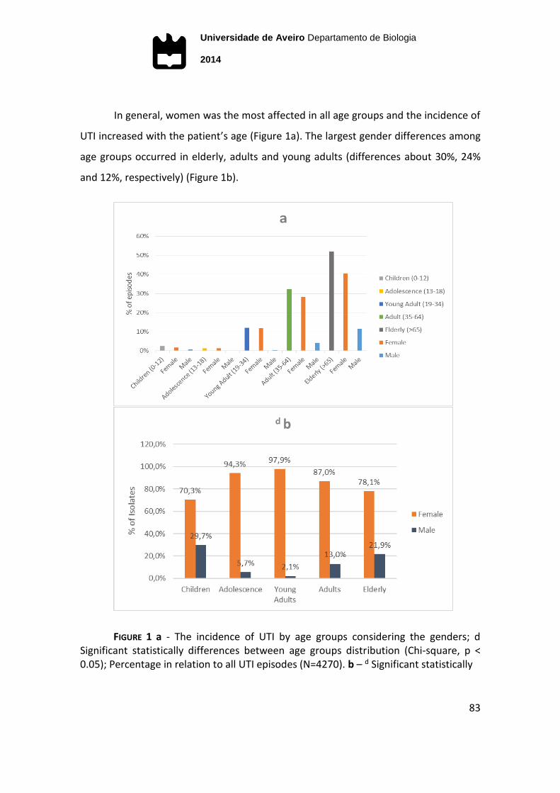
Universidade de Aveiro Departamento de Biologia
2014
83
In general, women was the most affected in all age groups and the incidence of
UTI increased with the patient’s age (Figure 1a). The largest gender differences among
age groups occurred in elderly, adults and young adults (differences about 30%, 24%
and 12%, respectively) (Figure 1b).
FIGURE 1 a - The incidence of UTI by age groups considering the genders; d Significant statistically differences between age groups distribution (Chi-square, p < 0.05); Percentage in relation to all UTI episodes (N=4270). b – d Significant statistically

Universidade de Aveiro Departamento de Biologia
2014
84
4.2. BACTERIAL IMPLIED IN UTI
The predominant uropathogens were E. coli (63.8%), Klebsiella spp (11.8%),
Enterococcus sp (6.8%), P. mirabilis (5.6%), Staphylococcus spp (2.4%), P. aeruginosa
(1.9%) and S. agalactiae (1.2%). The isolates under 1% of UTI classified as “other
bacteria” correspond to a 6.5% of the samples (Table 1).
TABLE 1 Frequency of isolates responsible for at least 1%; N – frequency of isolates; Total % - percentage of isolates in relation to N; Female (%) - percentage of isolates in relation to n women; Male (%) - percentage of isolates in relation to n men.
Order Bacteria Total (%) Female (%) Male (%)
N = 4270 n = 3561 n = 709
1º Escherichia coli 2723 63.8 67.80 43.40
2º Klebsiella spp 502 11.8 11.30 14.00
3º Enterococcus spp 290 6.8 6.20 9.60
4º Proteus mirabilis 240 5.6 4.60 10.70
5º Staphylococcus spp 103 2.4 2.20 3.40
6º Pseudomonas aeruginosa 83 1.9 1.30 5.10
7º Streptococcus agalactiae 50 1.2 1.30 0.70
… Other Bacteria 279 6.5 5.20 13.10
The main bacteria implicated in women UTI was statistically different from the
main bacteria implicated in men UTI (Mann-Whitney U test, p< 0.05). E. coli was the
bacterium most implicated in both genders UTI, however its incidence was higher in
women, 67.8%, than in men, 43.4% (Table 2). To the remaining bacteria in women, the
second most implicated bacterium was Klebsiella spp (11.3%) followed by
Enterococcus sp (6.2%), P. mirabilis (4.6%), Staphylococcus spp (2.2%), P. aeruginosa
(1.3%) and S. agalactiae (1.3%), other bacteria represented 5.2% of the isolates. For
men, the second uropathogen most frequent was Klebsiella spp (14%) followed by P.
mirabilis (10.7%), Enterococcus sp (9.6%), P. aeruginosa (5.1%), Staphylococcus spp
(3.4%) and S. agalactiae (1.3%), other bacteria represented 13.1% of the isolates
(Table 2).
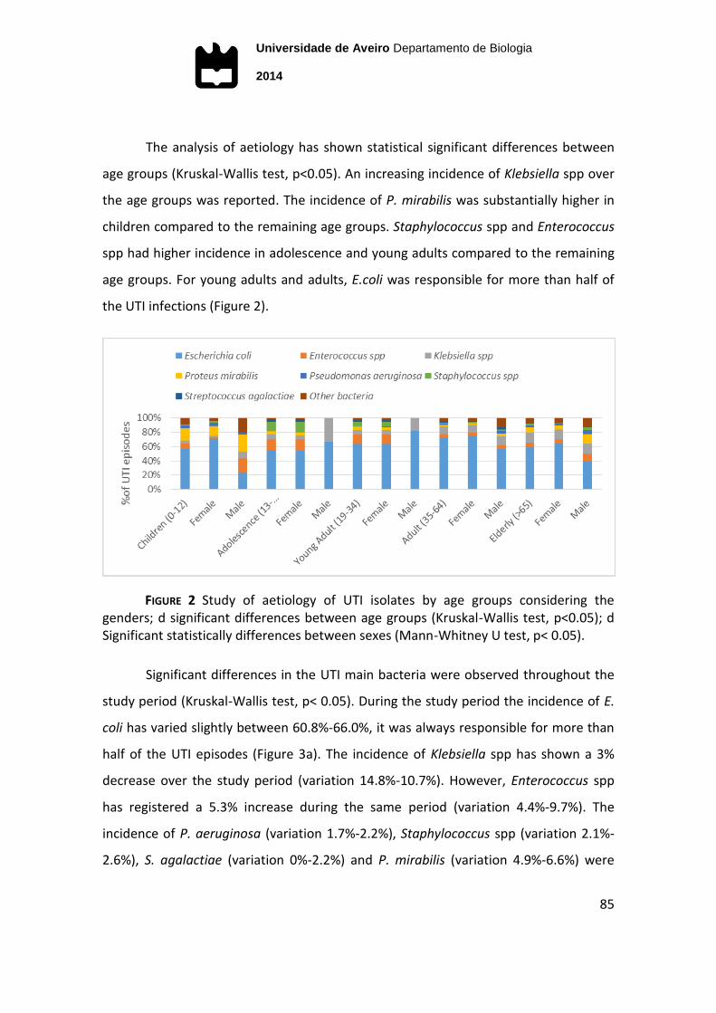
Universidade de Aveiro Departamento de Biologia
2014
85
The analysis of aetiology has shown statistical significant differences between
age groups (Kruskal-Wallis test, p<0.05). An increasing incidence of Klebsiella spp over
the age groups was reported. The incidence of P. mirabilis was substantially higher in
children compared to the remaining age groups. Staphylococcus spp and Enterococcus
spp had higher incidence in adolescence and young adults compared to the remaining
age groups. For young adults and adults, E.coli was responsible for more than half of
the UTI infections (Figure 2).
FIGURE 2 Study of aetiology of UTI isolates by age groups considering the genders; d significant differences between age groups (Kruskal-Wallis test, p<0.05); d Significant statistically differences between sexes (Mann-Whitney U test, p< 0.05).
Significant differences in the UTI main bacteria were observed throughout the
study period (Kruskal-Wallis test, p< 0.05). During the study period the incidence of E.
coli has varied slightly between 60.8%-66.0%, it was always responsible for more than
half of the UTI episodes (Figure 3a). The incidence of Klebsiella spp has shown a 3%
decrease over the study period (variation 14.8%-10.7%). However, Enterococcus spp
has registered a 5.3% increase during the same period (variation 4.4%-9.7%). The
incidence of P. aeruginosa (variation 1.7%-2.2%), Staphylococcus spp (variation 2.1%-
2.6%), S. agalactiae (variation 0%-2.2%) and P. mirabilis (variation 4.9%-6.6%) were
Universidade de Aveiro Departamento de Biologia
2014
86
irregular but almost constant over the study period (Figure 3b). Significant gender
differences in the main bacteria responsible for UTI were observed during the study
period into the years 2011 and 2013 (Mann–Whitney U test, p <0.05) (Figure 4).

Universidade de Aveiro Departamento de Biologia
2014
87
FIGURE 3 Etiology over the study period; a Percentage scale; b Logarithmic scale; d significant statistical differences between the main bacteria throughout the study period (Kruskal-Wallis test, p< 0.05).
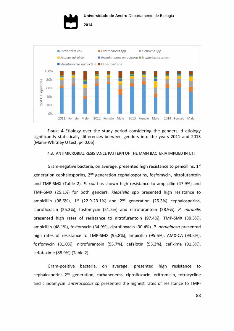
Universidade de Aveiro Departamento de Biologia
2014
88
FIGURE 4 Etiology over the study period considering the genders; d etiology significantly statistically differences between genders into the years 2011 and 2013 (Mann-Whitney U test, p< 0.05).
4.3. ANTIMICROBIAL RESISTANCE PATTERN OF THE MAIN BACTERIA IMPLIED IN UTI
Gram-negative bacteria, on average, presented high resistance to penicillins, 1st
generation cephalosporins, 2nd generation cephalosporins, fosfomycin, nitrofurantoin
and TMP-SMX (Table 2). E. coli has shown high resistance to ampicillin (47.9%) and
TMP-SMX (25.1%) for both genders. Klebsiella spp presented high resistance to
ampicillin (98.6%), 1st (22.9-23.1%) and 2nd generation (25.3%) cephalosporins,
ciprofloxacin (25.3%), fosfomycin (51.5%) and nitrofurantoin (28.9%). P. mirabilis
presented high rates of resistance to nitrofurantoin (97.4%), TMP-SMX (39.3%),
ampicillin (48.1%), fosfomycin (34.9%), ciprofloxacin (30.4%). P. aeruginosa presented
high rates of resistance to TMP-SMX (95.8%), ampicillin (95.6%), AMX-CA (93.3%),
fosfomycin (81.0%), nitrofurantoin (95.7%), cefalotin (93.3%), cefixime (91.3%),
cefotaxime (88.9%) (Table 2).
Gram-positive bacteria, on average, presented high resistance to
cephalosporins 2nd generation, carbapenems, ciprofloxacin, eritromicin, tetracycline
and clindamycin. Enterococcus sp presented the highest rates of resistance to TMP-
Universidade de Aveiro Departamento de Biologia
2014
89
SMX (94.8%), cefaclor (94.3%), cefuroxime (94.4%), clindamycin (92.7%) and
tetracycline (80.9%). Staphylococcus spp showed the highest resistance to cefotaxime
(100%), cefuroxime (85.7%), imipenem (83.9%), cefaclor (82.5%) and fosfomycin
(80.0%). S. agalactiae showed high resistance to tetracycline (87.8%), ciprofloxacin
(100%), clindamycin (51.1%) and eritromicin (46%) (Table 2).
The other bacteria group, on average, showed the highest resistances to
tetracycline (100%), vancomycin (100%), linezolid (100%), ampicillins (87.5%), cefalotin
(72.7%) and AMX-CA (60.7%) (Table 2).
The average resistance presented by bacteria implicated in UTI was statistically
different (Mann-Whitney U test, p< 0.05) between genders. E. coli, Klebsiella spp, P.
mirabilis and Staphylococcus spp, were the bacteria responsible for the major
difference between genders. No differences in the resistance patterns between
women and men were observed for S. agalactiae and the other bacteria group (Table
2).
Considering all the uropathogens isolated, on average, they were resistant to 3
antimicrobials and to 2 antimicrobial of different classes of drug. Considering the
isolates from both genders: the isolates from women were resistant on average to 2
antimicrobials and to 2 antimicrobial classes of drugs; the isolates from men were
resistant, on average, to 5 antimicrobials and to 4 antimicrobial classes of drug.
The bacterial resistance changed significantly over the study period (Kruskal-
Wallis test, p < 0.05) (Figure 5). The bacterial resistance to penicillins, 2nd and 3rd
generation cephalosporins, lincosamides, fosfomycins, carbapenems and macrolides
decreased. However, for S. agalactiae was observed an increase in resistance to the
antibiotics tetracyclines, glycopeptides, lincosamides and macrolides over the study
period. The pattern of resistance of P. aeruginosa was different from that of the other
gram-negative (E.coli, P. mirabilis, Klebsiella spp) from 2011-2013 for 1st to 3rd
Universidade de Aveiro Departamento de Biologia
2014
90
generation cephalosporins, fosfomycins and sulfamides. The resistance of E coli was
constant throughout the study period to each antimicrobials tested (Figure 5).
The pattern of bacterial resistance to antibiotics was significantly different
among age groups (Kruskal-Wallis test, p<0.05). The pattern of resistance of P.
aeruginosa was different from that of the other gram-negative (E. coli, P. mirabilis,
Klebsiella spp), namely in the group of young adults, for penicillins, 1st to 3rd generation
cephalosporins, fosfomycins, carbapenems, quinolones, nitrofurans and sulfamides
(Figure 6).
Since 63.8% of the infections were caused by E. coli and 36.2% by the remaining
bacteria, resistance were achieved through the weighted average. The weighted
average was calculated through arithmetic average, but having in to account the
incidence of each bacterium. The values of antimicrobial resistance of each bacterium
were multiplied by its incidence.
Considering the first line treatment recommended by EAU, the weighted
average for ciprofloxacin was > 20%, exceeding the recommended value. However,
considering only the women, the resistance was < 20%. For fosfomycin the resistance
was > 20% for isolates of both genders. Contrarily, the resistance to nitrofurantoin was
< 20% for isolates of both genders.
Considering the second line treatment recommended by EAU, the weighted
average resistance to ampicillin for both sexes was > 20%, higher than the value
recommended value by EAU. The weighted average resistance to TMP-SMX and AMX-
CA was > 20% considering all the isolates but it was < 20% when only the women were
considered. The resistance to imipenem and gentamicin was < 20% for both genders,
below the recommended value of the EAU (Table 3).
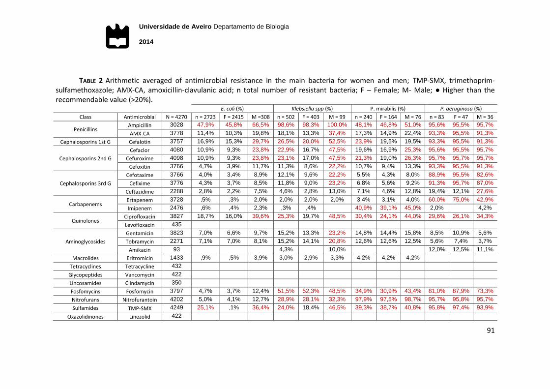
Universidade de Aveiro Departamento de Biologia
2014
91
TABLE 2 Arithmetic averaged of antimicrobial resistance in the main bacteria for women and men; TMP-SMX, trimethoprim-sulfamethoxazole; AMX-CA, amoxicillin-clavulanic acid; n total number of resistant bacteria; F – Female; M- Male; ● Higher than the recommendable value (>20%).
E. coli (%) Klebsiella spp (%) P. mirabilis (%) P. aeruginosa (%)
Class Antimicrobial N = 4270 n = 2723 F = 2415 M =308 n = 502 F = 403 M = 99 n = 240 F = 164 M = 76 n = 83 F = 47 M = 36
Penicillins Ampicillin 3028 47,9% 45,8% 66,5% 98,6% 98,3% 100,0% 48,1% 46,8% 51,0% 95,6% 95,5% 95,7%
AMX-CA 3778 11,4% 10,3% 19,8% 18,1% 13,3% 37,4% 17,3% 14,9% 22,4% 93,3% 95,5% 91,3%
Cephalosporins 1st G Cefalotin 3757 16,9% 15,3% 29,7% 26,5% 20,0% 52,5% 23,9% 19,5% 19,5% 93,3% 95,5% 91,3%
Cephalosporins 2nd G
Cefaclor 4080 10,9% 9,3% 23,8% 22,9% 16,7% 47,5% 19,6% 16,9% 25,3% 95,6% 95,5% 95,7%
Cefuroxime 4098 10,9% 9,3% 23,8% 23,1% 17,0% 47,5% 21,3% 19,0% 26,3% 95,7% 95,7% 95,7%
Cefoxitin 3766 4,7% 3,9% 11,7% 11,3% 8,6% 22,2% 10,7% 9,4% 13,3% 93,3% 95,5% 91,3%
Cephalosporins 3rd G
Cefotaxime 3766 4,0% 3,4% 8,9% 12,1% 9,6% 22,2% 5,5% 4,3% 8,0% 88,9% 95,5% 82,6%
Cefixime 3776 4,3% 3,7% 8,5% 11,8% 9,0% 23,2% 6,8% 5,6% 9,2% 91,3% 95,7% 87,0%
Ceftazidime 2288 2,8% 2,2% 7,5% 4,6% 2,8% 13,0% 7,1% 4,6% 12,8% 19,4% 12,1% 27,6%
Carbapenems Ertapenem 3728 ,5% ,3% 2,0% 2,0% 2,0% 2,0% 3,4% 3,1% 4,0% 60,0% 75,0% 42,9%
Imipenem 2476 ,6% ,4% 2,3% ,3% ,4% 40,9% 39,1% 45,0% 2,0% 4,2%
Quinolones Ciprofloxacin 3827 18,7% 16,0% 39,6% 25,3% 19,7% 48,5% 30,4% 24,1% 44,0% 29,6% 26,1% 34,3%
Levofloxacin 435
Aminoglycosides
Gentamicin 3823 7,0% 6,6% 9,7% 15,2% 13,3% 23,2% 14,8% 14,4% 15,8% 8,5% 10,9% 5,6%
Tobramycin 2271 7,1% 7,0% 8,1% 15,2% 14,1% 20,8% 12,6% 12,6% 12,5% 5,6% 7,4% 3,7%
Amikacin 93 4,3% 10,0% 12,0% 12,5% 11,1%
Macrolides Eritromicin 1433 ,9% ,5% 3,9% 3,0% 2,9% 3,3% 4,2% 4,2% 4,2%
Tetracyclines Tetracycline 432
Glycopeptides Vancomycin 422
Lincosamides Clindamycin 350
Fosfomycins Fosfomycin 3797 4,7% 3,7% 12,4% 51,5% 52,3% 48,5% 34,9% 30,9% 43,4% 81,0% 87,9% 73,3%
Nitrofurans Nitrofurantoin 4202 5,0% 4,1% 12,7% 28,9% 28,1% 32,3% 97,9% 97,5% 98,7% 95,7% 95,8% 95,7%
Sulfamides TMP-SMX 4249 25,1% ,1% 36,4% 24,0% 18,4% 46,5% 39,3% 38,7% 40,8% 95,8% 97,4% 93,9%
Oxazolidinones Linezolid 422
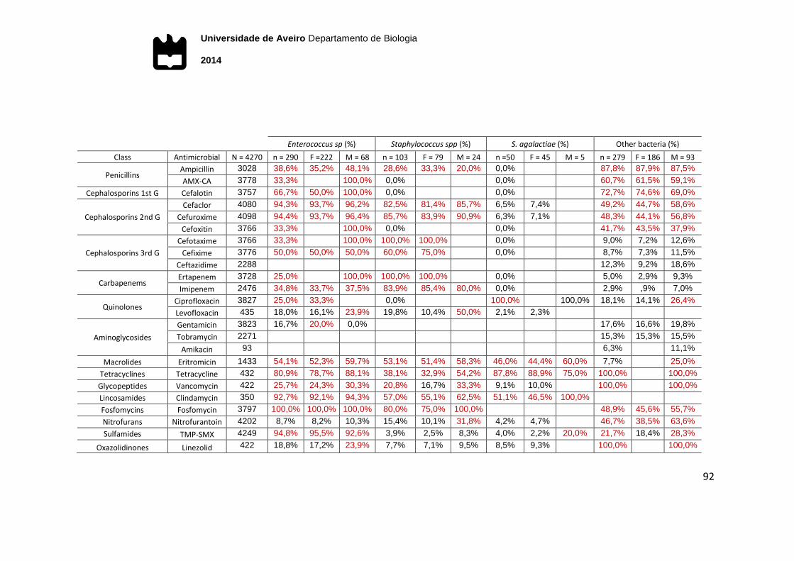
Universidade de Aveiro Departamento de Biologia
2014
92
Enterococcus sp (%) Staphylococcus spp (%) S. agalactiae (%) Other bacteria (%)
Class Antimicrobial N = 4270 n = 290 F =222 M = 68 n = 103 F = 79 M = 24 n =50 F = 45 M = 5 n = 279 F = 186 M = 93
Penicillins Ampicillin 3028 38,6% 35,2% 48,1% 28,6% 33,3% 20,0% 0,0% 87,8% 87,9% 87,5%
AMX-CA 3778 33,3% 100,0% 0,0% 0,0% 60,7% 61,5% 59,1%
Cephalosporins 1st G Cefalotin 3757 66,7% 50,0% 100,0% 0,0% 0,0% 72,7% 74,6% 69,0%
Cephalosporins 2nd G
Cefaclor 4080 94,3% 93,7% 96,2% 82,5% 81,4% 85,7% 6,5% 7,4% 49,2% 44,7% 58,6%
Cefuroxime 4098 94,4% 93,7% 96,4% 85,7% 83,9% 90,9% 6,3% 7,1% 48,3% 44,1% 56,8%
Cefoxitin 3766 33,3% 100,0% 0,0% 0,0% 41,7% 43,5% 37,9%
Cephalosporins 3rd G
Cefotaxime 3766 33,3% 100,0% 100,0% 100,0% 0,0% 9,0% 7,2% 12,6%
Cefixime 3776 50,0% 50,0% 50,0% 60,0% 75,0% 0,0% 8,7% 7,3% 11,5%
Ceftazidime 2288 12,3% 9,2% 18,6%
Carbapenems Ertapenem 3728 25,0% 100,0% 100,0% 100,0% 0,0% 5,0% 2,9% 9,3%
Imipenem 2476 34,8% 33,7% 37,5% 83,9% 85,4% 80,0% 0,0% 2,9% ,9% 7,0%
Quinolones Ciprofloxacin 3827 25,0% 33,3% 0,0% 100,0% 100,0% 18,1% 14,1% 26,4%
Levofloxacin 435 18,0% 16,1% 23,9% 19,8% 10,4% 50,0% 2,1% 2,3%
Aminoglycosides
Gentamicin 3823 16,7% 20,0% 0,0% 17,6% 16,6% 19,8%
Tobramycin 2271 15,3% 15,3% 15,5%
Amikacin 93 6,3% 11,1%
Macrolides Eritromicin 1433 54,1% 52,3% 59,7% 53,1% 51,4% 58,3% 46,0% 44,4% 60,0% 7,7% 25,0%
Tetracyclines Tetracycline 432 80,9% 78,7% 88,1% 38,1% 32,9% 54,2% 87,8% 88,9% 75,0% 100,0% 100,0%
Glycopeptides Vancomycin 422 25,7% 24,3% 30,3% 20,8% 16,7% 33,3% 9,1% 10,0% 100,0% 100,0%
Lincosamides Clindamycin 350 92,7% 92,1% 94,3% 57,0% 55,1% 62,5% 51,1% 46,5% 100,0%
Fosfomycins Fosfomycin 3797 100,0% 100,0% 100,0% 80,0% 75,0% 100,0% 48,9% 45,6% 55,7%
Nitrofurans Nitrofurantoin 4202 8,7% 8,2% 10,3% 15,4% 10,1% 31,8% 4,2% 4,7% 46,7% 38,5% 63,6%
Sulfamides TMP-SMX 4249 94,8% 95,5% 92,6% 3,9% 2,5% 8,3% 4,0% 2,2% 20,0% 21,7% 18,4% 28,3%
Oxazolidinones Linezolid 422 18,8% 17,2% 23,9% 7,7% 7,1% 9,5% 8,5% 9,3% 100,0% 100,0%
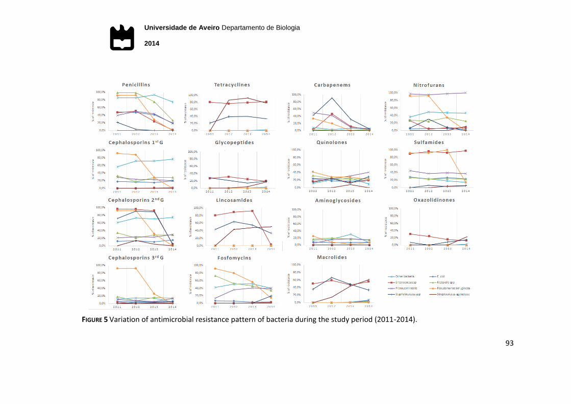
Universidade de Aveiro Departamento de Biologia
2014
93
FIGURE 5 Variation of antimicrobial resistance pattern of bacteria during the study period (2011-2014).
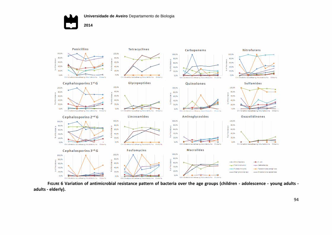
Universidade de Aveiro Departamento de Biologia
2014
94
FIGURE 6 Variation of antimicrobial resistance pattern of bacteria over the age groups (children - adolescence - young adults -
adults - elderly).
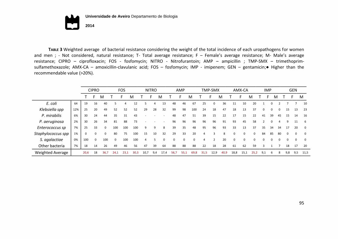
Universidade de Aveiro Departamento de Biologia
2014
95
TABLE 3 Weighted average of bacterial resistance considering the weight of the total incidence of each uropathogens for women and men ; - Not considered, natural resistance; T- Total average resistance; F – Female’s average resistance; M- Male’s average resistance; CIPRO – ciprofloxacin; FOS - fosfomycin; NITRO - Nitrofurantoin; AMP – ampicillin ; TMP-SMX – trimethoprim-sulfamethoxazole; AMX-CA – amoxicillin-clavulanic acid; FOS – fosfomycin; IMP - imipenem; GEN – gentamicin;● Higher than the recommendable value (>20%).
CIPRO FOS NITRO AMP TMP-SMX AMX-CA IMP GEN
T F M T F M T F M T F M T F M T F M T F M T F M
E. coli 64 19 16 40 5 4 12 5 4 13 48 46 67 25 0 36 11 10 20 1 0 2 7 7 10
Klebsiella spp 12% 25 20 49 52 52 52 29 28 32 99 98 100 24 18 47 18 13 37 0 0 0 15 13 23
P. mirabilis 6% 30 24 44 35 31 43 - - - 48 47 51 39 15 22 17 15 22 41 39 45 15 14 16
P. aeruginosa 2% 30 26 34 81 88 73 - - - 96 96 96 96 96 91 93 45 58 2 0 4 9 11 6
Enterococcus sp 7% 25 33 0 100 100 100 9 9 8 39 35 48 95 96 93 33 13 37 35 34 34 17 20 0
Staphylococcus spp 1% 0 0 0 80 75 100 15 10 32 29 33 20 4 3 8 0 0 0 84 85 80 0 0 0
S. agalactiae 0% 100 0 100 0 100 100 4 5 0 0 0 0 4 2 20 0 0 0 0 0 0 0 0 0
Other bacteria 7% 18 14 26 49 46 56 47 39 64 88 88 88 22 18 28 61 62 59 3 1 7 18 17 20
Weighted Average 20,6 18 36,7 24,1 23,1 30,3 10,7 9,4 17,4 56,7 55,1 69,8 31,5 12,9 40,9 18,8 15,1 25,2 9,1 6 8 9,8 9,5 11,5
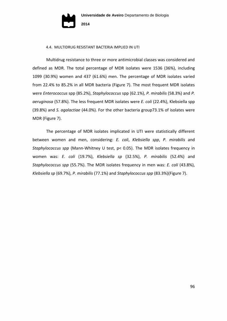
Universidade de Aveiro Departamento de Biologia
2014
96
4.4. MULTIDRUG RESISTANT BACTERIA IMPLIED IN UTI
Multidrug resistance to three or more antimicrobial classes was considered and
defined as MDR. The total percentage of MDR isolates were 1536 (36%), including
1099 (30.9%) women and 437 (61.6%) men. The percentage of MDR isolates varied
from 22.4% to 85.2% in all MDR bacteria (Figure 7). The most frequent MDR isolates
were Enterococcus spp (85.2%), Staphylococcus spp (62.1%), P. mirabilis (58.3%) and P.
aeruginosa (57.8%). The less frequent MDR isolates were E. coli (22.4%), Klebsiella spp
(39.8%) and S. agalactiae (44.0%). For the other bacteria group73.1% of isolates were
MDR (Figure 7).
The percentage of MDR isolates implicated in UTI were statistically different
between women and men, considering: E. coli, Klebsiella spp, P. mirabilis and
Staphylococcus spp (Mann-Whitney U test, p< 0.05). The MDR isolates frequency in
women was: E. coli (19.7%), Klebsiella sp (32.5%), P. mirabilis (52.4%) and
Staphylococcus spp (55.7%). The MDR isolates frequency in men was: E. coli (43.8%),
Klebsiella sp (69.7%), P. mirabilis (77.1%) and Staphylococcus spp (83.3%)(Figure 7).
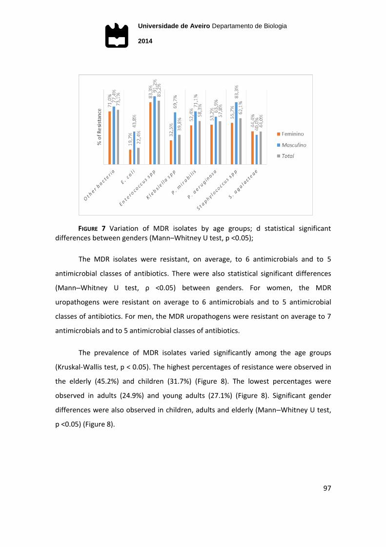
Universidade de Aveiro Departamento de Biologia
2014
97
FIGURE 7 Variation of MDR isolates by age groups; d statistical significant differences between genders (Mann–Whitney U test, p <0.05);
The MDR isolates were resistant, on average, to 6 antimicrobials and to 5
antimicrobial classes of antibiotics. There were also statistical significant differences
(Mann–Whitney U test, ρ <0.05) between genders. For women, the MDR
uropathogens were resistant on average to 6 antimicrobials and to 5 antimicrobial
classes of antibiotics. For men, the MDR uropathogens were resistant on average to 7
antimicrobials and to 5 antimicrobial classes of antibiotics.
The prevalence of MDR isolates varied significantly among the age groups
(Kruskal-Wallis test, p < 0.05). The highest percentages of resistance were observed in
the elderly (45.2%) and children (31.7%) (Figure 8). The lowest percentages were
observed in adults (24.9%) and young adults (27.1%) (Figure 8). Significant gender
differences were also observed in children, adults and elderly (Mann–Whitney U test,
p <0.05) (Figure 8).
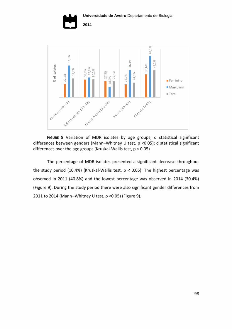
Universidade de Aveiro Departamento de Biologia
2014
98
FIGURE 8 Variation of MDR isolates by age groups; d statistical significant differences between genders (Mann–Whitney U test, p <0.05); d statistical significant differences over the age groups (Kruskal-Wallis test, p < 0.05)
The percentage of MDR isolates presented a significant decrease throughout
the study period (10.4%) (Kruskal-Wallis test, p < 0.05). The highest percentage was
observed in 2011 (40.8%) and the lowest percentage was observed in 2014 (30.4%)
(Figure 9). During the study period there were also significant gender differences from
2011 to 2014 (Mann–Whitney U test, ρ <0.05) (Figure 9).
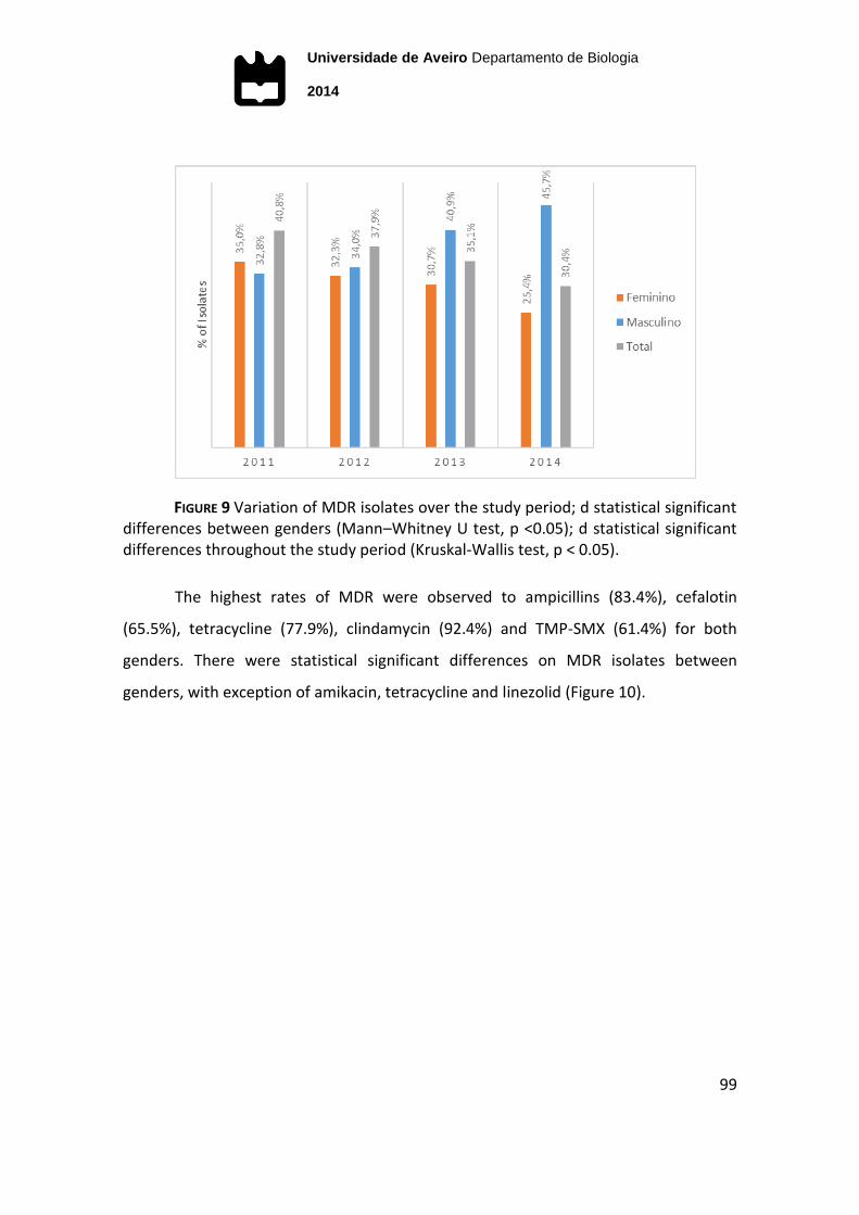
Universidade de Aveiro Departamento de Biologia
2014
99
FIGURE 9 Variation of MDR isolates over the study period; d statistical significant differences between genders (Mann–Whitney U test, p <0.05); d statistical significant differences throughout the study period (Kruskal-Wallis test, p < 0.05).
The highest rates of MDR were observed to ampicillins (83.4%), cefalotin
(65.5%), tetracycline (77.9%), clindamycin (92.4%) and TMP-SMX (61.4%) for both
genders. There were statistical significant differences on MDR isolates between
genders, with exception of amikacin, tetracycline and linezolid (Figure 10).
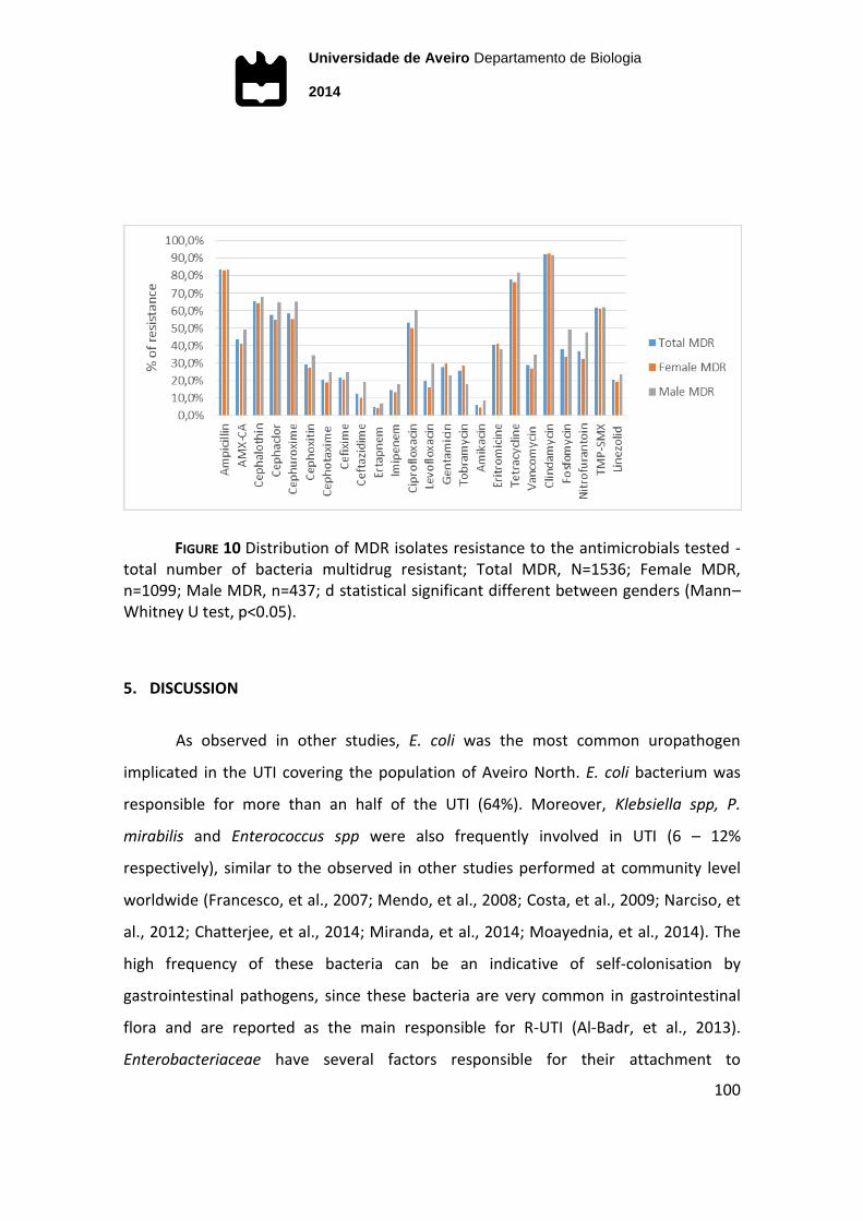
Universidade de Aveiro Departamento de Biologia
2014
100
FIGURE 10 Distribution of MDR isolates resistance to the antimicrobials tested - total number of bacteria multidrug resistant; Total MDR, N=1536; Female MDR, n=1099; Male MDR, n=437; d statistical significant different between genders (Mann–Whitney U test, p<0.05).
5. DISCUSSION
As observed in other studies, E. coli was the most common uropathogen
implicated in the UTI covering the population of Aveiro North. E. coli bacterium was
responsible for more than an half of the UTI (64%). Moreover, Klebsiella spp, P.
mirabilis and Enterococcus spp were also frequently involved in UTI (6 – 12%
respectively), similar to the observed in other studies performed at community level
worldwide (Francesco, et al., 2007; Mendo, et al., 2008; Costa, et al., 2009; Narciso, et
al., 2012; Chatterjee, et al., 2014; Miranda, et al., 2014; Moayednia, et al., 2014). The
high frequency of these bacteria can be an indicative of self-colonisation by
gastrointestinal pathogens, since these bacteria are very common in gastrointestinal
flora and are reported as the main responsible for R-UTI (Al-Badr, et al., 2013).
Enterobacteriaceae have several factors responsible for their attachment to

Universidade de Aveiro Departamento de Biologia
2014
101
urothelium, which transform them into the main bacteria responsible for UTI (Dash, et
al., 2013). Being the three most reported UTI main bacteria (81.2%) in the community
studies worldwide, would be significant for the empiric treatment success an action
spectrum for them (Costa, et al., 2009).
As stated before UTI is more frequent in women than in men (67.8% and
43.4%, respectively), and the highest difference between women and men patients
was observed for the P. mirabilis (incidence 10.7%, 2.3 times higher in men) and P.
aeruginosa (incidence 5.1%, 3.9 times higher in men). E. coli bacterium was the most
predominant in both genders, however its incidence was significantly higher in women
(67.8%) than in men (43.4%) (Mann-Whitney U test, p<0.05), which was also observed
in other similar studies (Linhares, et al., 2013; Chatterjee, et al., 2014). According to
the literature, P. aeruginosa is more frequent in men due to characteristics inherent to
the patient, including sex, age, previous use of antimicrobials, previous interventions in
the urinary tract and patients with neurogenic bladder (Grabe, et al., 2008; Koeijers, et
al., 2010; Rodrigues, et al., 2011; Linhares, et al., 2013).
The higher incidence of P. mirabilis in men than in women was consistent with
published literature (Orrett, et al., 2001). P. mirabilis due to biofilms production
capacity is a serious medical problem in catheter-associated UTI, which is consistent
with its higher rates in elderly (Czerwonka, et al., 2014). As reported by Nicolle (2014).
The elderly age group, especially in men (incidence in men 16% and women 3%), were
the most frequent catheter-associated UTI patients (Nicolle, 2014). Moreover,
Pseudomonas and Proteus spp have been found with high frequency in complicated
UTI, which may justify their high incidence in men, as well as their higher rates in
children and elderly age groups (Grabe, et al., 2008).
The average age of men with UTI was significantly higher than women
(averaged age 68 and 59 years, respectively), which can also explain the aetiological
differences among genders (Mann–Whitney U test, ρ-value <0.05). The higher
Universidade de Aveiro Departamento de Biologia
2014
102
incidence of UTI in older men can be due to benign prostate hyperplasia (BPH) and
neurogenic bladder more common at this age (Koeijers, et al., 2010; Dash, et al., 2013).
Moreover, the higher incidence of UTI caused by P. aeruginosa bacterium is frequently
associated with the hospital environment, in men seems to be related to the increased
hospitalizations due to their higher age (Koeijers, et al., 2010; Nicolle, 2014). Despite
hospital-acquired urinary infection caused by P. aeruginosa in community is still
uncommon (1.9% in this study). However, it should not be ignored, since other
community-acquired UTI studies have reported its increasing incidence (Koeijers, et al.,
2010; Rodrigues, et al., 2011; Linhares, et al., 2013).
The distribution into age groups was not random (Chi-square test, p<0.05), the
incidence of UTI increased with age in both genders. Being the elderly the most
affected (52%), followed by adults (32%), young adults (12%), children (2%) and
adolescents (1%). In the literature, it is well documented that the incidence of
community-acquired UTI increases with age, affecting both genders in the elderly
population (Cove-Smith, et al., 2007; Linhares, et al., 2013; Chatterjee, et al., 2014).
The increase in young adult and adult women is well established with the sexual
activity, the use of contraceptives (spermicides, diaphragm and oral contraceptives),
the use of antimicrobials and pregnancy (Narciso, et al., 2012). In adult and elder
women, the increase of UTI has been explained by menopause. After menopause the
estrogen levels and Lactobacilli number drop and makes post-menopausal women
susceptible to UTI (Al-Badr, et al., 2013). Also to elder women the frequent use of
antimicrobials, the increase of surgical urogenital interventions, urinary incontinence,
post void residual (PVR), more frequent hospitalization and other pathologies
associations explain the (Molander, et al., 2000; Rowe, et al., 2013; Rowe, et al., 2014).
In adult and elder men, the increase of urinary infections has been mainly related to
benign prostate enlargement causing voiding obstruction which leads to PVR, the use
of antimicrobials, more frequent hospitalization, urogenital interventions, neurogenic

Universidade de Aveiro Departamento de Biologia
2014
103
bladder and other pathologies (Narciso, et al., 2012; Dash, et al., 2013; Rowe, et al.,
2014).
According to the literature (Weir, et al., 2000; Nwokocha, et al., 2014), there is
an increase of urinary tract infections from children to adolescence, related to the
onset of sexual activity, which was not observed in this study. The number of urine
samples from adolescents was much lower compared to those performed by children,
which may explain the higher frequency of urinary infections in this age group
compared to the adolescent group. Despite the low incidence, it was observed the
highest prevalence of gram positive infection in this age group. According to Blumer
(2009), older children and adolescents also share many features commonly seen in
adult patients with infections at the same sites, however gram-positive organisms an
opportunistic agent are the most common cause of bacterial infections in infants,
children and adolescents, taking advantage of their vulnerability (Blumer, 2009).
Following the Blumer (2009) study, the most responsible gram negative uropathogen
in cystitis is the Staphylococci coagulase-negative, which may justify the increase of
Staphylococcus spp in the adolescent group in this study.
The incidence of E. coli, responsible for more than an half of the UTI isolates,
was constant during the study period (variation 60.8-66.0%) but Klebsiella spp has
shown a 3% decrease and, contrarily, Enterococcus spp registered a 5.3% increase.
Francesco et al (2007) also registered a slight increase for Enterococcus (0.7%) in his 4
years study (2002-2005) among the community outpatients. In the study of Kuzdan
(2014) was also observed a decrease and the increase, respectively, for the species
Klebsiella pneumonia and Enterococcus faecium (Kuzdan, et al., 2014). For E. coli and
the remaining bacteria were almost constant throughout the study period.
The more predominant uropathogen, E. coli, presented the lowest
antimicrobial average resistance comparing with the remaining bacteria implicated in
UTI. The resistance to the first line treatment recommended by EAU for uncomplicated
Universidade de Aveiro Departamento de Biologia
2014
104
UTI was tested for E. coli and revealed low resistance to the three antimicrobials
recommended (5.0% nitrofurantoin, 18.7% ciprofloxacin and 4.7% fosfomycin). To the
second line treatment recommended by EUA, E. coli isolates exceeding the
recommendable value (<20%) for two of the second line treatment, ampicillin and
TMP-SMX (31.5% and 25.1% respectively), contrary to AMX-CA (11.4%). E. coli high
resistance to ampicillin and moderate resistance to AMX-CA and SMX have been also
reported in other studies (Francesco, Ravizzola, Peroni, Negrini, & Manca, 2007).
Having into account the high incidence of E. coli (63.8%) and the drug
resistance pattern of the most implicated bacteria in UTI, it was calculated the
weighted average of resistance. Among the first line choice, that cause little collateral
damage, only the nitrofurantoin is suitable to be used in the empiric treatment of UTI
in the community (weighted average resistance of 10.7%, 20.6% and 24.1%,
respectively, for nitrofurantoin, ciprofloxacin and fosfomycin). The weighted average
of nitrofurantoin resistance was not determined for P. mirabilis and P. aeruginosa
because they are naturally resistant to this antimicrobial. Considering the second line
therapy recommended by EAU, the ciprofloxacin and TMP-SMX are not appropriate to
treat UTI in the community but the antimicrobial AMX-CA is appropriated to be used in
UTI empiric treatment (weighted average resistance of 56.7%, 31.5% and 18.8%,
respectively for ampicillin, TMP-SMX and AMX-CA).
The antimicrobial resistance also suggest that the choice of empirical
antimicrobial therapy should have into account the sex of the patient. The
uropathogens isolated from men presented, on average, resistance to a higher number
of antibiotics (on average 5) compared to those isolated from women (on average 4).
This difference was clearly evident in the average resistance of E. coli and Klebsiella
spp, which presented significant differences for at least 12 of the 18 antimicrobials
tested (corresponding to 66.7% and 72.2% of the antibiotics, respectively). For the
remaining bacteria the difference between women and men was also evident, namely
Universidade de Aveiro Departamento de Biologia
2014
105
for the antibiotics ciprofloxacin, AMX-CA and TMP-SMX. Consequently, the
antimicrobials AMX-CA and TMP-SMX should be suitable to treat women (resistance
<20% for the bacteria more implicated in UTI), but should not be used to treat men
(resistance >20% for all of the bacteria more implicated in UTI, except E. coli). Two
recent studies also found differences in antimicrobial resistance among genders. In a
recent Portuguese study the fluoroquinolones were found unsuitable to treat men,
and also in an Indian study E.coli isolates showed resistance to the majority of
antibiotics in men (Linhares, et al., 2013; Chatterjee, et al., 2014).
The resistance of the main bacteria among age groups has presented significant
differences to the antimicrobial classes tested on young adults and elderly (Kruskal-
Wallis test, p<0.05). For these two age groups, significant statistical differences were
observed in all antimicrobials classes. As it was observed before in other studies, the
resistance tendency significantly increases with age (Cove-Smith, et al., 2007; Linhares,
et al., 2013). The higher rates of resistance in elder patients can be due to the more
frequent hospitalization of this age group, as a result of the increase of average life
expectancy, weak immune system and recurring infections. It is recognized that
hospitalizations increase the transmission of bacterial strains between hospital and
community (Ott, et al., 2013).
In general, the resistance by the main bacteria implicated in UTI to penicillins,
2nd and 3rd generation cephalosporins, lincosamides, fosfomycins, carbapenems and
macrolides decreased during the study period. However, for glycopeptides,
quinolones, aminoglycosides, nitrofurans, sulfamides and oxazolidinones the
resistance was almost constant throughout the study period to the main
uropathogens. Nevertheless, S. agalactiae presented a resistance increase over the
study period to the tetracyclines, glycopeptides, lincosamides and macrolides. P.
aeruginosa presented a different pattern of resistance relatively to the antimicrobials
1st to 3rd generation cephalosporins and sulfamides, comparing to the other gram-
Universidade de Aveiro Departamento de Biologia
2014
106
negative (E .coli, P. mirabilis, Klebsiella spp). E coli resistance was constant throughout
the study period to all antimicrobials tested. According to Linhares et al (2013), the
pattern of resistance to the main bacteria also showed significant changes during a ten
years period, but E. coli did not show significant changes in the resistance to the tested
antimicrobials. Similarly, P. aeruginosa had also a different behaviour compared to the
gram negative bacteria (E.coli, P. mirabilis, P. vulgaris, Klebsiella spp, Enterobacter spp
and Providencia spp) throughout the study period, specially observed in the classes of
cephalosporins and quinolones (Linhares et al., 2013).
The resistance to first and second line therapy it is probably the reflection of
their misuse and/or overuse (Bartoloni, et al., 2004). Although most organisms remain
susceptible to nitrofurantoin, Pallett (2010) defends that this agent is licensed for
lower UTI only and it has shown that resistance may develop during treatment (Pallett,
et al., 2010). The oral options available for the treatment of UTI with simultaneous
resistance to trimethoprim and quinolones are small. As a suitable alternative to first
and second line recommended therapy, this study recommends imipenem and
gentamicin to empiric treatment, due to the low bacterial resistance to these drugs in
both genders, and also considering the low percentage of bacterial resistance to both
antimicrobials in the MDR isolates. However, gentamicin is contraindicated in patients
with significant renal impairment, which is more common in the elderly. It is also
required the regular monitoring of pre-dose serum concentrations to assess further
dosing (Pallett, et al., 2010). Imipenem can be used in empiric treatment of
complicated UTI in view its broad spectrum of action and wide diffusion in the body
(intravenous) (Pallett, et al., 2010). These drugs involve the inconvenience of
parenteral administration and it can be more expensive than those indicated for UTI.
The multidrug resistant isolates were observed among the most prevalent
bacteria involved in the community-acquired UTI, being a 36% of these bacteria
resistant to three or more antimicrobials of distinct classes. However, the most
Universidade de Aveiro Departamento de Biologia
2014
107
implicated bacteria in UTI, E. coli, responsible for more than half of the UTI of the
studied patients, presented the lowest MDR bacterial percentage (22.4%). The second
bacteria more implicated in the community-acquired UTI, Klebsiella spp, presented
also a low percentage of MDR isolates (39.8%). Considering the main bacteria more
implicated in UTI, the percentage of MDR bacteria in this study was higher than that
found in the Antimicrobial Resistance Epidemiological Survey on Cystitis (ARESC) study,
which 10.3% of bacteria isolated at community level in nine European countries and in
Brazil were MDR (Heijer, et al., 2010). However, it was very similar to that obtained in
a similar study performed at the community level in which 36.7% of isolates
responsible for UTI were MDR (Heijer, et al., 2010; Sheerin, 2011). Keeping in view the
more implicated bacterium in UTI, E. coli, higher percentages of MDR isolates have
been detected in other studies (35.9-72.7%) done at the community level (Sulikowska,
et al., 2001; Narciso, et al., 2011; Sheerin, 2011). In this study, the most predominant
gram negative bacteria and S. agalactiae presented lower percentages of MDR isolates
(between 22.4% and 44%). The highest percentages were observed among the other
gram positive bacteria (Enterococcus spp, P. aeruginosa and Staphylococcus spp) and
the other bacteria group (between 62.1% and 85.2%).
The MDR bacteria isolated from men were, on average, resistant to a higher
number of antimicrobial classes than those isolated from women and the higher
percentage of MDR was also observed in men (61.6%). Most of the UTI in men are
complicated (associated with structural or functional abnormality in urinary tract),
requiring prolonged antimicrobial therapy and antibiotics able to reach high
therapeutic concentrations in the prostatic tissues (Hooton, 2000; Kuroda, et al., 2005;
Magiorakos, et al., 2011). For these reasons, men are more likely to develop resistance
to other antibiotics, usually used with success in the treatment of UTI in women
(Sheerin, 2011). Corroborating this hypothesis, Baral and colleagues showed that the
percentage of MDR was higher when bacteria were isolated from men (58.7%) (Baral,
et al., 2012).
Universidade de Aveiro Departamento de Biologia
2014
108
Nearly 45.2% of the bacteria isolated from elderly patients were MDR. This may
be explained by the increase of number and duration of hospital admissions with the
increasing age of the patient. During hospitalization some invasive procedures as
catheterization and other interventions may be associated with the transmission of
resistant hospital bacteria to the elderly patients. The immune system fragility of the
elderly patients may also explain the increase of urinary infection incidence and also of
UTI caused by MDR bacteria (Kisich, et al., 2008). In this study, patients with
community and hospital-acquired UTI were included which can validate the MDR high
percentages in isolates from men.
The incidence of MDR bacteria decreased about 10.4% throughout the study
period which may be related with the decrease of the antimicrobial resistance of some
MDR bacteria during the study period, such as, Enterococcus spp, Staphylococcus spp
and other bacteria group that showed high averaged percentage of MDR, 85.2% and
62.1%, respectively.
6. CONCLUSIONS
The most prevalent bacteria responsible for UTI were gram negative
Enterobacteriaceae (E.coli, Klebsiella spp and P. mirabilis) and Enterococcus spp. Being
E.coli, Klebsiella spp and P. mirabilis the three most common bacteria responsible for
UTI in the community studies worldwide, would be significant for the empiric
treatment success an action spectrum for them.
The results of this study emphasized the relevance to consider sex and age as
differentiator factors in the choice of UTI empirical treatment, due to differences in
aetiology and in antimicrobial resistance. The episodes of UTI are undoubtedly higher
in women and in the elderly group, increasing the differences in the incidence
between women and men with age. In general, men were more resistant to
Universidade de Aveiro Departamento de Biologia
2014
109
antimicrobials (on average 5) and to antimicrobial classes resistance (on average 4)
than women (2 and 2, respectively).
The results obtained in this study suggest that from first line treatment
recommended by the EAU to empirical treatment of uncomplicated UTI only
nitrofurantoin is appropriated for both sexes and ciprofloxacin may be only considered
to treat women. In the second line antibiotic, ampicillin is not an appropriate drug to
empirical treatment of uncomplicated UTI, though the antimicrobials AMX-CA and
TMP-SMX should not be used to empirical treatment of men, due to the local high
incidence of resistance (> 20%). As alternative to first and second line recommended
treatment, imipenem and gentamicin should be used due to their low weighted
average percentage of resistance.
Universidade de Aveiro Departamento de Biologia
2014
110
7. CHAPTER II - REFERENCES
Akram M, Shahid M & Khan A U (2007). Etiology and antibiotic resistance patterns of
community-acquired urinary tract infections in J N M C Hospital Aligarh, India.
Ann Clin Microbiol Antimicrob, 6:4.
Al-Badr A & Al-Shaikh G (2013). Recurrent Urinary Tract Infections Management in
Women: A review. Sultan Qaboos University Med J, 13(3), 359-367.
Baral P, Neupane S, Marasini, B P, Ghimire K R, Lekhak B & Shrestha B (2012). High
prevalence of multidrug resistance in bacterial uropathogens from Kathmandu,
Nepal. BMC Research Notes, 5(38).
Bartoloni A, Bartalesi F, Mantella A, Barahona H G, Barrón V P, Paradisi F & Rossolini G
M (2004). High Prevalence of Acquired Antimicrobial Resistance Unrelated to
Heavy Antimicrobial Consumption. J Infect Dis 189, 1291-1294.
bioMérieux Portugal (2014). bioMériux Portugal. Retrieved 10 17, 2014, from Lista de
produtos bioMérieux 2014:
http://www.biomerieux.pt/upload/Biomerieux_vers%C3%A3o_3___definitiva
bioMérieux SA (2013). chromID to directly identify micro-organisms. Retrieved 10 21,
2014, from bioMérieux: http://www.biomerieux-culturemedia.com/product/9-
chromid-cps-elite
bioMérieux SA (2014 a). VITEK® 2 Advanced Expert System. Retrieved 10 17, 2014,
from bioMérieux: http://www.biomerieux-diagnostics.com/vitek-2-advanced-
expert-system
Universidade de Aveiro Departamento de Biologia
2014
111
bioMérieux SA (2014 b). VITEK® 2 Identification Cards. Retrieved 10 17, 2014, from
bioMérieux: http://www.biomerieux-diagnostics.com/vitek-2-identification-
cards
Blumer J (2009). Treatment of pediatric Gram-positive multidrug-resistant infections.
59(1), J Infect., 51-58.
Chatterjee N, Ray R, Chatterjee M & Chattopadhyay S (2014). Correlation of
demographic profile and antibiotic resistance in patients with urinary tract
infection attending a teaching hospital in Kolkata. Journal of Medical Science
and Clinical Research 2(1), 2806-2816.
Correia C, Costa E, Peres A, Alves M, Pombo G & Estevinho L (2007). Etiologia das
Infecções do Tracto Urinário e sua Susceptibilidade aos Antimicrobianos. In
Acta Médica Portuguesa, 543–549.
Costa M, Pereira P M, Bolotinha C, Ferreira A, Cardoso R, Monteiro C, Gomes C F &
Gomes J F (2009). Frequência e Susceptibilidade Bacteriana em Infecções
Urinárias – dados de um laboratório de Lisboa. Parte II. Rev Lusófona de
Ciências e Tecnologias da Saúde, 6, 87-103.
Cove-Smith A & Almond M (2007). Management of urinary tract infections in the
elderly. Trends in Urology Gynaecology & Sexual Health 12(4), 31-34.
Czerwonka G, Arabski M, Wąsik S, Jabłońska-Wawrzycka A, Rogala P & Kaca W (2014).
Morphological changes in Proteus mirabilis O18 biofilm under the influence of a
urease inhibitor and a homoserine lactone derivative. Arch Microbiol., 196(3),
169–177.
Universidade de Aveiro Departamento de Biologia
2014
112
Dash M, Padhi S, Mohanty I, Panda P & Parida, B (2013). Antimicrobial resistance in
pathogens causing urinary tract infections in a rural community of Odisha,
India. J Family Community Med. 20(1), 20-26.
European Confederation of Laboratory Medicine (2000). European Urinalysis
Guidelines. 60, 1-96.
Foxman B (2002). Epidemiology of urinary tract infections: incidence, morbidity, and
economic costs. Am J Med, 113(1A).
Francesco M A, Ravizzola G, Peroni L, Negrini R & Manca N (2007). Urinary tract
infections in Brescia, Italy: Etiology of uropathogens and antimicrobial
resistance of common uropathogens. Med Sci Monit, 6:136–144.
Funke G & Funke-Kissling P (2004). Evaluation of the New VITEK 2 Card for
Identification of Clinically Relevant Gram-Negative Rods. Journal of Clinical
Microbiology 42(9), 4067–4071.
Grabe M, Bishop MC, Bjerklund-Johansen T E, Botto H, Çek M, Lobel B, Naber K G,
Palou J & Tenke P (2008). The Management of Male Urinary and Genital Tract
Infections. Retrived 10 22, 2014, from European Association of Urology:
http://www.uroweb.org/fileadmin/user_upload/Guidelines/The%20Managem
ent%20of%20Male%20Urinary%20and%20Genital%20Tract%20Infections.pdf
Grabe M, Bjerklund-Johansen T E, Botto H, Çek M, Naber K G, Pickard R S, Tenke P,
Wagenlehner F & Wullt B (2014). Guidelines on Urological Infections. Retrieved
10 20, 2014, from European Association of Urology (EAU):
http://www.uroweb.org/guidelines/online-guidelines/
Universidade de Aveiro Departamento de Biologia
2014
113
Graziottin A (2014). Recurrent cystitis after intercourse: why the gynaecologist has a
say. 2, TreeLife Media 319-336.
Gupta K, Hooton T M, Naber K G, Wullt B, Colgan R, Miller L G, Moran G J, Nicolle L E,
Raz R, Schaeffer A J & Soper D E (2011). International Clinical Practice
Guidelines for the Treatment of Acute Uncomplicated Cystitis and
Pyelonephritis in Women: A 2010 Update by the Infectious Diseases Society of
America and the European Society for Microbiology and Infectious Diseases.
Clinical Practice Guidelines 52, e103-e120.
Heijer C, Donker G, Maes J & Stobberingh E (2010). Antibiotic susceptibility of
unselected uropathogenic Escherichia coli from female Dutch general practice
patients: a comparison of two surveys with a 5 year interval. J Antimicrob
Chemother 65, 2128–2133.
Hooton T (2000). Pathogenesis of urinary tract infections: an update. J Antimicrob
Chemother 46, 1–7.
Kisich K, Carspecken C, Fieve S, Boguniewicz M & Leung D (2008). Defective killing of
Staphylococcus aureus in atopic dermatitis is associated with reduced
mobilization of human beta-defensin-31. J Allergy Clin Immunol 122, 62–68.
Koeijers J J, Verbon A, Kessels A, Bartelds A, Donkers G, Nys S & Stobberingh E E (2010).
Urinary tract infection in male general pratice patients: uropathogens and
antibiotic susceptibility. Urology 76(2), 336-340.
Koningstein M, Bij A K, Kraker M E A, Monen J C, Muilwijk J, Greeff S C, Geerlings S E &
Hall M A L (2013). Recommendations for the Empirical Treatment of
Universidade de Aveiro Departamento de Biologia
2014
114
Complicated Urinary Tract Infections Using Surveillance Data on Antimicrobial
Resistance in the Netherlands. Plos One 9(1), e86634.
Kuroda M, Yamashita A, Hirakawa H, Kumano M, Morikawa K, Higashide M, Maruyama
A, Inose Y, Matoba K, Toh H, Kuhara S, Hatori M & Ohta T (2005). Whole
genome sequence of Staphylococcus saprophyticus reveals the pathogenesis of
uncomplicated urinary tract infection. Proc Natl Acad Sci U S A 102, 13272–
13277.
Kuzdan C, Soysal A, Çulha G, Altinkanat G, Söyletir G & Bakir M (2014). Three-year
study of health care-associated infections in a Turkish pediatric. J Infect Dev
Ctries 8(11), 1415-1420.
Laupland K, Ross T, Pitout J, Church D & Gregson D (2007). Community-onset urinary
tract infections: a population-based assessment. Infection 35, 150-153.
Linhares I, Raposo T, Rodrigues A & Almeida A (2013). Frequency and antimicrobial
resistance patterns implicated in community urinary tract infections: a ten-year
surveillance study (2000–2009). BCM Infectious Diseases 13(19).
Magiorakos A P, Srinivasan A, Carey R B, Carmeli Y, Falagas M E, Giske C G, Harbarth S,
Hindler J S, Kahlmeter G, Olsson-Liljequist B, Paterson D L, Rice L B, Stelling J,
Struelens M J, Vatopoulos A, Weber J T & Monnet D L (2011).
Multidrugresistant, extensively drug-resistant and pandrug-resistant bacteria:
an international expert proposal for interim standard definitions for acquired
resistance. Eur Soc Clin Microbiol Infect Dis 18, 268–281.
Universidade de Aveiro Departamento de Biologia
2014
115
Martins F, Vitorino J & Abreu A (2010) Avaliação do Perfil de Susceptibilidade aos
Antimicrobianos de Microorganismos Isolados em Urinas na Região do Vale do
Sousa e Tâmega. Acta Med Port, 23 641–646.
Mendo A, Antunes J, Costa M, Pereira P M, Monteiro C, Gomes C F & Gomes JF (2008)
Frequência de Infecções urinárias em Ambulatório - dados de um laboratório
de Lisboa. Parte I. Revista Lusófona de Ciências e Tecnologia da Saúde, 5 216–
223.
Miranda É J, Oliveira G S, Roque F L, Santos S R, Olmos R D & Lotufo P A (2014).
Susceptibility to antibiotics in urinary tract infections in a secondary care
setting from 2005-2006 and 2010-2011, in são paulo, brazil: data from 11,943
urine cultures. Rev Bras Ginecol Obstet 36(3), 102-106.
Moayednia R, Shokri D, Mobasherizadeh S, Baradaran A, Fatemi S M & Merrikhi A
(2014). Frequency assessment of β-lactamase enzymes in Escherichia coli and
Klebsiella isolates in patients with urinary tract infection. J Res Med Sci 19(1),
S41–S45.
Molander U, Arvidsson L, Milsom I & Sandberg T (2000). A longitudinal cohort study of
elderly women with urinary tract infections. The European Menopause Journal
34, 127–131.
Narciso A, Eusébio A, Fonseca F & Duarte A (2012). Urinary infections in community:
multicenter study. Revista Portuguesa de Doenças Infecciosas 8(1), 7-12.
Narciso A, Fonseca F, Cerqueira & Duarte A (2011). Susceptibilidade aos antibióticos
de bactérias responsáveis por cistites não complicadas: estudo comparativo
dos isolados de 2008 e 2010. Acta Urol 1, 16–21.

Universidade de Aveiro Departamento de Biologia
2014
116
Neto J A D, Martins A C P, Silva L D M, Tiraboshi R B, Domingos A L A, Cologna A J,
Paschoalia E L & Junior S T (2003) Community acquired urinary tract infection
etiology and bacterial susceptibility. Acta Cir Bras, 18:33–36.
Nicolle L E (2014). Catheter associated urinary tract infections. Antimicrobial
Resistance and Infection Control 3(23), 1-8.
Nimri L (2010). Community-acquired urinary tract infections in a rural area in Jordan:
redominant uropathogens, and their antimicrobial resistance. Webmed Central
microbiology, 1:1–10.
Orrett F & Shurland S (2001). Bacteriuria in the elderly population in a developing
country. Journal of the National Medical Association 93(7/8), 238-242.
Ott E, Saathoff S, Graf K, Schwab F & Chaberny I F (2013). The Prevalence of
Nosocomial and Community Acquired Infections in a University Hospital.
Deutsches Ärzteblatt International 110(31–32), 533−540.
Pallett A & Hand K (2010). Complicated urinary tract infections: practical solutions for
the treatment of multiresistant Gram-negative bacteria. J Antimicrob
Chemother 65(3), iii25–iii33.
Pires M C S, Frota K S, Junior P O M, Correia A F, Cortez-Escalantes J J & Silveira C A
(2007). Prevalence and bacterial susceptibility of community acquired urinary
tract infection in University Hospital of Brasília, 2001 to 2005. Rev Soc Bras Med
Trop, 40:643–647.
Universidade de Aveiro Departamento de Biologia
2014
117
Rahman F, Chowdhury S, Rahman M M, Ahmed D & Hossain A (2009). Antimicrobial
resistance pattern of gram-negative bacteria causing urinary tract infection. S J
Phar Sci. 2:44–55.
Rodrigues F J & Barroso A P (2011). Etiologia e sensibilidade bacteriana em infecções
do tracto urinário. Rev Port Saúde Pública 123-131.
Roriz-Filho J S, Vilar F C, Mota L M, Leal C L & Pisi P C (2010). Infecção do trato urinário.
Condutas em enfermaria de clínica médica de hospital de média complexidade
118-125. Ribeirão Preto: Medicina.
Rowe T A & Juthani-Mehta M (2013). Urinary tract infection in older adults. Nacional
Institute of Health 9(5), 1-15.
Rowe T A & Juthani-Mehta M (2014). Diagnosis and Management of Urinary Tract
Infection in Older Adults. Infect Dis Clin North Am 28(1), 75–89.
Sheerin N (2011). Urinary tract infection. Medicine 39, 384–389.
Stamm W (2006). Host-Pathogen Interactions in community-acquired urinary tract
infections. Transactions of the American Clinical and Climatological Association
117.
Stokkou S, Tammer I, Zibolka S, Grabau C & Geginat G (2014). Impact of minimal
inhibitory concentration breakpoints on local cumulative bacterial susceptibility
data and antibiotic consumption. BMC Research Notes 7(603), 1-7.
Sujatha R & Nawani M (2014). Prevalence of Asymptomatic Bacteriuria and its
Antibacterial Susceptibility Pattern Among Pregnant Women Attending the
Universidade de Aveiro Departamento de Biologia
2014
118
Antenatal Clinic at Kanpur, India. Journal of Clinical and Diagnostic Research 8,
DC01-DC03.
Sulikowska A, Jankowski K, Betltjewska K & Hryniewicz W (2001). Antibiotic
susceptibility of bacterial strains isolated from urinary tract. J Antimicrob
Chemother 47, 773–780.
Thermo Fisher Scientific Inc (2008). Saline (0.45%). Retrieved 10 31, 2014, from
http://www.thermoscientific.com/content/dam/tfs/SDG/MBD/MBD%20Docu
ments/Instructions%20For%20Use/Prepared%20Media/IFU64462.pdf
Thermo Fisher Scientific Inc (2009). Inoculation tubes and quality control products
designed for use with Vitek® and Vitek® 2. Retrieved 10 31, 2014, from
http://www.remel.com/PDF%5CVitek%20Saline%20Sell%20Sheet%20P2.pdf
WHO (2001). Global Strategy for Containment of Antimicrobial Resistance. Retrieved 9
19, 14, from World Health Organization:
http://www.who.int/drugresistance/WHO_Global_Strategy_English.pdf
WHO (2014). Antimicrobial Resistance: Global Report on Surveillance. Retrieved 9 19,
2014, from World Health Organization:
http://apps.who.int/iris/bitstream/10665/112647/1/WHO_HSE_PED_AIP_2014
.2_eng.pdf?ua=1
Wiles T, Kulesus R & Mulvey M (2008). Origins and virulence mechanisms of
uropathogenic Escherichia coli. Exp Mol Pathol 85, 11-19.