synthesis,characterization,andbsa-bindingstudiesofnovel...
TRANSCRIPT

Research ArticleSynthesis, Characterization, and BSA-Binding Studies of NovelSulfonated Zinc-Triazine Complexes
Nalin Abeydeera,1 Inoka C. Perera,2 and Theshini Perera 1
1Department of Chemistry, University of Sri Jayewardenepura, Nugegoda, Sri Lanka2Department of Zoology and Environmental Science, University of Colombo, Colombo, Sri Lanka
Correspondence should be addressed to �eshini Perera; [email protected]
Received 18 August 2017; Revised 9 November 2017; Accepted 28 November 2017; Published 18 February 2018
Academic Editor: Francesco Paolo Fanizzi
Copyright © 2018 Nalin Abeydeera et al. �is is an open access article distributed under the Creative Commons AttributionLicense, which permits unrestricted use, distribution, and reproduction in any medium, provided the original work isproperly cited.
Four Zn(II) complexes containing a pyridyl triazine core (L1�3-(2-pyridyl)-5,6-di(2-furyl)-1,2,4-triazine-5′,5″-disulfonic aciddisodium salt and L2� 3-(2-pyridyl)-5,6-diphenyl-1,2,4-triazine-4′,4″-disulfonic acid sodium salt) were synthesized, and theirchemical formulas were finalized as [Zn(L1)Cl2]·5H2O·ZnCl2 (1), [Zn(L1)2Cl2]·4H2O·2CH3OH (2), [Zn(L2)Cl2]·3H2O·CH3OH(3), and [Zn(L2)2Cl2] (4). The synthesized complexes are water soluble, making them good candidates for biological applications.All four complexes have been characterized by elemental analysis and 1H NMR, IR, and UV-Vis spectroscopy. �e IR stretchingfrequency of N�N and C�N bonds of complexes 1–4 have shifted to lower frequencies in comparison with free ligands, anda bathochromic shift was observed in UV-Vis spectra of all four complexes. The binding studies of ligands and complexes 1–4withbovine serum albumin (BSA) resulted binding constants (Kb) of 3.09×10⁴M−1, 12.30×10⁴M−1, and 16.84×10⁴M−1 for ferene,complex 1, and complex 2, respectively, indicating potent serum distribution via albumins.
1. Introduction
�e potential use of zinc complexes as antidiabetic insulinmimetics [1], antimicrobial [2], and anticancer agents [3]have garnered a renewed interest in such complexesamong other applications, such as serving as tumor photosensitizers [4], radioprotective agents [5], and antidandruffagents (Zn pyrithione-ZPT) [6]. Our interest in sulfa drugmoieties has been fueled by the fact that they possessa wide range of pharmaceutical applications [7]. Of par-ticular interest are 1,2,4-triazine derivatives because theyhave been reported to possess many biological activitiessuch as kinase inhibition [8], antihypertensivity [9], an-timicrobial [10], anticancer [11], anti-HIV [12, 13], andanti-inflammatory activities [8].
Novel polyanionic sulfonated aromatic synthetic plati-num chelates were preliminarily evaluated for their HIV-1virucidal activity, due to the presence of sulfonated aromaticgroups and metals in the most active members [14, 15].Furthermore, we recently reported that rhenium tricarbonyl
complexes of ferene and ferrozine have exhibited the po-tential to be used as biological imaging agents [16].(Chemical structures of ferene and ferrozine are illustratedin Figure 1.) However, to the best of our knowledge, noreports exist of zinc complexes of sulfonated 1,2,4-triazinederivatives.
It is noteworthy that although zinc complexes bearingthe 5,6-diphenyl-3, 2-pyridyl-1 2,4-triazine ligand werefirst synthesized more than a decade ago [17, 18], biologicalstudies have been reported for only one such complex,albeit only recently [19]. �erefore, our goal has been tosynthesize zinc complexes bearing sulfonated pyridyl tri-azine derivatives and to assess their interaction with bi-ological targets.
Being the most abundant protein in blood, serum al-bumin maintains the colloid osmotic pressure, while playinga major role in transport and sustained release of manybiomolecules such as steroids, fatty acids, and hormones.Serum albumin also serves as a carrier protein for drugmolecules [20]. Small molecule interaction with serum
HindawiBioinorganic Chemistry and ApplicationsVolume 2018, Article ID 7563820, 7 pageshttps://doi.org/10.1155/2018/7563820
albumin is thus exploited in pharmaceutical research, whereaffinity to albumins is indicative of drugs with high serumdistribution [21]. �rough the interaction between bovineserum albumin (BSA), an analog of human serum albumin,
and the novel compounds, we seek to investigate theirpharmacokinetic associations.
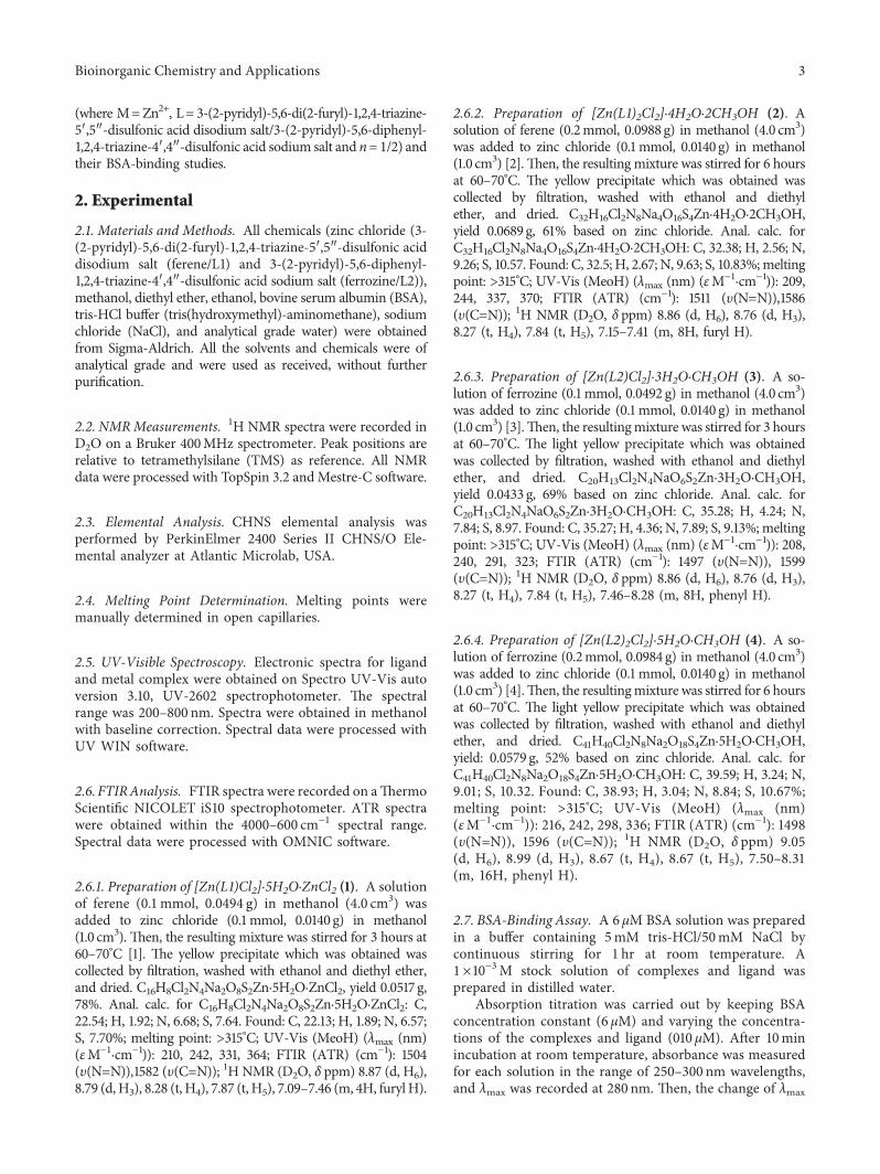
�us, we report here the synthesis and characterizationof four novel metal complexes of the type MLnCl2 (Figure 2)
5′ 5′
4′
6′
3′4′ 3′
6′ N N N N N
N
N
NO
O
O
O
O
O
OO
O
O
O
S
S
S
S
O Na
O Na
+−
+−
O Na+−
H
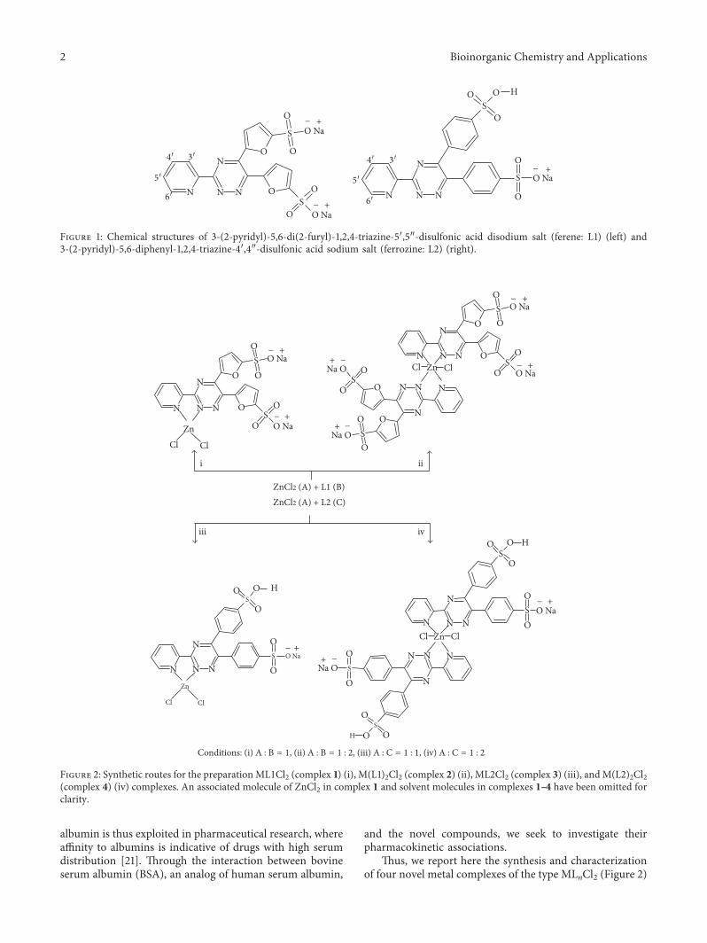
Figure 1: Chemical structures of 3-(2-pyridyl)-5,6-di(2-furyl)-1,2,4-triazine-5′,5″-disulfonic acid disodium salt (ferene: L1) (left) and3-(2-pyridyl)-5,6-diphenyl-1,2,4-triazine-4′,4″-disulfonic acid sodium salt (ferrozine: L2) (right).
N N N
N
N NN
N N N
N
N
NN N
N
ZnCl Cl
i
iii iv
ii
O OO
O
O
O
O
O
O
O
O
O
O
OO
O
O
O O
O
O
OS
S
S
S
O Na
O Na
+−
+−
O Na+−
O Na+−
+−
+ −
Na O
+ −
Na O
+ −
Na O
Cl ClZnO
O
O
O
O
O
S
S
ZnCl2 (A) + L1 (B)ZnCl2 (A) + L2 (C)
Cl Cl
Zn
S
S
S
S
O Na
H
N
N NN
N N N
N
Cl ClZn
O
O
O
O
OS
S+−
O Na
H
Conditions: (i) A : B = 1, (ii) A : B = 1 : 2, (iii) A : C = 1 : 1, (iv) A : C = 1 : 2
H
Figure 2: Synthetic routes for the preparationML1Cl2 (complex 1) (i), M(L1)2Cl2 (complex 2) (ii), ML2Cl2 (complex 3) (iii), andM(L2)2Cl2(complex 4) (iv) complexes. An associated molecule of ZnCl2 in complex 1 and solvent molecules in complexes 1–4 have been omitted forclarity.
2 Bioinorganic Chemistry and Applications
(where M�Zn2+, L� 3-(2-pyridyl)-5,6-di(2-furyl)-1,2,4-triazine-5′,5″-disulfonic acid disodium salt/3-(2-pyridyl)-5,6-diphenyl-1,2,4-triazine-4′,4″-disulfonic acid sodium salt and n� 1/2) andtheir BSA-binding studies.
2. Experimental
2.1. Materials and Methods. All chemicals (zinc chloride (3-(2-pyridyl)-5,6-di(2-furyl)-1,2,4-triazine-5′,5″-disulfonic aciddisodium salt (ferene/L1) and 3-(2-pyridyl)-5,6-diphenyl-1,2,4-triazine-4′,4″-disulfonic acid sodium salt (ferrozine/L2)),methanol, diethyl ether, ethanol, bovine serum albumin (BSA),tris-HCl buffer (tris(hydroxymethyl)-aminomethane), sodiumchloride (NaCl), and analytical grade water) were obtainedfrom Sigma-Aldrich. All the solvents and chemicals were ofanalytical grade and were used as received, without furtherpurification.
2.2. NMRMeasurements. 1H NMR spectra were recorded inD2O on a Bruker 400MHz spectrometer. Peak positions arerelative to tetramethylsilane (TMS) as reference. All NMRdata were processed with TopSpin 3.2 andMestre-C software.
2.3. Elemental Analysis. CHNS elemental analysis wasperformed by PerkinElmer 2400 Series II CHNS/O Ele-mental analyzer at Atlantic Microlab, USA.
2.4. Melting Point Determination. Melting points weremanually determined in open capillaries.
2.5. UV-Visible Spectroscopy. Electronic spectra for ligandand metal complex were obtained on Spectro UV-Vis autoversion 3.10, UV-2602 spectrophotometer. �e spectralrange was 200–800 nm. Spectra were obtained in methanolwith baseline correction. Spectral data were processed withUV WIN software.
2.6. FTIRAnalysis. FTIR spectra were recorded on a�ermoScientific NICOLET iS10 spectrophotometer. ATR spectrawere obtained within the 4000–600 cm−1 spectral range.Spectral data were processed with OMNIC software.
2.6.1. Preparation of [Zn(L1)Cl2]·5H2O·ZnCl2 (1). A solutionof ferene (0.1mmol, 0.0494 g) in methanol (4.0 cm3) wasadded to zinc chloride (0.1mmol, 0.0140 g) in methanol(1.0 cm3). �en, the resulting mixture was stirred for 3 hours at60–70°C [1]. �e yellow precipitate which was obtained wascollected by filtration, washed with ethanol and diethyl ether,and dried. C16H8Cl2N4Na2O8S2Zn·5H2O·ZnCl2, yield 0.0517 g,78%. Anal. calc. for C16H8Cl2N4Na2O8S2Zn·5H2O·ZnCl2: C,22.54; H, 1.92; N, 6.68; S, 7.64. Found: C, 22.13; H, 1.89; N, 6.57;S, 7.70%; melting point: >315°C; UV-Vis (MeoH) (λmax (nm)(εM−1·cm−1)): 210, 242, 331, 364; FTIR (ATR) (cm−1): 1504(υ(N�N)),1582 (υ(C�N)); 1H NMR (D2O, δ ppm) 8.87 (d, H6),8.79 (d,H3), 8.28 (t, H4), 7.87 (t, H5), 7.09–7.46 (m, 4H, furylH).
2.6.2. Preparation of [Zn(L1)2Cl2]·4H2O·2CH3OH (2). Asolution of ferene (0.2mmol, 0.0988 g) in methanol (4.0 cm3)was added to zinc chloride (0.1mmol, 0.0140 g) in methanol(1.0 cm3) [2].�en, the resulting mixture was stirred for 6 hoursat 60–70°C. �e yellow precipitate which was obtained wascollected by filtration, washed with ethanol and diethylether, and dried. C32H16Cl2N8Na4O16S4Zn·4H2O·2CH3OH,yield 0.0689g, 61% based on zinc chloride. Anal. calc. forC32H16Cl2N8Na4O16S4Zn·4H2O·2CH3OH: C, 32.38; H, 2.56; N,9.26; S, 10.57. Found: C, 32.5; H, 2.67; N, 9.63; S, 10.83%;meltingpoint: >315°C; UV-Vis (MeoH) (λmax (nm) (εM−1·cm−1)): 209,244, 337, 370; FTIR (ATR) (cm−1): 1511 (υ(N�N)),1586(υ(C�N)); 1H NMR (D2O, δ ppm) 8.86 (d, H6), 8.76 (d, H3),8.27 (t, H4), 7.84 (t, H5), 7.15–7.41 (m, 8H, furyl H).
2.6.3. Preparation of [Zn(L2)Cl2]·3H2O·CH3OH (3). A so-lution of ferrozine (0.1mmol, 0.0492 g) in methanol (4.0 cm3)was added to zinc chloride (0.1mmol, 0.0140 g) in methanol(1.0 cm3) [3].�en, the resultingmixture was stirred for 3 hoursat 60–70°C. �e light yellow precipitate which was obtainedwas collected by filtration, washed with ethanol and diethylether, and dried. C20H13Cl2N4NaO6S2Zn·3H2O·CH3OH,yield 0.0433 g, 69% based on zinc chloride. Anal. calc. forC20H13Cl2N4NaO6S2Zn·3H2O·CH3OH: C, 35.28; H, 4.24; N,7.84; S, 8.97. Found: C, 35.27; H, 4.36; N, 7.89; S, 9.13%;meltingpoint: >315°C; UV-Vis (MeoH) (λmax (nm) (εM−1·cm−1)): 208,240, 291, 323; FTIR (ATR) (cm−1): 1497 (υ(N�N)), 1599(υ(C�N)); 1H NMR (D2O, δ ppm) 8.86 (d, H6), 8.76 (d, H3),8.27 (t, H4), 7.84 (t, H5), 7.46–8.28 (m, 8H, phenyl H).
2.6.4. Preparation of [Zn(L2)2Cl2]·5H2O·CH3OH (4). A so-lution of ferrozine (0.2mmol, 0.0984 g) in methanol (4.0 cm3)was added to zinc chloride (0.1mmol, 0.0140 g) in methanol(1.0 cm3) [4].�en, the resultingmixture was stirred for 6 hoursat 60–70°C. �e light yellow precipitate which was obtainedwas collected by filtration, washed with ethanol and diethylether, and dried. C41H40Cl2N8Na2O18S4Zn·5H2O·CH3OH,yield: 0.0579 g, 52% based on zinc chloride. Anal. calc. forC41H40Cl2N8Na2O18S4Zn·5H2O·CH3OH: C, 39.59; H, 3.24; N,9.01; S, 10.32. Found: C, 38.93; H, 3.04; N, 8.84; S, 10.67%;melting point: >315°C; UV-Vis (MeoH) (λmax (nm)(εM−1·cm−1)): 216, 242, 298, 336; FTIR (ATR) (cm−1): 1498(υ(N�N)), 1596 (υ(C�N)); 1H NMR (D2O, δ ppm) 9.05(d, H6), 8.99 (d, H3), 8.67 (t, H4), 8.67 (t, H5), 7.50–8.31(m, 16H, phenyl H).
2.7. BSA-Binding Assay. A 6 µM BSA solution was preparedin a buffer containing 5mM tris-HCl/50mM NaCl bycontinuous stirring for 1 hr at room temperature. A1×10−3M stock solution of complexes and ligand wasprepared in distilled water.
Absorption titration was carried out by keeping BSAconcentration constant (6 µM) and varying the concentra-tions of the complexes and ligand (010 µM). After 10minincubation at room temperature, absorbance was measuredfor each solution in the range of 250–300 nm wavelengths,and λmax was recorded at 280 nm. �en, the change of λmax
Bioinorganic Chemistry and Applications 3
was recorded for each solution. All absorbance measure-ments were triplicated and corrected for background ab-sorbance by the compounds. �e plot of 1/(A−A0) (whereA0 is the initial absorbance of the free BSA at 280 nm andA isthe absorbance of BSA in the presence of different con-centrations of the complex) versus 1/[complex] is a linearcurve, and the binding constant (Kb) can be obtained fromthe ratio of the intercept to slope [19].
3. Results and Discussion
3.1. Synthesis. In order to synthesize the metal complexes,zinc chloride and the relevant ligands in 1 :1 and 1 : 2 ratioswere used (Figure 2).
3.2. UV-Visible Spectroscopy. UV-visible spectra of ligandsand complexes 1–4 recorded in methanol showed significantdifferences between the absorption peaks of ligands andtheir complexes (Table 1, Figures S1 and S2, SupportingInformation). Since both ligands bear conjugated systems,π–π∗ transition is possible. In all four complexes, thewavelengths have shifted towards the longer wavelengthrange (bathochromic shift) because of changes in the con-jugated electron system due to formation of metal ligandbonds. �ese observations are in agreement with previouslyreported zinc pyridyl triazine derivatives [18] and copperpyridyl triazine derivatives [22], upon coordination of ligandto metal.
3.3. 1H NMR Analysis. Complexes 1–4 were characterizedusing 1H NMR spectroscopy in D2O. All the peaks were
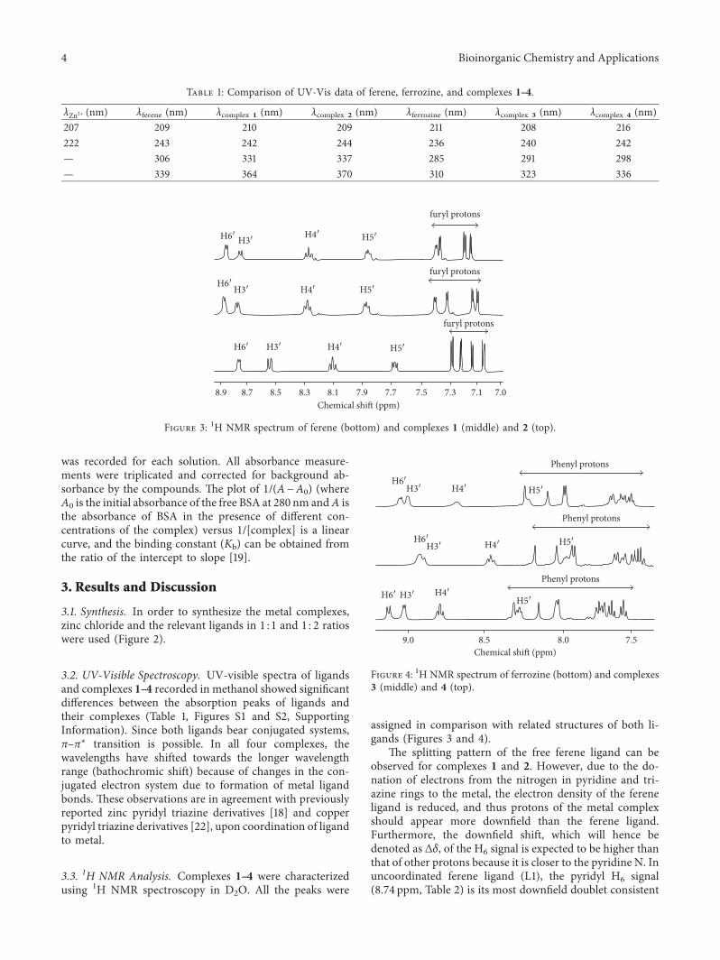
assigned in comparison with related structures of both li-gands (Figures 3 and 4).
�e splitting pattern of the free ferene ligand can beobserved for complexes 1 and 2. However, due to the do-nation of electrons from the nitrogen in pyridine and tri-azine rings to the metal, the electron density of the fereneligand is reduced, and thus protons of the metal complexshould appear more downfield than the ferene ligand.Furthermore, the downfield shift, which will hence bedenoted as ∆δ, of the H6 signal is expected to be higher thanthat of other protons because it is closer to the pyridine N. Inuncoordinated ferene ligand (L1), the pyridyl H6 signal(8.74 ppm, Table 2) is its most downfield doublet consistent
Table 1: Comparison of UV-Vis data of ferene, ferrozine, and complexes 1–4.
λZn2+ (nm) λferene (nm) λcomplex 1 (nm) λcomplex 2 (nm) λferrozine (nm) λcomplex 3 (nm) λcomplex 4 (nm)207 209 210 209 211 208 216222 243 242 244 236 240 242— 306 331 337 285 291 298— 339 364 370 310 323 336
H6′
H6′
H3′
H3′ H4′ H5′
H6′ H3′ H4′ H5′
H5′H4′
furyl protons
furyl protons
furyl protons
8.9 8.7 8.5 8.3 8.1 7.9 7.7 7.5 7.3 7.1 7.0Chemical shift (ppm)
Figure 3: 1H NMR spectrum of ferene (bottom) and complexes 1 (middle) and 2 (top).
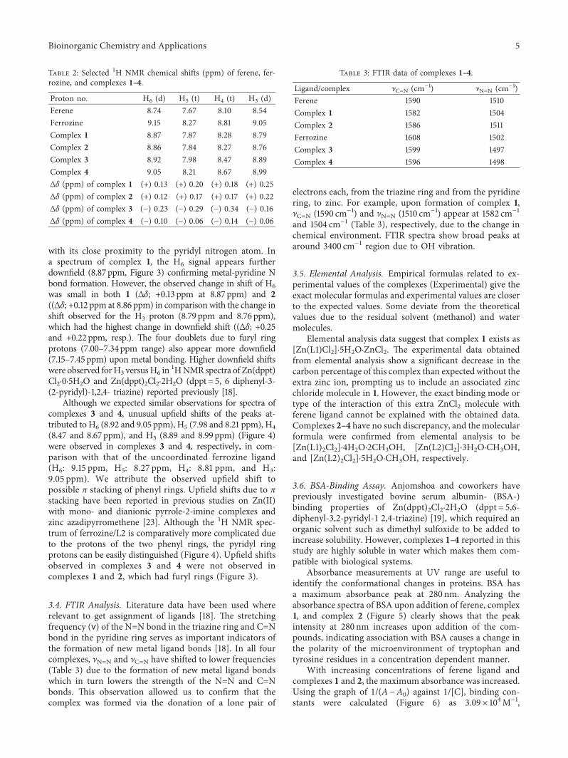
9.0 8.5 8.0 7.5Chemical shi� (ppm)
Phenyl protons
Phenyl protons
Phenyl protons
H6′H3′ H4′ H5′
H6′H3′ H4′ H5′
H6′ H3′ H4′H5′
Figure 4: 1H NMR spectrum of ferrozine (bottom) and complexes3 (middle) and 4 (top).
4 Bioinorganic Chemistry and Applications
with its close proximity to the pyridyl nitrogen atom. Ina spectrum of complex 1, the H6 signal appears furtherdownfield (8.87 ppm, Figure 3) confirming metal-pyridine Nbond formation. However, the observed change in shift of H6was small in both 1 (∆δ; +0.13 ppm at 8.87 ppm) and 2((∆δ; +0.12 ppm at 8.86ppm) in comparison with the change inshift observed for the H3 proton (8.79 ppm and 8.76ppm),which had the highest change in downfield shift ((∆δ; +0.25and +0.22ppm, resp.). �e four doublets due to furyl ringprotons (7.00–7.34 ppm range) also appear more downfield(7.15–7.45ppm) upon metal bonding. Higher downfield shiftswere observed forH3 versusH6 in 1HNMR spectra of Zn(dppt)Cl2·0·5H2O and Zn(dppt)2Cl2·2H2O (dppt� 5, 6 diphenyl-3-(2-pyridyl)-1,2,4- triazine) reported previously [18].
Although we expected similar observations for spectra ofcomplexes 3 and 4, unusual upfield shifts of the peaks at-tributed to H6 (8.92 and 9.05ppm), H5 (7.98 and 8.21ppm), H4(8.47 and 8.67ppm), and H3 (8.89 and 8.99ppm) (Figure 4)were observed in complexes 3 and 4, respectively, in com-parison with that of the uncoordinated ferrozine ligand(H6: 9.15 ppm, H5: 8.27 ppm, H4: 8.81 ppm, and H3:9.05 ppm). We attribute the observed upfield shift topossible π stacking of phenyl rings. Upfield shifts due to πstacking have been reported in previous studies on Zn(II)with mono- and dianionic pyrrole-2-imine complexes andzinc azadipyrromethene [23]. Although the 1H NMR spec-trum of ferrozine/L2 is comparatively more complicated dueto the protons of the two phenyl rings, the pyridyl ringprotons can be easily distinguished (Figure 4). Upfield shiftsobserved in complexes 3 and 4 were not observed incomplexes 1 and 2, which had furyl rings (Figure 3).
3.4. FTIR Analysis. Literature data have been used whererelevant to get assignment of ligands [18]. �e stretchingfrequency (ѵ) of the N�N bond in the triazine ring and C�Nbond in the pyridine ring serves as important indicators ofthe formation of new metal ligand bonds [18]. In all fourcomplexes, ]N�N and ]C�N have shifted to lower frequencies(Table 3) due to the formation of new metal ligand bondswhich in turn lowers the strength of the N�N and C�Nbonds. �is observation allowed us to confirm that thecomplex was formed via the donation of a lone pair of
electrons each, from the triazine ring and from the pyridinering, to zinc. For example, upon formation of complex 1,]C�N (1590 cm−1) and ]N�N (1510 cm−1) appear at 1582 cm−1and 1504 cm−1 (Table 3), respectively, due to the change inchemical environment. FTIR spectra show broad peaks ataround 3400 cm−1 region due to OH vibration.
3.5. Elemental Analysis. Empirical formulas related to ex-perimental values of the complexes (Experimental) give theexact molecular formulas and experimental values are closerto the expected values. Some deviate from the theoreticalvalues due to the residual solvent (methanol) and watermolecules.
Elemental analysis data suggest that complex 1 exists as[Zn(L1)Cl2]·5H2O·ZnCl2. �e experimental data obtainedfrom elemental analysis show a significant decrease in thecarbon percentage of this complex than expected without theextra zinc ion, prompting us to include an associated zincchloride molecule in 1. However, the exact binding mode ortype of the interaction of this extra ZnCl2 molecule withferene ligand cannot be explained with the obtained data.Complexes 2–4 have no such discrepancy, and the molecularformula were confirmed from elemental analysis to be[Zn(L1)2Cl2]·4H2O·2CH3OH, [Zn(L2)Cl2]·3H2O·CH3OH,and [Zn(L2)2Cl2]·5H2O·CH3OH, respectively.
3.6. BSA-Binding Assay. Anjomshoa and coworkers havepreviously investigated bovine serum albumin- (BSA-)binding properties of Zn(dppt)2Cl2·2H2O (dppt� 5,6-diphenyl-3,2-pyridyl-1 2,4-triazine) [19], which required anorganic solvent such as dimethyl sulfoxide to be added toincrease solubility. However, complexes 1–4 reported in thisstudy are highly soluble in water which makes them com-patible with biological systems.
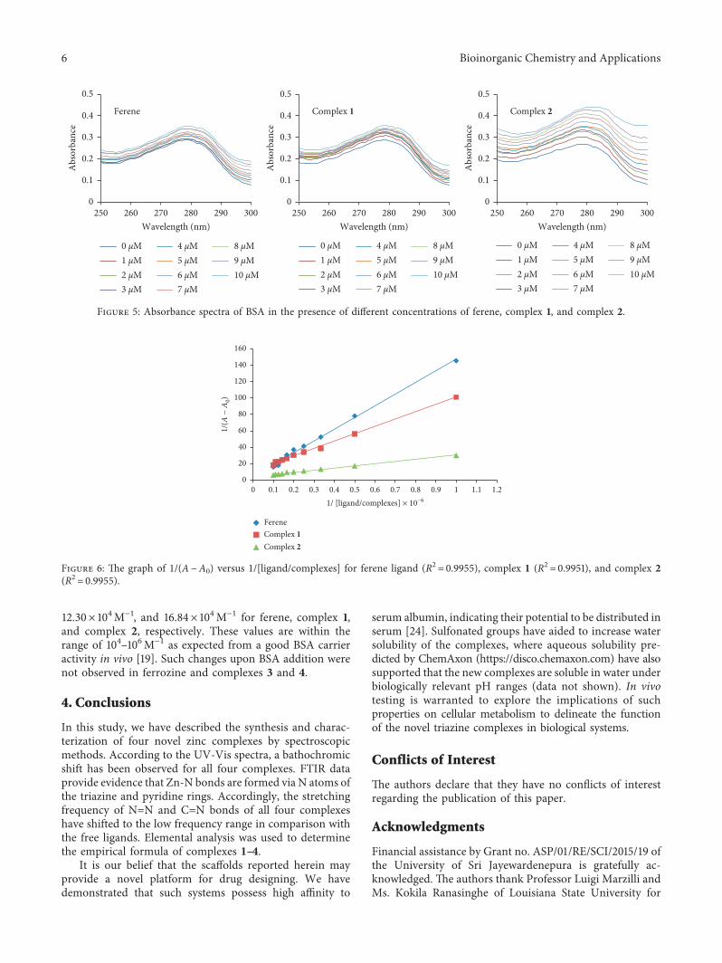
Absorbance measurements at UV range are useful toidentify the conformational changes in proteins. BSA hasa maximum absorbance peak at 280 nm. Analyzing theabsorbance spectra of BSA upon addition of ferene, complex1, and complex 2 (Figure 5) clearly shows that the peakintensity at 280 nm increases upon addition of the com-pounds, indicating association with BSA causes a change inthe polarity of the microenvironment of tryptophan andtyrosine residues in a concentration dependent manner.
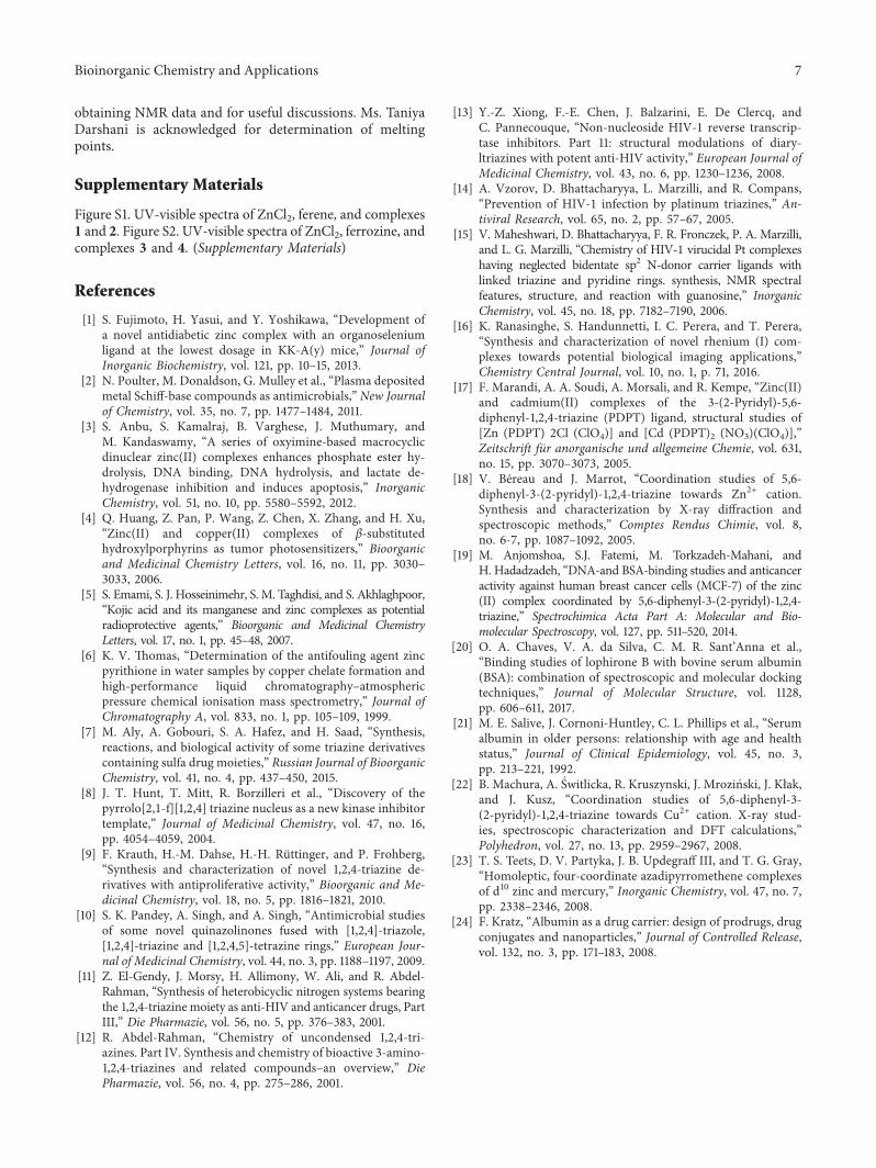
With increasing concentrations of ferene ligand andcomplexes 1 and 2, the maximum absorbance was increased.Using the graph of 1/(A−A0) against 1/[C], binding con-stants were calculated (Figure 6) as 3.09×10⁴M−1,
Table 2: Selected 1H NMR chemical shifts (ppm) of ferene, fer-rozine, and complexes 1–4.
Proton no. H6 (d) H5 (t) H4 (t) H3 (d)Ferene 8.74 7.67 8.10 8.54Ferrozine 9.15 8.27 8.81 9.05Complex 1 8.87 7.87 8.28 8.79Complex 2 8.86 7.84 8.27 8.76Complex 3 8.92 7.98 8.47 8.89Complex 4 9.05 8.21 8.67 8.99∆δ (ppm) of complex 1 (+) 0.13 (+) 0.20 (+) 0.18 (+) 0.25∆δ (ppm) of complex 2 (+) 0.12 (+) 0.17 (+) 0.17 (+) 0.22∆δ (ppm) of complex 3 (−) 0.23 (−) 0.29 (−) 0.34 (−) 0.16∆δ (ppm) of complex 4 (−) 0.10 (−) 0.06 (−) 0.14 (−) 0.06
Table 3: FTIR data of complexes 1–4.
Ligand/complex ]C�N (cm−1) ]N�N (cm−1)Ferene 1590 1510Complex 1 1582 1504Complex 2 1586 1511Ferrozine 1608 1502Complex 3 1599 1497Complex 4 1596 1498
Bioinorganic Chemistry and Applications 5
12.30×10⁴M−1, and 16.84×10⁴M−1 for ferene, complex 1,and complex 2, respectively. These values are within therange of 104–106M−1 as expected from a good BSA carrieractivity in vivo [19]. Such changes upon BSA addition werenot observed in ferrozine and complexes 3 and 4.
4. Conclusions
In this study, we have described the synthesis and charac-terization of four novel zinc complexes by spectroscopicmethods. According to the UV-Vis spectra, a bathochromicshift has been observed for all four complexes. FTIR dataprovide evidence that Zn-N bonds are formed via N atoms ofthe triazine and pyridine rings. Accordingly, the stretchingfrequency of N�N and C�N bonds of all four complexeshave shifted to the low frequency range in comparison withthe free ligands. Elemental analysis was used to determinethe empirical formula of complexes 1–4.
It is our belief that the sca�olds reported herein mayprovide a novel platform for drug designing. We havedemonstrated that such systems possess high a�nity to
serum albumin, indicating their potential to be distributed inserum [24]. Sulfonated groups have aided to increase watersolubility of the complexes, where aqueous solubility pre-dicted by ChemAxon (https://disco.chemaxon.com) have alsosupported that the new complexes are soluble in water underbiologically relevant pH ranges (data not shown). In vivotesting is warranted to explore the implications of suchproperties on cellular metabolism to delineate the functionof the novel triazine complexes in biological systems.
Conflicts of Interest
�e authors declare that they have no con�icts of interestregarding the publication of this paper.
Acknowledgments
Financial assistance by Grant no. ASP/01/RE/SCI/2015/19 ofthe University of Sri Jayewardenepura is gratefully ac-knowledged. �e authors thank Professor Luigi Marzilli andMs. Kokila Ranasinghe of Louisiana State University for
0 µM1 µM2 µM3 µM
4 µM5 µM6 µM7 µM
8 µM9 µM10 µM
0
0.1
0.2
0.3
0.4
0.5
250 260 270 280 290 300
Abs
orba
nce
Wavelength (nm)
Ferene Complex 2
0
0.1
0.2
0.3
0.4
0.5
250 260 270 280 290 300
Abs
orba
nce
Wavelength (nm)
Complex 1
0
0.1
0.2
0.3
0.4
0.5
250 260 270 280 290 300
Abs
orba
nce
Wavelength (nm)
0 µM1 µM2 µM3 µM
4 µM5 µM6 µM7 µM
8 µM9 µM10 µM
0 µM1 µM2 µM3 µM
4 µM5 µM6 µM7 µM
8 µM9 µM10 µM
Figure 5: Absorbance spectra of BSA in the presence of di�erent concentrations of ferene, complex 1, and complex 2.
00 0.2 0.4 0.6 0.8 1
20
40
60
80
100
120
140
160
1/(A
− A
0)
1/ [ligand/complexes] × 10−6
FereneComplex 1Complex 2
0.1 0.3 0.5 0.90.7 1.1 1.2
Figure 6: �e graph of 1/(A−A0) versus 1/[ligand/complexes] for ferene ligand (R2� 0.9955), complex 1 (R2� 0.9951), and complex 2(R2� 0.9955).
6 Bioinorganic Chemistry and Applications
obtaining NMR data and for useful discussions. Ms. TaniyaDarshani is acknowledged for determination of meltingpoints.
Supplementary Materials
Figure S1. UV-visible spectra of ZnCl2, ferene, and complexes1 and 2. Figure S2. UV-visible spectra of ZnCl2, ferrozine, andcomplexes 3 and 4. (Supplementary Materials)
References
[1] S. Fujimoto, H. Yasui, and Y. Yoshikawa, “Development ofa novel antidiabetic zinc complex with an organoseleniumligand at the lowest dosage in KK-A(y) mice,” Journal ofInorganic Biochemistry, vol. 121, pp. 10–15, 2013.
[2] N. Poulter, M. Donaldson, G. Mulley et al., “Plasma depositedmetal Schiff-base compounds as antimicrobials,” New Journalof Chemistry, vol. 35, no. 7, pp. 1477–1484, 2011.
[3] S. Anbu, S. Kamalraj, B. Varghese, J. Muthumary, andM. Kandaswamy, “A series of oxyimine-based macrocyclicdinuclear zinc(II) complexes enhances phosphate ester hy-drolysis, DNA binding, DNA hydrolysis, and lactate de-hydrogenase inhibition and induces apoptosis,” InorganicChemistry, vol. 51, no. 10, pp. 5580–5592, 2012.
[4] Q. Huang, Z. Pan, P. Wang, Z. Chen, X. Zhang, and H. Xu,“Zinc(II) and copper(II) complexes of β-substitutedhydroxylporphyrins as tumor photosensitizers,” Bioorganicand Medicinal Chemistry Letters, vol. 16, no. 11, pp. 3030–3033, 2006.
[5] S. Emami, S. J. Hosseinimehr, S. M. Taghdisi, and S. Akhlaghpoor,“Kojic acid and its manganese and zinc complexes as potentialradioprotective agents,” Bioorganic and Medicinal ChemistryLetters, vol. 17, no. 1, pp. 45–48, 2007.
[6] K. V. �omas, “Determination of the antifouling agent zincpyrithione in water samples by copper chelate formation andhigh-performance liquid chromatography–atmosphericpressure chemical ionisation mass spectrometry,” Journal ofChromatography A, vol. 833, no. 1, pp. 105–109, 1999.
[7] M. Aly, A. Gobouri, S. A. Hafez, and H. Saad, “Synthesis,reactions, and biological activity of some triazine derivativescontaining sulfa drug moieties,” Russian Journal of BioorganicChemistry, vol. 41, no. 4, pp. 437–450, 2015.
[8] J. T. Hunt, T. Mitt, R. Borzilleri et al., “Discovery of thepyrrolo[2,1-f][1,2,4] triazine nucleus as a new kinase inhibitortemplate,” Journal of Medicinal Chemistry, vol. 47, no. 16,pp. 4054–4059, 2004.
[9] F. Krauth, H.-M. Dahse, H.-H. Ruttinger, and P. Frohberg,“Synthesis and characterization of novel 1,2,4-triazine de-rivatives with antiproliferative activity,” Bioorganic and Me-dicinal Chemistry, vol. 18, no. 5, pp. 1816–1821, 2010.
[10] S. K. Pandey, A. Singh, and A. Singh, “Antimicrobial studiesof some novel quinazolinones fused with [1,2,4]-triazole,[1,2,4]-triazine and [1,2,4,5]-tetrazine rings,” European Jour-nal of Medicinal Chemistry, vol. 44, no. 3, pp. 1188–1197, 2009.
[11] Z. El-Gendy, J. Morsy, H. Allimony, W. Ali, and R. Abdel-Rahman, “Synthesis of heterobicyclic nitrogen systems bearingthe 1,2,4-triazinemoiety as anti-HIV and anticancer drugs, PartIII,” Die Pharmazie, vol. 56, no. 5, pp. 376–383, 2001.
[12] R. Abdel-Rahman, “Chemistry of uncondensed 1,2,4-tri-azines. Part IV. Synthesis and chemistry of bioactive 3-amino-1,2,4-triazines and related compounds–an overview,” DiePharmazie, vol. 56, no. 4, pp. 275–286, 2001.
[13] Y.-Z. Xiong, F.-E. Chen, J. Balzarini, E. De Clercq, andC. Pannecouque, “Non-nucleoside HIV-1 reverse transcrip-tase inhibitors. Part 11: structural modulations of diary-ltriazines with potent anti-HIV activity,” European Journal ofMedicinal Chemistry, vol. 43, no. 6, pp. 1230–1236, 2008.
[14] A. Vzorov, D. Bhattacharyya, L. Marzilli, and R. Compans,“Prevention of HIV-1 infection by platinum triazines,” An-tiviral Research, vol. 65, no. 2, pp. 57–67, 2005.
[15] V. Maheshwari, D. Bhattacharyya, F. R. Fronczek, P. A. Marzilli,and L. G. Marzilli, “Chemistry of HIV-1 virucidal Pt complexeshaving neglected bidentate sp2 N-donor carrier ligands withlinked triazine and pyridine rings. synthesis, NMR spectralfeatures, structure, and reaction with guanosine,” InorganicChemistry, vol. 45, no. 18, pp. 7182–7190, 2006.
[16] K. Ranasinghe, S. Handunnetti, I. C. Perera, and T. Perera,“Synthesis and characterization of novel rhenium (I) com-plexes towards potential biological imaging applications,”Chemistry Central Journal, vol. 10, no. 1, p. 71, 2016.
[17] F. Marandi, A. A. Soudi, A. Morsali, and R. Kempe, “Zinc(II)and cadmium(II) complexes of the 3-(2-Pyridyl)-5,6-diphenyl-1,2,4-triazine (PDPT) ligand, structural studies of[Zn (PDPT) 2Cl (ClO4)] and [Cd (PDPT)2 (NO3)(ClO4)],”Zeitschrift fur anorganische und allgemeine Chemie, vol. 631,no. 15, pp. 3070–3073, 2005.
[18] V. Bereau and J. Marrot, “Coordination studies of 5,6-diphenyl-3-(2-pyridyl)-1,2,4-triazine towards Zn2+ cation.Synthesis and characterization by X-ray diffraction andspectroscopic methods,” Comptes Rendus Chimie, vol. 8,no. 6-7, pp. 1087–1092, 2005.
[19] M. Anjomshoa, S.J. Fatemi, M. Torkzadeh-Mahani, andH. Hadadzadeh, “DNA-and BSA-binding studies and anticanceractivity against human breast cancer cells (MCF-7) of the zinc(II) complex coordinated by 5,6-diphenyl-3-(2-pyridyl)-1,2,4-triazine,” Spectrochimica Acta Part A: Molecular and Bio-molecular Spectroscopy, vol. 127, pp. 511–520, 2014.
[20] O. A. Chaves, V. A. da Silva, C. M. R. Sant’Anna et al.,“Binding studies of lophirone B with bovine serum albumin(BSA): combination of spectroscopic and molecular dockingtechniques,” Journal of Molecular Structure, vol. 1128,pp. 606–611, 2017.
[21] M. E. Salive, J. Cornoni-Huntley, C. L. Phillips et al., “Serumalbumin in older persons: relationship with age and healthstatus,” Journal of Clinical Epidemiology, vol. 45, no. 3,pp. 213–221, 1992.
[22] B. Machura, A. Switlicka, R. Kruszynski, J. Mrozinski, J. Kłak,and J. Kusz, “Coordination studies of 5,6-diphenyl-3-(2-pyridyl)-1,2,4-triazine towards Cu2+ cation. X-ray stud-ies, spectroscopic characterization and DFT calculations,”Polyhedron, vol. 27, no. 13, pp. 2959–2967, 2008.
[23] T. S. Teets, D. V. Partyka, J. B. Updegraff III, and T. G. Gray,“Homoleptic, four-coordinate azadipyrromethene complexesof d10 zinc and mercury,” Inorganic Chemistry, vol. 47, no. 7,pp. 2338–2346, 2008.
[24] F. Kratz, “Albumin as a drug carrier: design of prodrugs, drugconjugates and nanoparticles,” Journal of Controlled Release,vol. 132, no. 3, pp. 171–183, 2008.
Bioinorganic Chemistry and Applications 7
TribologyAdvances in
Hindawiwww.hindawi.com Volume 2018
Hindawiwww.hindawi.com Volume 2018
International Journal ofInternational Journal ofPhotoenergy
Hindawiwww.hindawi.com Volume 2018
Journal of
Chemistry
Hindawiwww.hindawi.com Volume 2018
Advances inPhysical Chemistry
Hindawiwww.hindawi.com
Analytical Methods in Chemistry
Journal of
Volume 2018
Bioinorganic Chemistry and ApplicationsHindawiwww.hindawi.com Volume 2018
SpectroscopyInternational Journal of
Hindawiwww.hindawi.com Volume 2018
Hindawi Publishing Corporation http://www.hindawi.com Volume 2013Hindawiwww.hindawi.com
The Scientific World Journal
Volume 2018
Medicinal ChemistryInternational Journal of
Hindawiwww.hindawi.com Volume 2018
NanotechnologyHindawiwww.hindawi.com Volume 2018
Journal of
Applied ChemistryJournal of
Hindawiwww.hindawi.com Volume 2018
Hindawiwww.hindawi.com Volume 2018
Biochemistry Research International
Hindawiwww.hindawi.com Volume 2018
Enzyme Research
Hindawiwww.hindawi.com Volume 2018
Journal of
SpectroscopyAnalytical ChemistryInternational Journal of
Hindawiwww.hindawi.com Volume 2018
MaterialsJournal of
Hindawiwww.hindawi.com Volume 2018
Hindawiwww.hindawi.com Volume 2018
BioMed Research International Electrochemistry
International Journal of
Hindawiwww.hindawi.com Volume 2018
Na
nom
ate
ria
ls
Hindawiwww.hindawi.com Volume 2018
Journal ofNanomaterials
Submit your manuscripts atwww.hindawi.com