syndrome of inappropriate anti-diuretic hormone … a sixty-four year lady who underwent an elective...
TRANSCRIPT
dRe Case Report
Malta Medical Journal Volume 28 Issue 02 2016
Abstract
Neurosurgical procedures in cases of Type 1
Arnold Chiari Malformation (ACM) may result in a
wide spectrum of complications.1 We report a case
of a sixty-four year lady who underwent an elective
posterior fossa decompression for Type 1 ACM.
The procedure was complicated by syndrome of
inappropriate anti-diuretic hormone secretion
(SIADH) and an ischaemic cerebrovascular event
affecting the posterior cerebral artery. The
association of these complications with the
procedure is rarely described in the literature. In
spite of the poor prognosis associated with such
complications, the patient made a relatively quick
and uneventful recovery.
Keywords
Neurosurgery, Arnold Chiari Malformation
(ACM), Hyponatraemia, Syndrome of inappropriate
anti-diuretic hormone secretion (SIADH),
Cerebrovascular accident
Case Presentation
A sixty-four year old lady was admitted for an
elective posterior fossa decompression for Type 1
Arnold Chiari Malformation (ACM). The patient
had presented with a longstanding history of
headaches and lower limb weakness and numbness.
The only positive finding on neurological
examination was clonus. The diagnosis of Type 1
ACM was confirmed on magnetic resonance
imaging (MRI) of the brain which revealed low
lying cerebellar tonsils associated with cervico-
medullary kinking (Figure 1). The patient had a past
medical history of hypothyroidism and
hypertension which were well controlled on
medications.
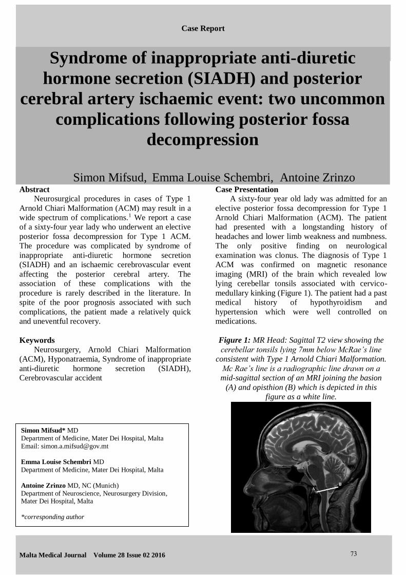
Figure 1: MR Head: Sagittal T2 view showing the
cerebellar tonsils lying 7mm below McRae’s line
consistent with Type 1 Arnold Chiari Malformation.
Mc Rae’s line is a radiographic line drawn on a
mid-sagittal section of an MRI joining the basion
(A) and opisthion (B) which is depicted in this
figure as a white line.
Syndrome of inappropriate anti-diuretic
hormone secretion (SIADH) and posterior
cerebral artery ischaemic event: two uncommon
complications following posterior fossa
decompression
Simon Mifsud, Emma Louise Schembri, Antoine Zrinzo
Simon Mifsud* MD
Department of Medicine, Mater Dei Hospital, Malta
Email: [email protected]
Emma Louise Schembri MD
Department of Medicine, Mater Dei Hospital, Malta
Antoine Zrinzo MD, NC (Munich)
Department of Neuroscience, Neurosurgery Division, Mater Dei Hospital, Malta
*corresponding author
73

dRe Case Report
Malta Medical Journal Volume 28 Issue 02 2016
Posterior fossa decompression was carried out
uneventfully. Following the procedure, the patient
was well and did not have apnoeic episodes
throughout the night. On the first post-operative
day, she was tolerating oral liquids and solids and
her speech was normal. All of the former were
indicative of intact brainstem function. However, by
the second day post-operatively she started to
complain of persistent headaches, nausea and
fatigue.
Physical examination revealed a drowsy patient
who was afebrile, normotensive and was not
tachycardic. She was not cooperative for a full
neurological examination; however there was no
pronator drift and no apparent focal neurological
deficit. The rest of the examination was
unremarkable.
Peripheral blood investigations revealed a
serum sodium level of 120mmol/L (normal values:
135-145mmol/L). This had dropped from
140mmol/L overnight. Serum osmolality was
253mOsm/kg (normal values: 275-299mOsm/kg),
urine osmolality was 728mOsm/kg (normal values:
50-1200mOsm/kg) and urine sodium was
291mmol/L (normal values: 54-190mmol/L).
Serum cortisol level was elevated at 1804nmol/L
(119-618nmol/L). Complete blood count, thyroid
function tests, lipid profile, and total protein and
albumin levels were normal. The aforementioned
blood tests in addition with the patient’s normal
blood pressure satisfied the Bartter-Schwartz
Diagnostic Criteria for the syndrome of
inappropriate anti-diuretic hormone secretion
(SIADH).2 An urgent computed tomography (CT)
scan of the brain revealed hypo-density in the left
occipito-temporal region but no haemorrhage.
In view of the hyponatraemia, the patient was
kept nil by mouth and started on 0.9% saline
infusion which was restricted to 1.5 litres daily.
Despite this management, the patient’s sodium level
was on the decline. After four hours, the sodium
level decreased further to 115mmol/L and clinical
symptoms worsened. In view of the risks of seizing,
the patient was transferred to the intensive therapy
unit for administration of intra-venous 1.8%
hypertonic saline.
Twelve hours after the administration of
hypertonic saline, the patient’s clinical condition
improved and her serum sodium level increased to
120mmol/L. Neurological examination was
repeated since the patient was now more co-
operative. The only positive finding was a right
sided homonymous hemi-anopia. She underwent an
urgent MRI brain which revealed an acute
ischaemic stroke in the left posterior cerebral artery
territory with a small focus in the medial aspect of
the right cerebellar hemisphere (Figure 2). A
magnetic resonance angiogram (MRA) revealed
that a thrombus had occluded the left posterior
cerebral artery. She was therefore started on aspirin
and dipyridamole.
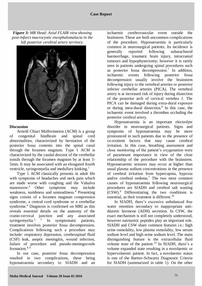
Figure 2: MR Head: Axial FLAIR view showing
hyper-intensity in the left posterior cerebral artery
territory indicative of an acute ischaemic stroke.
After four days of hypertonic saline
administration, the serum sodium level gradually
increased to 136mmol/L. At this point, the patient
was transferred to the neurosurgical ward. She
made a steady recovery with the help of the multi-
disciplinary team. The patient was discharged
fifteen days post-operatively with a serum sodium
level of 137mmol/L and a visual field assessment
which revealed right sided superior quadrant-
anopia. By the time of discharge she was
completely independent.
The patient was reviewed one month later at an
outpatient appointment. She remained well. Her
serum sodium level was 144mmol/L. A repeat MRI
brain revealed post-infarct macrocystic
encephalomalacia in the left posterior cerebral
artery territory (Figure 3).
74
dRe Case Report
Malta Medical Journal Volume 28 Issue 02 2016
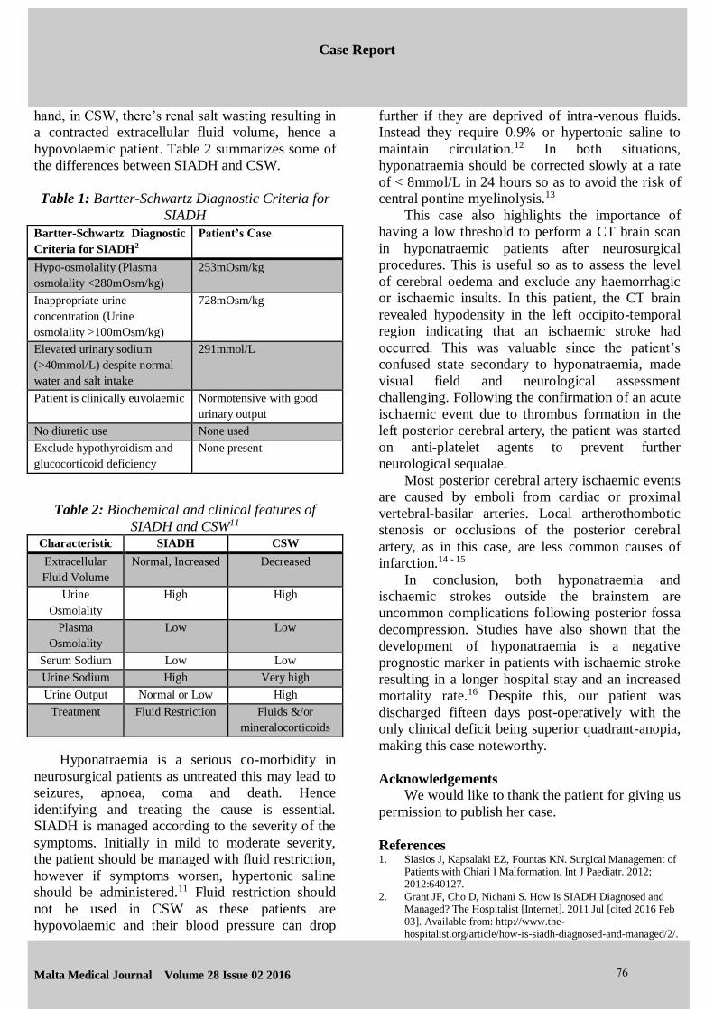
Figure 3: MR Head: Axial FLAIR view showing
post-infarct macrocystic encephalomalacia in the
left posterior cerebral artery territory.
Discussion
Arnold Chiari Malformation (ACM) is a group
of congenital hindbrain and spinal cord
abnormalities, characterized by herniation of the
posterior fossa contents into the spinal canal
through the foramen magnum. Type 1 ACM is
characterized by the caudal descent of the cerebellar
tonsils through the foramen magnum by at least 3-
5mm. It may be associated with an elongated fourth
ventricle, syringomyelia and medullary kinking.1
Type 1 ACM classically presents in adult life
with symptoms of headaches and neck pain which
are made worse with coughing and the Valsalva
manoeuvre.3 Other symptoms may include
weakness, numbness and unsteadiness.4 Presenting
signs consist of a foramen magnum compression
syndrome, a central cord syndrome or a cerebellar
syndrome.4 Diagnosis is confirmed on MRI as this
reveals essential details on the anatomy of the
cranio-cervical junction and any associated
syringomyelia.1, 5 In symptomatic patients,
treatment involves posterior fossa decompression.
Complications following such a procedure may
include: respiratory depression, cerebrospinal fluid
(CSF) leak, aseptic meningitis, wound infection,
failure of procedure and pseudo-meningocoele
formation.4, 6
In our case, posterior fossa decompression
resulted in two complications, these being
hyponatraemia secondary to SIADH and an
ischaemic cerebrovascular event outside the
brainstem. These are both uncommon complications
of the procedure. Hyponatraemia is particularly
common in neurosurgical patients. Its incidence is
generally reported following subarachnoid
haemorrhage, traumatic brain injury, intracranial
tumours and hypophysectomy; however it is rarely
seen in patients undergoing spinal procedures such
as posterior fossa decompression.7 In addition,
ischaemic events following posterior fossa
decompression usually involve the brainstem
following injury to the vertebral arteries or posterior
inferior cerebellar arteries (PICA). The vertebral
artery is at increased risk of injury during dissection
of the posterior arch of cervical vertebra 1. The
PICA can be damaged during extra-dural exposure
or during intra-dural dissection.8 In this case, the
ischaemic event involved a thrombus occluding the
posterior cerebral artery.
Hyponatraemia is an important electrolyte
disorder in neurosurgical patients. Signs and
symptoms of hyponatraemia may be more
pronounced in such patients due to the presence of
co-existent factors that may cause cerebral
irritation. In this case, breathing assessment and
close monitoring of the patient’s oxygenation were
of paramount importance in view of the close
relationship of the procedure with the brainstem.
Hyponatraemic seizures may occur at higher than
usual plasma sodium concentrations in the presence
of cerebral irritation from hypercapnia, hypoxia
and/or cerebral oedema.7 The two most common
causes of hyponatraemia following neurosurgical
procedures are SIADH and cerebral salt wasting
(CSW).9 Differentiating the two conditions is
essential, as their treatment is different.10
In SIADH, there’s excessive unbalanced free
water retention secondary to inappropriate anti-
diuretic hormone (ADH) secretion. In CSW, the
exact mechanism is still not completely understood,
however natriuretic peptides play an important role.
SIADH and CSW share common features i.e.: high
urine osmolality, low plasma osmolality, low serum
sodium level and high urine sodium level. The main
distinguishing feature is the extracellular fluid
volume state of the patient.10 In SIADH, there’s a
volume expanded state resulting in a euvolaemic or
hypervolaemic patient. In fact, a euvolaemic status
is one of the Bartter-Schwartz Diagnostic Criteria
for SIADH (summarized in table 1). On the other
75
dRe Case Report
Malta Medical Journal Volume 28 Issue 02 2016
hand, in CSW, there’s renal salt wasting resulting in
a contracted extracellular fluid volume, hence a
hypovolaemic patient. Table 2 summarizes some of
the differences between SIADH and CSW.
Table 1: Bartter-Schwartz Diagnostic Criteria for
SIADH
Table 2: Biochemical and clinical features of
SIADH and CSW11 Characteristic SIADH CSW
Extracellular
Fluid Volume
Normal, Increased Decreased
Urine
Osmolality
High High
Plasma
Osmolality
Low Low
Serum Sodium Low Low
Urine Sodium High Very high
Urine Output Normal or Low High
Treatment Fluid Restriction Fluids &/or
mineralocorticoids
Hyponatraemia is a serious co-morbidity in
neurosurgical patients as untreated this may lead to
seizures, apnoea, coma and death. Hence
identifying and treating the cause is essential.
SIADH is managed according to the severity of the
symptoms. Initially in mild to moderate severity,
the patient should be managed with fluid restriction,
however if symptoms worsen, hypertonic saline
should be administered.11 Fluid restriction should
not be used in CSW as these patients are
hypovolaemic and their blood pressure can drop
further if they are deprived of intra-venous fluids.
Instead they require 0.9% or hypertonic saline to
maintain circulation.12 In both situations,
hyponatraemia should be corrected slowly at a rate
of < 8mmol/L in 24 hours so as to avoid the risk of
central pontine myelinolysis.13
This case also highlights the importance of
having a low threshold to perform a CT brain scan
in hyponatraemic patients after neurosurgical
procedures. This is useful so as to assess the level
of cerebral oedema and exclude any haemorrhagic
or ischaemic insults. In this patient, the CT brain
revealed hypodensity in the left occipito-temporal
region indicating that an ischaemic stroke had
occurred. This was valuable since the patient’s
confused state secondary to hyponatraemia, made
visual field and neurological assessment
challenging. Following the confirmation of an acute
ischaemic event due to thrombus formation in the
left posterior cerebral artery, the patient was started
on anti-platelet agents to prevent further
neurological sequalae.
Most posterior cerebral artery ischaemic events
are caused by emboli from cardiac or proximal
vertebral-basilar arteries. Local artherothombotic
stenosis or occlusions of the posterior cerebral
artery, as in this case, are less common causes of
infarction.14 - 15
In conclusion, both hyponatraemia and
ischaemic strokes outside the brainstem are
uncommon complications following posterior fossa
decompression. Studies have also shown that the
development of hyponatraemia is a negative
prognostic marker in patients with ischaemic stroke
resulting in a longer hospital stay and an increased
mortality rate.16 Despite this, our patient was
discharged fifteen days post-operatively with the
only clinical deficit being superior quadrant-anopia,
making this case noteworthy.
Acknowledgements
We would like to thank the patient for giving us
permission to publish her case.
References 1. Siasios J, Kapsalaki EZ, Fountas KN. Surgical Management of
Patients with Chiari I Malformation. Int J Paediatr. 2012; 2012:640127.
2. Grant JF, Cho D, Nichani S. How Is SIADH Diagnosed andManaged? The Hospitalist [Internet]. 2011 Jul [cited 2016 Feb03]. Available from: http://www.the-hospitalist.org/article/how-is-siadh-diagnosed-and-managed/2/.
Bartter-Schwartz Diagnostic
Criteria for SIADH2
Patient’s Case
Hypo-osmolality (Plasma
osmolality <280mOsm/kg)
253mOsm/kg
Inappropriate urine
concentration (Urine
osmolality >100mOsm/kg)
728mOsm/kg
Elevated urinary sodium
(>40mmol/L) despite normal
water and salt intake
291mmol/L
Patient is clinically euvolaemic Normotensive with good
urinary output
No diuretic use None used
Exclude hypothyroidism and
glucocorticoid deficiency
None present
76
dRe Case Report
Malta Medical Journal Volume 28 Issue 02 2016
3. Taylor FR, Larkins MV. Headache and Chiari I malformation: clinical presentation, diagnosis, and controversies inmanagement. Curr Pain Headache Rep. 2002; 6(4): 331-7.
4. Paul KS, Lye RH, Strang FA, Dutton J. Arnold-Chiarimalformation. Review of 71 cases. J Neurosurg. 1983; 58(2): 183-7.
5. Nash J, Cheng JS, Meyer GA, Remler BF. Chiari Type I malformation: overview of diagnosis and treatment. WMJ.2002; 101(8): 35-40.
6. Menger R, Connor DE Jr., Hefner M, Caldito G, Nanda A.Pseudomeningocele formation following Chiaridecompression: 19-year retrospective review of predisposingand prognostic factors. Surg Neurol Int. 2015; 6: 70.
7. Hannon MJ, Thompson CJ. Neurosurgical Hyponatraemia. J
Clin Med. 2014; 3(4): 1084-1104.8. Bejjani GK, Cockerham KP. Adult Chiari Malformation.
Greater Pittsburgh Neurosurgical Associates (GPNA)[Internet]. 2001 [cited 2016 Feb 03]. Available from: http://www.neurosurgery-web.com/Chiari.pdf
9. Cole CD, Gottfried ON, Liu JK, Couldwell WT.Hyponatraemia in the neurosurgical patient: diagnosis andmanagement. Neurosurg Focus. 2004; 16(4): 1-10.
10. Palmer BF. Hyponatraemia in a neurosurgical patient; syndrome of inappropriate antidiuretic hormone secretionversus cerebral salt wasting. Nephrol Dial Transplant. 2000;15: 262-268.
11. Zaki SA, Lad V, Shanbag P. Cerebral salt wasting followingtuberculous meningoencephalitis in an infant. Ann Indian AcadNeurol. 2012; 15(2): 148-50.
12. Gross P. Clinical Management of SIADH. Ther Adv
Endocrinol Metab. 2012; 3(2): 61-73.13. George V, Mullhi D, Jones AF. Central pontine myelinolysis
following ‘optimal’ rate of correction of hyponatraemia with agood clinical outcome. Ann Clin Biochem. 2007: 44(5): 488-90.
14. Brandt T, Steinke W, Thie A, Pessin MS, Caplan LR. Posteriorcerebral artery territory infarcts: clinical features, infarcttopography, causes and outcome. Multicenter results and areview of literature. Cerebrovasc Dis. 2000; 10(3): 170-82.
15. Yamamoto Y, Georgiadis AL, Chang HM, Caplan LR.Posterior cerebral artery territory infracts in the New EnglandMedical Center Posterior Circulation Registry. Arch Neurol.1999; 56(7): 824-32.
16. Rodrigues B, Staff I, Fortunato G, McCullough LD.Hyponatraemia in the prognosis of acute ischemic stroke. JStroke Cerebrovasc Dis. 2014: 23(5): 850-4.
77