surgical management of renal cell carcinoma with inferior vena cava tumor thrombus
TRANSCRIPT
Scientific paper
Surgical management of renal cell carcinoma with inferior vena cavatumor thrombus
Sadi Kaplan, M.D.a,c,*, Sinan Ekici, M.D.b, Rıza Dogan, M.D.a, Metin Demircin, M.D.a,Haluk Ozen, M.D.b, Ilhan Pasaoglu, M.D.a
aDepartment of Thoracic and Cardiovascular Surgery, Faculty of Medicine, Hacettepe University, Ankara, TurkeybDepartment of Urology, Faculty of Medicine, Hacettepe University, Ankara, Turkey
c2, Dedeefendi Altay sok, 4/11, Kurtulu’I, 06600, Ankara, Turkey
Manuscript received July 9, 2001; revised manuscript November 30, 2001
Presented at the 10th National Vascular Surgery Congress, Belek, Antalya, April 20–23, 2000.
Abstract
Background: The successful excision of a renal cell carcinoma (RCC) invading the inferior vena cava (IVC) remains a technicalintraoperative challenge and requires a careful preoperative surgical management planning. Although a radical operation remains themainstay of the therapy for RCC, the optimal management of the patients with RCC causing IVC tumor thrombus remains unresolved. Inthis study, we reviewed our experience in this group of patients and herein report the results.Methods: Between July 1990 and August 1998, 11 patients with RCC with IVC tumor thrombus underwent surgical treatment. The meanpatient age was 54.2 years and the male to female ratio was 1.75. The cephalad extension of the tumor was suprarenal in all cases, beinginfrahepatic in 6 patients, intrahepatic in 2, and suprahepatic with right atrial extension in 3 patients. All tumors were resected via inferiorvena cava isolation and, when necessary, extended hepatic mobilization and Pringle maneuver, with primary or patch closure of venacavotomy. Cardiopulmonary bypass (CPB) and deep hypothermic circulatory arrest (DHCA) were used in 3 patients.Results: The mortality rate was 9.1% (1 patient was lost on the 11th postoperative day). Complications occurred in 3 patients. The remaining10 patients (90.9%) could be successfully discharged from hospital. Two of them were lost during follow-up because of tumor progressionat the 43rd and 54th postoperative months. The 10-year Kaplan-Meier survival estimate was 71.4%, with a mean follow-up of 4.6 years.The presence of lymph node metastases and perinephric spread seemed to possess an adverse effect on the survival. Although the groupsincluded small numbers of patients, there was no significant difference in survival in regard to the different levels of tumor thrombusextension into the vena cava.Conclusions: Surgical treatment is the preferred approach to patients with RCC and IVC tumor thrombi as it provides markedly betterresults when compared with the other therapeutical modalities. We believe that complete surgical excision of the tumor and the resultingthrombus with appropriate preoperative staging and a well-planned surgical approach, using CPB and DHCA when necessary, provide anacceptable long-term survival with a good quality of life expectation. © 2002 Excerpta Medica, Inc. All rights reserved.
Keywords: Renal cell carcinoma; Caval thrombus; Surgery; Prognosis
Vena cava involvement by intraluminal extension of tumormass has been reported to occur in 4% to 10% of patientswith renal neoplasms [1–4]. This intracaval extension oftumor thrombus is rather unique, because in the majority, itjust extends along the cava without involvement of the walland has no bearing on distant spread or survival. Althoughsuch intravascular growth implies a heightened biologic
behavior of the tumor, the presence of tumor thrombusassociated with renal cell carcinoma (RCC) has not beenshown to be determinant of survival when treated surgically[1,4–7]. But when present and if not treated these patientshave unfortunately poor survival rates [8]. Previous reportshave shown that total resection of this tumor affords the bestchance of cure and long-term survival when no distantmetastases are present [1,4,9]. However, recent reports rec-ommend surgical treatment even in the presence of distantmetastases [7,10]. Hence, in the absence of effective alter-native treatment, complete surgical removal of the primary
* Corresponding author. Tel.: �90-312-433-8801; fax: �90-312-229-0148.
E-mail address: [email protected]
The American Journal of Surgery 183 (2002) 292–299
0002-9610/02/$ – see front matter © 2002 Excerpta Medica, Inc. All rights reserved.PII: S0002-9610(02)00782-1
tumor with its extension along the vena cava is the onlyhope for a potential cure. For this reason, an aggressiveapproach for resection has been advocated for several de-cades and has been remained the mainstay of the treatment.
The diagnosis of vena caval invasion and the level oftumoral extension in RCC are important determinants whenplanning the surgical approach. If present, certain clinicalmanifestations may indicate complete occlusion of the venacava by tumor thrombus [11]. However most often thetumor thrombus is nonobstructive or sufficient collateralshave developed, so that these signs are seldom detected[4,9,11,12]. Because of that the diagnosis of vena cavalinvasion and the level of tumoral extension in RCC ismainly based on radiological examinations [7,10,11,13–15].The superior margin of the tumor in the inferior vena cava(IVC) is the basis for classification, and several differentoperations have been advocated depending on the proximalextends of tumor thrombus [1,5,6,8,16]. Traditional ap-proaches have included resection with or without the use ofcardiopulmonary bypass (CPB). When the thrombus is lo-cated within the IVC (level I), tumor extraction is usuallyaccomplished after proximal and distal control of the IVC.When the thrombus extends into the intrahepatic IVC (levelII) or higher to the right atrium (level III), exposure andisolation of the IVC are more extensive, and sometimesrequiring mobilization of the liver with or without the use ofCPB and in some circumstances, this must be accompaniedby deep hypothermic circulatory arrest (DHCA) [5]. CPB isusually required when the tumor thrombus extends into theheart and required atriotomy for removal because it im-proves control of immediate blood loss and with circulatoryarrest allows for tumor removal in a bloodless field [5,17].We reviewed our experience over the past 8 years with 11patients who had renal cancer extending to the IVC andherein report the results.
Patients and methods
From June 1990 to August 1998, 196 patients underwentnephrectomy for cancer in Hacettepe University Hospital.Within this group, 11 patients (5.6%) had RCC and IVCtumor thrombus. There were 7 males and 4 females, with amean age of 54.2 years (range 40 to 66 years). The tumorwas right sided in 9 patients and left sided in 2 patients.
On initial presentation, 7 patients (63.6%) had gross ormicroscopic hematuria, 7 patients (63.6%) had flank pain,and 5 patients (45%) had a palpable abdominal mass. Theclassic triad of hematuria, pain, and lump was present inonly 1 patient. None of them had the clinical evidence ofIVC obstruction.
Apart from a detailed history and clinical examination,all patients underwent urine examination for albuminuria,complete biochemistry assessment, and radiological imag-ing tests to demonstrate the tumor and tumor thrombus. Allpatients were examined preoperatively with chest radiogra-
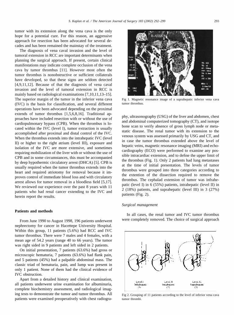
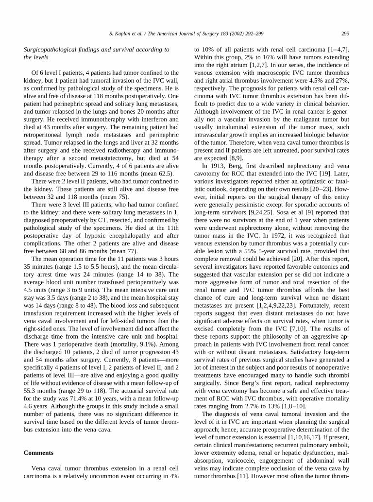
phy, ultrasonography (USG) of the liver and abdomen, chestand abdominal computerized tomography (CT), and isotopebone scan to verify absence of gross lymph node or meta-static disease. The renal tumor with its extension to thevenous system was assessed primarily by USG and CT, andin case the tumor thrombus extended above the level ofhepatic veins, magnetic resonance imaging (MRI) and echo-cardiography (ECO) were performed to examine any pos-sible intracardiac extension, and to define the upper limit ofthe thrombus (Fig. 1). Only 2 patients had lung metastasesat the time of initial presentation. The levels of tumorthrombus were grouped into three categories according tothe extention of the dissection required to remove thethrombus. The cephalad extension of tumor was infrahe-patic (level I) in 6 (55%) patients, intrahepatic (level II) in2 (18%) patients, and suprahepatic (level III) in 3 (27%)patients (Fig. 2).
Surgical management
In all cases, the renal tumor and IVC tumor thrombuswere completely removed. The choice of surgical approach
Fig 1. Magnetic resonance image of a suprahepatic inferior vena cavatumor thrombus.
Fig 2. Grouping of 11 patients according to the level of inferior vena cavatumor thrombi.
293S. Kaplan et al. / The American Journal of Surgery 183 (2002) 292–299

was dependent upon the categories based on the cephaladextent of the tumor thrombus. The operation was performedthrough a midline, anterior, abdominal, or chevron incision,and through extended sternotomy in 3 patients for level IIItumors. CPB and DHCA were used in 3 patients with levelIII tumors. Pulmonary metastastectomy was performed in 2patients, through present sternotomy in 1 and left thoracot-omy in the other.
Surgery
For level I tumor, after establishing vascular control ofvena cava distal and proximal to the thrombus and theopposite renal vein, vena cavotomy and extraction of tumorthrombus was performed. Partial vena caval resection wasperformed in 4 patients. Patch closure was necessary in 3 ofthese patients. In 1 patient, tumor invaded the wall of thevena cava. Regional lymph node resection was performed in1 patient for the palpable, enlarged, and suspicious-lookinglymph nodes and pulmonary metastastectomy was per-formed in another via left thoracotomy.
For level II tumor, control of the IVC above the throm-bus at the suprahepatic infradiaphragmatic level was alwaysachieved before any maneuver was performed on the renalvessels. After complete hepatic mobilization, a Pringle’smaneuver was performed; the suprahepatic IVC, the infra-renal IVC, the contralateral renal vein, and hepatic veinwere occluded in sequence. IVC was opened along its an-terolateral aspect to the level of the hepatic veins. The tumorthrombus was removed en bloc as completely as possibleand adherent tissue was peeled from the endothelial surface.Suture closure began proximally. When the inferior vena-cavotomy below the hepatic veins was closed, a vascularcross-clamp was applied immediately below the veinsacross the IVC and across the suture line, the Pringle’smaneuver was released, the hepatic vein tourniquet andsuprahepatic cross-clamp was removed to minimize hepaticischemia time. When IVC was closed to the level of theinfrarenal segment, cross-clamps were repositioned to allowdrainage of the contra lateral renal vein. The mean liver andrenal ischemia time was 16 minutes (range 8 to 24).
For level III tumor, CPB and DHCA were used in allpatients. The surgical technique followed previously de-scribed methods [5,18]. Briefly, during operation, an indexfinger was inserted through a purse-string–controlled atri-otomy to examine the intra atrial extent and the adherenceof the tumor. After confirmation of the extension, a decisionfor CPB and DHCA was made. The aorta, the IVC portionbelow the thrombus, and the right atrium were cannulated,and CPB was instituted with core cooling. An attempt topush the tumor with a finger backward to the abdominal partof the cava is never made, to avoid inadvertent pulmonaryembolism. Besides, the dumbbell situation does not allowsfor such an attempt for tumor removal. In all cases, renaltumor and the vena caval tumor thrombus could be com-
pletely removed. In addition to this, solitary lung metastaseswere resected in 1 patient. The circulatory arrest timeranged from 14 to 38 minutes, with a mean of 24 minutes
All patients were staged by pathological examinationaccording to the Robson classification system [19]. A com-plete blood count, serum biochemistry panel, chest radio-graph, bone scan, and abdominal USG and CT were re-peated at 6- to 12-month intervals postoperatively. Thefollow-up interval ranged from 29 to 118 months, with amean of 55.3 months and complete follow-up data wereobtained for all patients. Cancer-free status was determinedby negative findings on these follow-up examinations. Sur-vival times were calculated from date of operation to date ofdeath or last contact and were analyzed by the Kaplan-Meier method.
Results
In all cases, renal tumor and the vena caval tumor throm-bus could be completely removed. As a result of tumorextension into the renal vein or IVC, all patients in thisstudy had stage IIIa, IIIb, or IVb tumors according to theRobson classification. Nine patients had stage III and 2patients had stage IV renal cell carcinoma. Preoperativeevaluation with USG and CT raised suspicion for renal veinand vena caval invasion in all cases. In 3 cases, the upperlevel could not be visualized, therefore MRI and ECO werenecessary for confirmation. Mean renal tumor size measuredby CT was 88.5 mm and there was no correlation betweenthe renal tumor size, stage, location, and the level of thetumor thrombus.
Pathological study of the surgical specimens confirmedRCC along with the inferior vena caval tumor thrombus.Further histopathological study revealed capsular penetra-tion of the tumor in 1 patient and perinephric fat involve-ment in 2 patient. The enlarged retroperitoneal lymph nodesresected at the time of operation, was positive for renal cellcarcinoma in one patient. The excised metastatic lung nod-ules, which were detected preoperatively, contained RCC intwo patients. Local caval resection was performed in 4patients, necessitating patch closure in 3 of them. Onepatient had tumor invasion of the inferior vena cava wall. Inour study, IVC wall invasion, perinephric spread and retro-peritoneal lymph node metastases could not be recognizedby preoperative radiological examinations. Complicationsoccurred in 3 patients; one patient developed profound hy-potension with the induction of anesthesia, resulting inhypoxic encephalopathy, and died at the 11th postoperativeday. Liver laceration in one patient and mediastinal bleed-ing requiring immediate reoperation in another with theresultant prolonged ventilatory support were the other majorcomplications. Transient renal insufficiency was observedin several patients but none of them required dialysis.
294 S. Kaplan et al. / The American Journal of Surgery 183 (2002) 292–299
Surgicopathological findings and survival according tothe levels
Of 6 level I patients, 4 patients had tumor confined to thekidney, but 1 patient had tumoral invasion of the IVC wall,as confirmed by pathological study of the specimens. He isalive and free of disease at 118 months postoperatively. Onepatient had perinephric spread and solitary lung metastases,and tumor relapsed in the lungs and bones 20 months aftersurgery. He received immunotheraphy with interferon anddied at 43 months after surgery. The remaining patient hadretroperitoneal lymph node metastases and perinephricspread. Tumor relapsed in the lungs and liver at 32 monthsafter surgery and she received radiotherapy and immuno-therapy after a second metastatectomy, but died at 54months postoperatively. Currently, 4 of 6 patients are aliveand disease free between 29 to 116 months (mean 62.5).
There were 2 level II patients, who had tumor confined tothe kidney. These patients are still alive and disease freebetween 32 and 118 months (mean 75).
There were 3 level III patients, who had tumor confinedto the kidney; and there were solitary lung metastases in 1,diagnosed preoperatively by CT, resected, and confirmed bypathological study of the specimens. He died at the 11thpostoperative day of hypoxic encephalopathy and aftercomplications. The other 2 patients are alive and diseasefree between 68 and 86 months (mean 77).
The mean operation time for the 11 patients was 3 hours35 minutes (range 1.5 to 5.5 hours), and the mean circula-tory arrest time was 24 minutes (range 14 to 38). Theaverage blood unit number transfused perioperatively was4.5 units (range 3 to 9 units). The mean intensive care unitstay was 3.5 days (range 2 to 38), and the mean hospital staywas 14 days (range 8 to 48). The blood loss and subsequenttransfusion requirement increased with the higher levels ofvena caval involvement and for left-sided tumors than theright-sided ones. The level of involvement did not affect thedischarge time from the intensive care unit and hospital.There was 1 perioperative death (mortality, 9.1%). Amongthe discharged 10 patients, 2 died of tumor progression 43and 54 months after surgery. Currently, 8 patients—morespecifically 4 patients of level I, 2 patients of level II, and 2patients of level III—are alive and enjoying a good qualityof life without evidence of disease with a mean follow-up of55.3 months (range 29 to 118). The actuarial survival ratefor the study was 71.4% at 10 years, with a mean follow-up4.6 years. Although the groups in this study include a smallnumber of patients, there was no significant difference insurvival time based on the different levels of tumor throm-bus extension into the vena cava.
Comments
Vena caval tumor thrombus extension in a renal cellcarcinoma is a relatively uncommon event occurring in 4%
to 10% of all patients with renal cell carcinoma [1–4,7].Within this group, 2% to 16% will have tumors extendinginto the right atrium [1,2,7]. In our series, the incidence ofvenous extension with macroscopic IVC tumor thrombusand right atrial thrombus involvement were 4.5% and 27%,respectively. The prognosis for patients with renal cell car-cinoma with IVC tumor thrombus extension has been dif-ficult to predict due to a wide variety in clinical behavior.Although involvement of the IVC in renal cancer is gener-ally not a vascular invasion by the malignant tumor butusually intraluminal extension of the tumor mass, suchintravascular growth implies an increased biologic behaviorof the tumor. Therefore, when vena caval tumor thrombus ispresent and if patients are left untreated, poor survival ratesare expected [8,9].
In 1913, Berg, first described nephrectomy and venacavotomy for RCC that extended into the IVC [19]. Later,various investigators reported either an optimistic or fatal-istic outlook, depending on their own results [20–23]. How-ever, initial reports on the surgical therapy of this entitywere generally pessimistic except for sporadic accounts oflong-term survivors [9,24,25]. Sosa et al [9] reported thatthere were no survivors at the end of 1 year when patientswere underwent nephrectomy alone, without removing thetumor mass in the IVC. In 1972, it was recognized thatvenous extension by tumor thrombus was a potentially cur-able lesion with a 55% 5-year survival rate, provided thatcomplete removal could be achieved [20]. After this report,several investigators have reported favorable outcomes andsuggested that vascular extension per se did not indicate amore aggressive form of tumor and total resection of therenal tumor and IVC tumor thrombus affords the bestchance of cure and long-term survival when no distantmetastases are present [1,2,4,9,22,23]. Fortunately, recentreports suggest that even distant metastases do not havesignificant adverse effects on survival rates, when tumor isexcised completely from the IVC [7,10]. The results ofthese reports support the philosophy of an aggressive ap-proach in patients with IVC involvement from renal cancerwith or without distant metastases. Satisfactory long-termsurvival rates of previous surgical studies have generated alot of interest in the subject and poor results of nonoperativetreatments have encouraged many to handle such thrombisurgically. Since Berg’s first report, radical nephrectomywith vena cavotomy has become a safe and effective treat-ment of RCC with IVC thrombus, with operative mortalityrates ranging from 2.7% to 13% [1,8–10].
The diagnosis of vena caval tumoral invasion and thelevel of it in IVC are important when planning the surgicalapproach; hence, accurate preoperative determination of thelevel of tumor extension is essential [1,10,16,17]. If present,certain clinical manifestations; recurrent pulmonary emboli,lower extremity edema, renal or hepatic dysfunction, mal-absorption, varicocele, engorgement of abdominal wallveins may indicate complete occlusion of the vena cava bytumor thrombus [11]. However most often the tumor throm-
295S. Kaplan et al. / The American Journal of Surgery 183 (2002) 292–299

bus is nonobstructive or sufficient collaterals have devel-oped, therefore these signs are seldom detected [4,9,11,12].For this reason, the diagnosis of vena caval invasion andlevel of tumoral extension is mainly based on radiologicalexaminations [7,10,11,13–15]. Ultrasonography and CT areextremely useful and quite precise in demonstrating theextent of the thrombus in the majority of the patients [7,10,11,13,14]. Additionally, CT can be helpful in preoperativedetection of otherwise occult metastatic disease [14]. How-ever, CT is not always accurate in delineating the superiormargin of the tumor in the IVC. MRI can reliably demon-strate a tumor thrombus and its extension, and it can rule outIVC wall invasion so the exact surgical procedure can beplanned [15]. It is noteworthy that avoidance of contrastagent is particularly important in this group of patients whoare about to lose one kidney.
In our study, 7 patients presented with symptoms: 7patients (63.6%) had hematuria, 7 patients (63.6%) hadflank pain, and 5 patients (45%) had palpable abdominalmass. The classic triad of hematuria, pain, and lump existedonly in 1 patient. None of our patients had clinical evidenceof IVC obstruction. In our study, 100% of the vena cavalthrombi were diagnosed by the combined use of USG andCT, which is a percentage in line with reports by others[7,10,26]. In 3 patients, based on CT findings of IVC in-volvement, MRI and ECO were needed for better definitionof the upper extend of intracaval disease. However, in ourstudy, none of the radiological examinations were able todemonstrate IVC wall invasion by the tumoral process.Contrast studies, like vena cavography have been used inother series, but we have not found them to be additive toUSG, CT, and MRI, and it increases the risk of contrast-associated renal injury in a patient with one functioningkidney [1,4,7,10]. We also performed the cardiac evaluationof patients with level III tumor thrombus preoperatively.
The key point in the surgical management of RCC withIVC tumor thrombus is the correct assessment of the exten-sion of the endocaval thrombus. Once the diagnosis is made,the level of tumor thrombus involvement determines plan-ning of the operation. Objectives of operative managementinclude: (1) maintenance of complete resection of tumor andtumor thrombus (2) prevention of tumoral embolism, (3)minimizing blood loss, (4) maintainance of hemodynamicstability, and (5) prevention of vital organ ischemia. Inperforming surgical removal of IVC thrombus, it is essentialto obtain control of the cava above the thrombus beforemanipulating the intracaval tumor mass, to prevent intraop-erative embolization of tumor fragments. Although level Itumors do not require any complex maneuvers except forcontrol of the IVC above and below the tumor, level II andIII tumors require a complex maneuver and multiteam ap-proach. Whereas temporary occlusion of the infrahepaticIVC can safely be done for level I tumors, the occlusion ofthe suprahepatic IVC often causes a profound decrease invenous return resulting in hypotension. Additional disad-vantages of the latter maneuver when removing a caval
thrombus include back bleeding from hepatic and lumbarveins and occasional swelling of the liver from venouscongestion, which interferes with the exposure. Because ofthat, numerous different method of managements have beenreported for level II and III tumors [1,5,6,8,10,16,17].
In our experience, a midline, anterior, abdominal, orchevron incision was used; and they provided an adequatesurgical field for the safe dissection in the abdomen, distalcontrol of the vena cava, and mobilization of the kidneywith the tumor in the majority of our cases. Therefore, thesewere our standard surgical approaches. In this study, controlof the IVC above the thrombus was always achieved beforeany maneuver was performed on the renal vessels in level Iand level II. Tumor extraction was accomplished after prox-imal and distal control of the IVC in level I tumor thrombus.In level II tumor thrombus, the suprahepatic IVC just belowthe diaphragm (with transabdominal, suprahepatic, infradia-phragmatic dissection), the infrarenal IVC, the contralateralrenal vein, and hepatic vein were occluded, and the extrac-tion of the tumor thrombus was performed. Lumbar venousbleeding, seen after the removal of the tumor, was con-trolled with a curved vascular clamp. In our experience,resection of level II tumors required interruption of thehepatic and contralateral renal circulation. In our series,liver and renal ischemia time varied from 8 to 23 minutes(mean 16), which is acceptable when compared with re-ported tolerable normothermic continuous hepatic ischemiatime of 15 to 30 minutes [12,17]. Hence, none of ourpatients had significant postoperative hepatic and renal dys-function. Although dissection in all cases required signifi-cant manipulation of the vena cava and, thus a greater riskof embolization, we have not encountered tumor emboliza-tion in any case.
The management of patients with cardiac extension ofthe tumor thrombus (level III), that is clearly beyond simpleminimal extension into the atrium, must be individualized.CPB is necessary with or without DHCA to safely andcompletely extract the level III tumor thrombus as well as tovisualize and remove all sites of adherent tissue within theright atrium. The advantages and potential complications ofCPB and DHCA were well defined previously [1,5,16,17].It should be kept in mind that digital manipulation of thetumor in an attempt to push it down into the IVC without theprotection by CPB may be hazardous and may result intumor embolism. In our study, we used CPB and DHCA in3 patients with level III tumor thrombus and did not en-counter any problem inherent to these tools [1,5,16,17].Based on our data, we believe that this approach is safe andeffective, and allows extensive IVC thrombi to be com-pletely removed with an excellent exposure in a controlledoperative setting.
Various centers have reported mortality rates of 6% to9% for IVC extension of RCC [1,2,5,7]. The major cause ofdeath reported to be pulmonary embolism and myocardialinfarction or due to complications related to the bypassprocedures. However, with better perioperative manage-
296 S. Kaplan et al. / The American Journal of Surgery 183 (2002) 292–299

ment and standardization of the surgical techniques, themortality rates can be decreased considerably [7,10,16]. Wehave only one operative mortality (9.1%) due to hypoxicencephalopathy. Of 11 patients, 3 with intraatrial thrombirequired sternotomy for CPB and DHCA and the survivalwas not different from those presenting with invasion of thelower levels of IVC alone.
Although there is controversy concerning the prognosticsignificance of certain factors—namely, the presence oflymph node involvement, perinephric fat invasion, meta-static disease, renal vein or IVC wall invasion, or the levelof the vena caval tumor extension and possibility of com-plete tumoral tissue excision—all these factors may show animpact on prognosis and survival. The presence of tumoralinvasion in the regional lymph node and the perinephric fatis reported to be a definitive poor prognostic factor [4,7,9,10,22,23]. It has been shown that the patients with RCCextending only into the IVC have significantly better sur-vival rates than those with local spread of the tumor to thelymph nodes or perinephric tissue [7,10,22,23]. Metastaticspread to lymph nodes results in rapid relapse and a shortoverall survival [23]. Reissigl et al [27] reported a 15.5-month mean survival rate for patient who had lymph nodemetastases, with an overall 5-year survival of 62.5%. Lib-ertino et al [8] found an overall 5-year survival of 59% withan overall 10-year survival of 47%. Patients with no evi-dence of metastatic disease had a 60% survival rate withfollow-up to 16 years but no patient with metastatic diseasesor nodal involvement survived beyond 5 years. Hatcher et al[4] found a 5-year survival of 62% for their patients withcompletely resected stage IIIa disease. For patients withlymph node metastases, the 5-year survival rate dropped to17%. However, whether the perinephric spread is the onlyfactor to affect the prognosis adversely in patients with RCCwith IVC extension or not is controversial [8,20,22,23,28].Libertino et al [8] reported that perinephric spread, per se,does not affect the survival in patients with IVC thrombus.In contrast, others have reported that, the presence of peri-nephric spread in RCC is a strong poor prognostic param-eter [20,22,23,28]. In the report of Skinner et al [20] 4 of the5 nonsurvivors had perinephric spread. Recently, Glazerand Novick [28] found that mean postoperative survival wassignificantly improved in patients with no renal capsularpenetration by tumor compared with those with perinephricfat involvement (58.1 versus 19.7 months). The prognosticsignificance of lymph node metastases and perinephricspread in our study confirms the results of the abovemen-tioned reports [20,22,23,28]. In our series, both patientswho had lymph node metastases and perinephric spreaddied of tumor progression 43 and 54 months after surgery.In contrast, none of the patients without nodal disease andperinephric spread were dead at follow-up.
Distant metastatic disease at diagnosis also implies poorsurvival. Patients with metastatic RCC have a poor progno-sis, with a median survival expectation of less than 1 yearand an associated 5-year mortality rate ranging from 80% to
100%. Most researchers note worse survival for such pa-tients [1,6,29,30]. Using Cox proportional hazards regres-sion model, Montie et al [30] found that a patient withmetastatic disease was 2.76 times more likely to die thanpatients without metastases. Some reports suggest that find-ing a metastatic disease at presentation is an ominous sign,as most patients die within a year of diagnosis, and thesurvival is not increased by doing an adjunctive nephrec-tomy. Therefore, an aggressive operation is not recom-mended for this group [2,9,21,22]. However, controversialreports from large cancer centers indicating distant metas-tases did not have a significant adverse effect on survivalrates have been published recently [7,10]. In some reports,as is the case in our study, some patients with metastaticdisease have prolonged survival for unexplained reasons[1,2,7,10]. Our series included 2 patients with metastaticdisease; 1 of them died just after the operation whereas theother lived for more than 3 years. He was given interferonin the postoperative period for tumor relapse. It is difficultto determine whether this approach influenced his outcomeor not. We cannot find a reasonable explanation for theimproved survival observed in this isolated case. Whateverthe reason, irrespective of the implication of the distantdisease and in consideration of the potential for a survivalbenefit, we believe that metastatic disease should not pre-clude operative intervention. We recommend the removal ofthe primary neoplasm along with the extraction of the IVCtumor thrombus to palliate symptoms, to remove an imme-diately life-threatening focus of disease, and to reduce as-sociated problems such as coagulopathies and hepatic dys-functions.
Renal tumor often invades the wall of the vena cava andthis invasion itself, regardless of the level of invasion andthrombus development, reflects the biological properties ofit [26]. Because of that, the influence of vein invasion inRCC on metastatic spread and survival is theoretically in-teresting and has been the subject of much debate. It is wellknown that vein invasion is an adverse prognostic factor inmany tumors [31,32]. However, the prognostic impact ofrenal vein and IVC wall invasion in RCC has been unclear,and reports concerning the significance of it and the role ofpartial vena cavectomy to achieve a surgical cure, have beencontradictory [4,18,22,26,30,33,34]. Golimbu et al [33] re-ported that renal vein invasion alone did not alter the 5-yearsurvival, and noted that there is no survival difference if thevein was grossly or microscopically invaded by tumor. Incontrast, others have reported that the renal vein and IVCwall invasion in RCC is a strong poor prognostic parameterand when present, the risk of distant metastases and relapseis increased in such patients after surgery [4,18,22,26,30,34]. Additionally, these reports suggested that completeresection of the tumor thrombus, including complete resec-tion of the invaded wall of the IVC, provides a survivalbenefit [4,18,22,26,30,34]. In our study, IVC wall invasionwas present in only 1 patient with level I tumor thrombus.He is still alive and free of disease at 116 months postop-
297S. Kaplan et al. / The American Journal of Surgery 183 (2002) 292–299
eratively. Because the number is too small to analyze, wecannot comment on the prognostic significance of the wallinvasion in patients with RCC and IVC tumor thrombus.
The prognostic implication of the level of the IVC tumorthrombus is unclear and reports have been contradictory[4,18,22,31,35]. Several investigators have noted a worseprognosis for patients with a tumor thrombus that extendsinto the atrium than for those with disease that remainswithin the IVC, and they suggested that the risk of metas-tases and early death is increased with a more cephaladextend of IVC thrombi [1,9,30,36]. Sosa et al [9] reported a2-year survival rate of 80% in patients with intrahepaticIVC tumor thrombi compared with only 21% in those withsuprahepatic thrombi. Skinner et al [1] indicated a 5-yearsurvival rate of 35%, after surgical treatment for patientswith RCC and subhepatic IVC thrombus, and 5-year sur-vival rates for patients with intrahepatic or atrial tumorthrombi were 18% and 0%, respectively. In the series byMontie et al [30], patients with intrahepatic extension were0.49 times less likely to die than those with renal or infra-hepatic extension as determined by the Cox proportionalhazards regression model. In the series reported by Marshallet al [36], the survival ratio was worse in the group ofpatients with a higher-level tumor thrombus, but the differ-ence was not statistically different. In contrast, most reportsin the literature have not shown a significant difference insurvival when the tumor thrombus remains below the atriumand therefore have concluded that extension to the venacava alone has a limited or no impact on survival [1,4,7,12,17,21,22]. Cherrie et al [22], Novick et al [17], and Hatcheret al [4] have also reviewed their experience in similargroups of patients with RCC and IVC thrombi and have notfound the level of the tumor thrombus to impact survival. Inthe present report, there was no significant difference insurvival based on the level of the tumor thrombus. Thisfinding in our study, confirms the results of many others[1,4,7,12,15,17,21,22,34], while actually contradicting thereport of Sosa et al [9].
Patients with incomplete resections (excluding patientswith metastatic disease) have a significantly worse progno-sis. Similar findings have been shown in many reports,stressing the importance of surgical eradication of disease[1,2,4,7,10]. Hatcher et al [4] noted that the prognosis wasdetermined by the ability to perform a complete resection ofthe tumor, not by the level of tumor thrombus. They alsoshowed the prognostic importance of complete resection inpatients with nonmetastatic disease, as the 5-year survivalrate drops from 57% to 0%. Skinner et al [1] identified theimportance of complete resection by reporting a 34% 5-yearsurvival for those patients undergoing a complete resectionand a 1-year survival of only 8% for those who had incom-plete resections. Furthermore, Neves and Zincke [2] noted adifference in 5-year survival between patients with com-plete thrombus removal (68%) and those with incompletethrombus removal (17.5%). Because of that, it has been sug-
gested that every attempt should be done for complete resec-tion and surgical eradication of disease [1,2,4,7,10,11,17].
The 52% 5-year survival rate of patients with localizedrenal cancer [23] and 25% to 75% overall 5-year survivalrates of patients with RCC with tumor thrombus extensionto the IVC were reported previously [4,7,10,28,35]. In ourstudy, the overall 10-year survival rate of 71.4% for patientswith RCC and IVC tumor thrombus is comparable with theabove-mentioned results. Although each group in our studyincluded a few number of patients, lymph node involvementand perinephric spread seem to influence the prognosisadversely, whereas the level of tumor thrombus, stage ofdisease, distant metastatic disease, and the presence of IVCwall invasion do not.
In conclusion, all patients with RCC and IVC tumorthrombi should be considered for operation. No other ther-apeutical modality has achieved comparable results. Thesetumors can be totally resected by an aggressive approach,using CPB and DHCA when necessary with an acceptablemorbidity and mortality, and satisfactory long-term survivalrates can be achieved. We believe that complete surgical ex-cision of the tumor and its thrombus with accurate preoperativestaging and a well-planned surgical approach provides a highsurvival chance and offers a good quality of life.
References
[1] Skinner DG, Pritchett TR, Lieskovsky G, et al. Vena caval involve-ment by renal cell carcinoma. Surgical resection provides meaningfullong-term survival. Ann Surg 1989;210:387–94.
[2] Neves RJ, Zincke H. Surgical treatment of renal cancer with venacava extension. Br J Urol 1987;59:390–5.
[3] O’Donohoe MK, Flanagan F, Fitzpatrick JM, Smith JM. Surgicalapproach to inferior vena caval extension of renal cell carcinoma. Br JUrol 1987;60:492–6.
[4] Hatcher PA, Anderson EE, Paulson DF, et al. Surgical managementand prognosis of renal cell carcinoma invading the vena cava. J Urol1991;145:20–4.
[5] Klein EA, Kaye MC, Novick AC. Management of renal cell carci-noma with vena caval tumor thrombi via cardiopulmonary bypass anddeep hypothermic circulatory arrest. Urol Oncol 1991;18:445–7.
[6] Swierzewski DJ, Swierzewski JA. Radical nephrectomy in patientswith renal cell carcinoma with venous, vena caval, and atrial exten-sion. Am J Surg 1994;168:205–9.
[7] Nesbitt JC, Soltero ER, Dinney CP, et al. Surgical management ofrenal cell carcinoma with inferior vena cava tumor thrombus. AnnThorac Surg 1997;63:1592–600.
[8] Libertino JA, Zinman L, Watkins E. Long term results of renal cellcancer with extension into inferior vena cava. J Urol 1987;137:21–4.
[9] Sosa RE, Muecke EC, Vaughan ED, McCorron JP. Renal cell carci-noma extending into the inferior vena cava: the prognostic significanceof the level of the vena cava involvement. J Urol 1984;132:1097–100.
[10] Tongaonkar HB, Dandekar NP, Dalal AV, et al. Renal cell carcinomaextending to the renal vena cava: results of surgical treatment andprognostic factors. J Surg Oncol 1995;59:94–100.
[11] Libertino JA. Renal cell cancer with extension into vena cava. In:Dudley H, Carter D, editors. Rob and Smith’s operative surgery(urology). 4th ed. London: Butterworths, 1986, p 122–7.
[12] Clayman RV, Gonzalez R, Fraley EE. Renal cell carcinoma invadingthe inferior vena cava: clinical review and anatomical approach.J Urol 1980;123:157–63.
298 S. Kaplan et al. / The American Journal of Surgery 183 (2002) 292–299
[13] Hubsch P, Schurawitzke H, Susani M, et al. Colar Doppler imagingof the inferior vena cava: identification of tumor thrombus. J Ultra-sound Med 1992;11:639–45.
[14] Marks WM, Korobkin M, Callan PW, Kaysar JA. Computed tomo-graphic diagnosis of tumour thrombus of renal vein and the inferiorvena cava. AJR Am J Roentgenol 1978;131:843–6.
[15] Myneni L, Hricak H, Carroll PR. Magnetic resonance imaging ofrenal carcinoma with extension into the vena cava: staging, occuracyand recent advances. Br J Urol 1991;68:571–8.
[16] Babu SC, Mianoni T, Shah PM, et al. Malignant renal tumor withextension to the inferior vena cava. Am J Surg 1998;176:137–9.
[17] Novick AC, Kaye MC, Cosgrove DE, et al. Experience with cardio-pulmonary bypass and deep hypothermic circulatory arrest in themanagement of retroperitoneal tumors with large vena caval thrombi.Ann Surg 1990;212:472–7.
[18] Robson CJ, Churchill BM, Anderson W. The results of radical ne-phrectomy for renal cell carcinoma. J Urol 1969;101:297–301.
[19] Berg AA. Malignant hypernephroma of the kidney, its clinical courseand diagnosis, with a descriptions of the author’s method of radicaloperative cure. Surg Gynecol Obstet 1913;17:463–71.
[20] Skinner DG, Pfister RF, Colvin R. Extention of renal cell carcinomainto the vena cava: the rationale for agressive surgical management.J Urol 1972;107:711–16.
[21] Kearney GP, Waters WB, Klein LA, et al. Results of inferior venacava resection for renal cell carcinoma. J Urol 1981;125:769–73.
[22] Cherrie RJ, Goldman RG, Linder A, deKernion JB. Prognostic im-plications of vena caval extension of renal cell carcinoma. J Urol1982;128:910–12.
[23] Heney NM, Nocks BN. Influence of perinephric fat involvement onsurvival in patients with renal cell carcinoma extending into theinferior vena cava. J Urol 1982;128:18–20.
[24] McDonals JR, Priestley JT. Malignant tumor of the kidney: surgicaland prognostic significance of the tumor thrombosis of renal vein.Surg Gynecol Obstet 1943;77:295–9.
[25] Myers GH, Fehrenbaker LG, Kelalis PP. Prognostic significance ofrenal vein invasion by hypernephroma. J Urol 1968;100:420–3.
[26] Ljunberg B, Stenling R, Osterdahl B, et al. Vein invasion in renal cellcarcinoma: impact on metastatic behavior and survival. J Urol 1995;154:1681–4.
[27] Reissigl A, Janetschek G, Eberle J, et al. Renal cell carcinomaextending into the vena cava: surgical approach, technique and re-sults. J Urol 1995;75:138–42.
[28] Glazer AA, Novick AC. Long-term followup after surgical treatmentfor renal cell carcinoma extending into the right atrium. J Urol1996;155:448–50.
[29] Linehan WM, Shipley WU, Longo DL, et al. Cancer of the kidneyand ureter. In: DeVita VT, Hellman S, Rosenberg SA, editors. Principlesand practice of oncology. Philadelphia: Lippincott, 1993, p 1023–51.
[30] Montie JE, EL Ammar R, Pontes JE, et al. Renal cell carcinoma withinferior vena cava tumor thrombi. Surg Gynecol Obstet 1991;173:107–15.
[31] Sanchez MP, Zudaire JJ, Robles JE, et al. Renal cell carcinoma: venacaval invasion and prognostic factors. Eur Urol 1991;19:284–8.
[32] Hoetl W, Pont J, Kosak D, et al. Treatment decisions for stage Inonseminomatous germ cell tumors based on the risk factor “vascularinvasion.” Br J Urol 1992;69:83–6.
[33] Golimbu M, Joshi P, Sperber A, et al. Renal cell carcinoma: survivaland prognostic factors. Urology 1986;27:291–301.
[34] Mrstik C, Salamon J, Weber R, Stogermayer F. Microscopic venousinfiltration as predictor of relapse in renal cell carcinoma. J Urol1992;148:271–4.
[35] Pagano F, Dal Bianco M, Artibani W, et al. Renal cell carcinoma withextension into the inferior vena cava: problems in diagnosis, stagingand treatment. Eur Urol 1992;22:200–3.
[36] Marshall FF, Dietrick DD, Baumgartner WA, et al. Surgical manage-ment of renal cell carcinoma with intracaval neoplastic extensionabove the hepatic veins. J Urol 1988;139:1166–72.
299S. Kaplan et al. / The American Journal of Surgery 183 (2002) 292–299