surgical management of colorectal cancer · a. carcinoma in a polip. non-invasive carcinoma (tis)...
TRANSCRIPT

SURGICAL MANAGEMENT OF COLORECTAL CANCER
Irinel Popescu, MD, FACS, FEBSProfessor of Surgery
Dan Setlacec Center of General Surgery and Liver TransplantationFundeni Clinical Institute, Bucharest

Colorectal cancer – multimodal treatment
▪ Interventional endoscopy▪ Surgery▪ Chemotherapy▪ Radiotherapy▪ Interventional radiology

Metastatic
Loca
lized
Non-invasive
Inva
sive
Colorectal carcinoma-TNM staging

I. COLON CANCER

A. Carcinoma in a polip
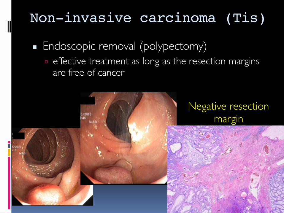
Non-invasive carcinoma (Tis)
▪ Endoscopic removal (polypectomy) ▫ effective treatment as long as the resection margins
are free of cancer
Negative resectionmargin

B. Localized invasive colon cancer (T1-4, N0-2, M0)

T1 carcinoma (in a polyp)▪ Endoscopic resection▫ Reasonable alternative to radical surgery◾Favorable risk, T1 colon cancers arising in a polyp
▪ Higher risk of residual cancer or nodal metastases:▫ Poorly-differentiated histology▫ Lymphovascular invasion▫ Cancer at the resection or stalk margin▫ Invasion into the muscularis propria (T2)▫ Sessile polyp with lower third submucosal penetration
Radical surgeryTattoo the area of endoscopic resection

Radical surgery▪ The only curative treatment modality for localized
invasive colon cancer (T1-4, N0-2, M0)
▪ Goals of surgery:1. Complete removal of:
1. the tumor2. the major vascular pedicle(s)3. the lymphatic drainage basin of the affected colonic
segment2. Restoration of bowel continuity

1. Complete resection

The concept of “Complete Mesocolic Excision with Central Vascular Ligation” – Hohenberger
▪ Complete mesocolic excision (CME) – “en bloc” removal of ▫ the tumor bearing colon▫ its associated lympho-vascular suply▫ ligation of the mesocolic vessels near their origin
By taking the colon and mesocolon in an intact “envelope “of visceral peritoneum

CME – Three essential components
1. Dissection between mesenteric plane andparietal fascia
2. Central vascular tie1. Complete removal of LN
towards central direction (A, B)3. Adequate length of bowel
1. Complete removal of pericolic LN – longitudinal direction (C)
▪ Mesocolic plane▪ Intramesocolic plane▪ Muscularis propria plane
▪ Adequate lymphadenectomy

CME – Mesocolic plane
▪ CME allows for higher quality of surgical specimens when compared to less radical “standard” surgery
▪ Disection plane significantly impacts loco-regional control and thus overall survival
P value < 0.05

CME – Central vascular ligation
▪ CORECT vascular ligation for cancer
▪ INADEQUATE vascular ligation for cancer
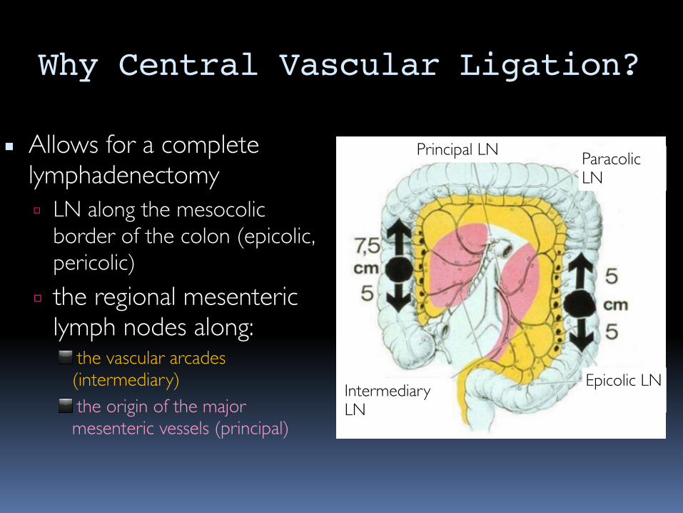
Why Central Vascular Ligation?
▪ Allows for a complete lymphadenectomy▫ LN along the mesocolic
border of the colon (epicolic, pericolic)
▫ the regional mesenteric lymph nodes along:◾ the vascular arcades
(intermediary) ◾ the origin of the major
mesenteric vessels (principal)
Epicolic LN
Paracolic LN
Principal LN
Intermediary LN
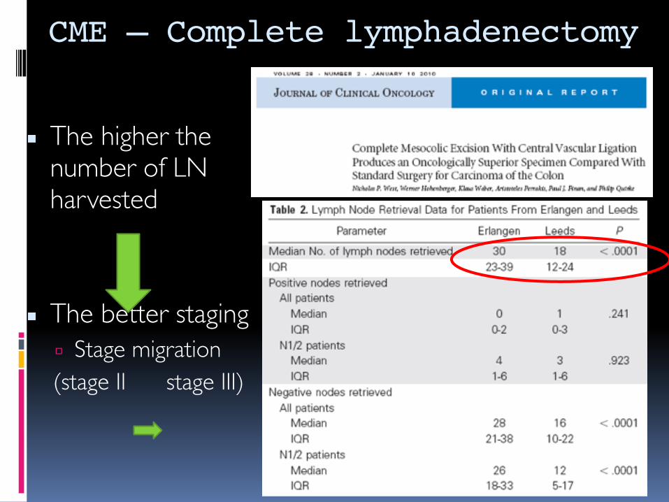
CME – Complete lymphadenectomy
▪ The higher the number of LN harvested
▪ The better staging▫ Stage migration(stage II stage III)

■ Stage I (T2, N0, M0): NO adjuvant chemotherapy■ Stage II (T3/T4, N0, M0): Chemotherapy – high-risk patients■ Stage III (any T, N1/N2, M0): Chemotherapy – all patients
Inadequate surgical resection/pathologic evaluation
Inadequate adjuvant chemotherapy allocation
CME – Better staging
Better staging allows a more correct adjuvant treatment

The number of LN evaluated was positively associated with survival
The number of LN evaluated – measure of the quality of CRC care
CME – Better survival

Rationale for CME
▪ The “envelope” of visceral peritoneum contains potentially metastatic lymph nodes (LN)
▪ By keeping intact the “envelope” minimize the risk of spillage of cancer cells into the peritoneal cavity
▪ Central vascular ligation of the relevant blood supply improves the number of lymph nodes (LN) harvested
Decreases the 5 year local recurrence rateIncreases the 5 year cancer related survival

Colon cancer: CME – standard of care
▪ Complete mesocolic excision with central vascular ligation should become the standard approach to colon carcinoma
▪ These approach (CME) decreases the local recurrence rates and increases overall survival in stage I-III colon cancer patients

Types of colon resections
▪ Right colectomy ▪ Extended right colectomy

Types of colon resections
▪ Transverse colectomy ▪ Sub-total colectomy

Types of colon resections▪ Total colectomy▪ Left colectomy

Type of approach open vs laparoscopic▪ Laparoscopic CME is
feasible▪ Short-term and long-
term outcomes following laparoscopic CME were similar to those achieved by open approach in specialized centers

Locally advanced colon cancer (T4)
▪ Invasion of contiguous organs or inflammatory adhesions involving neighboring structures – 10%
▪ Treatment objectives:▫ “En-bloc” multivisceral resection with a negative margin of
the adjacent structure▫ The plane of adherence between the colonic tumor and
the adjacent organ(s) should not be disrupted◾40% of these adhesions are malignant ◾transection of tumor could further impair prognosis.

2. Restoration bowel continuity

Uncomplicated tumors
▪ Primary anastomosis▫ Ileo-colostomy▫ Colo-colostomy▫ Colo-rectostomy

Colonic obstruction
▪ resection of the tumor with a primary anastomosis▫ with or without a temporary proximal diversion
▪ resection without anastomosis – end colostomy
▪ proximal diversion with a mucous fistula or a loop colostomy, followed by elective definitive resection (second operation)

Colonic perforation
▪ Resection of the tumor with a primary anastomosis▫ localized fluid collection/abscess
▪ Resection without anastomosis – end colostomy▫ free perforation▫ diffuse peritonitis▫ patient is medically unstable

II. RECTAL CANCER

Non-invasive carcinoma (Tis)
▪ Endoscopic removal (polypectomy) ▫ effective treatment as long as the resection margins are
free of cancer
▪ Transanal excision (TAE) – lesions located in the anal canal (less than 4 cm from anal verge)

Localized invasive rectal cancer (T1-4, N0-2, M0)
SURGICAL RESECTION

A. Local excision procedures

Techniques
▪ Transanal excision (TAE)▫ ADK below 4 cm from anal
verge
▪ Goal:▫ full-thickness excision of the rectal
cancer▫ minimum lateral margin of 1 cm▫ histologically negative deep margin▫ primary closure
▪ Transanal endoscopic microsurgery (TEM)▫ ADK 4-15 cm from anal verge

Indications TAE/TEM
▪ Standard:▫ T1 cancers < 3 cm▫ No radiographic evidence of positive regional LN (N0)▫ Low risk pathologic features◾well differentiated◾no vascular or neural invasion
▫ Compliance with aggressive postoperative surveillance

Results TAE/TEM
▪ T1N0 patients – low risk factors:▫ Significantly fewer postoperative complications than
following major resections▫ Local recurrence rates are comparable to more
extensive operative procedures (4-6%)▫ 10-year OS – 98%▫ 10-year DFS – 92%

Higher risk patients (pathology)
▪ > T1▪ > 3 cm▪ Inadequate margins ▪ Lympho-vascular invasion
ImmediateReoperation
(sphincter-saving/APR)
Chemoradiotherapy(improves results)
+Follow-up (aggressive)
Re
fusal

Specific indications for TAE/TEM
▪ Patients > T1 or >3 cm:▫ Complete response following chemoradiation▫ Refusal of major abdominal surgery▫ Comorbidities that preclude a major intra-
abdominal operation▫ Distant disease (short life expectancy)
Higher recurrence rates than following major resectionsLower survival rates compared with major operations

Response to chemoradiation
▪ Complete clinical response▫ Normal CEA level▫ Digital rectal examination▫ CT/endorectal ultrasonography▫ Absence of any residual scar, mass
or ulcer
▪ Near-complete clinical response▫ Suspicious, small residual
lesions
Full-thickness local excision
Negative pathological
result
Positive pathological
result
Watch and Wait(A. Habr-Gama)
Rectalresection

Watch and Wait - results
▪ Aggressive follow-up – mandatory▪ Local recurrence rate: 11-31%▫ Disease recurrence may occur at any time
▪ Salvage therapy – local excision/ rectal resection▫ Salvage rate – 93%
▪ 5-yr local recurrence-free survival: 87%▪ 5-yr cancer specific DFS – 68%▪ 5-yr cancer-specific OS – 91%
Endoscopic/Local excision procedures – curative attempt?
▪ Tis/T1 N0 colorectal carcinomas▫ Endoscopic resections or local excision procedures
could be curative◾Lower morbidity rates◾Similar local recurrence rates vs. Rectal resections◾Similar survival rates
▪ “Watch and wait” could be an acceptable option in patients with complete clinical response after neoadjuvant chemoradiotherapy

B. Rectal resections

▪ Objectives1. Removal
1. Distal colon and tumor-bearing rectum1. at least 2 cm away from distal tumor edge
2. Lymph nodes and relevant vessels (IMA&IMV)3. Mesorectum (total mesorectal excision – TME)
2. Restoration of bowel continuity – whenever possible1. Sphincter-sparing procedures
Indications: T2-4,N0-2,M0

The concept of mesorectum;mesorectal fascia - Thoma Ionnescu
▪ Mesorectum – lipoma-like “envelope” of the rectum
▪ Mesorectal fat – surrounded by mesorectal fascia
▪ Mesorectal fascia:▫ 1894 – Thoma Ionnescu – “Gaine fibro-sereuse du rectum”▫ 1899 - Wilhelm von Waldeyer – “Fascia propria recti”

Total Mesorectal Excision (TME) – Heald
▪ Mesorectum – it may content carcinoma cells and metastatic LN
▪ TME – removes completely the tumor, regional spread cells and loco-regional LN
Decreases local recurrence rates

Heald’s “Holly-plane” surrounding mesorectum
▪ Sharp & meticulous dissection surrounding mesorectal fascia▫ Posterior – easy to find▫ Anterior – less obvious▫ Lateral – the most difficult
▪ Correct TME – Intact circumferential resection margins (CRM) ▫ intact mesorectal fascia
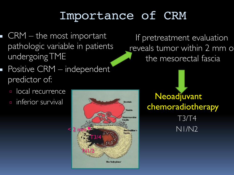
Importance of CRM
▪ CRM – the most important pathologic variable in patients undergoing TME
▪ Positive CRM – independent predictor of:▫ local recurrence▫ inferior survival
If pretreatment evaluation reveals tumor within 2 mm of
the mesorectal fascia
Neoadjuvant chemoradiotherapy
▫ T3/T4▫ N1/N2< 2 mm
T3/4
N1/2

TME – longitudinal extension
▪ The distal mesorectal excision margin – 5 cm below the lower border of the tumor

TME – Clinical relevance
▪ Total mesorectal excision significantly improved local recurrence and survival rates in rectal cancer patients

Low stapled (High anal)Manual transanal (Low anal)
Amputation (APR)
Tumor location – type of resection

1. Sphincter-sparing procedures

Indications
▪ Invasive rectal cancers beyond the submucosa▫ T2-4,N0-2,M0
▪ Histologically proven negative distal margin – R0
▪ Predicted adequate posttreatment anorectal sphincter function

Anterior Resection – AR▪ Indications▫ Cancers located in the upper third of the rectum◾11-15 cm
▪ Surgical technique▫ Removal of the sigmoid colon and rectum to a level
where the distal margin is free of cancer▫ Primary anastomosis between the descending colon and
the middle rectum
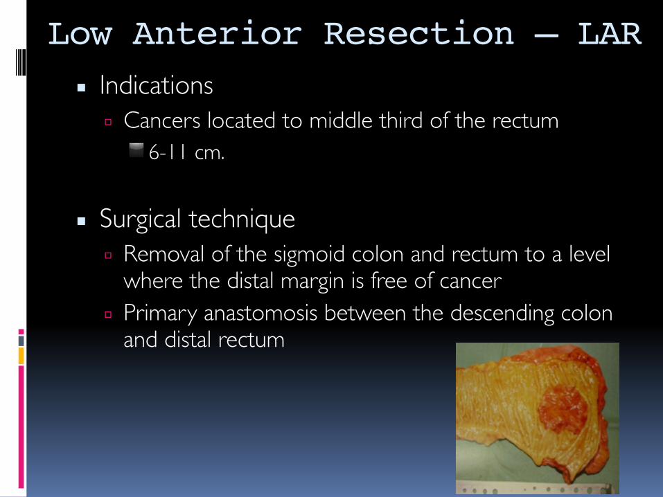
Low Anterior Resection – LAR▪ Indications▫ Cancers located to middle third of the rectum◾6-11 cm.
▪ Surgical technique▫ Removal of the sigmoid colon and rectum to a level
where the distal margin is free of cancer▫ Primary anastomosis between the descending colon
and distal rectum

Ultra-low anterior resection with “low-stapled” anastomosis
▪ Indications▫ distal rectal cancer – above the anal
sphincter (5-6 cm.)▪ Surgical technique▫ The rectum – transected just above the
pelvic floor musculature▫ Anastomosis: colon – low rectum◾Straight C-R anastomosis◾Colonic J-pouch reservoir◾Transverse coloplasty

Ultra-low anterior resection with transanal mucosectomy
▪ Indications▫ distal rectal cancer that does not invade
the anal sphincter (4-5 cm.)▪ Surgical technique▫ Muscularis propria of the rectum –
transected at pelvic floor musculature▫ Transanal mucosectomy▫ Anastomosis: colon - anal sphincter◾Straight colo-anal◾Colonic J-pouch reservoir◾Transverse coloplasty

ULAR with intersphincteric dissection + coloanal anastomosis
▪ Surgical technique▫ At the pelvic floor musculature the dissection continues
between internal and external anal sphincters▫ Perineal intersphincteric dissection▫ Anastomosis: colon – anal sphincter
Dissection plane APR ULAR + INTERSPHINTERIC

Sphincter-sparing procedures
▪ By improving surgical expertise, the number and the types of sphincter-saving procedures increased continuously in the last decade

2. Abdominal-perineal resection

Indications
▪ Low-lying rectal adenocarcinomas ▫ Negative distal margin of resection cannot be
achieved with sphincter-sparing procedures▪ Salvage procedure for local recurrence or locally
advanced rectal cancer
▪ It remains the standard against which sphincter sparing procedures and local excision procedures are compared

Surgical technique
▪ Resection of the ▫ Sigmoid colon▫ Rectum▫ Anus
▪ Construction of a permanent colostomy

Drawbacks – APR
▪ High rates of CRM involvement▪ Perforation of the bowel
▪ Higher rates of local recurrence▪ Lower rates of survival
▪ Absence of mesorectal margin “cushion”▪ Difficult technical dissection due to lack of planes▪ ‘Waist” of the specimen

Extralevator APR (ELAPE) - Holm
▪ Levator muscles are excised “en-bloc” with mesorectum, lower rectum and anus
▪ Avoids “waist” of the specimen (APR)
ELAPE APR

ELAPE - Advantages▪ Reduces bowel perforation▪ Reduces circumferential resection margins (CRM) positivity▪ Lower local recurrence rates

Type of approach: open vs. laparoscopic vs. robotic
▪ Short-term and long-term outcomes following laparoscopic/robotic rectal resections were similar to those achieved by open approach

Locally advanced rectal cancer-T4
▪ “En-bloc” multivisceral resections:▫ Rectal cancer▫ Adjacent organs invaded (R0)
▪ Total pelvic exenteration (TPE)▫ Rectum▫ Bladder ▫ Internal reproductive organs◾Prostate + seminal vesicles◾Uterus, ovaries and vagina

Modified exenterations
▪ Posterior pelvic exenteration (PPE)▫ rectum, anus▫ uterus, ovaries▫ posterior vaginal wall
▪ Supralevator exenteration▫ TPE/PPE with a primary
colo-rectal anastomosis
▪ Composite resections▫ Exenterative procedures
including resection of bony structures◾ Sacrum◾Coccyx

III. METASTATIC COLORECTAL CANCER

Initially resectable or initially unresectable: ESMO guidelines 2014
Van Cutsem E, et al. Ann Oncol 2014;25 (suppl 3):iii1–iii9
Patient Group 0• Primarily technically R0-
resectable liver or lung metastases
• No contraindications to resection
Patient Groups 1–3• Initially unresectable mCRC,
including:• Potentially resectable after
CT (Group 1)• Disseminated disease,
technically ‘never’/unlikely resectable (Group 2)
• Never-resectable metastatic disease (Group 3)
Curative intent resection:- Primary tumor and - Liver/lung Metastases

Synchronous resectable CLMs (Group 0 - ESMO)
Delayed liver resection
Simultaneous resection
Liver-first approach
Complications of the primary:
- Perforation- Obstruction
- Bleeding
All the patients, except for:
- Complications of the primary
- Difficult major hepatectomy
- Difficult rectal resection
-Borderline resectable SCLMs (dificult major
hepatectomy)- T3-4/N1-2 rectal carcinoma (requiring
radiotherapy)
Curative-intent resection of primary tumor

Potentially resectable CLMs (Group 1 - ESMO)
Curative-intent resection of primary tumor
FLR < 30% Few LM >30 mm in FLR < 3 LM, < 30 mm
in FLR Few bilobarCLMs
Hepatectomyfollowing
conversion CHT
DHepatectomy
+ RFAHepatectomy following
PVE/PVL or ALPPS“Two-stage”hepatectomy
A B C

mCRC – Groups 0 and 1 (ESMO)
▪ The presence of potentially resectable liver/lung metastases should not be a contraindication for a curative-intent surgery in colorectal carcinoma

Never resectable metastatic disease (Groups 2/3 - ESMO)
▪ Palliative surgery for complications of the primary▫ Perforation – palliative resection▫ Hemorrhage – palliative resection▫Obstruction – palliative resecti0n
– internal diversion – colostomy