supports & innervation of uterus
TRANSCRIPT
SUPPORTS AND INNERVATION
OF UTERUS
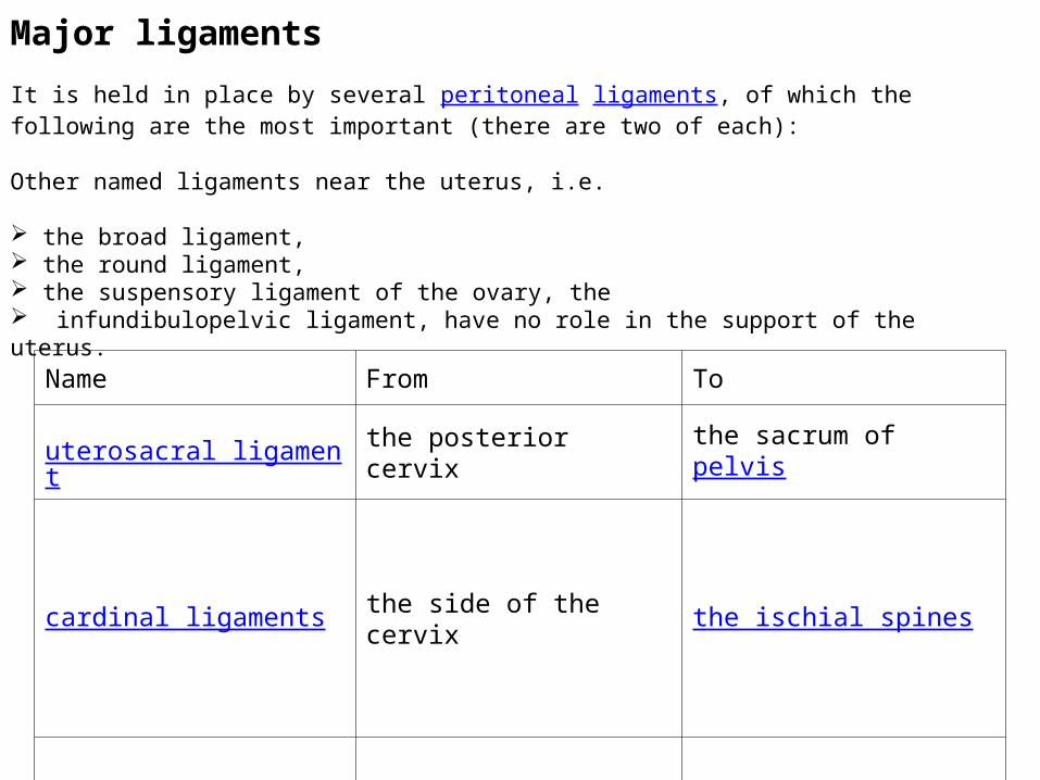
Name From To
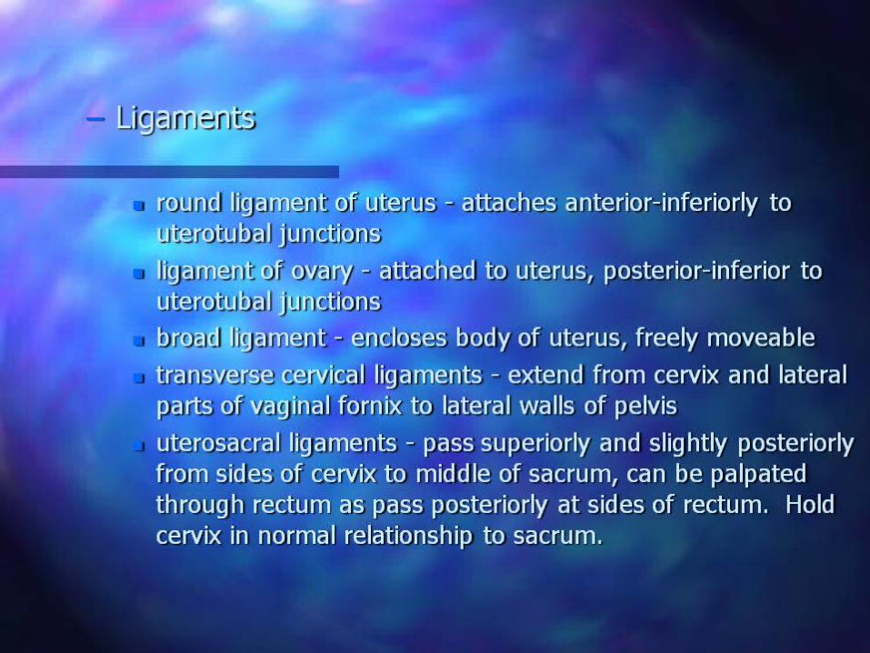
uterosacral ligament the posterior cervix the sacrum of pelvis
cardinal ligaments the side of the cervix the ischial spines
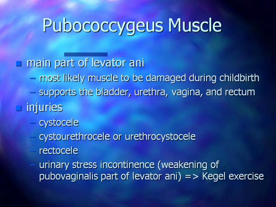
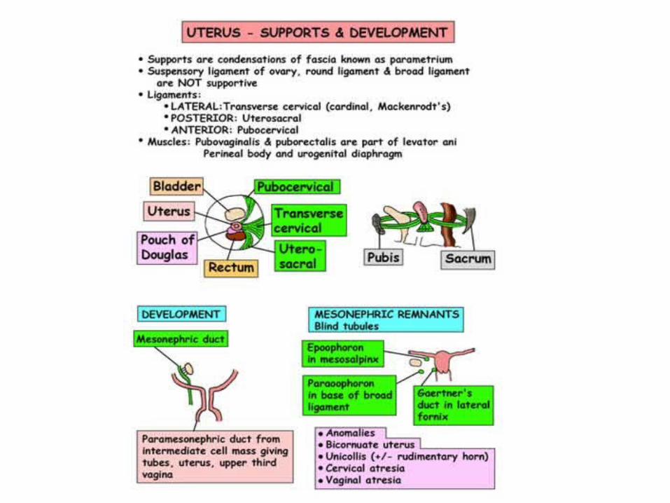
Major ligaments
It is held in place by several peritoneal ligaments, of which the following are the most important (there are two of each):
Other named ligaments near the uterus, i.e.
the broad ligament, the round ligament, the suspensory ligament of the ovary, the infundibulopelvic ligament, have no role in the support of the uterus.
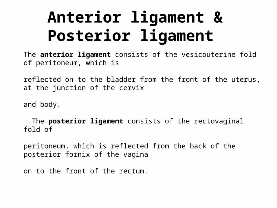
Anterior ligament & Posterior ligament
The anterior ligament consists of the vesicouterine fold of peritoneum, which is reflected on to the bladder from the front of the uterus, at the junction of the cervix
and body.
The posterior ligament consists of the rectovaginal fold of
peritoneum, which is reflected from the back of the posterior fornix of the vagina
on to the front of the rectum.
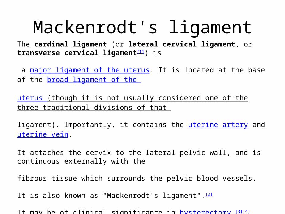
Mackenrodt's ligamentThe cardinal ligament (or lateral cervical ligament, or transverse cervical ligament[1]) is
a major ligament of the uterus. It is located at the base of the broad ligament of the
uterus (though it is not usually considered one of the three traditional divisions of that
ligament). Importantly, it contains the uterine artery and uterine vein.
It attaches the cervix to the lateral pelvic wall, and is continuous externally with the
fibrous tissue which surrounds the pelvic blood vessels.
It is also known as "Mackenrodt's ligament".[2]
It may be of clinical significance in hysterectomy.[3][4]
It provides support to the uterus.
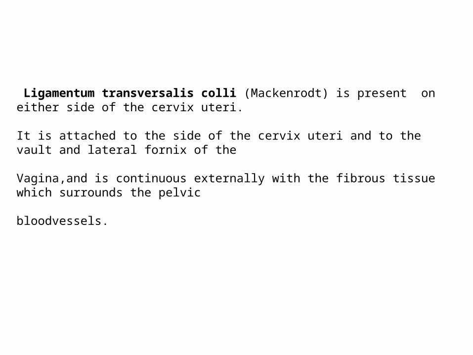
Ligamentum transversalis colli (Mackenrodt) is present on either side of the cervix uteri.
It is attached to the side of the cervix uteri and to the vault and lateral fornix of the
Vagina,and is continuous externally with the fibrous tissue which surrounds the pelvic
bloodvessels.
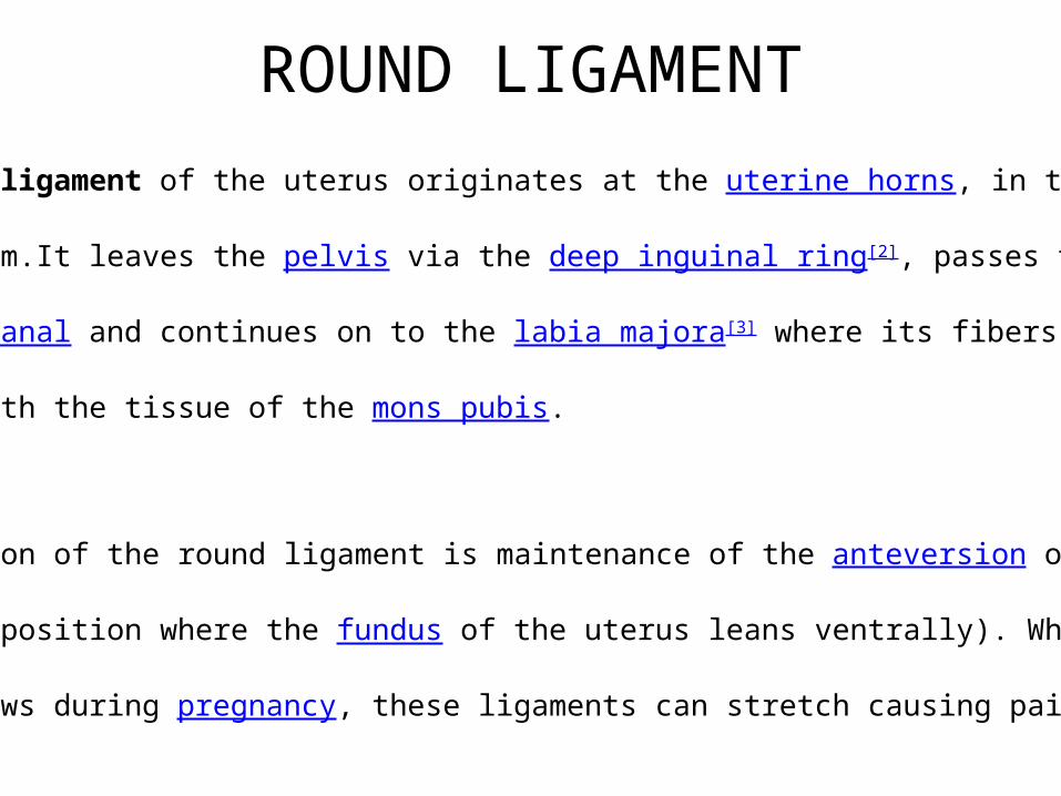
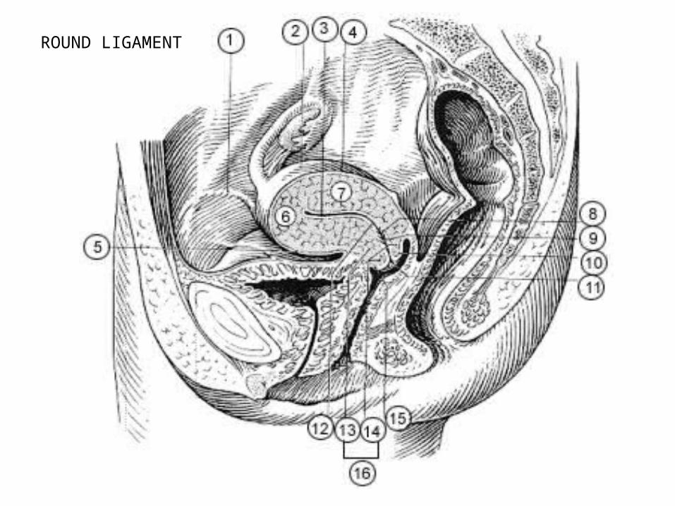
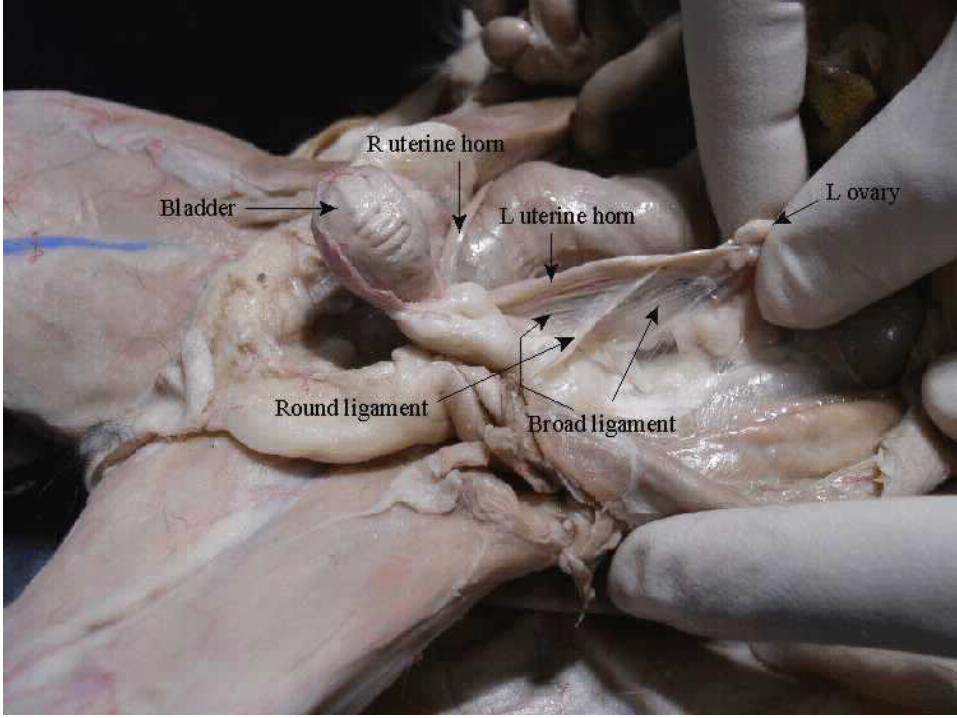
ROUND LIGAMENT
The round ligament of the uterus originates at the uterine horns, in the
Parametrium.It leaves the pelvis via the deep inguinal ring[2], passes through the inguinal canal and continues on to the labia majora[3] where its fibers spread
and mix with the tissue of the mons pubis.
Function
The function of the round ligament is maintenance of the anteversion of the
uterus (a position where the fundus of the uterus leans ventrally). When the
Uterus grows during pregnancy, these ligaments can stretch causing pain.[4]
ROUND LIGAMENT
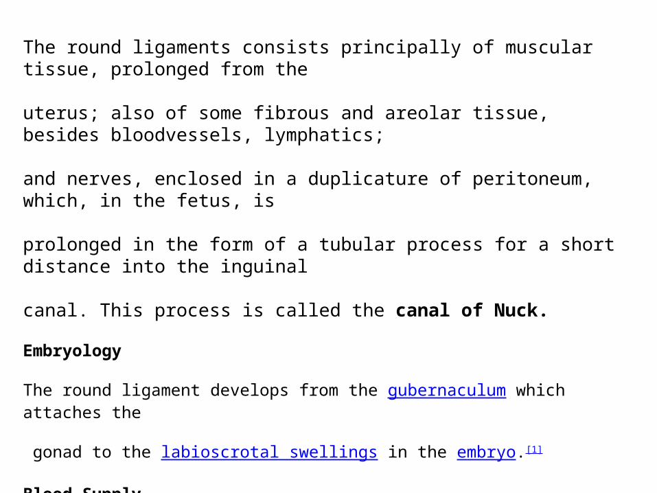
The round ligaments consists principally of muscular tissue, prolonged from the
uterus; also of some fibrous and areolar tissue, besides bloodvessels, lymphatics;
and nerves, enclosed in a duplicature of peritoneum, which, in the fetus, is
prolonged in the form of a tubular process for a short distance into the inguinal
canal. This process is called the canal of Nuck.
Embryology
The round ligament develops from the gubernaculum which attaches the
gonad to the labioscrotal swellings in the embryo.[1]
Blood Supply
The round ligament is supplied by the artery of the round ligament, otherwise
known as "Sampson's artery."
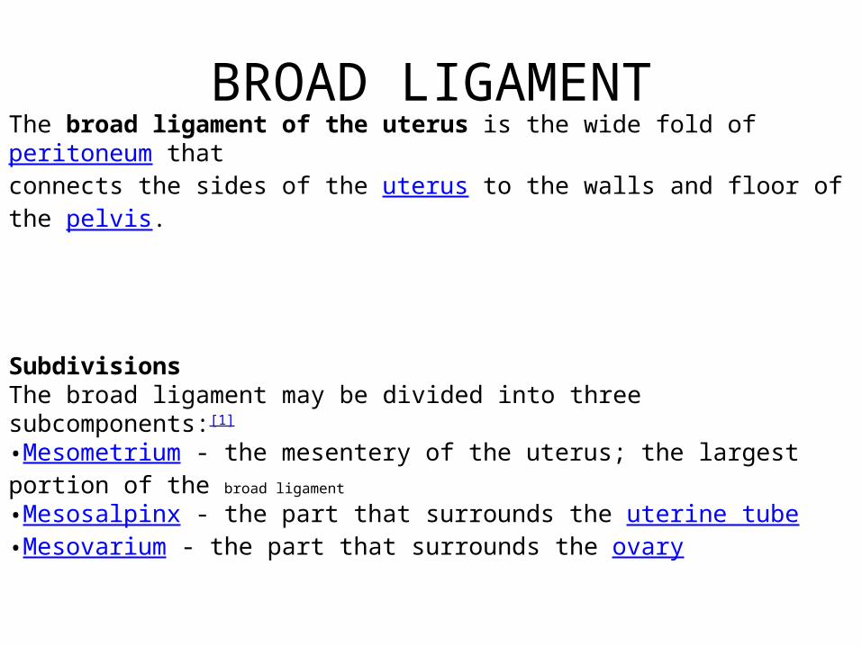
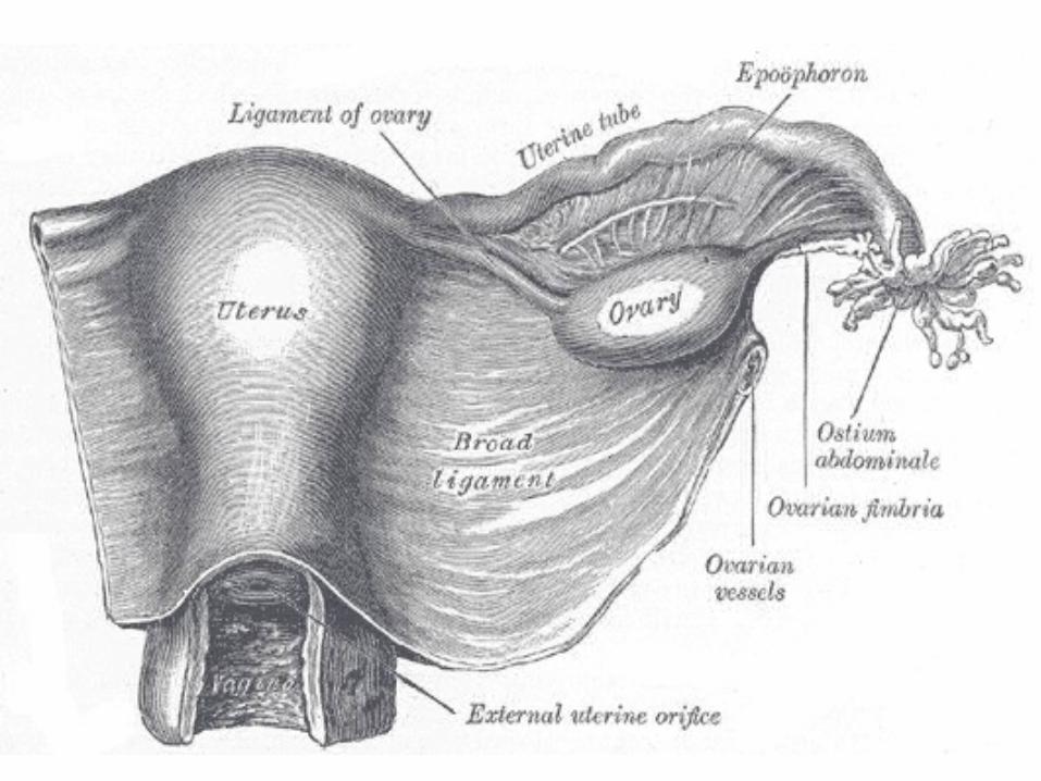
BROAD LIGAMENT
The broad ligament of the uterus is the wide fold of peritoneum that connects the sides of the uterus to the walls and floor of the pelvis.
SubdivisionsThe broad ligament may be divided into three subcomponents:[1]
•Mesometrium - the mesentery of the uterus; the largest portion of the broad ligament
•Mesosalpinx - the part that surrounds the uterine tube •Mesovarium - the part that surrounds the ovary
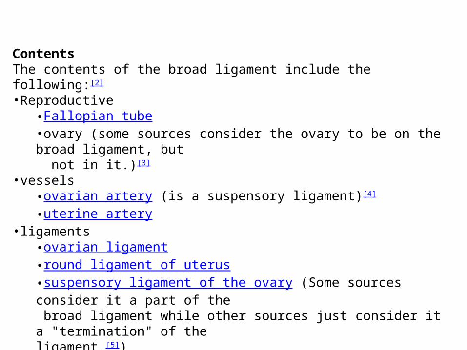
ContentsThe contents of the broad ligament include the following:[2]
•Reproductive •Fallopian tube •ovary (some sources consider the ovary to be on the broad ligament, but not in it.)[3]
•vessels •ovarian artery (is a suspensory ligament)[4] •uterine artery
•ligaments •ovarian ligament •round ligament of uterus •suspensory ligament of the ovary (Some sources consider it a part of the broad ligament while other sources just consider it a "termination" of the ligament.[5])
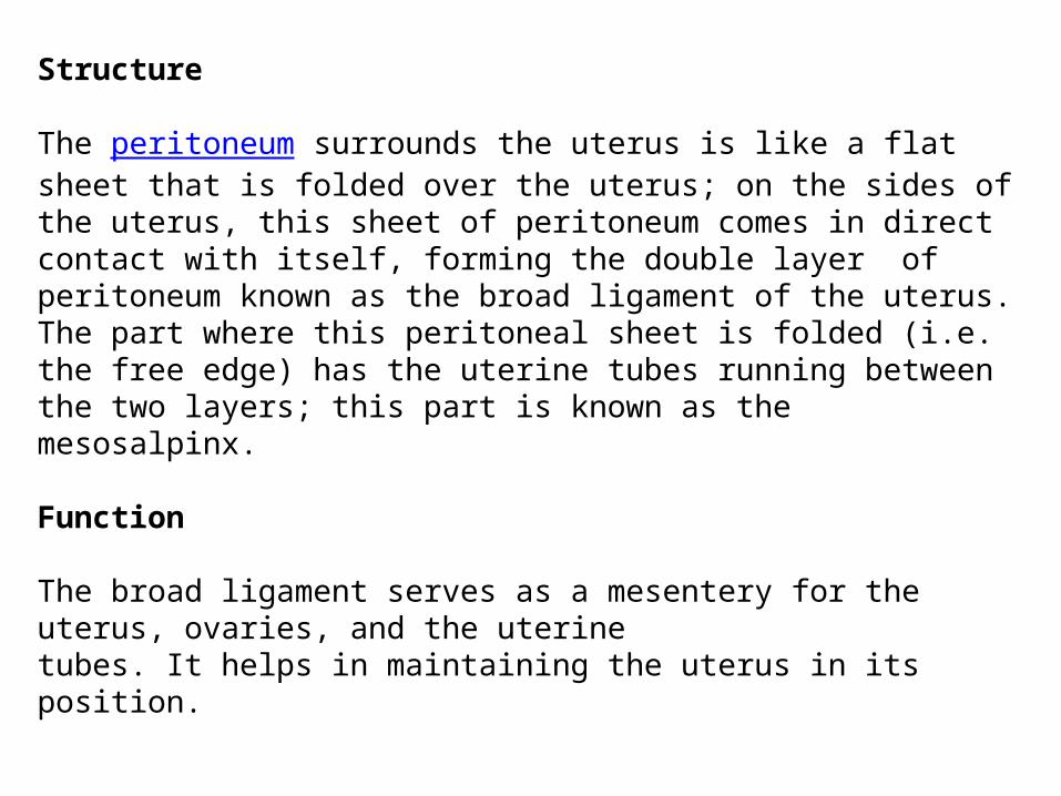
Structure
The peritoneum surrounds the uterus is like a flat sheet that is folded over the uterus; on the sides of the uterus, this sheet of peritoneum comes in direct contact with itself, forming the double layer of peritoneum known as the broad ligament of the uterus.The part where this peritoneal sheet is folded (i.e. the free edge) has the uterine tubes running between the two layers; this part is known as the mesosalpinx.
Function
The broad ligament serves as a mesentery for the uterus, ovaries, and the uterine tubes. It helps in maintaining the uterus in its position.
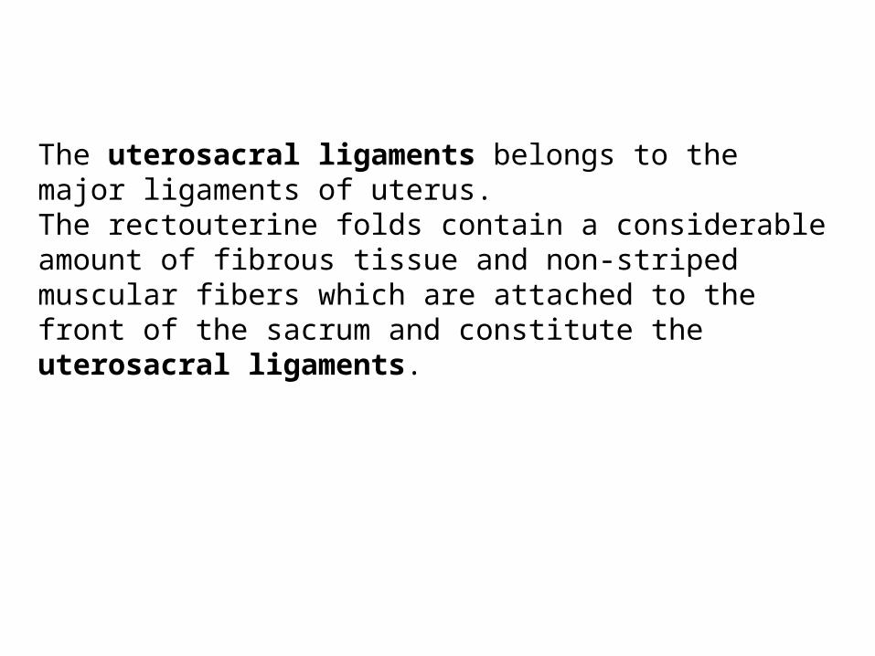
The uterosacral ligaments belongs to the major ligaments of uterus.The rectouterine folds contain a considerable amount of fibrous tissue and non-striped muscular fibers which are attached to the front of the sacrum and constitute the uterosacral ligaments.