supplementary information title - media.nature.com · biofísica, facultad de ciencias bioquímicas...
TRANSCRIPT
Supplementary Information
Title
Membrane-anchoring stabilizes and favors secretion of New Delhi Metallo-β-lactamase
Authors
Lisandro J. González1, Guillermo Bahr1, Toshiki G. Nakashige2, Elizabeth M. Nolan2,
Robert A. Bonomo3 and Alejandro J. Vila1*
Affiliations
1Instituto de Biología Molecular y Celular de Rosario (IBR, CONICET-UNR) and Área
Biofísica, Facultad de Ciencias Bioquímicas y Farmacéuticas, Universidad Nacional de
Rosario, Rosario, Argentina
2Department of Chemistry, Massachusetts Institute of Technology, Cambridge,
Massachusetts 02139, USA
3Research Service, Louis Stokes Cleveland Department of Veterans Affairs Medical
Center, Cleveland, OH; Departments of Medicine, Pharmacology, Microbiology and
Molecular Biology; Case Western Reserve University, Cleveland, OH, USA
*Corresponding author. E-mail: [email protected]
Nature Chemical Biology: doi:10.1038/nchembio.2083
Supplementary Results
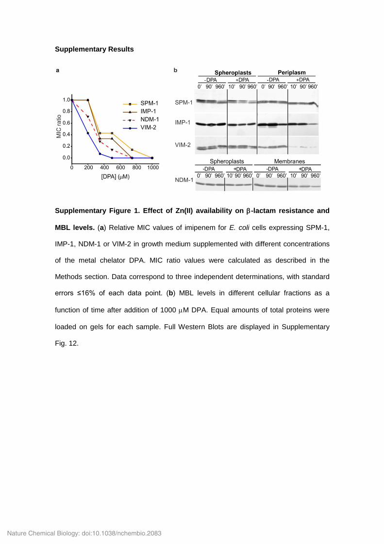
Supplementary Figure 1. Effect of Zn(II) availability on -lactam resistance and
MBL levels. (a) Relative MIC values of imipenem for E. coli cells expressing SPM-1,
IMP-1, NDM-1 or VIM-2 in growth medium supplemented with different concentrations
of the metal chelator DPA. MIC ratio values were calculated as described in the
Methods section. Data correspond to three independent determinations, with standard
errors ≤16% of each data point. (b) MBL levels in different cellular fractions as a
function of time after addition of 1000 M DPA. Equal amounts of total proteins were
loaded on gels for each sample. Full Western Blots are displayed in Supplementary
Fig. 12.
Nature Chemical Biology: doi:10.1038/nchembio.2083
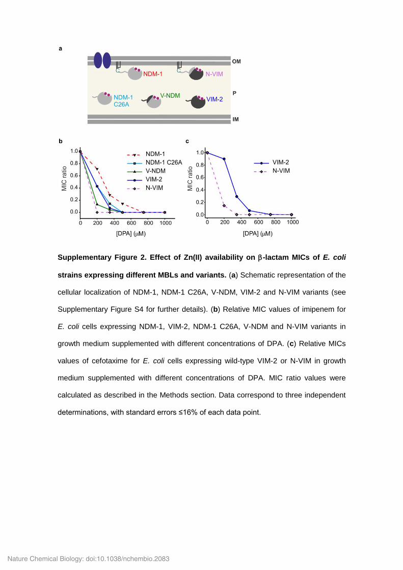
Supplementary Figure 2. Effect of Zn(II) availability on -lactam MICs of E. coli
strains expressing different MBLs and variants. (a) Schematic representation of the
cellular localization of NDM-1, NDM-1 C26A, V-NDM, VIM-2 and N-VIM variants (see
Supplementary Figure S4 for further details). (b) Relative MIC values of imipenem for
E. coli cells expressing NDM-1, VIM-2, NDM-1 C26A, V-NDM and N-VIM variants in
growth medium supplemented with different concentrations of DPA. (c) Relative MICs
values of cefotaxime for E. coli cells expressing wild-type VIM-2 or N-VIM in growth
medium supplemented with different concentrations of DPA. MIC ratio values were
calculated as described in the Methods section. Data correspond to three independent
determinations, with standard errors ≤16% of each data point.
Nature Chemical Biology: doi:10.1038/nchembio.2083
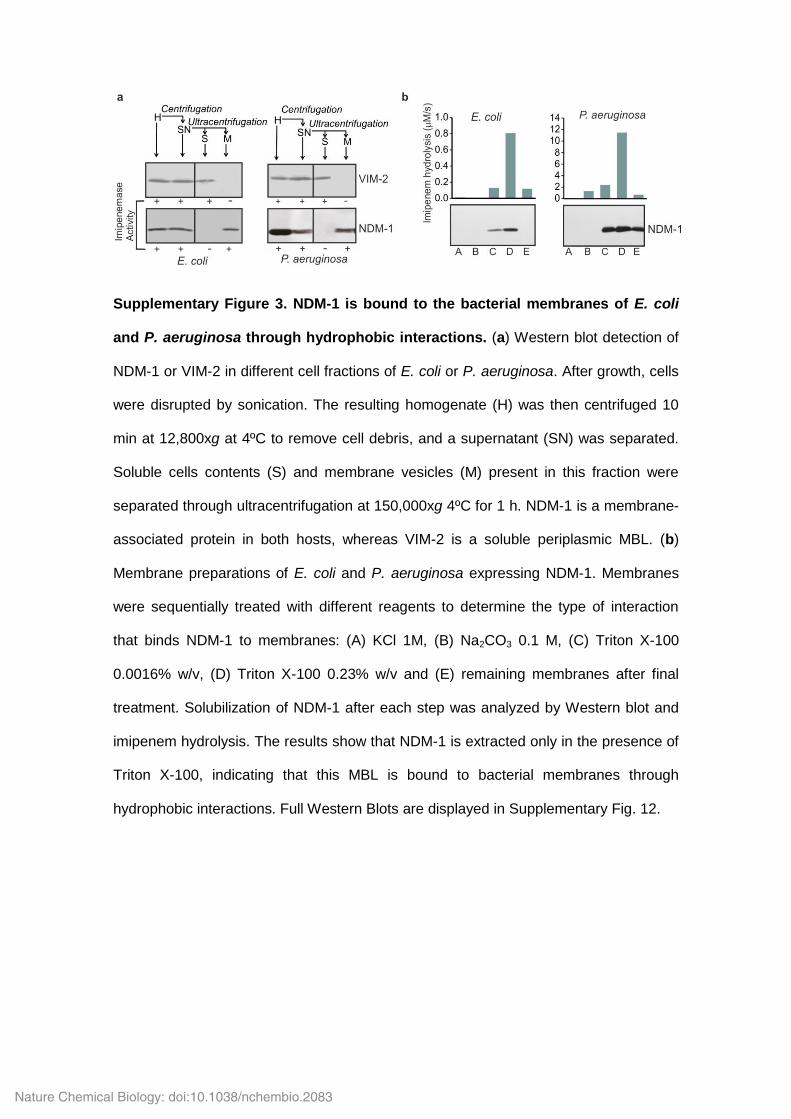
Supplementary Figure 3. NDM-1 is bound to the bacterial membranes of E. coli
and P. aeruginosa through hydrophobic interactions. (a) Western blot detection of
NDM-1 or VIM-2 in different cell fractions of E. coli or P. aeruginosa. After growth, cells
were disrupted by sonication. The resulting homogenate (H) was then centrifuged 10
min at 12,800xg at 4ºC to remove cell debris, and a supernatant (SN) was separated.
Soluble cells contents (S) and membrane vesicles (M) present in this fraction were
separated through ultracentrifugation at 150,000xg 4ºC for 1 h. NDM-1 is a membrane-
associated protein in both hosts, whereas VIM-2 is a soluble periplasmic MBL. (b)
Membrane preparations of E. coli and P. aeruginosa expressing NDM-1. Membranes
were sequentially treated with different reagents to determine the type of interaction
that binds NDM-1 to membranes: (A) KCl 1M, (B) Na2CO3 0.1 M, (C) Triton X-100
0.0016% w/v, (D) Triton X-100 0.23% w/v and (E) remaining membranes after final
treatment. Solubilization of NDM-1 after each step was analyzed by Western blot and
imipenem hydrolysis. The results show that NDM-1 is extracted only in the presence of
Triton X-100, indicating that this MBL is bound to bacterial membranes through
hydrophobic interactions. Full Western Blots are displayed in Supplementary Fig. 12.
Nature Chemical Biology: doi:10.1038/nchembio.2083
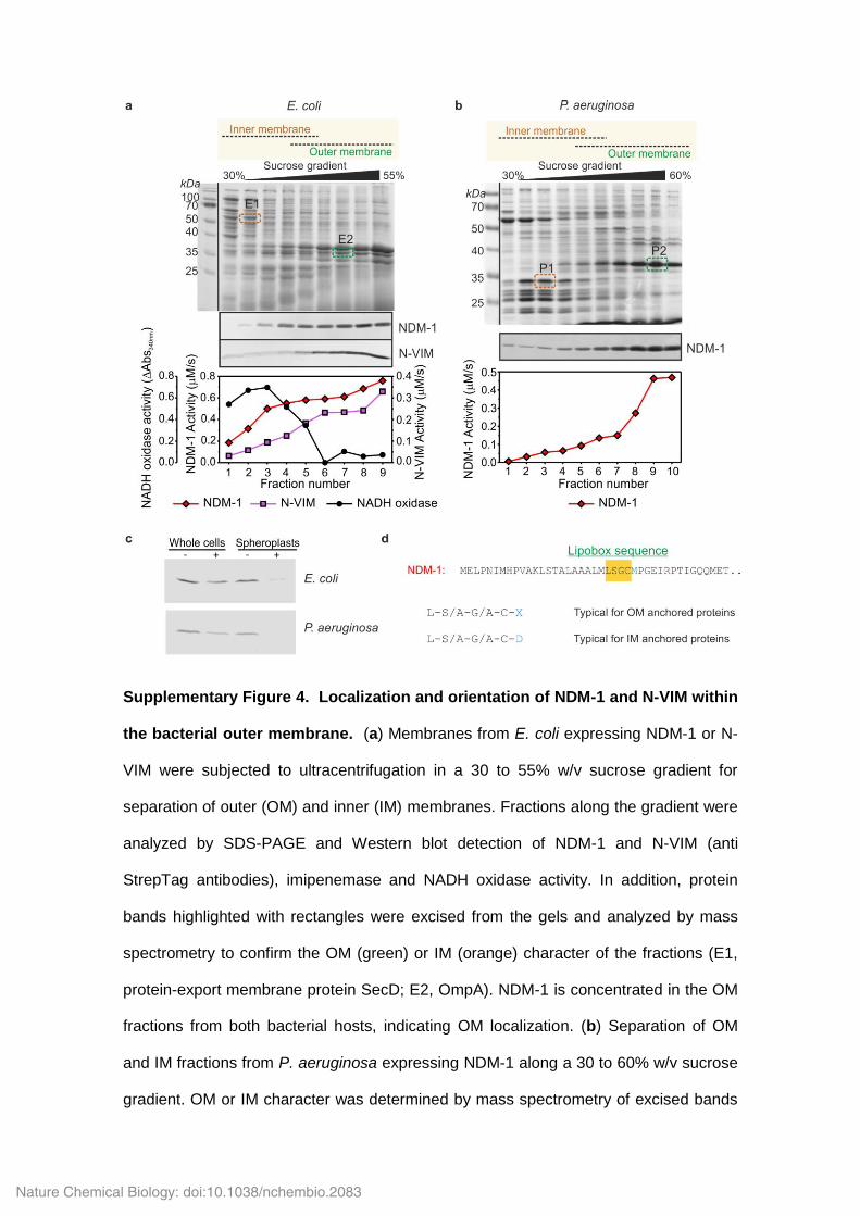
Supplementary Figure 4. Localization and orientation of NDM-1 and N-VIM within
the bacterial outer membrane. (a) Membranes from E. coli expressing NDM-1 or N-
VIM were subjected to ultracentrifugation in a 30 to 55% w/v sucrose gradient for
separation of outer (OM) and inner (IM) membranes. Fractions along the gradient were
analyzed by SDS-PAGE and Western blot detection of NDM-1 and N-VIM (anti
StrepTag antibodies), imipenemase and NADH oxidase activity. In addition, protein
bands highlighted with rectangles were excised from the gels and analyzed by mass
spectrometry to confirm the OM (green) or IM (orange) character of the fractions (E1,
protein-export membrane protein SecD; E2, OmpA). NDM-1 is concentrated in the OM
fractions from both bacterial hosts, indicating OM localization. (b) Separation of OM
and IM fractions from P. aeruginosa expressing NDM-1 along a 30 to 60% w/v sucrose
gradient. OM or IM character was determined by mass spectrometry of excised bands
Nature Chemical Biology: doi:10.1038/nchembio.2083
(P1, ATP synthase subunit b and Signal Peptidase I; P2, OmpF), and the presence of
NDM-1 was detected as in (a). (c) Whole cells (W) and permeable spheroplasts (S) of
E. coli and P. aeruginosa expressing NDM-1 were subjected to limited proteolysis with
proteinase K. Comparison of NDM-1 levels detected by Western blot in protease
treated samples (+) versus untreated controls (–) shows that NDM-1 is resistant to
proteolysis in whole cells, while being degraded in spheroplasts under identical
conditions. These results demonstrate that NDM-1 is inaccessible to external
proteases, and thus located in the inner leaflet of the outer membrane. (d) The N-
terminus of NDM-1 contains a canonical lipobox sequence (highlighted in orange),
which includes a cysteine residue that undergoes lipidation followed by peptide leader
processing in the bacterial IM1. The identity of the residue immediately posterior to the
C terminal end of posterior the lipobox determines whether the lipoprotein will be
localized to IM or OM, with an aspartic acid being required for retention in IM. The
presence of a methionine residue at this position in NDM-1 is suggestive of targeting to
the OM. Full Western Blots are displayed in Supplementary Fig. 12.
Nature Chemical Biology: doi:10.1038/nchembio.2083
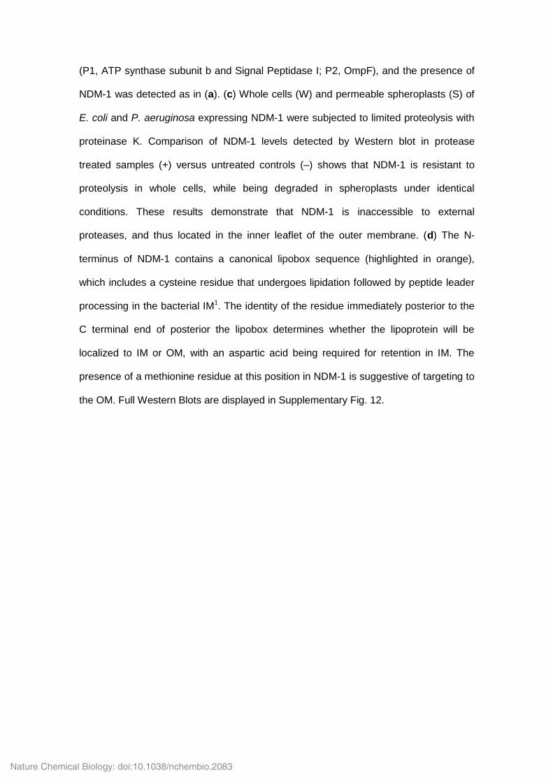
Supplementary Figure 5. Zn(II) deprivation causes selective degradation of MBLs
in the periplasm. Left, immunodetection of VIM-2 in spheroplasts and periplasmic
extracts of E. coli cells exposed to 1000 M DPA (or no DPA) at 20ºC for different time-
periods. Western blots of periplasmic maltose binding protein (MBP) and cytoplasmic
RNA polymerase (RNA pol) were performed as loading controls for periplasmic
extracts and spheroplasts, respectively. Right, Coomassie-stained SDS-PAGE of
periplasmic extracts. There is a clear and selective reduction in the intensity of the
band corresponding to VIM-2. The rest of the protein pattern remains unchanged. Full
Western Blots are displayed in Supplementary Figs. 12 and 13.
Nature Chemical Biology: doi:10.1038/nchembio.2083
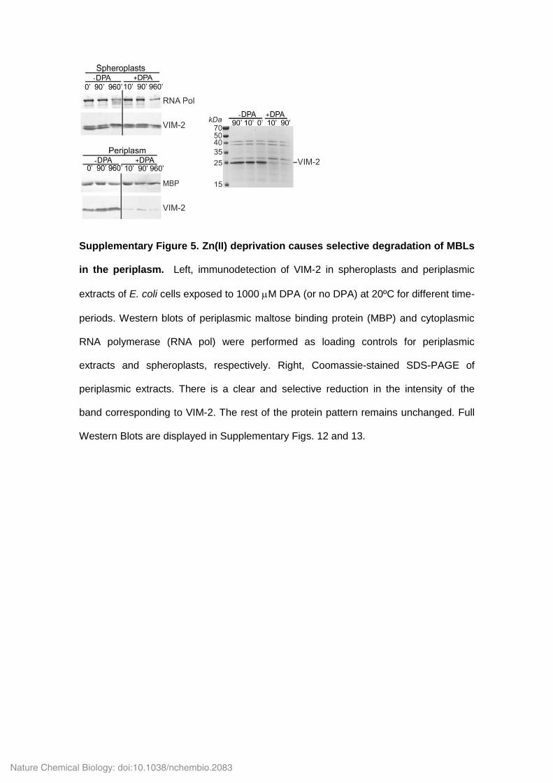
Supplementary Figure 6. Protein levels of MBLs in different cellular fractions
under conditions of Zn(II) deprivation. (a) Spheroplast MBL levels (full-length and
processed forms) determined by Western blot from E. coli expressing SPM-1, VIM-2,
IMP-1 or NDM-1 at different times after addition of 1000 μM DPA .(b) MBL levels of
NDM-1, VIM-2 and related mutants in periplasm and spheroplasts as a function time
after addition of 1000 μM DPA. Full Western Blots are displayed in Supplementary
Figs. 12 and 13. (c) Spheroplast MBL levels determined by Western blot from E. coli
cultures VIM-2, N-VIM, NDM-1, NDM-1 C26A or V-NDM at different times after addition
of 1000 μM DPA. Data correspond to three independent experiments and are shown as
mean ± s.e.m.
Nature Chemical Biology: doi:10.1038/nchembio.2083
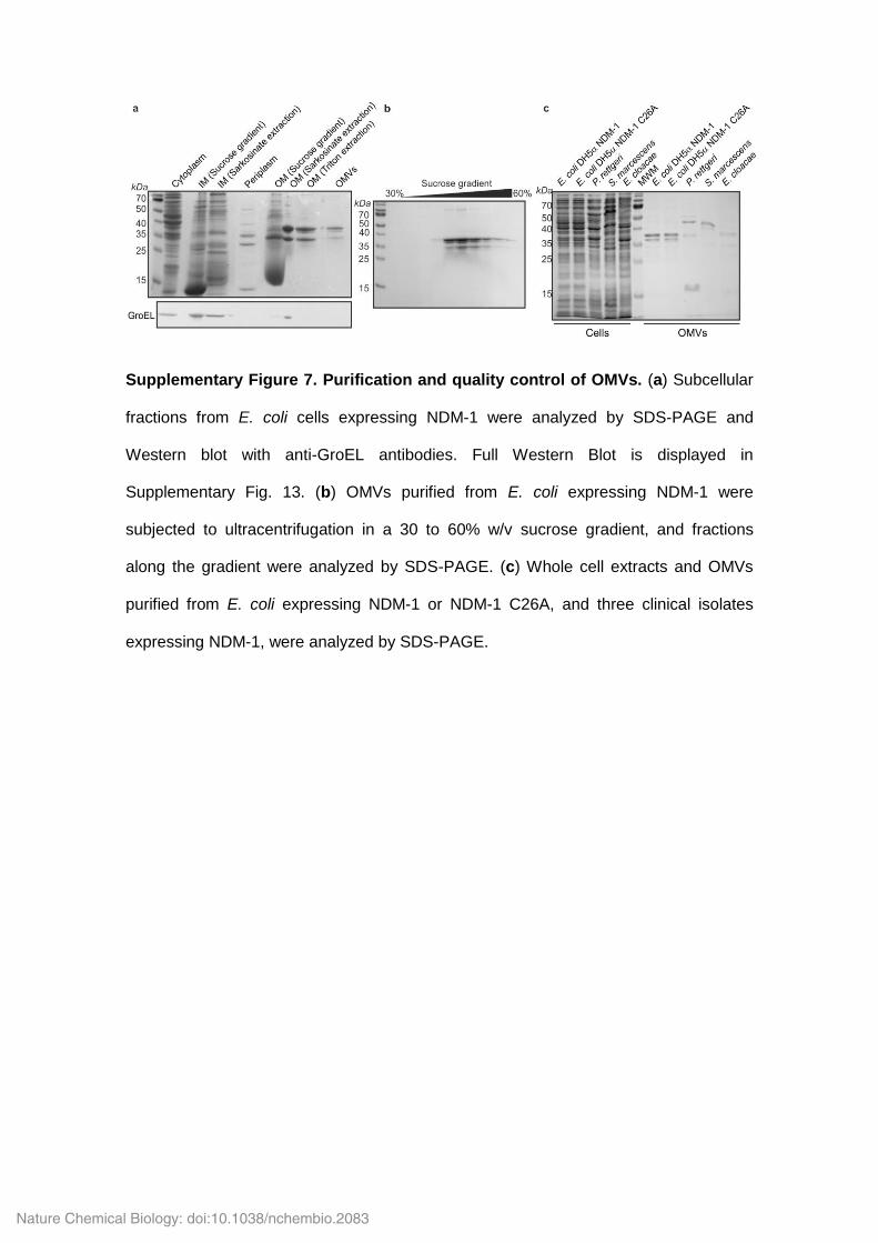
Supplementary Figure 7. Purification and quality control of OMVs. (a) Subcellular
fractions from E. coli cells expressing NDM-1 were analyzed by SDS-PAGE and
Western blot with anti-GroEL antibodies. Full Western Blot is displayed in
Supplementary Fig. 13. (b) OMVs purified from E. coli expressing NDM-1 were
subjected to ultracentrifugation in a 30 to 60% w/v sucrose gradient, and fractions
along the gradient were analyzed by SDS-PAGE. (c) Whole cell extracts and OMVs
purified from E. coli expressing NDM-1 or NDM-1 C26A, and three clinical isolates
expressing NDM-1, were analyzed by SDS-PAGE.
Nature Chemical Biology: doi:10.1038/nchembio.2083
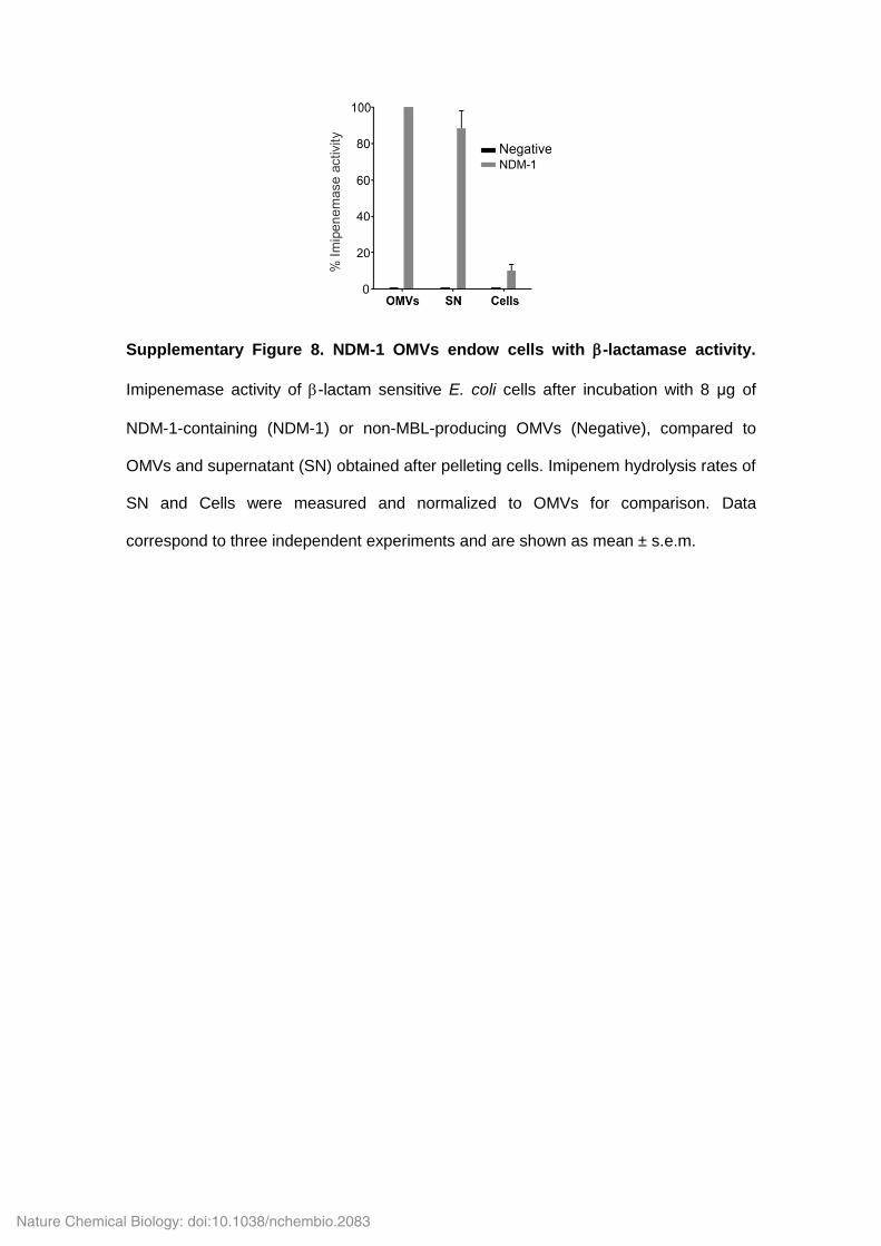
Supplementary Figure 8. NDM-1 OMVs endow cells with -lactamase activity.
Imipenemase activity of -lactam sensitive E. coli cells after incubation with 8 μg of
NDM-1-containing (NDM-1) or non-MBL-producing OMVs (Negative), compared to
OMVs and supernatant (SN) obtained after pelleting cells. Imipenem hydrolysis rates of
SN and Cells were measured and normalized to OMVs for comparison. Data
correspond to three independent experiments and are shown as mean ± s.e.m.
Nature Chemical Biology: doi:10.1038/nchembio.2083

Supplementary Figure 9. All NDM variants have a conserved lipobox. Sequence
alignment of N-terminus of NDM-1 to NDM-16. Conserved LSGC lipobox sequences
and residues at +2 position are shown in red and green, respectively. Alignment was
performed with the T-Coffee tool, available at www.tcoffee.org.
Nature Chemical Biology: doi:10.1038/nchembio.2083
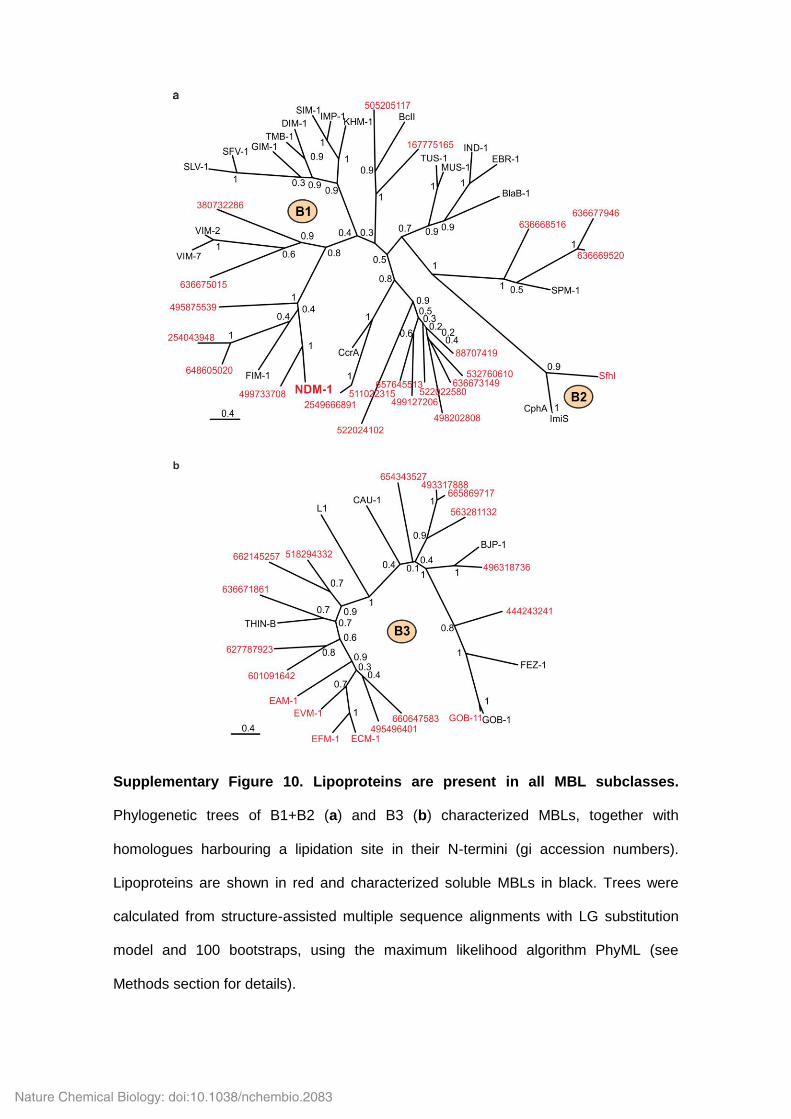
Supplementary Figure 10. Lipoproteins are present in all MBL subclasses.
Phylogenetic trees of B1+B2 (a) and B3 (b) characterized MBLs, together with
homologues harbouring a lipidation site in their N-termini (gi accession numbers).
Lipoproteins are shown in red and characterized soluble MBLs in black. Trees were
calculated from structure-assisted multiple sequence alignments with LG substitution
model and 100 bootstraps, using the maximum likelihood algorithm PhyML (see
Methods section for details).
Nature Chemical Biology: doi:10.1038/nchembio.2083
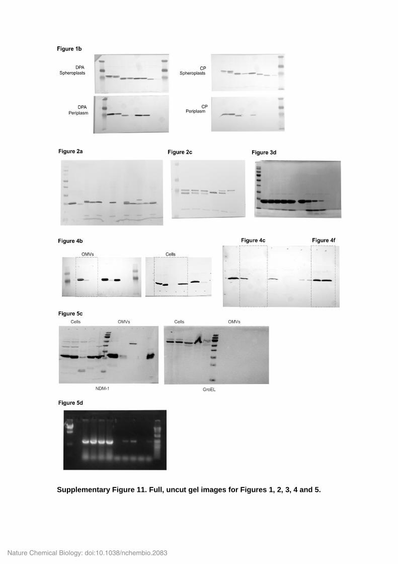
Supplementary Figure 11. Full, uncut gel images for Figures 1, 2, 3, 4 and 5.
Nature Chemical Biology: doi:10.1038/nchembio.2083
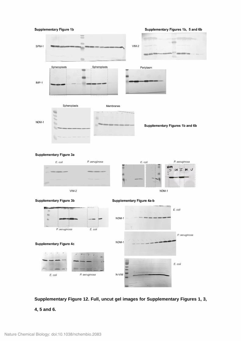
Supplementary Figure 12. Full, uncut gel images for Supplementary Figures 1, 3,
4, 5 and 6.
Nature Chemical Biology: doi:10.1038/nchembio.2083
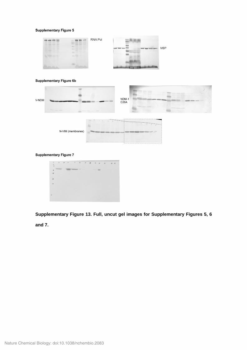
Supplementary Figure 13. Full, uncut gel images for Supplementary Figures 5, 6
and 7.
Nature Chemical Biology: doi:10.1038/nchembio.2083
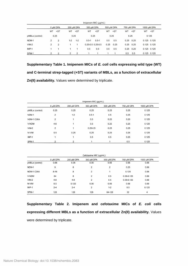
Supplementary Table 1. Imipenem MICs of E. coli cells expressing wild type (WT)
and C-terminal strep-tagged (+ST) variants of MBLs, as a function of extracellular
Zn(II) availability. Values were determined by triplicate.
Supplementary Table 2. Imipenem and cefotaxime MICs of E. coli cells
expressing different MBLs as a function of extracellular Zn(II) availability. Values
were determined by triplicate.
Nature Chemical Biology: doi:10.1038/nchembio.2083
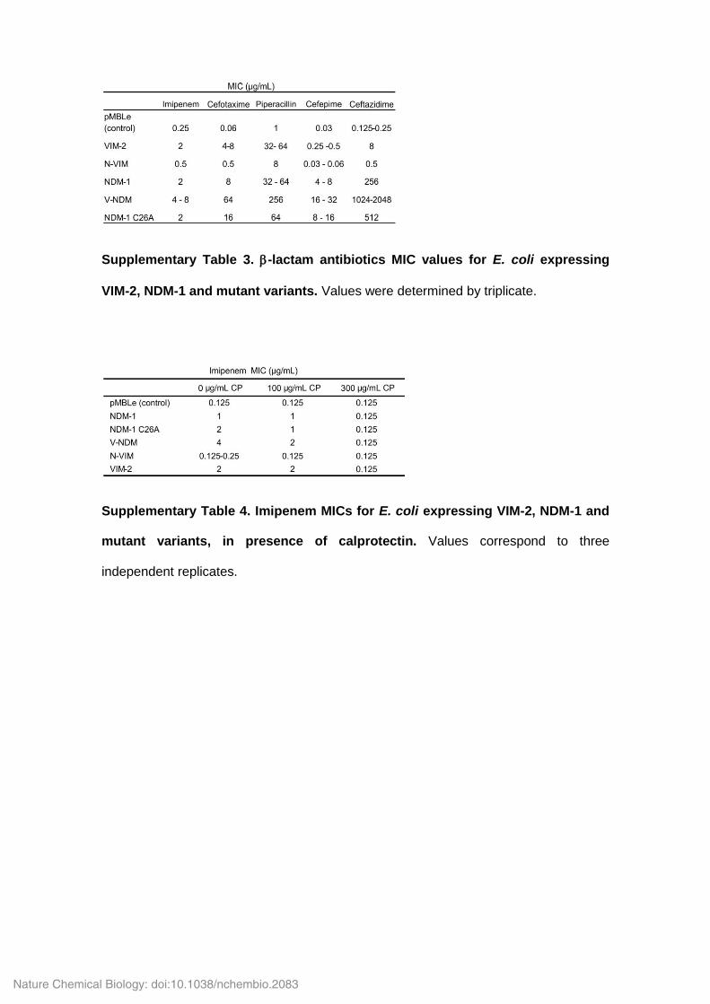
Supplementary Table 3. -lactam antibiotics MIC values for E. coli expressing
VIM-2, NDM-1 and mutant variants. Values were determined by triplicate.
Supplementary Table 4. Imipenem MICs for E. coli expressing VIM-2, NDM-1 and
mutant variants, in presence of calprotectin. Values correspond to three
independent replicates.
Nature Chemical Biology: doi:10.1038/nchembio.2083
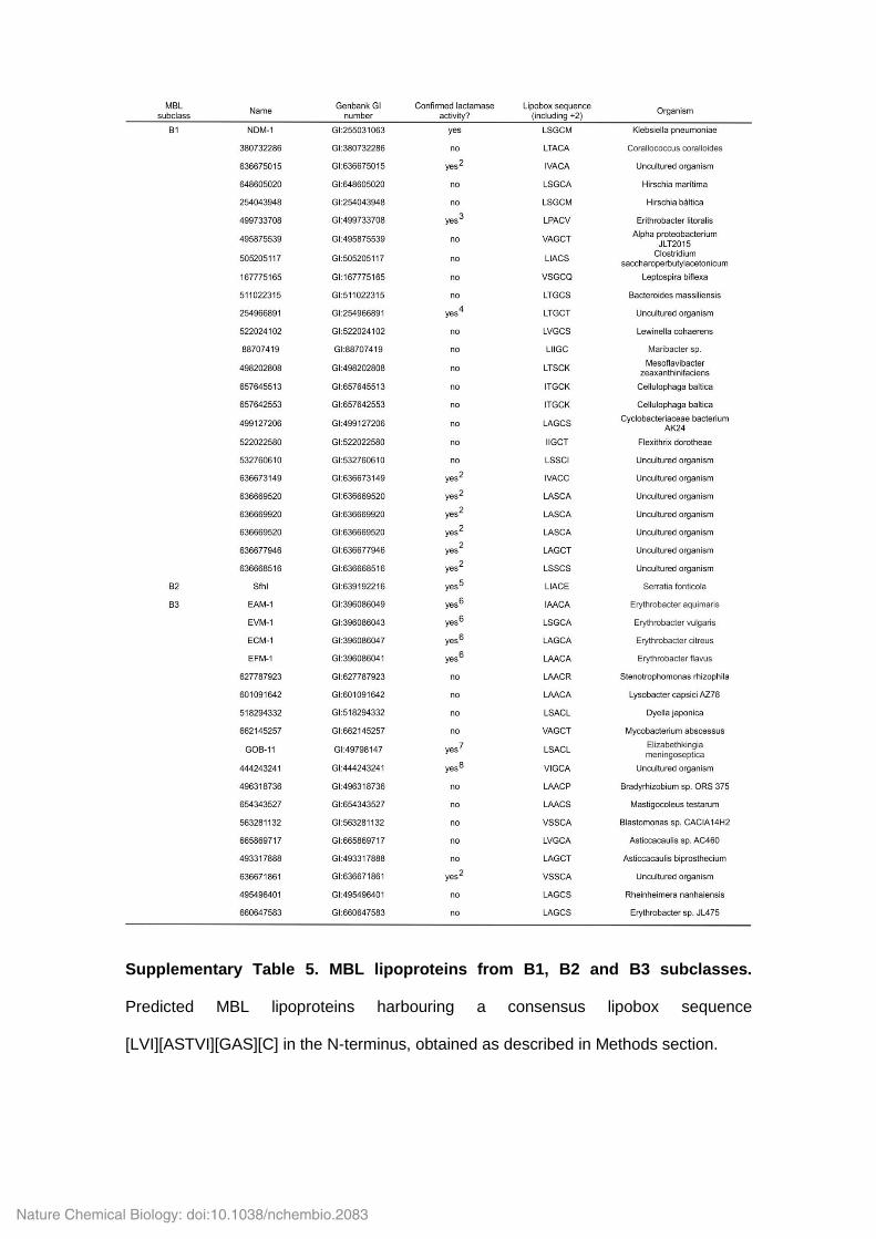
Supplementary Table 5. MBL lipoproteins from B1, B2 and B3 subclasses.
Predicted MBL lipoproteins harbouring a consensus lipobox sequence
[LVI][ASTVI][GAS][C] in the N-terminus, obtained as described in Methods section.
Nature Chemical Biology: doi:10.1038/nchembio.2083
Supplementary Information References
1 Kovacs-Simon, A., Titball, R. W. & Michell, S. L. Lipoproteins of bacterial
pathogens. Infect. Immun. 79, 548-561 (2011).
2 Forsberg, K. J. et al. Bacterial phylogeny structures soil resistomes across
habitats. Nature 509, 612–616 (2014).
3 Zheng, B. et al., An unexpected similarity between antibiotic-resistant NDM-1
and -lactamase II from Erythrobacter litoralis. Protein Cell 2, 250-258 (2011).
4 Sommer, M. O. A., Dantas, G. & Church, G. M. Functional Characterization of
the Antibiotic Resistance Reservoir in the Human Microflora. Science 325, 1128-1131
(2009).
5 Saavedra, M. J. et al. Sfh-I, a subclass B2 metallo--lactamase from a Serratia
fonticola environmental isolate. Antimicrob. Agents Chemother. 47, 2330-2333 (2003).
6 Girlich, D., Poirel, L. & Nordmann, P. Diversity of naturally occurring Ambler
class B metallo-β-lactamases in Erythrobacter spp. J. Antimicrob. Agents Chemother.
67, 2661-2664 (2012).
7 Yum, J. H. et al. Genetic diversity of chromosomal metallo--lactamase genes
in clinical isolates of Elizabethkingia meningoseptica from Korea. J. Microbiol. 48, 358-
64 (2010).
8 Terrón-González, L., Medina, C., Limón-Mortés, M. C. & Santero, E.
Heterologous viral expression systems in fosmid vectors increase the functional
analysis potential of metagenomic libraries. Sci. Rep. 3, 1107 (2013).
Nature Chemical Biology: doi:10.1038/nchembio.2083