supplemental material pulmonary hypertension secondary...
TRANSCRIPT
1
Supplemental Material
Pulmonary hypertension secondary to left‐heart failure involves peroxynitrite‐induced downregulation of PTEN in the lung
Yazhini Ravi, Karuppaiyah Selvendiran, Shan K. Naidu, Sarath Meduru, Lucas A. Citro, Balázs Bognár, Mahmood Khan, Tamás Kálai, Kálmán Hideg, Periannan Kuppusamy, Chittoor B. Sai‐Sudhakar
Materials and Methods
Reagents: Dimethyl sulfoxide (DMSO) and antibody directed against actin were obtained from Sigma‐Aldrich (St. Louis, MO). Polyvinylidene‐fluoride membrane and molecular‐weight markers were obtained from Bio‐Rad (Hercules, CA). Antibodies directed against Akt (pan), pAkt (Ser473), PTEN, and pPTEN (Ser380 and Thr381/382), were purchased from Cell Signaling Technology (Danvers, MA). Enhanced chemiluminescence reagents were obtained from GE Healthcare (Chalfont St. Giles, Buckinghamshire, UK). 3‐Morpholinosydnonimine hydrochloride (SIN‐1) (catalog# M5793) was purchased from Sigma Aldrich (St. Louis, MO) and peroxynitrite (catalog# 81565) was purchased from Cayman Chemicals (Ann Arbor, MI). HO‐3867 was synthesized as reported.1 Stock solutions (20 mM) of the compound were freshly prepared in dimethyl sulfoxide.
Induction of LHF‐PH by ligation of left‐anterior‐descending coronary artery: Left‐heart failure was induced in rats by permanent ligation of left‐anterior‐descending coronary artery (LAD). All the procedures were performed with the approval of the Institutional Animal Care and Use Committee of The Ohio State University and conformed to the Guide for Care and Use of Laboratory Animals (NIH publication no. 86‐23). Male Sprague‐Dawley rats (225‐250 g) were intubated orally, placed on a volume‐cycled ventilator (Rodent Ventilator, model 683; Harvard Apparatus Holliston, MA), and maintained under general anesthesia with 1 to 2% isoflurane in air. An oblique 12‐mm incision was made 8 mm away from the left sternal border toward the left armpit. The chest cavity was opened with scissors by a small incision (10‐mm long) at the level of the third or fourth intercostal space, 2 to 3 mm from the left sternal border. The LAD was visualized as a pulsating bright‐red spike running through the midst of the heart wall from underneath the left atrium toward apex. The LAD was ligated 1 to 2 mm below the tip of the left atrium using a tapered needle and a 6‐0 polypropylene ligature passed underneath the LAD. A double knot was made to occlude the LAD permanently. Occlusion was confirmed by a sudden change in color (pale) of the anterior wall of the left ventricle (LV). EKG changes were recorded, and ST (ventricular depolarization) elevation was observed after LAD ligation. The chest cavity was closed by bringing together the third and fourth ribs with one 4‐0 polypropylene silk suture. The layers of muscle and skin were closed with a 4‐0 polypropylene suture, and the rats were allowed to recover under a warm light. An age‐matched control group underwent sham surgeries and was used for comparison. The study used 8‐10 animals per group. No mortality was observed in the 4‐week period following LAD occlusion.
2
Treatment with HO‐3867: HO‐3867 was administered in the diet (100 ppm), beginning day 1 after LAD ligation, and continued for the entire treatment period of 4 weeks. The 100‐ppm dose of HO‐3867 was based upon our earlier studies that used oral administration of this compound in mice.2,3
Processing of lung tissues for histopathology and protein analysis: Before explantation, lungs were flushed with cold saline to remove intravascular blood. The explanted lungs were then, either flash‐frozen for mRNA and protein analysis or inflated with 4% paraformaldehyde at 20‐cm water pressure followed by immersion‐fixation using 4% paraformaldehyde for 48 hours at room temperature for histology.
Magnetic resonance imaging (MRI): Rats were anesthetized using isofluorane (1.5%)‐carbogen gas mixture and placed on a water‐heated imaging bed in the head first, supine position. Two metal EKG (electrocardiogram) pads were then placed on the animal’s front and rear paws and a respiratory pad was placed across the animal’s abdomen. During imaging, respiratory and heart rates were monitored and recorded using a PCSAM (Small Animal Instruments) monitoring program. A Bruker BioSpec 94/30USR 9.4T horizontal bore MRI system (Bruker, BioSpin, Germany), equipped with Paravision 4.0 and was used to acquire images. Multiple cardiac‐gated, T1‐weighted FLASH‐cine images were acquired in the transverse orientation to cover the entire left and right ventricle of each animal, from apex to aortic valve, using the following settings: TR/TE = 16/1.6 ms, slice thickness = 2 mm, α= 10o, FOV = 51.0 mm, 256 x 192 (in‐plane pixel size = 0.20 mm x 0.27 mm. All transverse cine images were analyzed using the image analysis program ImageJ (NIH). The end‐diastolic (ED) and end‐systolic (ES) time points for each acquired cine slice were identified visually. The endocardial and epicardial boundaries of the left and right ventricles were then manually traced for both end‐systolic (ES) and end‐diastolic (ED) time points of the cardiac cycle and used to calculate LV and RV end‐diastolic volume (EDV), end‐systolic volume (ESV), LV and LV ES wall thickness (LV ES‐WT) and LV ED wall thickness (LV ED‐WT). Using Segment (Medviso AB), the ES and ED cardiac timepoints were identified for the mid‐ventricular slice of each heart. The epicardial and endocardial boundaries of the LV and interventricular septal (IVS) walls were then manually traced. The LV wall was then divided into eight 45o sectors using the anterior right‐ventricular insertion point as the reference, with the first sector further from the RV. Septal wall sectors were then used to calculate fractional IVS wall thickness, while all eight sectors were used to calculate fractional LV wall thickness at both ED and ES. Papillary muscles were excluded from the endocardial boundary of the LV.
Ultrasound imaging (ECHO): M‐mode ultrasound echocardiography was performed using Vevo 2100 ultrasound system (VisualSonics, Ontario, Canada). Rats, under the influence of 1.5% isoflurane inhalation‐anesthesia, were placed in the supine position and EKG limb electrodes were attached to collect physiological data (heart rate, respiratory rate). The chest was carefully shaved and ultrasound gel was applied to the thorax to optimize visibility during the exam. The ultrasonic transducer was applied to the thorax and used to obtain two dimensional, M‐mode images from a parasternal short axis view. The images were analyzed to obtain ejection fraction (EF), fractional shortening (FS), LV‐ESV and LV‐EDV.
3
Hemodynamic measurements: Rats, anaesthetized with sodium pentobarbital (50 mg/kg, IP), were intubated orally, placed on a volume‐cycled ventilator (Rodent Ventilator, model 683; Harvard Apparatus Holliston, MA), and maintained under general anesthesia with 1.5% isofluorane throughout the measurements. A Millar catheter (SPR‐1000) (Millar Instruments, Houston, TX) was advanced directly into the LV, RV, or pulmonary artery through RV for pressure measurements using a PowerLab data acquisition system (model ML866; Colorado Springs, CO).
Histopathology and immunohistochemistry: Paraffin‐embedded tissues were cut into 5‐μm thick sections and stained with hematoxylin and eosin (H&E) using standard methods. Immunohistochemical (IHC) staining using PTEN antibody (catalog# 9559, 1:100 dilution; Cell Signaling Technology), Akt antibody (catalog# 9557, 1:100 dilution Cell Signaling Technology) and αSMA (catalog# sc‐53015 Santa Cruz Bio‐technology) were performed on the tissue sections with 3,3’‐diaminobenzidine peroxidase substrate (SK‐4100, Vector Labs). The samples were then counterstained with hematoxylin following the manufacturer’s protocol. Representative photomicrographs were taken using an inverted fluorescence microscope (Nikon TE 2000, Japan). To assess the density of muscular arteries, images of lung sections with elastin stain were taken in 10 consecutive fields. The arteries and alveoli in the images were counted, and the number of arteries per 100 alveoli was calculated.
Superoxide and peroxynitrite staining in the lung: Superoxide generation in the lung was assessed using frozen sections of rat lung. Dihydroethidium (DHE) staining was used to evaluate the superoxide production in the lung. Lungs were perfused blood‐free, and embedded in optimal cutting temperature (OCT) compound and frozen at ‐80oC. Sections (5 µm) were then prepared and stained with DHE (10 µM) by covering the section with 30 µl of DHE and a coverslip followed by incubation at 37oC in a humidified, 5% CO2 atmosphere for 30 minutes. Sections from each treatment group were examined by fluorescence microscopy, and images were acquired at 20x magnification using identical instrument settings. To examine nitrotyrosine protein levels in lung tissue, lungs were pressure perfused, then embedded in OCT and frozen at ‐80oC. Frozen lung sections (5 µm) were then prepared and stained with primary antibody to nitrotyrosine (1:50) followed by secondary rhodamine red–labeled goat anti‐mouse IgG FITC antibody. Sections from each treatment group were examined by fluorescence microscopy, and images were acquired using identical instrument settings. Quantitation of image intensity was performed using ImageJ software.
In vitro studies: Human pulmonary artery smooth muscle cells (PASMC) were obtained from Lonza (Walkersville, CA). The cells were briefly thawed according to the vendor’s protocol and maintained in a humidified incubator at 37°C/5% CO2. The SmGM‐2 “bullet kit” (Lonza CC‐3182) containing one 500 ml SMC basal medium, with growth factor supplements (hEGF 0.5 ml; insulin 0.5 ml; hFGF‐B 1ml; FBS 10 ml; GA‐1000 0.5 ml; penicillin 10 U/ml; streptomycin 1 mg/ml) was used. Cells were trypsinized and passaged at 95% confluence. Studies were performed on cells at passage 4 through 6 and at 60% to 90% confluence. The cells were exposed to 0.5 µM, 1 µM, 2 µM and 10 µM peroxynitrite for 24, 48 and 72 h and cells of the same passage were cultured for the same time periods and used as controls. Cell counting assays and protein analysis were performed using these cells. PASMCs were cultured with or
4
without fetal bovine serum (FBS) and were exposed to 1‐µM peroxynitrite and after 24 h were treated with 10‐µM HO‐3867. Cell counting was performed at 24 or 48 h using an automated cell counter (NucleoCounter, New Brunswick Scientific, NJ).
Transfection of PTEN siRNA: PASMCs were transfected with PTEN siRNA (Invitrogen) and negative control siRNA using FuGene (Roche) according to the manufacturer’s instructions. Gene‐silencing was used to inhibit the expression of PTEN gene in PASMCs. Using 6‐well plates, cells (1×10⁵/well) were seeded in 2 ml of antibiotic‐free medium supplemented with 10% FBS, 24 hours before the transfection. The cells were incubated at 37°C until cells reached about 60–70% confluence which was usually attained after overnight incubation. Transfection was done using FuGene. A mixture of FuGene, PTEN siRNA (1 or 2 µg) and serum‐free medium were prepared in 3:1, 3:2 and 6:1 dilutions. The FuGene and siRNA mixed together with serum‐free medium and was allowed to incubate at room temperature for 20 min and then the entire volume was added to each well along with additional medium prepared with FBS. The FuGene and RNA mixture was added and gently rocked back and forth to ensure adequate mixing. The control well was not treated with the RNA, but only with FuGene and culture medium. Six hours after transfection, the transfection agents were removed and replaced with culture medium containing FBS. The culture plates were returned to the incubator and assayed for target gene activity 24 and 48h after transfection. Cell count was performed on PASMCs transfected with PTEN siRNA using NucleoCounter.
PTEN overexpression using cDNA: PTEN‐overexpression experiments were performed using wild‐type PTEN cDNA. The FLAG‐tagged gene was transfected into PASMC cells using lipofectamine 2000 (Invitrogen) according to the manufacturer's protocol. Cell count was performed at 12, 24, and 48 h after the transfection of the PTEN gene using NucleoCounter. Laser‐capture microdissection (LCM) and RNA isolation: Cryopreserved lung specimens were cut into serial 6‐8 µm sections and mounted on pre‐chilled PEN membrane slides and stored at ‐80oC. Stored tissue sections were thawed in ice‐cold 70% ethanol for 2‐3 min prior to staining. A truncated hematoxylin staining procedure was followed: hematoxylin was quick stained for 30 sec, followed by wash in DEPC‐treated water for 30 sec twice, dehydrated in 70% and 95% ethanol for 30 sec each, followed by 100% ethanol for 45 sec twice. Slides were air‐dried under laminar flow for 10‐30 min and immediately processed for LCM. LCM was performed with a PALM MicroBeam IV instrument from Carl Zeiss. Vascular smooth muscle cells and endothelial cells in pulmonary arterioles were identified and captured using a 7.5‐µm laser beam at 50‐100 mV. Approximately 10000 cells were procured from each lung specimen. Samples were immediately transferred to lysis buffer and stored at ‐80oC until RNA extraction. Total RNA was extracted from LCM‐captured cells with Picopure RNA isolation kit (Arcturus, catalog # 12204‐01). One µl of samples was used for RNA quantitative and qualitative analysis and the rest was stored at ‐80oC prior to running RT‐PCR. Western blotting: Western blotting was used to determine the expression of proteins in the lung associated with PH. Whole lung tissues were homogenized with nondenaturing lysis buffer (10 mM Tris‐HCl pH 7.4; 150 mM NaCl; 1% Triton X‐100; 1 mM EDTA; 1 mM EGTA; 0.3 mM phenylmethylsulfonyl fluoride; 0.2 mM sodium orthovanadate; 0.5% NP40; 1 μg/ml aprotinin and 1 μg/ml leupetin). The lysates were centrifuged at 12,500 rpm for 20 min at 4°C, and the supernatant was separated from the solid material. The protein concentration in the lysates
5
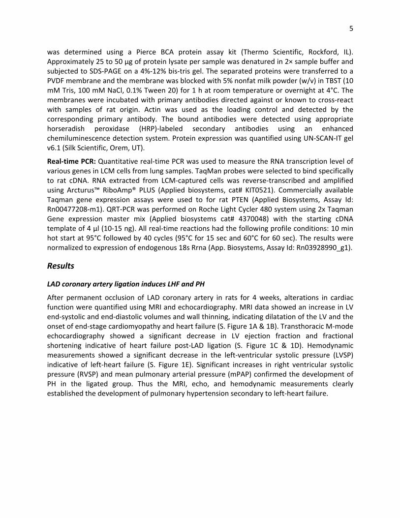
was determined using a Pierce BCA protein assay kit (Thermo Scientific, Rockford, IL). Approximately 25 to 50 μg of protein lysate per sample was denatured in 2× sample buffer and subjected to SDS‐PAGE on a 4%‐12% bis‐tris gel. The separated proteins were transferred to a PVDF membrane and the membrane was blocked with 5% nonfat milk powder (w/v) in TBST (10 mM Tris, 100 mM NaCl, 0.1% Tween 20) for 1 h at room temperature or overnight at 4°C. The membranes were incubated with primary antibodies directed against or known to cross‐react with samples of rat origin. Actin was used as the loading control and detected by the corresponding primary antibody. The bound antibodies were detected using appropriate horseradish peroxidase (HRP)‐labeled secondary antibodies using an enhanced chemiluminescence detection system. Protein expression was quantified using UN‐SCAN‐IT gel v6.1 (Silk Scientific, Orem, UT).
Real‐time PCR: Quantitative real‐time PCR was used to measure the RNA transcription level of various genes in LCM cells from lung samples. TaqMan probes were selected to bind specifically to rat cDNA. RNA extracted from LCM‐captured cells was reverse‐transcribed and amplified using Arcturus™ RiboAmp® PLUS (Applied biosystems, cat# KIT0521). Commercially available Taqman gene expression assays were used to for rat PTEN (Applied Biosystems, Assay Id: Rn00477208‐m1). QRT‐PCR was performed on Roche Light Cycler 480 system using 2x Taqman Gene expression master mix (Applied biosystems cat# 4370048) with the starting cDNA template of 4 µl (10‐15 ng). All real‐time reactions had the following profile conditions: 10 min hot start at 95°C followed by 40 cycles (95°C for 15 sec and 60°C for 60 sec). The results were normalized to expression of endogenous 18s Rrna (App. Biosystems, Assay Id: Rn03928990_g1).
Results
LAD coronary artery ligation induces LHF and PH
After permanent occlusion of LAD coronary artery in rats for 4 weeks, alterations in cardiac function were quantified using MRI and echocardiography. MRI data showed an increase in LV end‐systolic and end‐diastolic volumes and wall thinning, indicating dilatation of the LV and the onset of end‐stage cardiomyopathy and heart failure (S. Figure 1A & 1B). Transthoracic M‐mode echocardiography showed a significant decrease in LV ejection fraction and fractional shortening indicative of heart failure post‐LAD ligation (S. Figure 1C & 1D). Hemodynamic measurements showed a significant decrease in the left‐ventricular systolic pressure (LVSP) indicative of left‐heart failure (S. Figure 1E). Significant increases in right ventricular systolic pressure (RVSP) and mean pulmonary arterial pressure (mPAP) confirmed the development of PH in the ligated group. Thus the MRI, echo, and hemodynamic measurements clearly established the development of pulmonary hypertension secondary to left‐heart failure.
6
References
1. Kalai T, Kuppusamy ML, Balog M, Selvendiran K, Rivera BK, Kuppusamy P, Hideg K. Synthesis of n‐substituted 3,5‐bis(arylidene)‐4‐piperidones with high antitumor and antioxidant activity. J Med Chem. 2011;54:5414‐5421.
2. Selvendiran K, Tong L, Bratasz A, Kuppusamy ML, Ahmed S, Ravi Y, Trigg NJ, Rivera BK, Kalai T, Hideg K, Kuppusamy P. Anticancer efficacy of a difluorodiarylidenyl piperidone (ho‐3867) in human ovarian cancer cells and tumor xenografts. Mol Cancer Ther. 2010;9:1169‐1179.
3. Dayton A, Selvendiran K, Meduru S, Khan M, Kuppusamy ML, Naidu S, Kalai T, Hideg K, Kuppusamy P. Amelioration of doxorubicin‐induced cardiotoxicity by an anticancer‐antioxidant dual‐function compound, ho‐3867. J Pharmacol Exp Ther. 2011;339:350‐357.
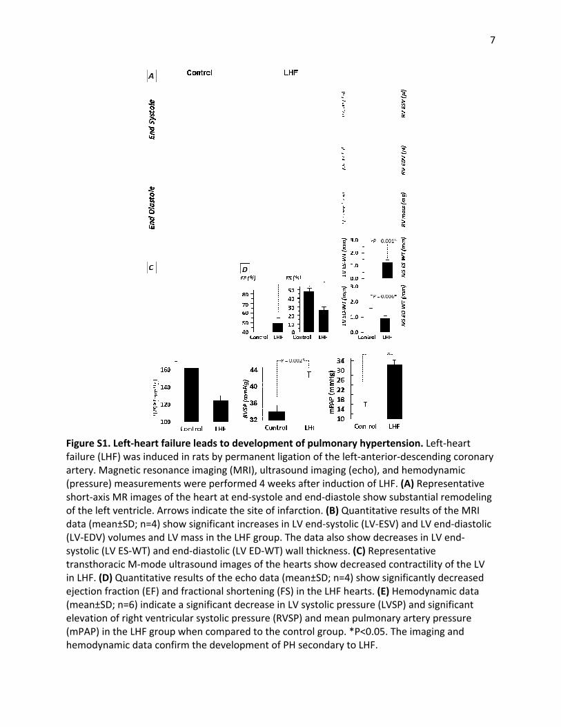
Figure S1failure (Lartery. M(pressureshort‐axiof the lefdata (me(LV‐EDV)systolic (transthoin LHF. (Dejection f(mean±Selevation(mPAP) ihemodyn
1. Left‐heartLHF) was indMagnetic resoe) measurems MR imageft ventricle. Aean±SD; n=4 volumes anLV ES‐WT) aracic M‐modD) Quantitatfraction (EF)SD; n=6) indicn of right venn the LHF grnamic data c
t failure leaduced in rats onance imagments were ps of the heaArrows indic) show signifnd LV mass innd end‐diasde ultrasountive results o) and fractiocate a signifntricular sysroup when cconfirm the
ds to develoby permaneging (MRI), uperformed 4rt at end‐syscate the siteficant increan the LHF grstolic (LV ED‐nd images ofof the echo dnal shortenificant decreatolic pressurompared todevelopmen
opment of puent ligation oultrasound im4 weeks aftestole and ene of infarctioases in LV enoup. The da‐WT) wall thf the hearts sdata (mean±ing (FS) in thase in LV sysre (RVSP) an the controlnt of PH seco
ulmonary hyof the left‐anmaging (echer induction nd‐diastole sn. (B) Quantnd‐systolic (Lta also showickness. (C)show decrea±SD; n=4) shohe LHF hearttolic pressurnd mean pulm group. *P<0ondary to LH
ypertensionnterior‐desco), and hemof LHF. (A) Rhow substantitative resuLV‐ESV) and w decreases Representatased contracow significans. (E) Hemodre (LVSP) anmonary arte0.05. The imHF.
n. Left‐heart cending coromodynamic Representatintial remodelts of the MRLV end‐diasin LV end‐tive ctility of the ntly decreasdynamic datd significantery pressure maging and
7
onary
ive eling RI tolic
LV ed ta t
8
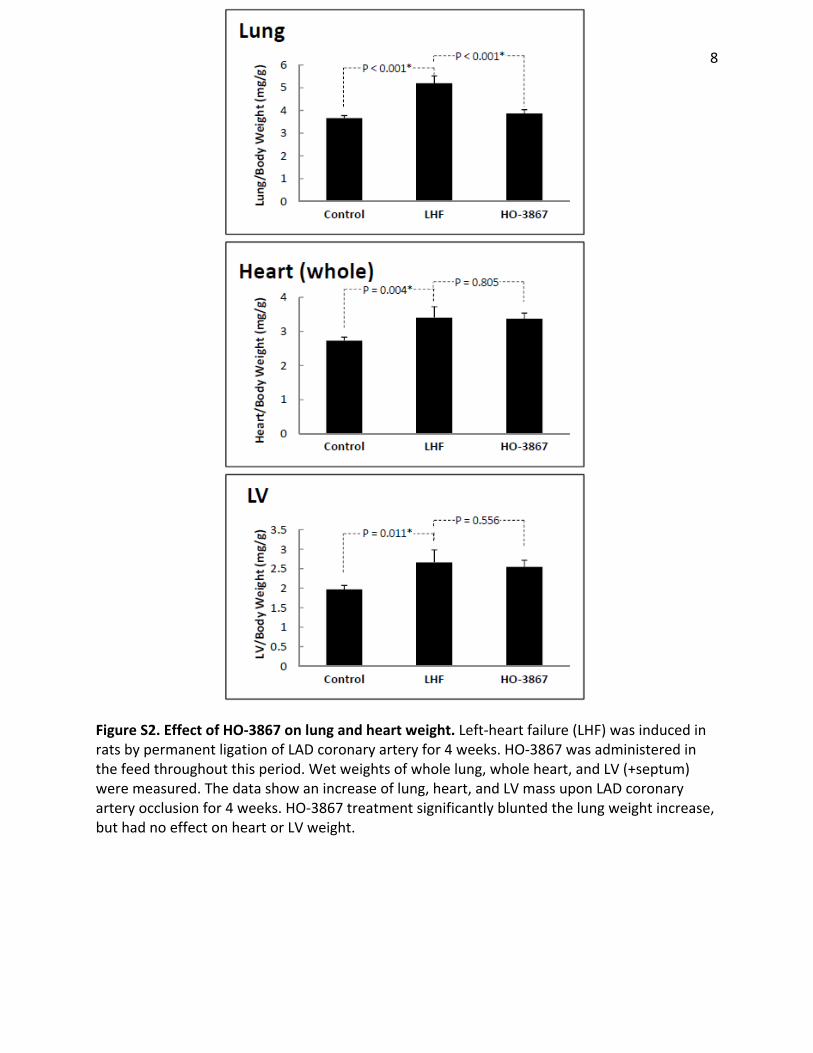
Figure S2. Effect of HO‐3867 on lung and heart weight. Left‐heart failure (LHF) was induced in rats by permanent ligation of LAD coronary artery for 4 weeks. HO‐3867 was administered in the feed throughout this period. Wet weights of whole lung, whole heart, and LV (+septum) were measured. The data show an increase of lung, heart, and LV mass upon LAD coronary artery occlusion for 4 weeks. HO‐3867 treatment significantly blunted the lung weight increase, but had no effect on heart or LV weight.