substrate rigidity regulates human t cell activation … rigidity regulates human t cell activation...
TRANSCRIPT
of June 23, 2018.This information is current as
Activation and ProliferationSubstrate Rigidity Regulates Human T Cell
Kam and Michael C. MiloneBashour, Tatiana Akimova, Wayne W. Hancock, Lance C. Roddy S. O'Connor, Xueli Hao, Keyue Shen, Keenan
ol.1102757http://www.jimmunol.org/content/early/2012/06/25/jimmun
published online 25 June 2012J Immunol
MaterialSupplementary
7.DC1http://www.jimmunol.org/content/suppl/2012/06/25/jimmunol.110275
average*
4 weeks from acceptance to publicationFast Publication! •
Every submission reviewed by practicing scientistsNo Triage! •
from submission to initial decisionRapid Reviews! 30 days* •
Submit online. ?The JIWhy
Subscriptionhttp://jimmunol.org/subscription
is online at: The Journal of ImmunologyInformation about subscribing to
Permissionshttp://www.aai.org/About/Publications/JI/copyright.htmlSubmit copyright permission requests at:
Email Alertshttp://jimmunol.org/alertsReceive free email-alerts when new articles cite this article. Sign up at:
Print ISSN: 0022-1767 Online ISSN: 1550-6606. Immunologists, Inc. All rights reserved.Copyright © 2012 by The American Association of1451 Rockville Pike, Suite 650, Rockville, MD 20852The American Association of Immunologists, Inc.,
is published twice each month byThe Journal of Immunology
by guest on June 23, 2018http://w
ww
.jimm
unol.org/D
ownloaded from
by guest on June 23, 2018
http://ww
w.jim
munol.org/
Dow
nloaded from
The Journal of Immunology
Substrate Rigidity Regulates Human T Cell Activation andProliferation
Roddy S. O’Connor,* Xueli Hao,* Keyue Shen,† Keenan Bashour,† Tatiana Akimova,*
Wayne W. Hancock,* Lance C. Kam,† and Michael C. Milone*
Adoptive immunotherapy using cultured T cells holds promise for the treatment of cancer and infectious disease. Ligands
immobilized on surfaces fabricated from hard materials such as polystyrene plastic are commonly employed for T cell culture.
The mechanical properties of a culture surface can influence the adhesion, proliferation, and differentiation of stem cells and
fibroblasts. We therefore explored the impact of culture substrate stiffness on the ex vivo activation and expansion of human
T cells. We describe a simple system for the stimulation of the TCR/CD3 complex and the CD28 receptor using substrates with
variable rigidity manufactured from poly(dimethylsiloxane), a biocompatible silicone elastomer. We show that softer (Young’s
Modulus [E] < 100 kPa) substrates stimulate an average 4-fold greater IL-2 production and ex vivo proliferation of human CD4+
and CD8+ T cells compared with stiffer substrates (E > 2 MPa). Mixed peripheral blood T cells cultured on the stiffer substrates
also demonstrate a trend (nonsignificant) toward a greater proportion of CD62Lneg, effector-differentiated CD4+ and CD8+
T cells. Naive CD4+ T cells expanded on softer substrates yield an average 3-fold greater proportion of IFN-g–producing Th1-
like cells. These results reveal that the rigidity of the substrate used to immobilize T cell stimulatory ligands is an important and
previously unrecognized parameter influencing T cell activation, proliferation, and Th differentiation. Substrate rigidity should
therefore be a consideration in the development of T cell culture systems as well as when interpreting results of T cell activation
based upon solid-phase immobilization of TCR/CD3 and CD28 ligands. The Journal of Immunology, 2012, 189: 000–000.
Adoptive immunotherapy holds great potential as a ther-apeutic modality for the treatment of a variety of dis-eases, including cancer and chronic viral infections (1).
Central to these therapeutic approaches are controllable plat-forms for ex vivo activation of T cells, and several cell-basedand artificial substrate systems have been described (2). AgonistAbs to CD3 and CD28 immobilized on rigid materials like poly-styrene plastic and glass are widely used in many of these systemsfor the activation and expansion of T cells. These artificial culturesubstrates are also widely used in basic studies of T cell activation,forming the foundation for much of our knowledge of T signaltransduction (3).The outcome of ex vivo culture for many types of adherent cells
is increasingly recognized to depend upon the mechanical prop-erties of the culture substrate. Fibroblast spreading and focal ad-hesion formation are highly dependent upon the force generatedby the fibroblast, as well as the elasticity of the material to whichthey attach (4, 5). The differentiation of pluripotent mesenchymal
stem cells is directly linked to the stiffness of the culture substrate(6). Similarly, expanding myogenic stem cells on soft hydrogelmaterials leads to enhanced self-renewal and improved engraft-ment into mice (7).T cells are unlikely to encounter a stimulatory surface with the
stiffness of plastic in vivo, and the stiffness of the solid supportsused for ex vivo culture of T cells may have important influenceson their activation, proliferation, and differentiation. It has longbeen recognized that anti-CD3ε agonist Abs such as OKT3 andpeptide/MHC complexes require immobilization on solid supportsfor robust T cell activation (8). T cell cytoskeleton integrity andcontractility also appear vital for T cell activation (9–11), andmodels wherein forces applied by the T cell cytoskeleton toligand-bound TCR modulate and/or trigger TCR/CD3 signalinghave been proposed (9, 12). More recent studies have provideddirect evidence for force as a mediator of TCR signal transduction(13, 14). The demonstration that the ITAM in the CD3ε chaincan be activated by conformational changes in ITAM interactionwith the inner leaflet of the plasma membrane provides at leastone possible mechanism by which force might be able to mediatesignals through the TCR/CD3 complex (15). In addition to thedirect role of force in TCR/CD3 complex signal transduction,many proteins involved in TCR and costimulatory receptor signaltransduction directly or indirectly interact with the actin cyto-skeleton (16). The structure and dynamics of the actin cytoskel-eton, which is affected by attachment substrate stiffness (17), havebeen reported to play an important role in supporting and regu-lating signal transduction at the immune synapse (18, 19).We describe a novel system for stimulating T cells under con-
ditions of variable substrate stiffness based upon a biocompatiblepolymer. We show that softer substrates used for immobilizingT cell ligands significantly enhance T cell activation and expansion.We also demonstrate effects of rigidity on Th cell differentiation.These results have implications for the ex vivo culture of T cells
*Department of Pathology and Laboratory Medicine, Perelman School of Medicine,University of Pennsylvania, Philadelphia, PA 19104; and †Department of BiomedicalEngineering, School of Engineering and Applied Science, Columbia University, NewYork, NY 10111
Received for publication September 26, 2011. Accepted for publication May 28,2012.
This work was supported by the National Institutes of Health Common Fund Nano-medicine Program (PN2 EY016586).
Address correspondence and reprint requests to Dr. Michael C. Milone, Hospital ofthe University of Pennsylvania, 7.103 Founders Pavilion, 3400 Spruce Street, Phil-adelphia, PA 19104. E-mail address: [email protected]
The online version of this article contains supplemental material.
Abbreviations used in this article: ECM, extracellular matrix; PDMS, poly(dimethyl-siloxane); qRT-PCR, quantitative real-time RT-PCR.
Copyright� 2012 by The American Association of Immunologists, Inc. 0022-1767/12/$16.00
www.jimmunol.org/cgi/doi/10.4049/jimmunol.1102757
Published June 25, 2012, doi:10.4049/jimmunol.1102757 by guest on June 23, 2018
http://ww
w.jim
munol.org/
Dow
nloaded from
that forms the foundation for many adoptive immunotherapyapproaches currently being pursued in clinical trials. They alsosuggest that the mechanical properties of the substrate used forimmobilizing T cell surface receptor ligands, especially TCR/CD3and CD28 receptor ligands, should be considered when interpretingbasic studies of signal transduction and T cell activation.
Materials and MethodsFabrication of silicone-based culture surfaces
Poly(dimethylsiloxane) (PDMS) surfaces were fabricated by mixing dime-thylsiloxane monomer (Dow Corning Sylgard 184) with its correspondingcross-linking agent, according to manufacturer instructions. The ratio ofcross-linking agent to base polymer was varied from 1:5 to 1:50. PDMSelastomer slabs of .1 mm were cured at 60˚C for 2 h in multiwell platesprior to use in T cell culture experiments. Young’s Modulus (E) of PDMSprepared at each ratio of cross-linking agent to elastomer base wasestimated using a custom-built indentation apparatus. Slabs of PDMS(32 mm 3 43 mm) with a thickness of ∼10 mm were deformed usinga flat, cylindrical head, which makes a no-slip contact with the PDMSsurface. A calibrated mass was applied to this head, producing a deflectionof the PDMS slab. Hertzian contact between the head and PDMS wasassumed (20), which allows estimation of the material’s Young’s modulusfrom the head diameter (D), deflection (h), weight (m), gravitationalconstant (g), and Poisson ratio (n), assumed to be 0.5 for PDMS using thefollowing equation:E ¼ ð12 n2Þpmp
g
D×h:
Coating of PDMS surfaces with Abs for T cell stimulation
Cured PDMS elastomer was incubated with a goat anti-mouse IgG (Cappel,MP Biomedicals) in PBS overnight at 4˚C. Unless otherwise indicated,a concentration of 1 mg/ml was used. Following PBS rinsing, a 1-h in-cubation with a blocking buffer containing 5% BSA was performed. Afterwashing, agonist mAbs to human CD3ε (5 mg/ml, OKT3; Roche Phar-maceuticals) and human CD28 (5 mg/ml, clone 9.3; gift of C. June,University of Pennsylvania) were captured by incubation in PBS for 2 h,followed by washing before use.
Primary cell preparation and cell culture
Primary PBLs were obtained under approval by the University of Penn-sylvania Institutional Review Board. Purified total T cells, CD4+ T cells,or CD8+ T cells were isolated using RosetteSep isolation kits (Stem CellTechnologies). Naive CD4+ T cells were obtained by further depletionof CD45RO+ cells using human CD45RO-specific magnetic microbe-ads, LD selection columns, and a VarioMACS system (Miltenyi Biotec).Lymphocytes were cultured in either X-VIVO 15 or RPMI 1640 (Lonza)supplemented with 5% human serum (GemCell) or 10% FBS (HyClone),respectively, with 10 mM HEPES, L-glutamine, penicillin G, and strep-tomycin. The 4.5-mm beads with immobilized anti-human CD3 and anti-human CD28 (a gift of B. Levine, University of Pennsylvania) were usedin some experiments at a ratio of 3 beads to 1 cell. T cells were maintainedin culture at a concentration of 0.8–1.0 3 106 cells/ml by regular countingon a Multisizer III particle counter (Beckman-Coulter). In some experi-ments, cells were also counted by flow cytometry using CountBright beads(BD Biosciences) and mAbs to human CD4 and CD8.
Quantitative real-time RT-PCR analyses for IL-2 mRNA
Total RNAwas isolated from cells using a RNeasy kit (Qiagen). cDNAwasgenerated by reverse transcription using a High Capacity cDNA ReverseTranscription kit (Applied Biosystems, Carlsbad, CA). cDNA was am-plified with a predesigned primer-probe set for human IL-2 (Hs00174114_m1;Applied Biosystems). A b-actin–specific primer-probe set (Hs999999 03_m1;Applied Biosystems) was used as a normalization control. Quantitativereal-time RT-PCR (qRT-PCR) assays were performed on a 7500 Fast Real-Time PCR system thermal cycler (Applied Biosystems) using the com-parative Ct model. In experiments in which the stability of IL-2 mRNAwas evaluated, CD4+ lymphocytes were seeded at 0.5 3 106 cells/well.After 6 h, these cultures were treated with/without cyclosporine A (1026 M;Sigma-Aldrich) or actinomycin D (5 mg/ml; Sigma-Aldrich) to inhibit denovo IL-2 mRNA transcription. Cells were harvested, as indicated, andIL-2 mRNA was determined by qRT-PCR analyses, as described above.
Abs and flow cytometry
At the indicated time following activation, cells were stained with a panelof mAbs to CD3, CD4, CD8, CD45RA, CD45RO, CD62L, CCR7, CD27,
and LIVE/DEAD Aqua dye (Invitrogen). Flow cytometry was performedusing a LSRII (BD Biosciences, San Jose, CA), and data were analyzedusing FlowJo software (Tree Star, Ashland, OR). In experiments in whichproliferation was assessed using CFSE dilution, cells were stained with5 mM CFSE for 5 min, washed twice, and resuspended in culture mediumprior to initiation of the culture, as indicated. Flow cytometry was per-formed on cells on day 3, and data were analyzed using the proliferationmodule as implemented in FlowJo.
Statistical analysis
Student’s t test for paired data, Wilcoxon rank sum, or a one-way ANOVAwere performed using GraphPad Prism version 4.0a (GraphPad Software).A p value ,0.05 was considered statistically significant.
ResultsPDMS as a substrate with controllable rigidity for T cellactivation and culture
PDMS, a biocompatible organosilicon polymer commonly usedas a lubricant, anticaking agent in foods, and antibloating agent,was selected as a substrate for Ab immobilization. Followingcross-linking of the base polymer, PDMS forms an elastomericmaterial with a highly hydrophobic surface (21). Proteins, in-cluding Ab, passively adsorb to this hydrophobic surface. Alter-ation of the cross-linking agent-to-base-polymer stoichiometry inthe commonly used Sylgard 184 preparation of PDMS providesa simple method for varying the elastic modulus of PDMS froma Young’s modulus of .2.3 MPa (stiff) to a range of 50–100 kPa(soft) (Fig. 1A). Prepared this way, this material has been used tostudy the effects of substrate rigidity on fibroblast focal adhesionformation (4). Adsorption of anti-CD3 (OKT3) and anti-CD28(clone 9.3) Abs to the surface of PDMS provides a system foractivation of T cells on substrates with varying elastic modulus,analogous to standard immobilization on more rigid polystyrenetissue culture plastic or glass. Quantitative measurement of en-zymatically coupled primary capture Ab (Fig. 1B) as well asfluorescently labeled OKT3 and clone 9.3 (data not shown) dem-onstrates that the amount of Ab adsorbed on PDMS surfaces withvarying elastic modulus is equivalent despite changes in the ratioof base polymer to cross-linking agent. Both OKT3 and clone9.3 also demonstrated stable binding over the course of 48 h with,20% loss of Ab at 37˚C in complete culture medium indepen-dent of the cross-linker ratio (Fig. 1C). Clone 9.3 appeared todemonstrate a slightly more rapid loss from stiff surfaces com-pared with soft surfaces; however, the quantity of bound clone 9.3was not significantly different between the PDMS surfaces at 48 h,after which T cells typically transfer to uncoated culture vesselsfor log-phase ex vivo expansion using planar-activating substrates.Initial evaluation of T cell activation demonstrated that softer
PDMS stimulatory substrate increased IL-2 secretion (Fig. 1D).Because the stiffer PDMS substrates contain more cross-linkingagent, we considered the possibility that one of the componentsin the cross-linking agent may be toxic, leading to nonspecificinhibition of T cell activation and IL-2 secretion. To evaluate thispossibility, we simultaneously stimulated T cells with Ab-coatedmagnetic microbeads in the presence of PDMS with variable ri-gidity. Unlike T cells stimulated with Abs immobilized on thePDMS substrate, microbead-stimulated IL-2 secretion was compa-rable across the different PDMS surfaces, arguing against a toxiceffect of PDMS elastomer or its cross-linking agent (Fig. 1D).Because Ab density is an important factor affecting T cell ac-
tivation and proliferation, we evaluated the ability of primaryhuman peripheral blood CD4+ T cells to undergo proliferationin response to varying density of OKT3 and clone 9.3 on PDMSsurfaces. We observed both Ab density-dependent and stiffness-dependent effects on T cell proliferation. Using a CFSE dilution
2 RIGIDITY REGULATES EX VIVO T CELL ACTIVATION
by guest on June 23, 2018http://w
ww
.jimm
unol.org/D
ownloaded from
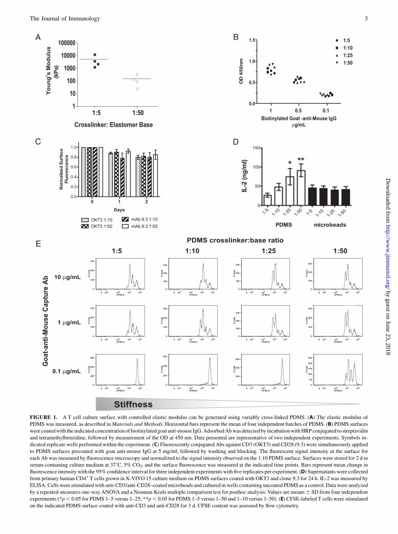
FIGURE 1. A T cell culture surface with controlled elastic modulus can be generated using variably cross-linked PDMS. (A) The elastic modulus of
PDMS was measured, as described inMaterials and Methods. Horizontal bars represent the mean of four independent batches of PDMS. (B) PDMS surfaces
were coatedwith the indicated concentration of biotinylated goat anti-mouse IgG.AdsorbedAbwasdetected by incubationwithHRPconjugated to streptavidin
and tetramethylbenzidine, followed by measurement of the OD at 450 nm. Data presented are representative of two independent experiments. Symbols in-
dicated replicate wells performed within the experiment. (C) Fluorescently conjugated Abs against CD3 (OKT3) and CD28 (9.3) were simultaneously applied
to PDMS surfaces precoated with goat anti-mouse IgG at 5 mg/ml, followed by washing and blocking. The fluorescent signal intensity at the surface for
each Ab was measured by fluorescence microscopy and normalized to the signal intensity observed on the 1:10 PDMS surface. Surfaces were stored for 2 d in
serum-containing culture medium at 37˚C, 5% CO2, and the surface fluorescence was measured at the indicated time points. Bars represent mean change in
fluorescence intensity with the 95% confidence interval for three independent experiments with five replicates per experiment. (D) Supernatants were collected
from primary human CD4+ T cells grown in X-VIVO 15 culture medium on PDMS surfaces coated with OKT3 and clone 9.3 for 24 h. IL-2 was measured by
ELISA. Cells were stimulatedwith anti-CD3/anti-CD28–coatedmicrobeads and cultured in wells containing uncoated PDMS as a control. Data were analyzed
by a repeated-measures one-way ANOVA and a Neuman-Keuls multiple comparison test for posthoc analysis. Values are means6 SD from four independent
experiments (*p, 0.05 for PDMS 1–5 versus 1–25; **p, 0.05 for PDMS 1–5 versus 1–50 and 1–10 versus 1–50). (E) CFSE-labeled T cells were stimulated
on the indicated PDMS surface coated with anti-CD3 and anti-CD28 for 3 d. CFSE content was assessed by flow cytometry.
The Journal of Immunology 3
by guest on June 23, 2018http://w
ww
.jimm
unol.org/D
ownloaded from
assay, greater proliferation was reproducibly observed at 72 h onsofter surfaces. The difference in proliferation became more pro-nounced as the coating concentration of the goat anti-mouse cap-ture Ab decreased ,1 mg/ml, with the proliferation completelylost on harder surfaces coated with Abs at low concentration(0.1 mg/ml; Fig. 1E).
Polyclonal expansion of peripheral blood T cells is enhancedby culture on softer substrates
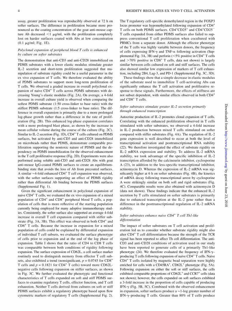
The demonstration that anti-CD3 and anti-CD28 immobilized onPDMS substrates with a lower elastic modulus stimulate greaterIL-2 secretion and short-term proliferation suggested that ma-nipulation of substrate rigidity could be a useful parameter in theex vivo expansion of T cells. We therefore evaluated the abilityof PDMS substrates to support more long-term proliferation ofT cells. We observed a graded increase in overall polyclonal ex-pansion of naive CD4+ T cells across PDMS substrates with de-creasing Young’s elastic modulus (Fig. 2A). On average, a 4-foldincrease in overall culture yield is observed when comparing thesoftest PDMS substrate (1:50 cross-linker to base ratio) with thestiffest PDMS substrate (1:5 cross-linker to base ratio). The dif-ference in overall expansion is primarily due to a more prolongedlog-phase growth rather than a difference in the rate of prolif-eration (Fig. 2B). This enhanced log-phase expansion correlateswith a more prolonged blast phase, as shown by the increase inmean cellular volume during the course of the culture (Fig. 2C).Similar to IL-2 secretion (Fig. 1D), CD4+ T cells cultured on PDMSsurfaces, but activated by anti-CD3 and anti-CD28 immobilizedon microbeads rather than PDMS, demonstrate comparable pro-liferation supporting the nontoxic nature of PDMS and the de-pendency on PDMS immobilization for the observed enhancementin the T cell proliferative response (Fig. 2D). Experiments were alsoperformed using soluble anti-CD3 and anti-CD28 Abs with goatanti-mouse IgG-coated PDMS surfaces to create an equilibrium-binding state that avoids the loss of Ab from precoated surfaces.A similar ∼4-fold enhanced CD4+ T cell expansion was observed,with the softer surfaces supporting an effect of PDMS rigidityrather than differential Ab binding between the PDMS surfaces(Supplemental Fig. 1).Given the significant enhancement in polyclonal expansion of
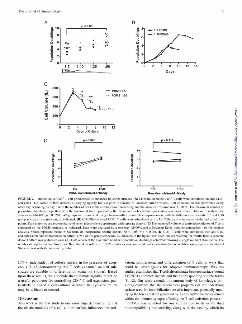
naive CD4+ T cells, we evaluated the ex vivo expansion of a mixedpopulation of CD4+ and CD8+ peripheral blood T cells, a pop-ulation of cells that is more reflective of the starting populationcurrently being employed for many adoptive immunotherapy stud-ies. Consistently, the softer surface also supported an average 4-foldincrease in overall T cell expansion compared with stiffer sub-strate (Fig. 3A, 3B). This effect was observed in both CD4+ andCD8+ T cells. Because the increase in expansion for a mixedpopulation of cells could be explained by differential expansionof individual T cell subsets, we evaluated the surface phenotypeof cells prior to expansion and at the end of the log phase ofexpansion. Table I shows that the ratio of CD4 to CD8 T cellswas comparable between both conditions of rigidity followingexpansion. The surface expression of CD62L, a cell surface markerroutinely used to distinguish memory from effector T cell sub-sets, also exhibited a trend (nonsignificant, p = 0.0745 for CD8+
T cells and p = 0.1821 for CD4+ T cells) toward more CD62L-negative cells following expansion on stiffer surfaces, as shownin Fig. 3C. We further evaluated the phenotypic and functionalcharacteristics of T cells expanded on soft and stiff PDMS sur-faces to examine regulatory T cells, effector function, and T cellexhaustion. Neither T cells derived from cultures on soft or stiffPDMS surfaces exhibit a regulatory phenotype based upon flowcytometric markers of regulatory T cells (Supplemental Fig. 2).
The T regulatory cell-specific demethylated region in the FOXP3locus promoter was hypomethylated following expansion of CD4+
T cells on both PDMS surfaces. CD4+CD25+ and CD4+CD252
T cells expanded from either PDMS surfaces also failed to sup-press conventional T cell proliferation when cocultured withPBMCs from a separate donor. Although the effector phenotypeof the T cells was highly variable between donors, the frequencyof cells expressing IFN-g and TNF-a following activation (Sup-plemental Fig. 3A, 3B) and perforin (,5% positive in CD4+ T cellsand .70% positive in CD8+ T cells, data not shown) is largelysimilar between cells cultured on soft and stiff surfaces. The cellsalso showed similar low expression of markers of T cell exhaus-tion, including 2B4, Lag-3, and PD-1 (Supplemental Fig. 3C, 3D).These findings show that a simple decrease in elastic modulus
of the substrate used to immobilize T cell-activating Abs cansignificantly enhance the T cell activation and proliferative re-sponse to these signals. Furthermore, the effects of stiffness arenot subset dependent, with similar effects observed in both CD4+
and CD8+ T cells.
Softer substrates stimulate greater IL-2 secretion primarilythrough enhanced transcription
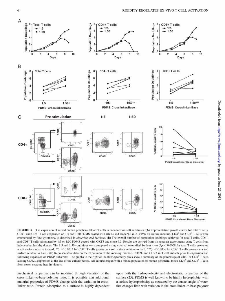
Autocrine production of IL-2 promotes clonal expansion of T cells.Correlating with the enhanced proliferation observed in T cellsstimulated with softer substrates, we observed a 4-fold increasein IL-2 production between mixed T cells stimulated on softercompared with stiffer substrates (Fig. 4A). The regulation of IL-2gene expression is well described to occur at both the level oftranscriptional activation and posttranscriptional RNA stability(22). We therefore investigated the effect of substrate rigidity onIL-2 mRNA expression and stability. To address IL-2 mRNAstability, we took advantage of the specific inhibition of IL-2transcription afforded by the calcineurin inhibitor, cyclosporineA (23), in addition to the less-specific transcriptional inhibitor,actinomycin D. Whereas the expression of IL-2 mRNA was sig-nificantly higher at 6 h on softer substrates (Fig. 4B), the kineticsof mRNA decay following transcriptional arrest by cyclosporineA were strikingly similar on both soft and rigid substrates (Fig.4C). Comparable results were also obtained with actinomycin D(data not shown). These findings indicate that the enhanced IL-2secretion by T cells stimulated on softer substrates is primarilydue to enhanced transcription at the IL-2 gene rather than adifference in the posttranscriptional regulation of IL-2 mRNAstability.
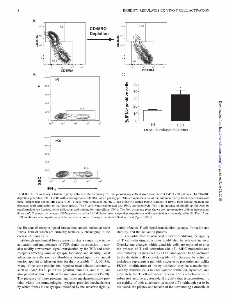
Softer substrates enhance naive CD4+ T cell Th1-likedifferentiation
The impact of softer substrates on T cell activation and prolif-eration led us to consider whether substrate rigidity might alsoalter CD4+ T cell differentiation because the strength of the TCRsignal has been reported to affect Th cell differentiation. The anti-CD3 and anti-CD28 conditions of activation used in our studyhave been reported to generate cells of a primarily Th1-likephenotype (24). We therefore evaluated the frequency of IFN-g–producing T cells following expansion of naive CD4+ T cells. NaiveCD4+ T cells isolated by magnetic bead separation were highlyenriched for cells with a CD45RA+, CD62L+ phenotype (Fig. 5A).Following expansion on either the soft or stiff surfaces, the cellsexhibited comparable proportions of CD62L+ and CCR7+ cells (datanot shown); however, the cells expanded on soft surfaces exhibiteda 3-fold increase in the proportion of cells capable of producingIFN-g (Fig. 5B, 5C). Combined with the observed enhancementin expansion, softer surfaces produce a ∼1-log greater number ofIFN-g–producing T cells. Greater than 80% of T cells produce
4 RIGIDITY REGULATES EX VIVO T CELL ACTIVATION
by guest on June 23, 2018http://w
ww
.jimm
unol.org/D
ownloaded from
IFN-g, independent of culture surface in the presence of exog-enous IL-12, demonstrating that T cells expanded on stiff sub-strates are capable of differentiation (data not shown). Basedupon these results, we conclude that substrate rigidity might bea useful parameter for controlling CD4+ T cell expansion, par-ticularly in mixed T cell cultures in which the cytokine milieumay be difficult to control.
DiscussionThis work is the first study to our knowledge demonstrating thatthe elastic modulus of a cell culture surface influences the acti-
vation, proliferation, and differentiation of T cells in ways thatcould be advantageous for adoptive immunotherapy. Previousstudies established that T cells discriminate between surface-boundTCR/CD3 complex ligands and their corresponding soluble forms(8, 12). Our work extends this current body of knowledge, pro-viding evidence that the mechanical properties of the underlyingsurface used for immobilization are also important, potentially mod-ifying the forces that are generated by T cells and/or the forces sensedwithin the immune synapse affecting the T cell activation process.PDMS was selected for our studies due to its established
biocompatibility and stability, along with the ease by which its
FIGURE 2. Human naive CD4+ T cell proliferation is enhanced by softer surfaces. (A) CD45RO-depleted CD4+ T cells were stimulated on anti-CD3–
and anti-CD28–coated PDMS surfaces of varying rigidity for 3 d prior to transfer to uncoated culture vessels. Cell enumeration was performed every
other day beginning on day 3 until the number of cells in the culture ceased increasing and the mean cell volume was ,350 fL. The maximum number of
population doublings is plotted, with the horizontal bars representing the mean and each symbol representing a separate donor. Data were analyzed by
a one-way ANOVA (p = 0.0261). All groups were compared using a Newman-Keuls multiple comparison test, with the difference between the 1:5 and 1:50
group statistically significant, as indicated. (B) CD45RO-depleted CD4+ T cells were stimulated as in (A). Cells were enumerated at the indicated time
points. Data presented are representative of seven independent experiments with separate donors. (C) The mean cell volume of a mixed population of T cells
expanded on the PDMS surfaces, as indicated. Data were analyzed by a one-way ANOVA and a Newman-Keuls multiple comparison test for posthoc
analysis. Values represent means 6 SD from six independent healthy donors (*p , 0.05, **p , 0.05). (D) CD4+ T cells were stimulated with anti-CD3
and anti-CD28 Abs immobilized on either PDMS or 4.5-mm microbeads, as indicated in the figure, with each line representing the results from a separate
donor. Culture was performed as in (A). Data represent the maximum number of population doublings achieved following a single round of stimulation. The
number of population doublings for cells cultured on soft or stiff PDMS surfaces was compared under each stimulation condition using a paired, two-tailed
Student t test with the indicated p value.
The Journal of Immunology 5
by guest on June 23, 2018http://w
ww
.jimm
unol.org/D
ownloaded from
mechanical properties can be modified through variation of thecross-linker–to-base-polymer ratio. It is possible that additionalmaterial properties of PDMS change with the variation in cross-linker ratio. Protein adsorption to a surface is highly dependent
upon both the hydrophobicity and electrostatic properties of thesurface (25). PDMS is well known to be highly hydrophobic, witha surface hydrophobicity, as measured by the contact angle of water,that changes little with variation in the cross-linker–to-base-polymer
FIGURE 3. The expansion of mixed human peripheral blood T cells is enhanced on soft substrates. (A) Representative growth curves for total T cells,
CD4+, and CD8+ T cells expanded on 1:5 and 1:50 PDMS coated with OKT3 and clone 9.3 in X-VIVO 15 culture medium. CD4+ and CD8+ T cells were
enumerated by flow cytometry, as described in Materials and Methods. (B) The overall number of population doublings achieved for total T cells, CD4+,
and CD8+ T cells stimulated by 1:5 or 1:50 PDMS coated with OKT3 and clone 9.3. Results are derived from six separate experiments using T cells from
independent healthy donors. The 1:5 and 1:50 conditions were compared using a paired, two-tailed Student t test (*p , 0.0008 for total T cells grown on
a soft surface relative to hard; **p , 0.0013 for CD4+ T cells grown on a soft surface relative to hard; ***p , 0.0036 for CD8+ T cells grown on a soft
surface relative to hard). (C) Representative data on the expression of the memory markers CD62L and CCR7 in T cell subsets prior to expansion and
following expansion on PDMS substrates. The graphs to the right of the flow cytometry plots show a summary of the percentage of CD4+ or CD8+ T cells
lacking CD62L expression at the end of the culture period. All cultures began with a mixed population of human peripheral blood CD4+ and CD8+ T cells
from seven separate healthy donors.
6 RIGIDITY REGULATES EX VIVO T CELL ACTIVATION
by guest on June 23, 2018http://w
ww
.jimm
unol.org/D
ownloaded from
ratio (26). We demonstrate comparable Ab adsorption and sta-bility across the cross-linker–to-base ratios used in our study,which is largely consistent with other studies of PDMS surfacehydrophobicity and passive adsorption of protein (27). Brownet al. (28), using a layer-by-layer polyanion coating on PDMSwith variable stiffness, reported some differences in surface rough-ness and water contact angle following their surface treatment.These differences appeared to impact vascular smooth musclecell spreading in the absence of serum; however, cell spreadingwas similar when serum was present.Physical forces at the TCR/CD3 complex have been linked to
intracellular signaling events. Dynamic imaging studies of T cellsinteracting with supported planar bilayers and solid glass sup-ports demonstrate that TCR microclusters and signaling com-plexes initially form at the periphery of the immune synapse(19). This peripheral area of the immune synapse is rich in actin
and myosin, with force exerted on many adhesive receptor-ligandinteractions by actin-driven, lamellipodial extension of the T cellmembrane as well as myosin-driven contraction of the actin net-work (9). The importance of actin-driven force in the generationof TCR signal transduction is highlighted by studies demonstrat-ing that inhibition of actin polymerization affects T cell activation(12, 19, 29, 30). Inhibition of myosin L chain kinase by blebbis-tatin or the depletion of myosin IIA has also been demonstratedto severely inhibit TCR signaling, supporting a critical role forcell-generated force in the T cell activation process (10).Changes in the stiffness of the TCR/CD3 ligand support sub-
strate would be expected to dampen the forces applied to indi-vidual adhesive receptor-ligand interactions. The range of forcenecessary to induce signal transduction by the TCR comparedwith the force necessary to disrupt the mechanical linkage be-tween the receptor and its ligand is currently unknown; however,bond strength and bond lifetime change with the application ofa loading force (31). Whereas the bond between avidin and biotin(Kd ∼10215) is one of the strongest noncovalent associations, witha lifetime (1/koff) on the order of ∼109 seconds, this bond’s life-time is reduced dramatically under load to ,1 s with a pN-rangeload (32). Much lower forces are therefore expected to havesignificant effects on the lifetime of bonds between Abs orTCRs and their ligands, which are several orders of magnitudeweaker in their binding affinity. Whereas the softer surfaces usedin our studies have sufficient stiffness to trigger TCR signaling, thenet effect of the softer surface may be to provide prolongation ofreceptor-ligand binding and signaling, leading to more effectivestimulation. Unfortunately, the testing of this hypothesis requiresthe ability to evaluate dynamic signaling in the context of measuring
Table I. Phenotypic analysis of mixed lymphocytes followingexpansion on stimulatory PDMS surfaces
Start ofCulture PDMS 1:5 PDMS 1:50
Donor CD4+ (%) CD8+ (%) CD4+ (%) CD8+ (%) CD4+ (%) CD8+ (%)
1 44.2 53.7 25.5 70.0 56.0 43.72 65.9 33.8 37.6 62.4 45.4 53.53 48.6 49.5 27.6 70.7 20.3 78.94 65.5 29.5 94.6 6.11 81.0 17.95 73.7 25.4 90.5 9.39 83.0 16.86 67.8 31.8 82.4 17.3 78.8 20.7
FIGURE 4. Softer substrates increase IL-2 expression independent of mRNA stability. (A) Cellular supernatants were collected from mixed populations
of human T cells grown on PDMS surfaces coated with OKT3 and clone 9.3 for 24 h in RPMI 1640 culture medium. IL-2 was measured by a human
IL-2–specific ELISA. The results shown are derived from five separate healthy donors. (B) CD4+ T cells were stimulated on PDMS substrates coated with
OKT3 and clone 9.3. Cells were harvested after 20–24 h, and IL-2 mRNA expression was measured by qRT-PCR analysis following normalization to
b-actin, as described in Materials and Methods. Values represent the means 6 SD from four independent experiments. (C) T cells were stimulated on
PDMS substrates coated with OKT3 and clone 9.3. After 6 h, cyclosporine Awas added (t = 0 h) to the culture to inhibit IL-2 transcription. IL-2 mRNAwas
measured by qRT-PCR analysis with normalization to the t = 6 h mRNA expression for each PDMS condition.
The Journal of Immunology 7
by guest on June 23, 2018http://w
ww
.jimm
unol.org/D
ownloaded from
the lifespan of receptor-ligand interactions and/or molecular-scaleforces, both of which are currently technically challenging in thecontext of living cells.Although mechanical force appears to play a central role in the
activation and maintenance of TCR signal transduction, it mayalso modify downstream signal transduction by the TCR and otherreceptors affecting immune synapse formation and stability. Focaladhesions in cells such as fibroblasts depend upon mechanicaltension applied to adhesion sites for their assembly (4, 5, 33, 34).Many of the same proteins that regulate focal adhesion assembly,such as Pyk2, FAK, p130Cas, paxillin, vinculin, and talin, arealso present within T cells at the immunological synapse (35–39).The presence of these proteins, and other mechanosensitive pro-teins within the immunological synapse, provides mechanism(s)by which forces at the synapse, modified by the substrate rigidity,
could influence T cell signal transduction, synapse formation andstability, and the activation process.It is possible that the observed effect of modifying the rigidity
of T cell-activating substrates could also be relevant in vivo.Cytoskeletal changes within dendritic cells are reported to alterthe process of T cell activation (40–43). MHC molecules andcostimulatory ligands such as CD80 also appear to be anchoredto the dendritic cell cytoskeleton (44, 45). Because the actin cy-toskeleton represents a gel with viscoelastic properties not unlikePDMS, modification of the cytoskeleton may be a mechanismused by dendritic cells to alter synapse formation, dynamics, andultimately the T cell activation process. Cells attached to solidsubstrates assume a cytoskeletal rigidity that is proportional tothe rigidity of their attachment substrate (17). Although yet to beevaluated, the pliancy and tension of the surrounding extracellular
FIGURE 5. Stimulatory substrate rigidity influences the frequency of IFN-g–producing cells derived from naive CD4+ T cell cultures. (A) CD45RO
depletion generates CD4+ T cells with a homogenous CD45RA+ naive phenotype. Data are representative of the minimum purity from experiments with
three independent donors. (B) Naive CD4+ T cells were stimulated on OKT3 and clone 9.3-coated PDMS surfaces in RPMI 1640 culture medium and
expanded until termination of log phase growth. The T cells were restimulated with PMA and ionomycin for 5 h in presence of GolgiStop, followed by
paraformaldehyde fixation, permeabilization, and staining for intracellular IFN-g. The flow cytometry plots shown are representative of three independent
donors. (C) The mean percentage of IFN-g–positive cells (6SEM) from three independent experiments with separate donors as analyzed in (B). The 1:5 and
1:50 conditions were significantly different when compared using a two-tailed Student t test (*p = 0.0219).
8 RIGIDITY REGULATES EX VIVO T CELL ACTIVATION
by guest on June 23, 2018http://w
ww
.jimm
unol.org/D
ownloaded from
matrix (ECM) within a lymph node most likely change quitedramatically during the course of an immune response as thelymph node undergoes marked hyperplasia and distention. Studiesto evaluate the dynamic mechanical properties of the ECM withina lymph node or lymphoid tissue during an immune response arecurrently lacking, but should be an area of future study.Our observation that substrate rigidity affects the Th cell dif-
ferentiation of T cells could be explained by a number of factors.Although cytokines certainly play a critical role, differentiationof Th cells toward different fates also depends upon TCR liganddensity and duration of signaling (46–49). Cell-ECM inter-actions have also been linked to the control of gene expressionin cells. The PDZ-domain–containing transcription factors YAPand TAZ were recently demonstrated to be important nuclearmediators of ECM-stiffness–induced mesenchymal cell differen-tiation (50). These pathways or other mechanosensitive pathwayscould affect lymphocyte differentiation in ways that have notbeen previously considered, and may depend upon the mechanicalproperties of culture systems employed to study T cells.In addition to the fundamental importance of our findings to the
basic study of T cells, a culture system with altered mechanicalproperties may also have useful applications to the field ofadoptive T cell immunotherapy. Anti-CD3 and anti-CD28 Absimmobilized either on planar plastic surfaces or plastic micro-beads are a commonly employed system for activating T cellsex vivo (3, 24). Efficient expansion of T cells, especially frompatients with cancer, represents a significant challenge. The ob-servation of enhanced expansion on softer surfaces with retentionof a mostly CD62Lhigh memory-like phenotype suggests thata softer substrate might increase the feasibility of adoptive im-munotherapy for more patients, especially in challenging diseaseslike leukemia, in which few peripheral blood T cells are availablein the circulation for expansion. The increased frequency of IFN-g–producing cells observed with softer substrates also suggeststhat T cells expanded on a softer surface may have improvedfunction following adoptive therapy in cancer. Th1-differentiated,IFN-g–producing cells have been shown in preclinical immuno-therapy models to be important for efficacy (51).In summary, these data highlight a novel role for the elastic
modulus of a T cell culture surface, a previously unrecognizedculture parameter for lymphocytes. Using PDMS elastomers, wedemonstrate that stimulatory substrate rigidity can be controlledto effect changes in T cells of biologic importance. Although notdirectly evaluated in this study, these data also support the role offorce in T cell activation by their Ag receptor. We provide evidencethat PDMS, a biocompatible polymer, could be used as a platformfor the ex vivo culture of T cells for adoptive immunotherapy withpotential advantages over currently used rigid plastic surfaces.
AcknowledgmentsWe thank Dr. Carl June at the University of Pennsylvania and Dr. Michael
Dustin at New York University for helpful discussions and critiques of
the manuscript. We also thank Nicholas Vena, Christina Fang, and Trevor
Cassidy for technical assistance with these studies. In addition, we thank
Korey Demers, Carolina Martinez, and Michael Betts for assistance with
flow cytometry.
DisclosuresThe authors have no financial conflicts of interest.
References1. June, C. H. 2007. Adoptive T cell therapy for cancer in the clinic. J. Clin. Invest.
117: 1466–1476.2. June, C. H. 2007. Principles of adoptive T cell cancer therapy. J. Clin. Invest.
117: 1204–1212.
3. Kruisbeek, A. M., E. Shevach, and A. M. Thornton. 2004. Proliferative assaysfor T cell function. Curr. Protoc. Immunol. Chapter 3: Unit 3.12.
4. Balaban, N. Q., U. S. Schwarz, D. Riveline, P. Goichberg, G. Tzur, I. Sabanay,D. Mahalu, S. Safran, A. Bershadsky, L. Addadi, and B. Geiger. 2001. Forceand focal adhesion assembly: a close relationship studied using elastic micro-patterned substrates. Nat. Cell Biol. 3: 466–472.
5. Choquet, D., D. P. Felsenfeld, and M. P. Sheetz. 1997. Extracellular matrixrigidity causes strengthening of integrin-cytoskeleton linkages. Cell 88: 39–48.
6. Engler, A. J., S. Sen, H. L. Sweeney, and D. E. Discher. 2006. Matrix elasticitydirects stem cell lineage specification. Cell 126: 677–689.
7. Gilbert, P. M., K. L. Havenstrite, K. E. Magnusson, A. Sacco, N. A. Leonardi,P. Kraft, N. K. Nguyen, S. Thrun, M. P. Lutolf, and H. M. Blau. 2010. Substrateelasticity regulates skeletal muscle stem cell self-renewal in culture. Science329: 1078–1081.
8. Geppert, T. D., and P. E. Lipsky. 1987. Accessory cell independent proliferationof human T4 cells stimulated by immobilized monoclonal antibodies to CD3.J. Immunol. 138: 1660–1666.
9. Sims, T. N., T. J. Soos, H. S. Xenias, B. Dubin-Thaler, J. M. Hofman, J. C. Waite,T. O. Cameron, V. K. Thomas, R. Varma, C. H. Wiggins, et al. 2007. Opposingeffects of PKCtheta and WASp on symmetry breaking and relocation of theimmunological synapse. Cell 129: 773–785.
10. Ilani, T., G. Vasiliver-Shamis, S. Vardhana, A. Bretscher, and M. L. Dustin. 2009.T cell antigen receptor signaling and immunological synapse stability requiremyosin IIA. Nat. Immunol. 10: 531–539.
11. Valitutti, S., M. Dessing, K. Aktories, H. Gallati, and A. Lanzavecchia. 1995.Sustained signaling leading to T cell activation results from prolonged T cellreceptor occupancy: role of T cell actin cytoskeleton. J. Exp. Med. 181: 577–584.
12. Ma, Z., K. A. Sharp, P. A. Janmey, and T. H. Finkel. 2008. Surface-anchoredmonomeric agonist pMHCs alone trigger TCR with high sensitivity. PLoS Biol.6: e43.
13. Kim, S. T., K. Takeuchi, Z. Y. Sun, M. Touma, C. E. Castro, A. Fahmy,M. J. Lang, G. Wagner, and E. L. Reinherz. 2009. The alphabeta T cell receptoris an anisotropic mechanosensor. J. Biol. Chem. 284: 31028–31037.
14. Li, Y. C., B. M. Chen, P. C. Wu, T. L. Cheng, L. S. Kao, M. H. Tao, A. Lieber,and S. R. Roffler. 2010. Cutting edge: mechanical forces acting on T cellsimmobilized via the TCR complex can trigger TCR signaling. J. Immunol.184: 5959–5963.
15. Xu, C., E. Gagnon, M. E. Call, J. R. Schnell, C. D. Schwieters, C. V. Carman,J. J. Chou, and K. W. Wucherpfennig. 2008. Regulation of T cell receptor ac-tivation by dynamic membrane binding of the CD3epsilon cytoplasmic tyrosine-based motif. Cell 135: 702–713.
16. Burkhardt, J. K., E. Carrizosa, and M. H. Shaffer. 2008. The actin cytoskeletonin T cell activation. Annu. Rev. Immunol. 26: 233–259.
17. Tee, S. Y., J. Fu, C. S. Chen, and P. A. Janmey. 2011. Cell shape and substraterigidity both regulate cell stiffness. Biophys. J. 100: L25–L27.
18. Gomez, T. S., S. D. McCarney, E. Carrizosa, C. M. Labno, E. O. Comiskey,J. C. Nolz, P. Zhu, B. D. Freedman, M. R. Clark, D. J. Rawlings, et al. 2006. HS1functions as an essential actin-regulatory adaptor protein at the immune synapse.Immunity 24: 741–752.
19. Campi, G., R. Varma, and M. L. Dustin. 2005. Actin and agonist MHC-peptidecomplex-dependent T cell receptor microclusters as scaffolds for signaling.J. Exp. Med. 202: 1031–1036.
20. Sneddon, I. 1965. The relation between load and penetration in the axisymmetricBoussinesq problem for a punch of arbitrary profile. Int. J. Eng. Sci. 3: 47–57.
21. Androit, M., et al. 2007. Silicones in industrial applications. In InorganicPolymers. R. D. Jaeger, and M. Gleria, eds. Nova Science Publishers, New York,p. 61–161.
22. Lindstein, T., C. H. June, J. A. Ledbetter, G. Stella, and C. B. Thompson. 1989.Regulation of lymphokine messenger RNA stability by a surface-mediated T cellactivation pathway. Science 244: 339–343.
23. Clipstone, N. A., and G. R. Crabtree. 1992. Identification of calcineurin as a keysignalling enzyme in T-lymphocyte activation. Nature 357: 695–697.
24. Levine, B. L., W. B. Bernstein, M. Connors, N. Craighead, T. Lindsten,C. B. Thompson, and C. H. June. 1997. Effects of CD28 costimulation on long-term proliferation of CD4+ T cells in the absence of exogenous feeder cells.J. Immunol. 159: 5921–5930.
25. Nakanishi, K., T. Sakiyama, and K. Imamura. 2001. On the adsorption of pro-teins on solid surfaces, a common but very complicated phenomenon. J. Biosci.Bioeng. 91: 233–244.
26. Mata, A., A. J. Fleischman, and S. Roy. 2005. Characterization of poly-dimethylsiloxane (PDMS) properties for biomedical micro/nanosystems. Biomed.Microdevices 7: 281–293.
27. Gray, D. S., J. Tien, and C. S. Chen. 2003. Repositioning of cells by mecha-notaxis on surfaces with micropatterned Young’s modulus. J. Biomed. Mater.Res. A 66: 605–614.
28. Brown, X. Q., K. Ookawa, and J. Y. Wong. 2005. Evaluation of poly-dimethylsiloxane scaffolds with physiologically-relevant elastic moduli: inter-play of substrate mechanics and surface chemistry effects on vascular smoothmuscle cell response. Biomaterials 26: 3123–3129.
29. Rivas, F. V., J. P. O’Keefe, M. L. Alegre, and T. F. Gajewski. 2004. Actin cy-toskeleton regulates calcium dynamics and NFAT nuclear duration. Mol. Cell.Biol. 24: 1628–1639.
30. Bunnell, S. C., V. Kapoor, R. P. Trible, W. Zhang, and L. E. Samelson. 2001.Dynamic actin polymerization drives T cell receptor-induced spreading: a rolefor the signal transduction adaptor LAT. Immunity 14: 315–329.
31. Evans, E. 2001. Probing the relation between force—lifetime—and chemistryin single molecular bonds. Annu. Rev. Biophys. Biomol. Struct. 30: 105–128.
The Journal of Immunology 9
by guest on June 23, 2018http://w
ww
.jimm
unol.org/D
ownloaded from
32. Merkel, R., P. Nassoy, A. Leung, K. Ritchie, and E. Evans. 1999. Energylandscapes of receptor-ligand bonds explored with dynamic force spectroscopy.Nature 397: 50–53.
33. Riveline, D., E. Zamir, N. Q. Balaban, U. S. Schwarz, T. Ishizaki, S. Narumiya,Z. Kam, B. Geiger, and A. D. Bershadsky. 2001. Focal contacts as mechano-sensors: externally applied local mechanical force induces growth of focalcontacts by an mDia1-dependent and ROCK-independent mechanism. J. CellBiol. 153: 1175–1186.
34. Wolfenson, H., A. Bershadsky, Y. I. Henis, and B. Geiger. 2011. Actomyosin-generated tension controls the molecular kinetics of focal adhesions. J. Cell Sci.124: 1425–1432.
35. Berg, N. N., and H. L. Ostergaard. 1997. T cell receptor engagement inducestyrosine phosphorylation of FAK and Pyk2 and their association with Lck.J. Immunol. 159: 1753–1757.
36. Simonson, W. T., S. J. Franco, and A. Huttenlocher. 2006. Talin1 regulatesTCR-mediated LFA-1 function. J. Immunol. 177: 7707–7714.
37. Collins, M., R. R. Bartelt, and J. C. Houtman. 2010. T cell receptor activationleads to two distinct phases of Pyk2 activation and actin cytoskeletal rear-rangement in human T cells. Mol. Immunol. 47: 1665–1674.
38. Robertson, L. K., L. R. Mireau, and H. L. Ostergaard. 2005. A role for phos-phatidylinositol 3-kinase in TCR-stimulated ERK activation leading to paxillinphosphorylation and CTL degranulation. J. Immunol. 175: 8138–8145.
39. Nolz, J. C., R. B. Medeiros, J. S. Mitchell, P. Zhu, B. D. Freedman, Y. Shimizu,and D. D. Billadeau. 2007. WAVE2 regulates high-affinity integrin binding byrecruiting vinculin and talin to the immunological synapse. Mol. Cell. Biol. 27:5986–6000.
40. Aldinucci, A., L. Rizzetto, L. Pieri, D. Nosi, P. Romagnoli, T. Biagioli,B. Mazzanti, R. Saccardi, L. Beltrame, L. Massacesi, et al. 2010. Inhibition ofimmune synapse by altered dendritic cell actin distribution: a new pathway ofmesenchymal stem cell immune regulation. J. Immunol. 185: 5102–5110.
41. Benvenuti, F., S. Hugues, M. Walmsley, S. Ruf, L. Fetler, M. Popoff,V. L. Tybulewicz, and S. Amigorena. 2004. Requirement of Rac1 and
Rac2 expression by mature dendritic cells for T cell priming. Science 305:1150–1153.
42. Al-Alwan, M. M., R. S. Liwski, S. M. Haeryfar, W. H. Baldridge,D. W. Hoskin, G. Rowden, and K. A. West. 2003. Cutting edge: dendriticcell actin cytoskeletal polarization during immunological synapse formation ishighly antigen-dependent. J. Immunol. 171: 4479–4483.
43. Al-Alwan, M. M., G. Rowden, T. D. Lee, and K. A. West. 2001. The dendriticcell cytoskeleton is critical for the formation of the immunological synapse.J. Immunol. 166: 1452–1456.
44. Doty, R. T., and E. A. Clark. 1996. Subcellular localization of CD80 receptorsis dependent on an intact cytoplasmic tail and is required for CD28-dependentT cell costimulation. J. Immunol. 157: 3270–3279.
45. Tseng, S. Y., M. Liu, and M. L. Dustin. 2005. CD80 cytoplasmic domain con-trols localization of CD28, CTLA-4, and protein kinase Ctheta in the immu-nological synapse. J. Immunol. 175: 7829–7836.
46. Constant, S., C. Pfeiffer, A. Woodard, T. Pasqualini, and K. Bottomly. 1995.Extent of T cell receptor ligation can determine the functional differentiationof naive CD4+ T cells. J. Exp. Med. 182: 1591–1596.
47. Hosken, N. A., K. Shibuya, A. W. Heath, K. M. Murphy, and A. O’Garra. 1995.The effect of antigen dose on CD4+ T helper cell phenotype development ina T cell receptor-alpha beta-transgenic model. J. Exp. Med. 182: 1579–1584.
48. Rogers, P. R., G. Huston, and S. L. Swain. 1998. High antigen density andIL-2 are required for generation of CD4 effectors secreting Th1 rather thanTh0 cytokines. J. Immunol. 161: 3844–3852.
49. Rogers, P. R., and M. Croft. 1999. Peptide dose, affinity, and time of differentiationcan contribute to the Th1/Th2 cytokine balance. J. Immunol. 163: 1205–1213.
50. Dupont, S., L. Morsut, M. Aragona, E. Enzo, S. Giulitti, M. Cordenonsi,F. Zanconato, J. Le Digabel, M. Forcato, S. Bicciato, et al. 2011. Role of YAP/TAZ in mechanotransduction. Nature 474: 179–183.
51. Nishimura, T., M. Nakui, M. Sato, K. Iwakabe, H. Kitamura, M. Sekimoto,A. Ohta, T. Koda, and S. Nishimura. 2000. The critical role of Th1-dominantimmunity in tumor immunology. Cancer Chemother. Pharmacol. 46: S52–S61.
10 RIGIDITY REGULATES EX VIVO T CELL ACTIVATION
by guest on June 23, 2018http://w
ww
.jimm
unol.org/D
ownloaded from