substitution of a tata box from a herpes simplex virus icp4
TRANSCRIPT
JOURNAL OF VIROLOGY, Sept. 1992, p. 5453-54630022-538X/92/095453-11$02.00/0Copyright © 1992, American Society for Microbiology
Substitution of a TATA Box from a Herpes Simplex VirusLate Gene in the Viral Thymidine Kinase Promoter Alters
ICP4 Inducibility but Not Temporal ExpressionANTHONY N. IMBALZANOt AND NEAL A. DELUCAt*
Department ofMicrobiology and Molecular Genetics, Harvard Medical School,and Dana-Farber Cancer Institute, Boston, Massachusetts 02115
Received 2 April 1992/Accepted 22 May 1992
The role of cis-acting promoter elements associated with herpes simplex virus type 1 (HSV-1) early and lategenes was evaluated during productive infection with regard to activation of gene expression by the HSV-1transactivator ICP4 and control of temporal regulation. A set ofrecombinant viruses was constructed such thatexpression of an HSV-1 early gene, thymidine kinase (tk), was placed under the control of either the tk TATAbox or the TATA box from the late gene, glycoprotein C (gC), in the presence or absence of the upstream Spland CCAAT sites normally found in the tk promoter. The presence of Spl sites in the promoter or replacementof the tk TATA box with the gC TATA box resulted in a decreased activation of tk mRNA expression by ICP4.Substitution of the A+T-rich region from the gC TATA box in the context of the remainder of the surroundingtk sequences resulted in a promoter that bound recombinant TATA-binding protein (TBP) better at lowerconcentrations than the wild-type tk promoter did. These results indicate that tk promoters that are better ableto utilize TBP are less responsive to ICP4 activation and suggest that activation by ICP4 involves the generaltranscription factors that interact with TBP or TBP itself. Additionally, all of the viruses expressed tk at earlytimes postinfection, indicating that cis-acting promoter elements that control the level of expression of HSV-1early and late genes do not determine temporal regulation.
Herpes simplex virus type 1 (HSV-1) genes can be roughlycategorized into three classes on the basis of their time ofexpression during productive infection (18). Immediate-early(IE) genes are expressed shortly after infection, and theirtranscription does not require prior protein synthesis (18,19). Early genes are the next class expressed; their synthesisrequires the activity of at least one IE protein (19). Amongthe products of early genes are the DNA replication en-zymes. Finally, late genes are expressed to maximum levelsfollowing viral DNA synthesis (18, 19). Late genes encodepredominantly the structural proteins of the virion. Expres-sion of the early and late genes is absolutely dependent onthe presence of functional IE proteins and, in particular, theIE protein ICP4 (11, 42).ICP4 is a 170-kDa phosphoprotein that is required for
activating transcription from most HSV-1 genes during viralinfection (11, 42, 58). Mutational analyses have identifieddomains of the ICP4 molecule that are associated with itstranscription-activating function (9, 10, 38, 39, 48); however,the mechanism by which ICP4 transactivates HSV-1 earlyand late gene promoters remains unclear. ICP4 binds specif-ically to diverse sequences present in many HSV-1 early andlate genes (14, 22, 27, 34, 36, 54); however, the ability ofICP4 to bind to DNA has not been clearly shown tocontribute to transactivation (2, 22, 40, 46, 47, 49, 54, 55).The viral thymidine kinase (tk) promoter is transactivated
by ICP4 (6, 8, 35). It is transcribed by RNA polymerase IIand contains a CCAAT box and two Spl sites upstream of a
* Corresponding author.t Present address: Department of Molecular Biology, Massachu-
setts General Hospital, Boston, MA 02114.t Present address: Department of Molecular Genetics and Bio-
chemistry, University of Pittsburgh School of Medicine, Pittsburgh,PA 15261.
TATA box (24, 33). Studies of this promoter have found thatonly the cis sequences that interact with cellular transcrip-tion factors are required for expression of tk during viralinfection; no induction-specific sequences have been identi-fied (3, 5, 12). A more recent study directly evaluated therole of these cis sites in ICP4 induction of the tk promoterand found that induction by ICP4 was not dependent on theintegrity of these cis sequences (21). Efficient induction byICP4 could be observed in the presence of only the tk TATAbox. Therefore, the induction of HSV-1 genes by ICP4 maybe mediated through TFIID, the other general cellular tran-scription factors (TFIIA, TFIIB, and TFIIE/F), or RNApolymerase II. TFIID is composed of TATA-binding protein(TBP) and a number of TBP-associated factors that oftenmediate interactions with activating proteins (lla). Bio-chemical studies on the IE transactivator of pseudorabiesvirus, which shows sequence conservation with ICP4 (4, 57),have shown that IE stabilizes the interaction of TFIID withan inducible promoter (1). Therefore, promoters that possessTATA boxes with a greater affinity for TFIID or TBP maynot be affected by the activity or presence of ICP4 as muchas are TATA boxes that have a lower affinity for thesefactors. In this study, the effects of ICP4 mediated inductionof two different TATA box sequences with different affinitiesfor TBP were evaluated to test this hypothesis.HSV-1 late gene promoters differ from early gene promot-
ers in that the cis-acting sequences 5' to the TATA box, suchas Spl or CCAAT sites, that are prevalent in early promoters(53) are usually not present in late promoters. In general,prior analyses of late gene promoters have indicated thatDNA sequences 5' to the TATA box are not required forinduction or for regulation as a late gene (14, 16, 17, 23, 45).Late gene promoters also differ from early promoters bytheir inability to be maximally expressed until viral DNAreplication occurs (18). However, a tk promoter lacking the
5453
Vol. 66, No. 9
on February 3, 2018 by guest
http://jvi.asm.org/
Dow
nloaded from
5454 IMBALZANO AND DELUCA
upstream CCAAT and Spl sites has been shown to beexpressed as an early gene (21). This promoter is similar toHSV-1 late gene promoters in that the only identified cis-acting sequence present is the TATA box; nevertheless, itwas not expressed as a late gene. This result may be due todifferences in the sequence of the TATA boxes. Previouswork has suggested that functional differences between earlyand late gene TATA boxes exist (16).
In this study, a set of chimeric promoters that combinedthe identified cis elements of the early tk promoter and of thelate gC promoter was constructed and each was subse-quently recombined in place of the wild-type tk promoter inan ICP4-deficient virus, n12 (10). This permitted evaluationof tk expression under the control of either the early gene tkTATA box or the late gene gC TATA box in the presence or
absence of the upstream Spl and CCAAT sites found in thetk promoter. By recombining these promoters into the tklocus of n12 (10), expression directed by each promotercould be monitored both in the absence of ICP4, by infectingVero cells, and in the presence of ICP4, by infecting E5cells. E5 cells are Vero cells that have been transformed withthe ICP4 gene and that, on HSV-1 infection, produce levelsof ICP4 that can complement ICP4-deficient viruses (7, 9).Thus the ability of ICP4 to induce each of these promoterscan be examined. In addition, the kinetics of tk expression asa function of each promoter can be monitored by infectingE5 cells for various times postinfection.
MATERUILS AND METHODS
Virus and cells. Cells were maintained under conditionspreviously described (7). The n12 viral strain contains a
nonsense mutation in both copies of the ICP4 gene at aminoacid 251 (10). E5 cells (7, 9) express complementing levels ofICP4 upon HSV-1 infection and were used to propagate n12and n12 viruses containing tk promoter mutations. n12d-111/-46 was previously described (21). n12 viruses con-
taining other tk promoter mutations are described below.Plasmids. pLS/ts-42/-32 and pLS/ts-111/-101 each con-
tain a linker-scanning mutation marked by a BamHI site inthe indicated position in the tk promoter (see Fig. 2D). Theseplasmids have been described previously (5, 33). pd/ts- 111/-32 was generated by ligation of the 4.4-kb BamHI fragmentof pLS/ts-111/-101 to the 3.0-kb BamHI fragment of pLS/ts-421-32.To generate plasmids containing gC TATA boxes in the
background of the -42/-32 and -111/-32 mutations, theHindIII-BglII (-500 to +54) promoter fragments of pLS/ts-421-32 and pd/ts-111/-32 were first cloned into a
pUC19 plasmid in which a BglII linker (New EnglandBioLabs) had been inserted at the SmaI site in the poly-linker. The resulting plasmids, p-42/-32HB and p-lll/-32HB, were then digested with BamHI and with MIuI,which cuts the tk promoter at - 13 relative to the start site oftk transcription. Two complementary oligonucleotides were
synthesized such that when they were annealed to formdouble-stranded DNA, the tk sequence between the BamHIsite at -42/-32 or at - 111/-32 and the MluI site at - 13 wasreconstituted, except that the nucleotides making up the tk
TATA box were precisely replaced by those encoding the gCTATA box (see Fig. 1B). The sequences of the oligonucle-otides are 5'-GATCCGGTTCGTATAAATTGGTGA-3' and
5'-CGCGTCACCAATlTTATACGAACCG-3'. Phosphory-lated, double-stranded oligonucleotides were ligated to
BamiHI-MluI-digested p-42/-32HB and p-lll/-32HB to
generate p-42/-32(C)HB and p-111/-32(C)HB, respec-
tively. The presence of the oligonucleotide encoding the gCTATA box was identified by the loss of an MseI site that isencoded within the tk TATA box and was verified by DNAsequencing. The HindIII-BglII promoter fragments of theseplasmids were then cloned back into the HindIII-BglIIvector fragment of pLSIts-111I-101 to generate pLS/ts-42/-32(C) and pdlts-111I-32(C).Generation and screening of recombinant viruses. Plasmids
LS/ts-42/-32 and dlts-111I-32 and their counterparts con-taining the gC TATA box were linearized at the Sall site inthe vector sequence and cotransfected into E5 cells withinfectious n12 DNA as described previously (15, 21, 37).Selection for the temperature-sensitive thymidine kinasephenotype with acyclovir was performed as described pre-viously (5, 21). Subsequent screening for the tk promotermutation was performed by Southern blot hybridization (50),using electrophoretically separated digests of DNA fromsmall-plaque cultures and nick-translated probes containingtk promoter sequences.
Isolation of infected-cell RNA and Northern blot analysis.Confluent monolayers of Vero or E5 cells were infected at amultiplicity of infection (MOI) of 10 PFU per cell. Infected-cell RNA was prepared and subjected to Northern (RNA)blot analysis exactly as described previously (21). The probefor tk mRNA levels was a gel-purified, 2P-labeled SacI-SmaI fragment from the coding region of the tk gene. Thisfragment spans +555 to +1217 relative to the start site of tktranscription and was chosen because it does not containsequences from the overlapping UL24 gene or the adjacentgH gene. Filters were stripped by boiling in distilled waterand were probed with a gel-purified 32P-labeled BamHI-SalIfragment internal to the ICP8 gene or with a gel-purified32P-labeled BamHI-SalI fragment internal to the gC codingregion. Probes were labeled by nick translation with Esche-nchia coli DNA polymerase I (30).
Primer extension. Infected-cell RNA (35 ,ug) was used forprimer extension analysis exactly as described previously(21).
Southern blot analysis of replicated viral DNA. Approxi-mately 5 x 10⁵ Vero or E5 cells were infected with n12 at aMOI of 10 PFU per cell in the presence or absence of 0.3 mgof phosphonoacetic acid (PAA) per ml. Samples were har-vested at 1, 6, or 12 h postinfection and lysed in 0.5% sodiumdodecyl sulfate (SDS)-0.2 mg of proteinase K per ml for 4 hat 37°C. Each sample was phenol-chloroform extractedrepeatedly, chloroform extracted, ethanol precipitated, andresuspended in 0.1 ml of TE (10 mM Tris-HCl [pH 8.0], 1mM EDTA). The samples were then treated with 0.02 mg ofRNase A for 1.5 h at 37°C and were phenol-chloroformextracted, ethanol precipitated, and resuspended in distilledH20. The A260 of each sample was measured, and 3 ,ug ofeach infected-cell DNAwas digested with BamHI, separatedby electrophoresis on a 0.9% agarose gel, and transferred tonitrocellulose (50). Blots were probed with a gel-isolated,32P-labeled, Sacl-SmaI (+555 to +1217) DNA fragmentinternal to the tk coding sequence. Densitometic measure-ments of appropriately exposed autoradiograms were madewith a Hoefer GS300 densitometer, and the data wereprocessed by using Hoefer software for the Macintoshcomputer.
Purification of human TBP expressed in bacteria. Thehuman TBP expression plasmid pETHIID and E. coli BL21were generous gifts of Arnold Berk and have been describedpreviously (25). pETHIID-transformed BL21 cells (2 liters)were grown in 2x YT medium (44)-0.4% glucose in thepresence of 50 ,ug of ampicillin per ml at 37°C until the A600
J. VIROL.
on February 3, 2018 by guest
http://jvi.asm.org/
Dow
nloaded from
INDUCTION OF tk BY HSV-1 ICP4 5455
reached approximately 0.8. Isopropyl-3-D-thiogalactoside(IPTG) was added to a final concentration of 0.5 mM, and thecells were grown for a further 3 h at 30°C. The cells werepelleted, resuspended in cold TBS (140 mM NaCl, 5 mMKCl, 25 mM Tricine [pH 7.4], 0.7 mM CaCl2, 0.5 mMMgCl2), and repelleted. All of the following procedures wereperformed at 4°C. The cells were resuspended in buffer D (20mM N-2-hydroxyethylpiperazine-N'-2-ethanesulfonic acid[HEPES; pH 7.9], 0.1 mM EDTA, 10% glycerol, 0.1%Nonidet P-40, 1 mM dithiothreitol, 0.1 mM phenylmethyl-sulfonyl fluoride, 1 ,ug of aprotinin per ml, 1 ,g of antipainper ml) containing 0.3 M KCI. After the total volume hadbeen measured, an appropriate volume of 2 M KCI wasadded to bring the final concentration of KCl to 0.3 M. Thecells were sonicated on ice four times for 30 s each,lysozyme was added to a final concentration of 0.05 mg/ml,and the cells were incubated on ice for 15 min. They weresonicated again on ice four times for 30 s each, and the lysatewas centrifuged for 15 min at 15,000 rpm. The supernatantwas drawn off and put into cold microcentrifuge tubes,which were then centrifuged for 5 min at 13,000 rpm.Supernatants were pooled and applied to a DEAE-Sephacelcolumn preequilibrated with buffer D containing 0.3 M KCl.The flowthrough was collected, diluted 1:1 with buffer D,
and applied at a rate of 0.8 ml/h to a heparin-Sepharosecolumn preequilibrated with buffer D plus 0.15 M KCl.Bound protein was eluted with a 0.15 to 1.0 M KCl gradient.Fractions were assayed by gel shift with a 29-bp doublestranded oligonucleotide encoding the adenovirus E1BTATA box, as described previously (25), and by immuno-reactivity to a mouse anti-TBP antibody (generously pro-vided by Robert Roeder, Rockefeller University). Relevantfractions were pooled, concentrated by Centricon 30 micro-concentrators (Amicon), and applied at a rate of 1 ml/min toa fast protein liquid chromatography (FPLC) Mono Q col-umn equilibrated with buffer D plus 0.1 M KCl. TFIIDimmunoreactive material was present in the flowthrough;this was applied at a rate of 1 ml/min to an FPLC Mono Scolumn equilibrated with buffer D plus 0.1 M KCl. Boundproteins were eluted with a continuous 0.1 to 1.0 M KCIgradient. Immunoreactive fractions were frozen in liquidnitrogen and stored at -80°C.
Gel shift assays. Complementary 29-bp oligonucleotidesencoding the tk and gC TATA boxes were annealed and endlabeled with [a-32P]ATP (New England Nuclear) and T4polynucleotide kinase (New England BioLabs) as describedpreviously (44). The DNA sequence of the top strand of thetk oligonucleotide is GATCCCACTTCGCATATTAAGGTGACGCG, and the DNA sequence of the top strand of thegC oligonucleotide is GATCGGCCCGGGTATAAATTCCGGAAGGG. The indicated amounts of bacterially ex-pressed human TBP were incubated with 50 pg of probe and10 ,ug of poly(dG-dC)-poly(dG-dC) per ml in the bindingbuffer described by Kao et al. (25). The total reaction volumewas 10 pLl. Following incubation at room temperature for 30min, 1 ,ul of 0.025% bromophenol blue was added. Sampleswere separated by electrophoresis on a 5% polyacrylamide-0.5 x TBE (44) gel at 100 V, vacuum dried, and exposed tofilm.DNase I assays. Plasmids -42/-32 HB, -42/-32(C)HB,
LS/ts-111/-101, and LS/ts-29/-18 were digested withBglII, treated with calf intestinal phosphatase (Boehringer),end labeled with [y-32P]ATP and T4 polynucleotide kinase(44), digested with EcoRI, and separated on an 8% poly-acrylamide gel. The liberated promoter fragment spans from-77 to +54 and was prepared as previously described (22).
Probe (0.5 ng) was incubated with bacterially expressedhuman TBP and 50 ng of poly(dG-dC)-poly(dG-dC) in thebinding buffer described by Kao et al. (25). After a 30-minincubation at room temperature, an equal volume of 10 mMMgCl2-5 mM CaCl2 was added and the samples were treatedwith DNase I (Worthington) for 1 min at room temperature.Reactions were stopped with 20 pl of STOP buffer (100 mMTris-HCl [pH 7.5], 2% SDS, 0.05 mg of tRNA per ml, 40 mMEDTA, 0.2 mg of proteinase K per ml) and incubated at 37°Cfor 15 min. The samples were then brought to 100 RI withdistilled H20, phenol extracted, ethanol precipitated, rinsedwith 70% ethanol, and resuspended in loading buffer (95%formamide, 20 mM EDTA, 0.05% bromophenol blue, 0.05%xylene cyanol). After being heated to 95°C, the samples wereseparated by electrophoresis on an 8% denaturing polyacryl-amide gel. Following electrophoresis, the gels were fixed in10% acetic acid-10% methanol, dried, and exposed to film.
RESULTS
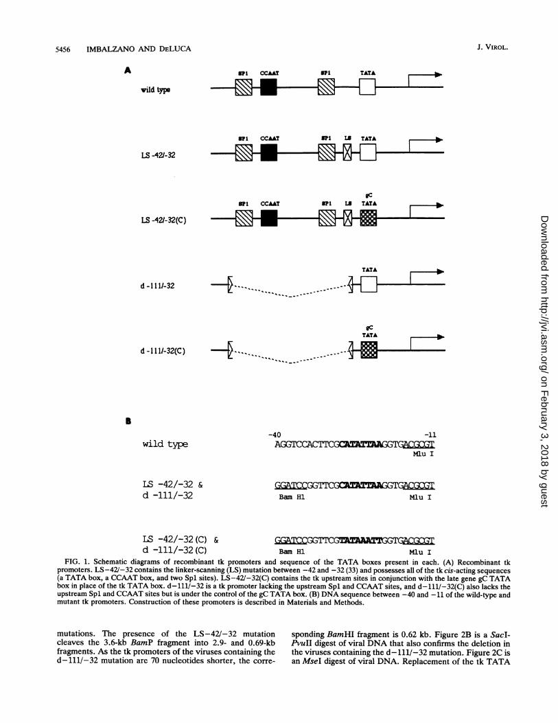
To facilitate precise substitution of the TATA box fromthe tk gene with the TATA box from the gC gene, we used aplasmid containing a linker-scanning mutation between nu-cleotides -42 and -32 (5, 33). This mutation alters thenucleotides between -42 and -32 to create a BamHI site.The effect of this mutation has been evaluated extensively intransient expression assays and in the context of viralinfection, and in no instance has this alteration been shownto affect tk expression (5, 12, 13, 24, 33). By digesting thisplasmid with BamHI and MluI, which cleaves the tk pro-moter just 3' to the TATA box, a short sequence containingthe tk TATA box was removed. A double-stranded oligonu-cleotide designed to restore the tk sequence excised, exceptfor replacing the tk TATA box with the gC TATA box, wascloned into this plasmid as well as into a similarly digestedplasmid lacking promoter sequences from -111 to -32. Thiseliminates the tk upstream Spl and CCAAT sites (seeMaterials and Methods and Fig. 1B). Because the tk TATAbox encodes an MseI site and the gC TATA box does not,clones were screened by restriction digestion for the loss ofan MseI site. Positive clones were then sequenced to verifythe substitution.The recombinant tk promoters are shown schematically in
Fig. 1A. LS-42/-32 contains the linker-scanning mutationbetween -42 and -32 and possesses all the tk cis-actingsequences: a TATA box, a CCAAT box, and two Spl sites.LS-42/-32(C) contains the tk upstream sites (Spl andCCAAT boxes) but possesses the TATA box from the lategene gC in place of the tk TATA box. d-111/-32(C) is a tkpromoter under the control of just the tk TATA box, andd-111/-32(C) is a tk promoter under the control of just thetk TATA box. These promoters were incorporated into thetk locus of the ICP4-deficient virus, n12, which possesses anonsense mutation at amino acid 251 in both copies of theICP4 gene. The n12 ICP4 molecule is severely truncated andhas no measurable ICP4 function (10). Promoter activity cantherefore be measured in the context of viral infection as afunction of the promoter alteration in the presence andabsence of ICP4.Southern blot hybridization was performed on selected
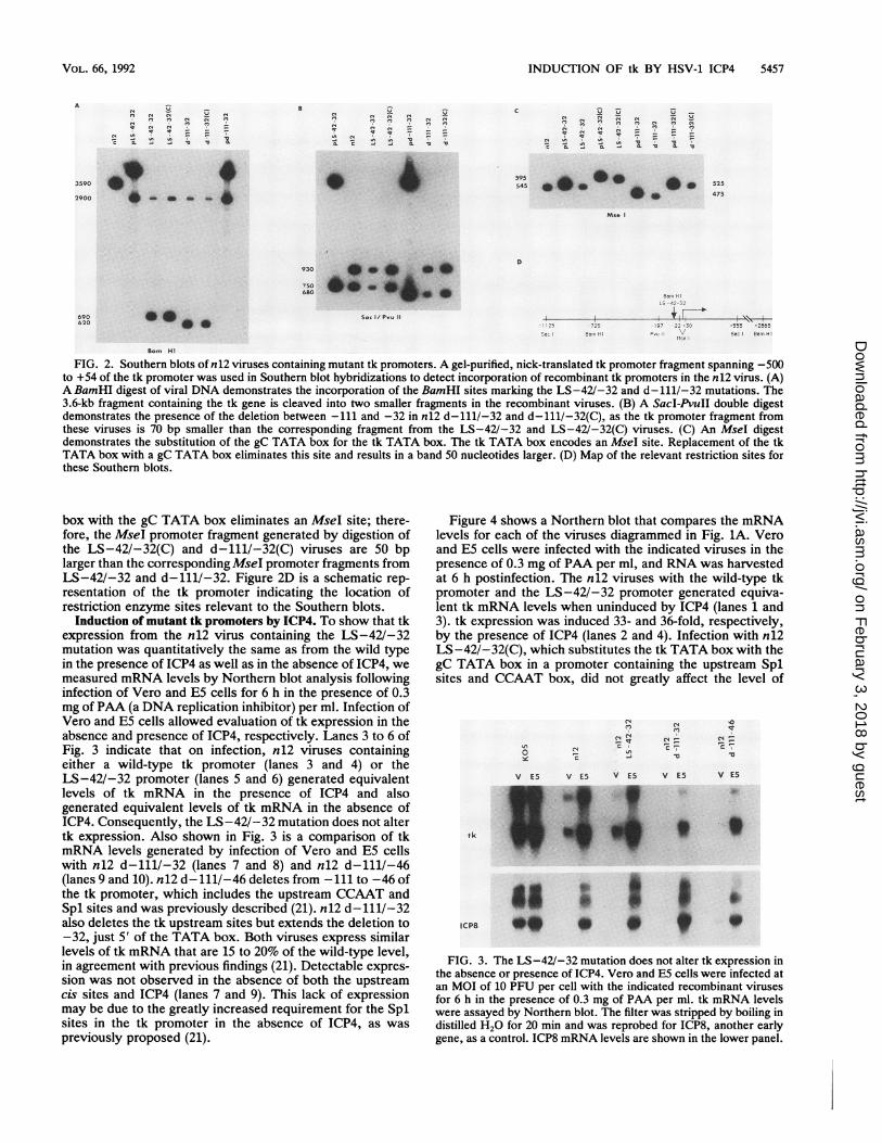
virus progeny to detect incorporation of the recombinant tkpromoters. The probe used was a gel-purified, nick-trans-lated fragment of the tk promoter spanning -500 to +54.Figure 2A shows a Southern blot of a BamHI digest ofrecombinant viral DNA that demonstrates the incorporationof the BamHI site marking the LS-42/-32 and d-111/-32
VOL. 66, 1992
on February 3, 2018 by guest
http://jvi.asm.org/
Dow
nloaded from
5456 IMBALZANO AND DELUCA
A Bpi
vild type
BPI
LS -42/-32
"I
B1
Bpi
BPI
CCAAT
CCAAT
CCAAT
TATA r
LB TA£TA ,- o
_
LG TATA , lo
LS -42/-32(C)
d -111/-32
DcTATA ,
d - 111-32(C) -{ - - -
B
wild type
LS -42/-32 &d -111/-32
-40 -11ACGGTCCACTT=G
Mlu I
Bam Hi Mlu I
LS -42/-32 (C) & G G GTTCd -111/-32(C) Bam Hi Mlu I
FIG. 1. Schematic diagrams of recombinant tk promoters and sequence of the TATA boxes present in each. (A) Recombinant tkpromoters. LS-42/-32 contains the linker-scanning (LS) mutation between -42 and -32 (33) and possesses all of the tk cis-acting sequences(a TATA box, a CCAAT box, and two Spl sites). LS-42/-32(C) contains the tk upstream sites in conjunction with the late gene gC TATAbox in place of the tk TATA box. d-111/-32 is a tk promoter lacking the upstream Spl and CCAAT sites, and d-111/-32(C) also lacks theupstream Spl and CCAAT sites but is under the control of the gC TATA box. (B) DNA sequence between -40 and -11 of the wild-type andmutant tk promoters. Construction of these promoters is described in Materials and Methods.
mutations. The presence of the LS-42/-32 mutationcleaves the 3.6-kb BamP fragment into 2.9- and 0.69-kbfragments. As the tk promoters of the viruses containing thed-111/-32 mutation are 70 nucleotides shorter, the corre-
sponding BamHI fragment is 0.62 kb. Figure 2B is a SacI-PvuII digest of viral DNA that also confirms the deletion inthe viruses containing the d-111/-32 mutation. Figure 2C isan MseI digest of viral DNA. Replacement of the tk TATA
-
J. VIROL.
TATA
I
on February 3, 2018 by guest
http://jvi.asm.org/
Dow
nloaded from
INDUCTION OF tk BY HSV-1 ICP4 5457
C? " f &
E4 In
a. .I.,C CL .1 - m v
_m_ m_ -
_ _ n Iq
In 7 - -
CL . -4 Cf la -1
_545 0 0 a
*
Mse
690620 aS..
930 *10 * . @
680 *-O -IP
S ac / P..u
Bam H1
FIG. 2. Southern blots of n12 viruses containing mutant tk promoters. A gel-purified, nick-translated tk promoter fragment spanning -500to +54 of the tk promoter was used in Southern blot hybridizations to detect incorporation of recombinant tk promoters in the n12 virus. (A)A BamHI digest of viral DNA demonstrates the incorporation of the BamHI sites marking the LS-42/-32 and d- 111/-32 mutations. The3.6-kb fragment containing the tk gene is cleaved into two smaller fragments in the recombinant viruses. (B) A SacI-PvuII double digestdemonstrates the presence of the deletion between -111 and -32 in n12 d-111/-32 and d-111/-32(C), as the tk promoter fragment fromthese viruses is 70 bp smaller than the corresponding fragment from the LS-42/-32 and LS-42/-32(C) viruses. (C) An MseI digestdemonstrates the substitution of the gC TATA box for the tk TATA box. The tk TATA box encodes an MseI site. Replacement of the tkTATA box with a gC TATA box eliminates this site and results in a band 50 nucleotides larger. (D) Map of the relevant restriction sites forthese Southern blots.
box with the gC TATA box eliminates an MseI site; there-fore, the MseI promoter fragment generated by digestion ofthe LS-42/-32(C) and d-111/-32(C) viruses are 50 bplarger than the corresponding MseI promoter fragments fromLS-42/-32 and d-111/-32. Figure 2D is a schematic rep-resentation of the tk promoter indicating the location ofrestriction enzyme sites relevant to the Southern blots.
Induction of mutant tk promoters by ICP4. To show that tkexpression from the n12 virus containing the LS-42/-32mutation was quantitatively the same as from the wild typein the presence of ICP4 as well as in the absence of ICP4, wemeasured mRNA levels by Northern blot analysis followinginfection of Vero and E5 cells for 6 h in the presence of 0.3mg of PAA (a DNA replication inhibitor) per ml. Infection ofVero and E5 cells allowed evaluation of tk expression in theabsence and presence of ICP4, respectively. Lanes 3 to 6 ofFig. 3 indicate that on infection, n12 viruses containingeither a wild-type tk promoter (lanes 3 and 4) or theLS-42/-32 promoter (lanes 5 and 6) generated equivalentlevels of tk mRNA in the presence of ICP4 and alsogenerated equivalent levels of tk mRNA in the absence ofICP4. Consequently, the LS-42/-32 mutation does not altertk expression. Also shown in Fig. 3 is a comparison of tkmRNA levels generated by infection of Vero and E5 cellswith n12 d-111/-32 (lanes 7 and 8) and n12 d-111/-46(lanes 9 and 10). n12 d-111/-46 deletes from -111 to -46 ofthe tk promoter, which includes the upstream CCAAT andSpl sites and was previously described (21). n12 d-111/-32also deletes the tk upstream sites but extends the deletion to-32, just 5' of the TATA box. Both viruses express similarlevels of tk mRNA that are 15 to 20% of the wild-type level,in agreement with previous findings (21). Detectable expres-sion was not observed in the absence of both the upstreamcis sites and ICP4 (lanes 7 and 9). This lack of expressionmay be due to the greatly increased requirement for the Splsites in the tk promoter in the absence of ICP4, as waspreviously proposed (21).
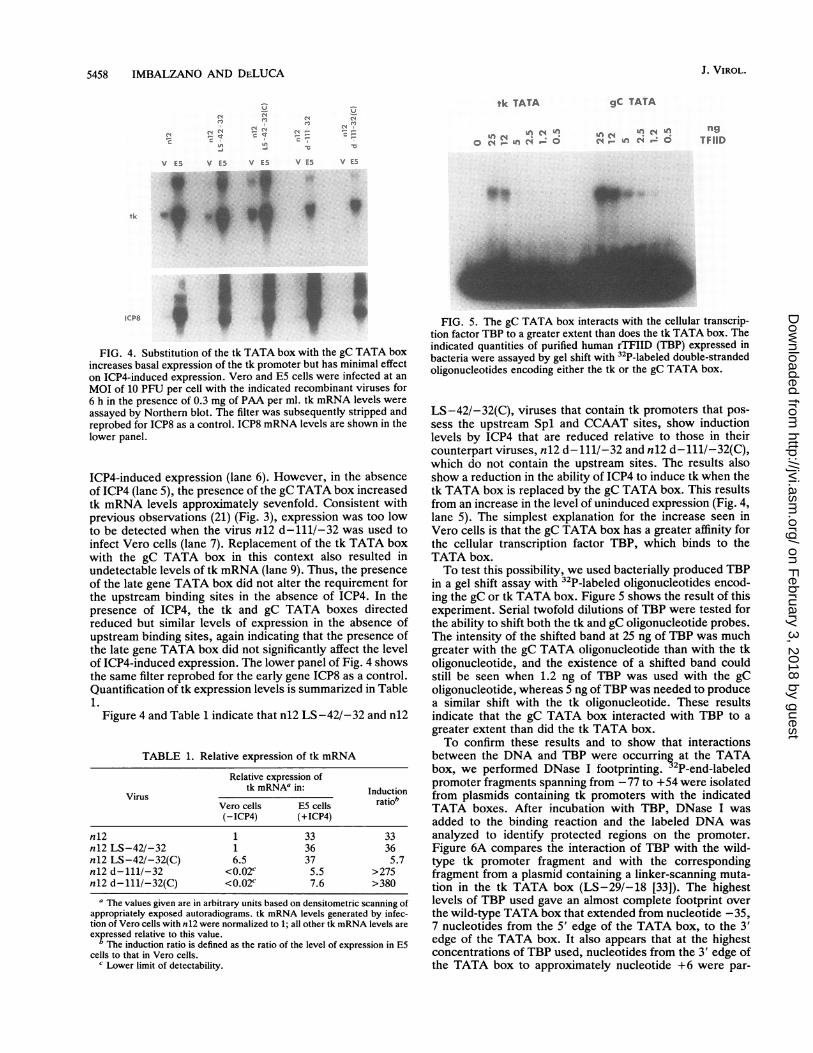
Figure 4 shows a Northern blot that compares the mRNAlevels for each of the viruses diagrammed in Fig. 1A. Veroand E5 cells were infected with the indicated viruses in thepresence of 0.3 mg of PAA per ml, and RNA was harvestedat 6 h postinfection. The n12 viruses with the wild-type tkpromoter and the LS-42/-32 promoter generated equiva-lent tk mRNA levels when uninduced by ICP4 (lanes 1 and3). tk expression was induced 33- and 36-fold, respectively,by the presence of ICP4 (lanes 2 and 4). Infection with n12LS-42/-32(C), which substitutes the tk TATA box with thegC TATA box in a promoter containing the upstream Splsites and CCAAT box, did not greatly affect the level of
ut C C, C0
V E5 V E5 V E5 V E5 V E5
_- - ,f
ICPB
FIG. 3. The LS-42/-32 mutation does not alter tk expression inthe absence or presence of ICP4. Vero and E5 cells were infected atan MOI of 10 PFU per cell with the indicated recombinant virusesfor 6 h in the presence of 0.3 mg of PAA per ml. tk mRNA levelswere assayed by Northern blot. The filter was stripped by boiling indistilled H20 for 20 min and was reprobed for ICP8, another earlygene, as a control. ICP8 mRNA levels are shown in the lower panel.
3590
2900
e,l C4i,, 04 C4CIAIn
a. .9 I.
525
475
VOL. 66, 1992
Ba" "r -. ., --
I. .*i 14"',
19' - 22 - 3-' -555 -2865,-5- 9,., -I! P,;.; ..;e Sac Bal.l HI
tk
on February 3, 2018 by guest
http://jvi.asm.org/
Dow
nloaded from
J. VIROL.5458 IMBALZANO AND DELUCA
(N (N(N (N
0 N r en _ o
(N (Ntu
..
V E5 V E5 V ES V E5 V E5
tk s.
FIG. 4. Substitution of the tk TATA box with the gC TATA boxincreases basal expression of the tk promoter but has minimal effecton ICP4-induced expression. Vero and E5 cells were infected at anMOI of 10 PFU per cell with the indicated recombinant viruses for6 h in the presence of 0.3 mg of PAA per ml. tk mRNA levels wereassayed by Northern blot. The filter was subsequently stripped andreprobed for ICP8 as a control. ICP8 mRNA levels are shown in thelower panel.
ICP4-induced expression (lane 6). However, in the absenceof ICP4 (lane 5), the presence of the gC TATA box increasedtk mRNA levels approximately sevenfold. Consistent withprevious observations (21) (Fig. 3), expression was too lowto be detected when the virus n12 d-111/-32 was used toinfect Vero cells (lane 7). Replacement of the tk TATA boxwith the gC TATA box in this context also resulted inundetectable levels of tk mRNA (lane 9). Thus, the presenceof the late gene TATA box did not alter the requirement forthe upstream binding sites in the absence of ICP4. In thepresence of ICP4, the tk and gC TATA boxes directedreduced but similar levels of expression in the absence ofupstream binding sites, again indicating that the presence ofthe late gene TATA box did not significantly affect the levelof ICP4-induced expression. The lower panel of Fig. 4 showsthe same filter reprobed for the early gene ICP8 as a control.Quantification of tk expression levels is summarized in Table1.
Figure 4 and Table 1 indicate that n12 LS-42/-32 and n12
TABLE 1. Relative expression of tk mRNA
Relative expression oftk mRNAa in: Induction
Virus *tbVero cells E5 cells ratio(-ICP4) (+ICP4)
n 12 1 33 33n12 LS-42/-32 1 36 36n12 LS-42/-32(C) 6.5 37 5.7nl2 d-111/-32 <0.02c 5.5 >275nl2 d-111/-32(C) <0.02c 7.6 >380
a The values given are in arbitrary units based on densitometric scanning ofappropriately exposed autoradiograms. tk mRNA levels generated by infec-tion of Vero cells with n12 were normalized to 1; all other tk mRNA levels areexpressed relative to this value.
The induction ratio is defined as the ratio of the level of expression in E5cells to that in Vero cells.
c Lower limit of detectability.
tk TATA gC TATA
(n c, f Ce c ngTFIID
FIG. 5. The gC TATA box interacts with the cellular transcrip-tion factor TBP to a greater extent than does the tk TATA box. Theindicated quantities of purified human rTFIID (TBP) expressed inbacteria were assayed by gel shift with 32P-labeled double-strandedoligonucleotides encoding either the tk or the gC TATA box.
LS-42/-32(C), viruses that contain tk promoters that pos-sess the upstream Spl and CCAAT sites, show inductionlevels by ICP4 that are reduced relative to those in theircounterpart viruses, n12 d-111/-32 and n12 d-111/-32(C),which do not contain the upstream sites. The results alsoshow a reduction in the ability of ICP4 to induce tk when thetk TATA box is replaced by the gC TATA box. This resultsfrom an increase in the level of uninduced expression (Fig. 4,lane 5). The simplest explanation for the increase seen inVero cells is that the gC TATA box has a greater affinity forthe cellular transcription factor TBP, which binds to theTATA box.To test this possibility, we used bacterially produced TBP
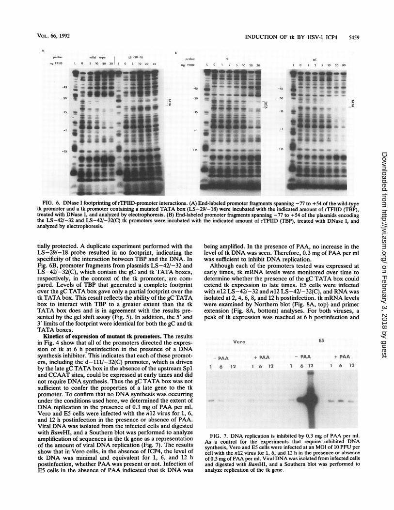
in a gel shift assay with 32P-labeled oligonucleotides encod-ing the gC or tk TATA box. Figure 5 shows the result of thisexperiment. Serial twofold dilutions of TBP were tested forthe ability to shift both the tk and gC oligonucleotide probes.The intensity of the shifted band at 25 ng of TBP was muchgreater with the gC TATA oligonucleotide than with the tkoligonucleotide, and the existence of a shifted band couldstill be seen when 1.2 ng of TBP was used with the gColigonucleotide, whereas 5 ng ofTBP was needed to producea similar shift with the tk oligonucleotide. These resultsindicate that the gC TATA box interacted with TBP to agreater extent than did the tk TATA box.To confirm these results and to show that interactions
between the DNA and TBP were occurring at the TATAbox, we performed DNase I footprinting. 2P-end-labeledpromoter fragments spanning from -77 to +54 were isolatedfrom plasmids containing tk promoters with the indicatedTATA boxes. After incubation with TBP, DNase I wasadded to the binding reaction and the labeled DNA wasanalyzed to identify protected regions on the promoter.Figure 6A compares the interaction of TBP with the wild-type tk promoter fragment and with the correspondingfragment from a plasmid containing a linker-scanning muta-tion in the tk TATA box (LS-29/-18 [33]). The highestlevels of TBP used gave an almost complete footprint overthe wild-type TATA box that extended from nucleotide -35,7 nucleotides from the 5' edge of the TATA box, to the 3'edge of the TATA box. It also appears that at the highestconcentrations of TBP used, nucleotides from the 3' edge ofthe TATA box to approximately nucleotide +6 were par-
on February 3, 2018 by guest
http://jvi.asm.org/
Dow
nloaded from
INDUCTION OF tk BY HSV-1 ICP4 5459
probe wild type LS-29-18
ng TFIID L 0 5 10 20 30 L 0 5 10 20 30
-45 __^* , ' |
-15Ad
*15 *:* |p< , I!
JA9Av.k7
mr-Y~
probe tk
ng TFIID L 0 1 2 5 10 20 30
-45 £ - M
-30
-15 _-
m -M
+15~~~~~4 d*
v
-+15 S _ _ 6-
-~~2-~~~~~~~'. A
gC
L 0 1 2 5 10 20 30
44---45 4l _0 _b GM do _
-30 _ +
-15
_*2 iY
15 'hii&ii
* *--*A
FIG. 6. DNase I footprinting of rTFIID-promoter interactions. (A) End-labeled promoter fragrnents spanning -77 to +54 of the wild-typetk promoter and a tk promoter containing a mutated TATA box (LS-29/-18) were incubated with the indicated amount of rTFIID (TBP),treated with DNase I, and analyzed by electrophoresis. (B) End-labeled promoter fragments spanning -77 to +54 of the plasmids encodingthe LS-42/-32 and LS-42/-32(C) tk promoters were incubated with the indicated amount of rTFIID (TBP), treated with DNase I, andanalyzed by electrophoresis.
tially protected. A duplicate experiment performed with theLS-29/-18 probe resulted in no footprint, indicating thespecificity of the interaction between TBP and the DNA. InFig. 6B, promoter fragments from plasmids LS-42/-32 andLS-42V-32(C), which contain the gC and tk TATA boxes,respectively, in the context of the tk promoter, are com-pared. Levels of TBP that generated a complete footprintover the gC TATA box gave only a partial footprint over thetk TATA box. This result reflects the ability of the gC TATAbox to interact with TBP to a greater extent than the tkTATA box does and is in agreement with the results pre-sented by the gel shift assay (Fig. 5). In addition, the 5' and3' limits of the footprint were identical for both the gC and tkTATA boxes.
Kinetics of expression of mutant tk promoters. The resultsin Fig. 4 show that all of the promoters directed the expres-sion of tk at 6 h postinfection in the presence of a DNAsynthesis inhibitor. This indicates that each of these promot-ers, including the d-111/-32(C) promoter, which is drivenby the late gC TATA box in the absence of the upstream Spland CCAAT sites, could be expressed at early times and didnot require DNA synthesis. Thus the gC TATA box was notsufficient to confer the properties of a late gene to the tkpromoter. To confirm that no DNA synthesis was occurringunder the conditions used here, we determined the extent ofDNA replication in the presence of 0.3 mg of PAA per ml.Vero and E5 cells were infected with the n12 virus for 1, 6,and 12 h postinfection in the presence or absence of PAA.Viral DNA was isolated from the infected cells and digestedwith BamHI, and a Southern blot was performed to analyzeamplification of sequences in the tk gene as a representationof the amount of viral DNA replication (Fig. 7). The resultsshow that in Vero cells, in the absence of ICP4, the level oftk DNA was minimal and equivalent for 1, 6, and 12 hpostinfection, whether PAA was present or not. Infection ofE5 cells in the absence of PAA indicated that tk DNA was
being amplified. In the presence of PAA, no increase in thelevel of tk DNA was seen. Therefore, 0.3 mg of PAA per mlwas sufficient to inhibit DNA replication.
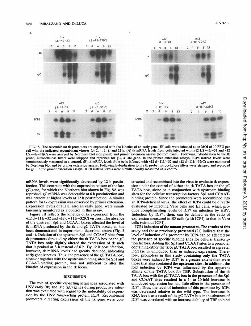
Although each of the promoters tested was expressed atearly times, tk mRNA levels were monitored over time todetermine whether the presence of the gC TATA box couldextend tk expression to late times. E5 cells were infectedwith n12 LS-42/-32 and n12 LS-42/-32(C), and RNA wasisolated at 2, 4, 6, 8, and 12 h postinfection. tk mRNA levelswere examined by Northern blot (Fig. 8A, top) and primerextension (Fig. 8A, bottom) analyses. For both viruses, apeak of tk expression was reached at 6 h postinfection and
Vero
- PAA
1 6 12
+ PAA - PAA
1 6 12 1 6 12
40
E5
+ PAA
6 12
I
FIG. 7. DNA replication is inhibited by 0.3 mg of PAA per ml.As a control for the experiments that require inhibited DNAsynthesis, Vero and E5 cells were infected at an MOI of 10 PFU percell with the n12 virus for 1, 6, and 12 h in the presence or absenceof 0.3 mg ofPAA per ml. Viral DNA was isolated from infected cellsand digested with BamHI, and a Southern blot was performed toanalyze replication of the tk gene.
VOL. 66, 1992
on February 3, 2018 by guest
http://jvi.asm.org/
Dow
nloaded from
5460 IMBALZANO AND DELUCA
Bn12
LS -42 - 32
2 4 6 8 12
n12LS-42-32
2 4 6 8 12
ebe.
n12LS -42-32(C)
2 4 6 8 12
nl2L5 -42-32(C)
2 4 6 8 12
n12d-111 -32
2 4 6 8 12
tk -
.....
gC
n12d -111- 32
M 2 4 6 8 12
ICP8
n12d -111-32(C)
2 4 6 8 12
n12d -111-32 (C)
2 4 6 8 12
m.e,....-1.
tk1. tk
FIG. 8. The recombinant tk promoters are expressed with the kinetics of an early gene. E5 cells were infected at an MOI of 10 PFU percell with the indicated recombinant viruses for 2, 4, 6, 8, and 12 h. (A) tk mRNA levels from cells infected with n12 LS-42/-32 and n12LS-42/-32(C) were assayed by Northern blot (top panel) and primer extension assays (bottom panel). Following hybridization to the tkprobe, nitrocellulose filters were stripped and reprobed for gC, a late gene. In the primer extension assays, ICP8 mRNA levels weresimultaneously measured as a control. (B) tk mRNA levels from cells infected with n12 d-111/-32 and n12 d-111/-32(C) were monitoredby Northern blot and by primer extension assays. Following hybridization to the tk probe, nitrocellulose filters were stripped and reprobedfor gC. In the primer extension assays, ICP8 mRNA levels were simultaneously measured as a control.
mRNA levels were significantly decreased by 12 h postin-fection. This contrasts with the expression pattern of the lategC gene, for which the Northern blot shown in Fig. 8A wasreprobed. gC mRNA was detectable at 6 h postinfection andwas present at higher levels at 12 h postinfection. A similarpattern for tk expression was observed by primer extension.Expression levels of ICP8, also an early gene, were simul-taneously monitored as a control in this assay.
Figure 8B reflects the kinetics of tk expression from then12 d-111/-32 and n12 d-111/-32(C) viruses. The absenceof the upstream Spl and CCAAT boxes affected the level oftk mRNA produced by the tk and gC TATA boxes, as hasbeen demonstrated in experiments described above (Fig. 3and 4). Deletion of the upstream Spl and CCAAT sites fromtk promoters directed by either the tk TATA box or the gCTATA box only slightly altered the expression of tk suchthat it peaked at 8 h instead of 6 h. By 12 h postinfection,however, tk mRNA levels had greatly declined, indicatingearly gene kinetics. Thus, the presence of the gC TATA box,alone or together with the upstream-binding sites for Spl andCCAAT-binding protein, was not sufficient to alter thekinetics of expression in the tk locus.
DISCUSSION
The role of specific cis-acting sequences associated withHSV early (tk) and late (gC) genes during productive infec-tion was evaluated with regard to the inducibility of expres-sion by the HSV trans-acting protein ICP4. Recombinantpromoters directing expression of the tk gene were con-
structed and recombined into the virus to evaluate tk expres-sion under the control of either the tk TATA box or the gCTATA box, alone or in conjunction with upstream bindingsites for the cellular transcription factors Spl and CCAAT-binding protein. Since the promoters were recombined intoan ICP4-deficient virus, the effect of ICP4 could be directlyevaluated by infecting Vero cells and E5 cells, which pro-duce complementing levels of ICP4 on infection by HSV.Induction by ICP4, then, can be defined as the ratio ofexpression measured in E5 cells (with ICP4) to that in Verocells (without ICP4).ICP4 induction of the mutant promoters. The results of this
study and those previously presented (21) indicate that thelevel of induction of a promoter by ICP4 can be affected bythe presence of specific binding sites for cellular transcrip-tion factors. Adding the Spl and CCAAT sites to a promotercontaining either the tk or gC TATA box resulted in a greaterincrease in uninduced than in induced expression. There-fore, promoters in this study containing only the TATAboxes were induced by ICP4 to a greater extent than werethose that also contained the upstream sites. In addition, thefold induction by ICP4 was influenced by the apparentaffinity of the TATA box for TBP. Substitution of the tkTATA box with the gC TATA box in the presence of the Spland CCAAT sites resulted in a 5- to 10-fold increase inuninduced expression but had little effect in the presence ofICP4. Thus, the level of induction of this promoter by ICP4was decreased relative to the wild type. The increase inRNA levels as a result of the gC TATA box in the absence ofICP4 was correlated with an increased ability of TBP to bind
A
tk
gc
ICP8
J. VIROL.
on February 3, 2018 by guest
http://jvi.asm.org/
Dow
nloaded from
INDUCTION OF tk BY HSV-1 ICP4 5461
to the gC TATA box relative to the tk TATA box. The gelshift assay (Fig. 5) and the DNase I footprinting assay (Fig.6B) indicate that equal levels of specific interaction betweenTBP and the promoter were achieved with approximatelyfourfold less TBP when the promoter contained the gCTATA box than when it contained the tk TATA box.
Despite the large variation in relative strengths of thesepromoters under uninduced conditions, the presence of ICP4still caused induction of gene expression such that significantlevels of tk mRNA were produced. Thus, no specific cis-acting sequence element is required for induction by ICP4.This is in agreement with previous findings (3, 5, 21).Furthermore, the relative strength of the promoter underuninduced conditions does not necessarily correlate with itsrelative strength in the presence of ICP4. Thali et al. (56)have proposed that the pseudorabies virus IE protein, whichshows structural similarity to the ICP4 molecule (4, 57),induces only "sub-optimally utilized" promoters, such thatIE function is limited to, or specific for, promoters that arenot maximally transcribed, such as those found in PRVgenes. The data presented here are in agreement with thisproposal, in that the weakest promoters under uninducedconditions, which are under the control of only the tk or gCTATA box, gave the highest induction levels and the stron-gest promoter under uninduced conditions, which containsthe tk upstream sites hooked to the gC TATA box, had thelowest induction level (Table 1; Fig. 4). Thus, an increase inthe affinity of the TATA box for TBP or the presence of Splsites in the promoter reduced the induction of tk expressionby ICP4. Since Spl has been shown to activate expressionvia a coactivator that functions through TBP (41, 43), theseresults suggest that a promoter that is better able to utilizeTBP is less responsive to induction by ICP4. Therefore,ICP4, either alone or in conjunction with other factors, maypromote or stabilize the interaction of TBP with the pro-moter, as has been shown for the IE protein of pseudorabiesvirus (1) and the Zta transactivator of Epstein-Barr virus(29). Given that previous results have shown that a mutationin the CCAAT box of the tk promoter had no effect on ICP4induction levels (21), the effect of the factor binding to theCCAAT box may be independent of TBP. The simplestmechanism by which ICP4 may promote or stabilize TFIIDinteractions at the promoter is by direct interaction betweenICP4 and TBP or TFIID. The ability of TBP to interact invitro with the viral trans-activators VP16 of HSV (52), Zta ofEpstein-Barr virus (29), and ElA of adenovirus (20, 28) hasbeen documented. In addition, ICP4 may stabilize TBP-promoter interactions by not only interacting with TBP, butalso interacting with other transcription factors that interactwith TBP, such as TBP-associated factors, TFIIB, orTFIIA. Preliminary studies suggest that this may be the case(5a).
Kinetics of expression. The data presented in this studydemonstrate that the presence of upstream binding sites forcellular transcription factors does not determine the tempo-ral pattern of tk mRNA accumulation resulting from tran-scription at the natural tk locus. Moreover, substitution ofT+A-rich core comprising the TATA box of a true late genehad no effect on the temporal pattern of tk expression fromthis locus in the presence or absence of the upstream factors.
Previous studies analyzing HSV late gene promoters havegenerally indicated that sequences from just 5' of the TATAbox to the translation initiation site are sufficient to directlate gene expression (14, 16, 17, 23, 45). Homa et al. (16)postulated that a specific 15-bp sequence including theTATA box mediated expression of the gC gene at late times,
because substitution of this TATA sequence with sequencescontaining the tk TATA box or random A+T-rich sequencesresulted in no expression and deletion of the 5' untranslatedleader sequences affected only the level of mRNA accumu-lation and not the time of expression. Explanations for thedifference between their results and those presented in thisstudy include the possibility that sequences flanking theTATA box contribute to the temporal pattern of expressionand that the position of the promoter in the genome may alsocontribute to the determination of kinetic class. It may beinteresting to examine the temporal pattern of expressionfrom a given promoter element at its natural site and atseveral ectopic sites in the viral genome. Results publishedby Steffy and Weir (51) showed that TATA boxes from IE,early, and late HSV genes were essentially interchangeablein a recombinant late gene promoter expressed from the viralgenome, further implying that structural features other thanthe TATA box determine late gene expression.The results suggest that the cis-acting binding sites for
cellular transcription factors act predominantly to determinethe level of expression and not when a promoter is activated.However, fusion of Spl and CCAAT sites to late genepromoters has been shown to result in expression at bothearly and late times (16, 32). If these sites, as our dataindicate, only increase expression levels, then perhaps thepresence of such sites in a late promoter increases thestrength of the promoter such that the restriction thatprevents expression of late genes until the onset of DNAreplication is overcome. The data presented here suggestthat this restriction is not due to any of the defined sequencesin early or late gene promoters that bind to cellular transcrip-tion factors. It has been proposed that sequences present inthe 5' untranslated leader sequences of late genes contributeto late gene expression (26, 32). Such sequences could bindto a cellular or viral protein that represses transcription bypreventing the formation of an active transcription initiationcomplex. Alternately, a cis sequence could affect the com-petence of a promoter for expression by affecting the sec-ondary structure of the template, as has been proposedpreviously (31). However, other reports have shown thatelimination of the gC leader sequences did not alter temporalregulation (59). It remains to be determined whether lategene leader sequences contribute to expression at late timesbecause of an effect on transcription initiation or an increasein mRNA stability. The potential role of transacting factorsand cis-acting sites operating in the leaders or at the startsites of late genes is an area for future studies.
ACKNOWLEDGMENT
This work was supported by Public Health Service grant AI27431to N. A. DeLuca.
REFERENCES1. Abmayr, S. M., J. L. Workman, and R. G. Roeder. 1988. The
pseudorabies immediate early protein stimulates in vitro tran-scription by facilitating TFIID:promoter interactions. GenesDev. 2:542-553.
2. Beard, P., S. Faber, K. W. Wilcox, and L. I. Pizer. 1986. Herpessimplex virus immediate early infected-cell polypeptide 4 bindsto DNA and promotes transcription. Proc. Natl. Acad. Sci.USA 83:4016-4020.
3. Boni, J., and D. M. Coen. 1989. Examination of the roles oftranscription factor Spl-binding sites and an octamer motif intrans-induction of the herpes simplex virus thymidine kinasegene. J. Virol. 63:4088-4092.
VOL. 66, 1992
on February 3, 2018 by guest
http://jvi.asm.org/
Dow
nloaded from
5462 IMBALZANO AND DELUCA
4. Cheung, A. K. 1989. DNA nucleotide sequence analysis of theimmediate early gene of pseudorabies virus. Nucleic Acids Res.17:4637-4646.
5. Coen, D. M., S. P. Weinheimer, and S. L. McKnight. 1986. Agenetic approach to promoter recognition during trans inductionof viral gene expression. Science 234:53-59.
Sa.DeLuca, N. A. Unpublished observations.6. DeLuca, N. A., M. A. Courtney, and P. A. Schaffer. 1984.
Temperature-sensitive mutants in HSV-1 ICP4 permissive forearly gene expression. J. Virol. 52:767-776.
7. DeLuca, N. A., A. McCarthy, and P. A. Schaffer. 1985. Isolationand characterization of deletion mutants of herpes simplex virustype 1 in the gene encoding immediate-early regulatory proteinICP4. J. Virol. 56:558-570.
8. DeLuca, N. A., and P. A. Schaffer. 1985. Activation of immedi-ate-early, early, and late promoters by temperature-sensitiveand wild-type forms of herpes simplex virus type 1 proteinICP4. Mol. Cell. Biol. 5:1997-2008.
9. DeLuca, N. A., and P. A. Schaffer. 1987. Activities of herpessimplex virus type 1 (HSV-1) ICP4 genes specifying nonsense
peptides. Nucleic Acids Res. 15:4491-4511.10. DeLuca, N. A., and P. A. Schaffer. 1988. Physical and functional
domains of the herpes simplex virus transcriptional regulatoryprotein ICP4. J. Virol. 62:732-743.
11. Dixon, R. A. F., and P. A. Schaffer. 1980. Fine-structuremapping and functional analysis of temperature-sensitive mu-
tants in the gene encoding the herpes simplex virus type 1immediate early protein VP175. J. Virol. 36:189-203.
11a.Dynlacht, D. B., T. Hoey, and R. Tjian. 1991. Isolation ofcofactors associated with the TATA-binding protein that medi-ates transcriptional activation. Cell 66:563-576.
12. Eisenberg, S. P., D. M. Coen, and S. L. McKnight. 1985.Promoter domains required for expression of plasmid-bornecopies of the herpes simplex virus thymidine kinase gene invirus-infected mouse fibroblasts and microinjected frog oocytes.Mol. Cell. Biol. 5:1940-1947.
13. El Kareh, A., A. J. M. Murphy, T. Fichter, A. Efstratiadis, andS. Silverstein. 1985. "Transactivation" control signals in thepromoter of the herpes simplex virus thymidine kinase gene.
Proc. Natl. Acad. Sci. USA 82:1002-1006.14. Flanagan, W. M., A. G. Papavassiliou, M. Rice, L. B. Hecht, S.
Silverstein, and E. K. Wagner. 1991. Analysis of the herpessimplex virus type 1 promoter controlling the expression ofUL38, a true late gene involved in capsid assembly. J. Virol.65:769-786.
15. Graham, F. L., and A. J. van der Eb. 1973. A new technique forthe assay of infectivity of human adenovirus S DNA. Virology52:456-457.
16. Homa, F. L., J. C. Glorioso, and M. Levine. 1988. A specific15-bp TATA box promoter element is required for expression ofa herpes simplex virus type 1 late gene. Genes Dev. 2:40-53.
17. Homa, F. L., T. M. Otal, J. C. Glorioso, and M. Levine. 1986.Transcriptional control signals of a herpes simplex virus type 1late (Y2) gene lie within bases -34 to +124 relative to the 5'terminus of the mRNA. Mol. Cell. Biol. 6:3652-3666.
18. Honess, R. W., and B. Roizman. 1974. Regulation of herpesvirus macromolecular synthesis. I. Cascade regulation of threegroups of viral proteins. J. Virol. 14:8-19.
19. Honess, R. W., and B. Roizman. 1975. Regulation of herpes
virus macromolecular synthesis: sequential transition of poly-
peptide synthesis requires functional viral polypeptides. Proc.Natl. Acad. Sci. USA 72:1276-1280.
20. Horikoshi, N., K. Maguire, A. Kralli, E. Maldonado, D. Rein-
berg, and R. Weinmann. 1991. Direct interaction betweenadenovirus ElA protein and the TATA box binding transcrip-
tion factor IID. Proc. Natl. Acad. Sci. USA 88:5124-5128.
21. Imbalzano, A. N., D. M. Coen, and N. A. DeLuca. 1991. Herpes
simplex virus transactivator ICP4 operationally substitutes for
the cellular transcription factor Spl for efficient expression of
the viral thymidine kinase gene. J. Virol. 65:565-574.
22. Imbalzano, A. N., A. A. Shepard, and N. A. DeLuca. 1990.
Functional relevance of specific interactions between herpes
simplex type 1 ICP4 and sequences from the promoter-regula-
tory domain of the viral thymidine kinase gene. J. Virol.64:2620-2631.
23. Johnson, P. A., and R. D. Everett. 1986. The control of herpessimplex virus type 1 late gene transcription: a TATA box/capsite region is sufficient for fully regulated activity. Nucleic AcidsRes. 14:8247-8256.
24. Jones, K. A., K. R. Yamomoto, and R. Tjian. 1985. Two distincttranscription factors bind to the HSV thymidine kinase pro-moter in vitro. Cell 42:559-572.
25. Kao, C. C., P. M. Lieberman, M. C. Schmidt, Q. Zhou, R. Pei,and A. J. Berk. 1990. Cloning of a transcriptionally activehuman TATA binding factor. Science 248:1646-1650.
26. Kibler, P. K., J. Duncan, B. D. Keith, T. Hupel, and J. R.Smiley. 1991. The regulation of herpes simplex virus true lategene expression: sequences downstream from the US11 TATAbox inhibit expression from an unreplicated template. J. Virol.65:6749-6760.
27. Kristie, T. M., and B. Roizman. 1986. a4, the major regulatoryprotein of herpes simplex virus type 1, is stably and specificallyassociated with promoter-regulatory domains ofa genes and ofselected other viral genes. Proc. Natl. Acad. Sci. USA 83:3218-3222.
28. Lee, W. S., C. C. Kao, G. 0. Bryant, X. Liu, and A. J. Berk.1991. Adenovirus ElA activation domain binds the basic repeatin the TATA box transcription factor. Cell 67:365-376.
29. Lieberman, P. M., and A. J. Berk. 1991. The Zta trans-activatorprotein stabilizes TFIID association with promoter DNA bydirect protein-protein interaction. Genes Dev. 5:2441-2454.
30. Maniatis, T., A. Jeffrey, and D. G. Kleid. 1975. Nucleotidesequence of the rightward operator of phage 1. Proc. Natl.Acad. Sci. USA 72:1184-1188.
31. Mavromara-Nazos, P., and B. Roizman. 1987. Activation ofherpes simplex virus 1 Y2 genes by viral DNA replication.Virology 161:593-598.
32. Mavromara-Nazos, P., and B. Roizman. 1989. Delineation ofregulatory domains of early (,) and late (Y2) genes by construc-tion of chimeric genes expressed in herpes simplex virus type 1genomes. Proc. Natl. Acad. Sci. USA 86:4071-4075.
33. McKnight, S. L., and R. Kingsbury. 1982. Transcriptionalcontrol signals of a eukaryotic protein-coding gene. Science217:316-324.
34. Michael, N., D. Spector, P. Mavromara-Nazos, T. M. Kristie,and B. Roizman. 1988. The DNA-binding properties of the majorregulatory protein a4 of herpes simplex virus. Science 239:1531-1534.
35. O'Hare, P., and G. S. Hayward. 1985. Evidence for a direct rolefor both the 175,000- and 110,000-molecular-weight immediate-early proteins of herpes simplex virus in the trans-activation ofdelayed-early promoters. J. Virol. 53:751-760.
36. Papavassiliou, A. G., and S. J. Silverstein. 1990. Interaction ofcell and virus proteins with DNA sequences encompassing thepromoter/regulatory and leader regions of the herpes simplexvirus thymidine kinase gene. J. Biol. Chem. 265:9402-9412.
37. Parris, D. S., R. A. F. Dixon, and P. A. Schaffer. 1980. Physicalmapping of herpes simplex virus type 1 ts mutants by markerrescue: correlation of the physical and genetic maps. Virology100:275-287.
38. Paterson, T., and R. D. Everett. 1988. Mutational dissection ofthe HSV-1 immediate early protein Vmwl75 involved in tran-scriptional transactivation and repression. Virology 166:186-196.
39. Paterson, T., and R. D. Everett. 1988. The regions of the herpessimplex virus type 1 immediate-early protein Vmwl75 requiredfor site specific DNA binding closely corresponds to thoseinvolved in transcriptional regulation. Nucleic Acids Res. 16:11005-11025.
40. Paterson, T., V. G. Preston, and R. D. Everett. 1990. A mutantof herpes simplex virus type 1 immediate early polypeptideVmwl75 binds to the cap site of its own promoter in vitro butfails to autoregulate in vivo. J. Gen. Virol. 71:851-861.
41. Peterson, M. G., N. Tanese, B. F. Pugh, and R. Tjian. 1990.Functional domains and upstream activator properties of clonedhuman TATA binding protein. Science 248:1625-1630.
J. VIROL.
on February 3, 2018 by guest
http://jvi.asm.org/
Dow
nloaded from
INDUCTION OF tk BY HSV-1 ICP4 5463
42. Preston, C. M. 1979. Abnormal properties of an immediate earlypolypeptide in cells infected with herpes simplex virus type 1mutant tsK J. Virol. 32:357-369.
43. Pugh, B. F., and R. Tjian. 1990. Mechanism of transcriptionalactivation by Spl: evidence for coactivators. Cell 61:1187-1197.
44. Sambrook, J., E. F. Fritsch, and T. Maniatis. 1989. Molecularcloning: a laboratory manual, 2nd ed. Cold Spring HarborLaboratory, Cold Spring Harbor, N.Y.
45. Shapira, M., F. L. Homa, J. C. Glorioso, and M. Levine. 1987.Regulation of the herpes simplex virus type 1 late (y2) glyco-protein gene: sequences between base pairs -34 and +29control transient expression and responsiveness to transactiva-tion by the products of the immediate early (a) 4 and 0 genes.Nucleic Acids Res. 15:3097-3111.
46. Shepard, A. A., and N. A. DeLuca. 1991. Activities of het-erodimers composed of DNA-binding and transactivation-defi-cient subunits of herpes simplex virus regulatory protein ICP4.J. Virol. 65:299-307.
47. Shepard, A. A., and N. A. DeLuca. 1991. A second-site revertantof a defective herpes simplex virus ICP4 protein with restoredregulatory activities and impaired DNA-binding properties. J.Virol. 65:787-795.
48. Shepard, A. A., A. N. Imbalzano, and N. A. DeLuca. 1989.Separation of primary structural components conferring auto-regulation, transactivation, and DNA-binding properties to theherpes simplex virus transcriptional regulatory protein ICP4. J.Virol. 63:3714-3728.
49. Smiley, J. R., D. C. Johnson, L. I. Pizer, and R. D. Everett. 1992.The ICP4 binding sites in the herpes simplex virus type 1glycoprotein D (gD) promoter are not essential for efficient gDtranscription during virus infection. J. Virol. 66:623-631.
50. Southern, E. M. 1975. Detection of specific sequences among
DNA fragments separated by gel electrophoresis. J. Mol. Biol.98:503-517.
51. Steffy, K. R., and J. P. Weir. 1991. Upstream promoter elementsof the herpes simplex virus type 1 glycoprotein H gene. J. Virol.65:972-975.
52. Stringer, K. F., C. J. Ingles, and J. Greenblatt. 1990. Direct andselective binding of an acidic transcriptional activation domainto the TATA-box factor TFIID. Nature (London) 345:783-786.
53. Su, L., and D. M. Knipe. 1987. Mapping of the transcriptionalinitiation site of the herpes simplex virus type 1 ICP8 gene ininfected and transfected cells. J. Virol. 61:615-620.
54. Tedder, D. G., R. D. Everett, K. W. Wilcox, P. Beard, and L. I.Pizer. 1989. ICP4 binding sites in the promoter and codingregions of the herpes simplex virus gD gene contribute toactivation of in vitro transcription by ICP4. J. Virol. 63:2510-2520.
55. Tedder, D. G., and L. I. Pizer. 1988. Role for DNA-proteininteraction in activation of the herpes simplex virus glycopro-tein D gene. J. Virol. 62:4661-4672.
56. Thali, M., S. Rusconi, and W. Schaffner. 1990. Immediate earlyprotein of pseudorabies virus is a general transactivator butstimulates only suboptimally utilized promoters. A clue tospecificity? J. Mol. Biol. 215:301-311.
57. Vlcek, C., V. Paces, and M. Schwyzer. 1989. Nucleotide se-quence of the pseudorabies virus immediate early gene, encod-ing a strong transactivator protein. Virus Genes 2:335-346.
58. Watson, R., and J. B. Clements. 1980. A herpes simplex virustype 1 function continuously required for early and late virusRNA synthesis. Nature (London) 285:329-330.
59. Weir, J. P., and P. R. Narayanan. 1990. Expression of theherpes simplex virus type 1 glycoprotein C gene requiressequences in the 5' noncoding region of the gene. J. Virol.64:445-449.
VOL. 66, 1992
on February 3, 2018 by guest
http://jvi.asm.org/
Dow
nloaded from